Journal of Clinical Medicine Review Peptic Ulcer Disease: A Brief Review of Conventional Therapy and Herbal Treatment Options Lucija Kuna 1 , Jelena Jakab 2,3 , Robert Smolic 2,4,5 , Nikola Raguz-Lucic 1 , Aleksandar Vcev 2,5 and Martina Smolic 1,4, * 1 Department of Pharmacology and Biochemistry, Faculty of Dental Medicine and Health Osijek, Josip Juraj Strossmayer University of Osijek, 31000 Osijek, Croatia; [email protected] (L.K.); [email protected] (N.R.-L.) 2 Department of Pathophysiology and Physiology with Immunology, Faculty of Dental Medicine and Health Osijek, Josip Juraj Strossmayer University of Osijek, 31000 Osijek, Croatia; [email protected] (J.J.); [email protected] (R.S.); [email protected] (A.V.) 3 Department of Internal Medicine, Faculty of Medicine Osijek, Josip Juraj Strossmayer University of Osijek, 31000 Osijek, Croatia 4 Department of Pharmacology, Faculty of Medicine Osijek, Josip Juraj Strossmayer University of Osijek, 31000 Osijek, Croatia 5 Department of Internal Medicine, University Hospital Osijek, 31000 Osijek, Croatia * Correspondence: [email protected]; Tel.: +385-31-512-800 Received: 31 December 2018; Accepted: 31 January 2019; Published: 3 February 2019 Abstract: Peptic ulcer is a chronic disease affecting up to 10% of the world’s population. The formation of peptic ulcers depends on the presence of gastric juice pH and the decrease in mucosal defenses. Non-steroidal anti-inflammatory drugs (NSAIDs) and Helicobacter pylori (H. pylori) infection are the two major factors disrupting the mucosal resistance to injury. Conventional treatments of peptic ulcers, such as proton pump inhibitors (PPIs) and histamine-2 (H2) receptor antagonists, have demonstrated adverse effects, relapses, and various drug interactions. On the other hand, medicinal plants and their chemical compounds are useful in the prevention and treatment of numerous diseases. Hence, this review presents common medicinal plants that may be used for the treatment or prevention of peptic ulcers. Keywords: peptic ulcer disease; Helicobacter pylori infection; herbal treatment 1. Introduction Peptic ulcer is an acid-induced lesion of the digestive tract that is usually located in the stomach or proximal duodenum, and is characterized by denuded mucosa with the defect extending into the submucosa or muscularis propria [1]. The estimated prevalence of peptic ulcer disease in the general population is 5–10% [2], but recent epidemiological studies have shown a decrease in the incidence, rates of hospital admissions, and mortality associated with peptic ulcer [3,4]. This is most likely secondary to the introduction of new therapies and improved hygiene, which resulted in a decline in Helicobacter pylori (H. pylori) infections. Traditionally, mucosal disruption in patients with the acid peptic disease is considered to be a result of a hypersecretory acidic environment together with dietary factors or stress. Risk factors for developing peptic ulcer include H. pylori infection, alcohol and tobacco consumption, non-steroidal anti-inflammatory drugs (NSAIDs) use, and Zollinger–Ellison syndrome [5]. The main risk factors for both gastric and duodenal ulcers are H. pylori infection and NSAID use [6]. However, only a small proportion of people affected with H. pylori or using NSAIDs develop peptic ulcer disease, meaning that individual susceptibility is important in the beginning of mucosal damage. Functional polymorphisms J. Clin. Med. 2019, 8, 179; doi:10.3390/jcm8020179 www.mdpi.com/journal/jcm
Transcript
Journal of
Clinical Medicine
Review
Peptic Ulcer Disease: A Brief Review of ConventionalTherapy and Herbal Treatment Options
Lucija Kuna 1, Jelena Jakab 2,3, Robert Smolic 2,4,5, Nikola Raguz-Lucic 1, Aleksandar Vcev 2,5
and Martina Smolic 1,4,*1 Department of Pharmacology and Biochemistry, Faculty of Dental Medicine and Health Osijek, Josip Juraj
2 Department of Pathophysiology and Physiology with Immunology, Faculty of Dental Medicine and HealthOsijek, Josip Juraj Strossmayer University of Osijek, 31000 Osijek, Croatia; [email protected] (J.J.);[email protected] (R.S.); [email protected] (A.V.)
3 Department of Internal Medicine, Faculty of Medicine Osijek, Josip Juraj Strossmayer University of Osijek,31000 Osijek, Croatia
4 Department of Pharmacology, Faculty of Medicine Osijek, Josip Juraj Strossmayer University of Osijek,31000 Osijek, Croatia
5 Department of Internal Medicine, University Hospital Osijek, 31000 Osijek, Croatia* Correspondence: [email protected]; Tel.: +385-31-512-800
Received: 31 December 2018; Accepted: 31 January 2019; Published: 3 February 2019�����������������
Abstract: Peptic ulcer is a chronic disease affecting up to 10% of the world’s population. The formationof peptic ulcers depends on the presence of gastric juice pH and the decrease in mucosal defenses.Non-steroidal anti-inflammatory drugs (NSAIDs) and Helicobacter pylori (H. pylori) infection are thetwo major factors disrupting the mucosal resistance to injury. Conventional treatments of peptic ulcers,such as proton pump inhibitors (PPIs) and histamine-2 (H2) receptor antagonists, have demonstratedadverse effects, relapses, and various drug interactions. On the other hand, medicinal plants andtheir chemical compounds are useful in the prevention and treatment of numerous diseases. Hence,this review presents common medicinal plants that may be used for the treatment or prevention ofpeptic ulcers.
Peptic ulcer is an acid-induced lesion of the digestive tract that is usually located in the stomachor proximal duodenum, and is characterized by denuded mucosa with the defect extending into thesubmucosa or muscularis propria [1]. The estimated prevalence of peptic ulcer disease in the generalpopulation is 5–10% [2], but recent epidemiological studies have shown a decrease in the incidence,rates of hospital admissions, and mortality associated with peptic ulcer [3,4]. This is most likelysecondary to the introduction of new therapies and improved hygiene, which resulted in a decline inHelicobacter pylori (H. pylori) infections.
Traditionally, mucosal disruption in patients with the acid peptic disease is considered to be aresult of a hypersecretory acidic environment together with dietary factors or stress. Risk factors fordeveloping peptic ulcer include H. pylori infection, alcohol and tobacco consumption, non-steroidalanti-inflammatory drugs (NSAIDs) use, and Zollinger–Ellison syndrome [5]. The main risk factors forboth gastric and duodenal ulcers are H. pylori infection and NSAID use [6]. However, only a smallproportion of people affected with H. pylori or using NSAIDs develop peptic ulcer disease, meaning thatindividual susceptibility is important in the beginning of mucosal damage. Functional polymorphisms
J. Clin. Med. 2019, 8, 179; doi:10.3390/jcm8020179 www.mdpi.com/journal/jcm
in different cytokine genes are associated with peptic ulcers. For example, polymorphisms ofinterleukin 1 beta (IL1B) affect mucosal interleukin 1β production, causing H. pylori-associatedgastroduodenal diseases [7].
On the other hand, the risk of complications of peptic ulcer is increased four times in NSAID users,and two times in aspirin users [8]. The concomitant use of NSAIDs or aspirin with anticoagulants,corticosteroids, and selective serotonin reuptake inhibitors increase the risk of upper gastrointestinalbleeding [9]. Although many people who use NSAIDs or aspirin have concurrent H. pylori infection,their interaction in the pathogenesis of peptic ulcer disease remains controversial. A meta-analysis ofobservational studies resulted in a conclusion that NSAIDs, aspirin use, and H. pylori infection increasethe risk of peptic ulcer disease independently [10].
H. pylori-negative, NSAID-negative, and aspirin-negative peptic ulcer disease, which is classifiedas an idiopathic ulcer, can be diagnosed in about one-fifth of cases [11]. It is caused by the imbalancebetween factors that contribute to mucosal integrity and aggressive insults, but the pathogenicmechanisms behind the development of idiopathic peptic ulcer are still unknown [5]. A Danish studyshowed that psychological stress could increase the incidence of peptic ulcer [12]. Other etiologiesinclude ischemia, drugs (steroids, chemotherapeutic agents) and radiotherapy, viruses, histamine,eosinophilic infiltration, gastric bypass surgery, and metabolic disturbances [13].
2. Pathogenesis of Peptic Ulcer
Almost half of the world’s population is colonized by H. pylori, which remains one of the mostcommon causes of peptic ulcer disease [14]. The prevalence of H. pylori is higher in developingcountries, especially in Africa, Central America, Central Asia, and Eastern Europe [15]. Theorganism is usually acquired in childhood in an environment of unsanitary conditions and crowding,mostly in countries with lower socioeconomic status. H. pylori causes epithelial cell degeneration andinjury, which is usually more severe in the antrum, by the inflammatory response with neutrophils,lymphocytes, plasma cells, and macrophages.
The mechanism by which H. pylori induces the development of different types of lesions in thegastroduodenal mucosa is not fully explained. H. pylori infection can result in either hypochlorhydria orhyperchlorhydria, thus determining the type of peptic ulcer. The main mediators of H. pylori infectionare cytokines that inhibit parietal cell secretion, but H. pylori can directly affect the H+/K+ ATPaseα-subunit, activate calcitonin gene-related peptide (CGRP) sensory neurons linked to somatostatin,or inhibit the production of gastrin [16]. Although the formation of gastric ulcers is associated withhyposecretion, 10–15% of patients with H. pylori infection have increased gastric secretion caused byhypergastrinemia and reduced antral somatostatin content [17]. This leads to increased histaminesecretion, and subsequently the increased secretion of acid or pepsin from parietal and gastric cells.Additionally, the eradication of H. pylori leads to a decrease in gastrin mRNA expression and anincrease in somatostatin mRNA expression [18]. In the remaining majority of patients, gastric ulcersare associated with hypochlorhydria and mucosal atrophy.
The main mechanism of NSAID-associated damage of the gastroduodenal mucosa is thesystemic inhibition of constitutively expressed cyclooxygenase-1 (COX-1), which is responsiblefor prostaglandin synthesis, and is associated with decreased mucosal blood flow, low mucus andbicarbonate secretion, and the inhibition of cell proliferation. NSAIDs inhibit the enzyme reversiblyin a concentration-dependent manner. The co-administration of exogenous prostaglandins andcyclooxygenase-2 (COX-2)-selective NSAIDs use reduces mucosal damage and the risk of ulcers [19].However, the different physicochemical properties of NSAIDs cause differences in their toxicity [20].NSAIDs disrupt mucus phospholipids and lead to the uncoupling of mitochondrial oxidativephosphorylation, thus initiating mucosal damage. When exposed to acidic gastric juice (pH 2),NSAIDs become protonated and cross lipid membranes to enter epithelial cells (pH 7.4), where theyionize and release H+. In that form, NSAIDs cannot cross the lipid membrane, and are trapped inepithelial cells, leading to the uncoupling of oxidative phosphorylation, decreased mitochondrial
J. Clin. Med. 2019, 8, 179 3 of 19
energy production, increased cellular permeability, and reduced cellular integrity. Patients who havea history of peptic ulcers or hemorrhage, are over the age of 65, also use steroids or anticoagulants,and take high doses or combinations of NSAIDs are at the highest risk for acquiring NSAID-inducedulcers [1]
Main pathophysiological mechanisms and the sites of action of antiulcer treatment are shown inthe Figure 1.
J. Clin. Med. 2019, 8, x FOR PEER REVIEW 3 of 18
and take high doses or combinations of NSAIDs are at the highest risk for acquiring NSAID-induced ulcers [1]
Main pathophysiological mechanisms and the sites of action of antiulcer treatment are shown in the Figure 1.
Figure 1. Schematic presentation of main pathophysiological mechanisms involved in the development of peptic ulcer disease, and the sites of action of the most commonly used pharmacological options in the treatment of peptic ulcer disease. CCK2 = Cholecystokinin Receptor; PGE2 = Prostaglandin E2; PGI2 = Prostaglandin I2; EP3 = Prostaglandin E receptor 3; HIST = Histamine.
3. Treatment
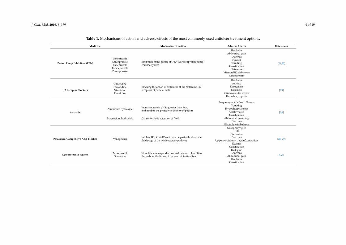
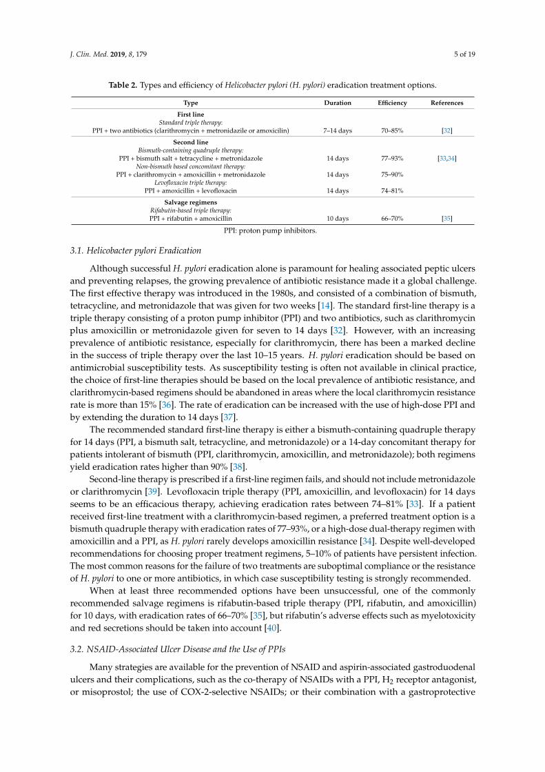
An overview of conventional antiulcer treatment options is summarized in Table 1 and Table 2.
Table 1. Mechanisms of action and adverse effects of the most commonly used antiulcer treatment options.
Medicine Mechanism of Action Adverse Effects References
Proton Pump Inhibitors (PPIs)
Omeprazole
Inhibition of the gastric H+/K+-ATPase (proton pump) enzyme system
Headache Abdominal pain
Diarrhea Nausea
Vomiting Constipation
Flatulence Vitamin B12 deficiency
Osteoporosis
[21,22]
Lansoprazole
Rabeprazole
Esomeprazole
Pantoprazole
H2 Receptor Blockers
Cimetidine
Blocking the action of histamine at the histamine H2 receptors of parietal cells
Headache Anxiety
Depression Dizziness
Cardiovascular events Thrombocytopenia
[23] Famotidine
Nizatidine
Ranitidine
Antacids Aluminum hydroxide
Increases gastric pH to greater than four, and inhibits the proteolytic activity of pepsin
Frequency not defined: Nausea
Vomiting Hypophosphatemia
[24]
Figure 1. Schematic presentation of main pathophysiological mechanisms involved in the developmentof peptic ulcer disease, and the sites of action of the most commonly used pharmacological optionsin the treatment of peptic ulcer disease. CCK2 = Cholecystokinin Receptor; PGE2 = Prostaglandin E2;PGI2 = Prostaglandin I2; EP3 = Prostaglandin E receptor 3; HIST = Histamine.
3. Treatment
An overview of conventional antiulcer treatment options is summarized in Tables 1 and 2.
J. Clin. Med. 2019, 8, 179 4 of 19
Table 1. Mechanisms of action and adverse effects of the most commonly used antiulcer treatment options.
Medicine Mechanism of Action Adverse Effects References
Proton Pump Inhibitors (PPIs)
OmeprazoleInhibition of the gastric H+/K+-ATPase (proton pump)enzyme system
HeadacheAbdominal pain
DiarrheaNausea
VomitingConstipation
FlatulenceVitamin B12 deficiency
Osteoporosis
[21,22]LansoprazoleRabeprazole
EsomeprazolePantoprazole
H2 Receptor Blockers
CimetidineBlocking the action of histamine at the histamine H2receptors of parietal cells
HeadacheAnxiety
DepressionDizziness
Cardiovascular eventsThrombocytopenia
[23]FamotidineNizatidineRanitidine
AntacidsAluminum hydroxide Increases gastric pH to greater than four,
and inhibits the proteolytic activity of pepsin
Frequency not defined: NauseaVomiting
HypophosphatemiaChalky tasteConstipation
Abdominal crampingDiarrhea
Electrolyte imbalance
[24]
Magnesium hydroxide Causes osmotic retention of fluid
Potassium-Competitive Acid Blocker Vonoprazan Inhibits H+, K+-ATPase in gastric parietal cells at thefinal stage of the acid secretory pathway
NasopharyngitisFall
ContusionDiarrhea
Upper respiratory tract inflammationEczema
ConstipationBack pain
[25–29]
Cytoprotective Agents Misoprostol Stimulate mucus production and enhance blood flowthroughout the lining of the gastrointestinal tract
DiarrheaAbdominal pain
HeadacheConstipation
[30,31]Sucralfate
J. Clin. Med. 2019, 8, 179 5 of 19
Table 2. Types and efficiency of Helicobacter pylori (H. pylori) eradication treatment options.
Type Duration Efficiency References
First lineStandard triple therapy:
PPI + two antibiotics (clarithromycin + metronidazile or amoxicilin) 7–14 days 70–85% [32]
Second lineBismuth-containing quadruple therapy:
PPI + bismuth salt + tetracycline + metronidazole 14 days 77–93% [33,34]Non-bismuth based concomitant therapy:
PPI + clarithromycin + amoxicillin + metronidazole 14 days 75–90%Levofloxacin triple therapy:
Although successful H. pylori eradication alone is paramount for healing associated peptic ulcersand preventing relapses, the growing prevalence of antibiotic resistance made it a global challenge.The first effective therapy was introduced in the 1980s, and consisted of a combination of bismuth,tetracycline, and metronidazole that was given for two weeks [14]. The standard first-line therapy is atriple therapy consisting of a proton pump inhibitor (PPI) and two antibiotics, such as clarithromycinplus amoxicillin or metronidazole given for seven to 14 days [32]. However, with an increasingprevalence of antibiotic resistance, especially for clarithromycin, there has been a marked declinein the success of triple therapy over the last 10–15 years. H. pylori eradication should be based onantimicrobial susceptibility tests. As susceptibility testing is often not available in clinical practice,the choice of first-line therapies should be based on the local prevalence of antibiotic resistance, andclarithromycin-based regimens should be abandoned in areas where the local clarithromycin resistancerate is more than 15% [36]. The rate of eradication can be increased with the use of high-dose PPI andby extending the duration to 14 days [37].
The recommended standard first-line therapy is either a bismuth-containing quadruple therapyfor 14 days (PPI, a bismuth salt, tetracycline, and metronidazole) or a 14-day concomitant therapy forpatients intolerant of bismuth (PPI, clarithromycin, amoxicillin, and metronidazole); both regimensyield eradication rates higher than 90% [38].
Second-line therapy is prescribed if a first-line regimen fails, and should not include metronidazoleor clarithromycin [39]. Levofloxacin triple therapy (PPI, amoxicillin, and levofloxacin) for 14 daysseems to be an efficacious therapy, achieving eradication rates between 74–81% [33]. If a patientreceived first-line treatment with a clarithromycin-based regimen, a preferred treatment option is abismuth quadruple therapy with eradication rates of 77–93%, or a high-dose dual-therapy regimen withamoxicillin and a PPI, as H. pylori rarely develops amoxicillin resistance [34]. Despite well-developedrecommendations for choosing proper treatment regimens, 5–10% of patients have persistent infection.The most common reasons for the failure of two treatments are suboptimal compliance or the resistanceof H. pylori to one or more antibiotics, in which case susceptibility testing is strongly recommended.
When at least three recommended options have been unsuccessful, one of the commonlyrecommended salvage regimens is rifabutin-based triple therapy (PPI, rifabutin, and amoxicillin)for 10 days, with eradication rates of 66–70% [35], but rifabutin’s adverse effects such as myelotoxicityand red secretions should be taken into account [40].
3.2. NSAID-Associated Ulcer Disease and the Use of PPIs
Many strategies are available for the prevention of NSAID and aspirin-associated gastroduodenalulcers and their complications, such as the co-therapy of NSAIDs with a PPI, H2 receptor antagonist,or misoprostol; the use of COX-2-selective NSAIDs; or their combination with a gastroprotective
J. Clin. Med. 2019, 8, 179 6 of 19
agent. PPIs are the most popular and effective prophylactic agents [41]. The mechanism of actionis reducing the production of gastric acid through irreversible binding to the hydrogen/potassiumATPase enzyme on gastric parietal cells. The combination of COX-2-selective NSAIDs and a PPI offersthe best protection against peptic ulcer complications [42]. Standard doses of H2 receptor antagonistscannot reduce the risk of gastric ulcers [43]. Gastrointestinal upset and abortifacient actions limit theuse of misoprostol for gastric protection, despite its effective prevention of peptic ulcer complications.Ulcers heal in more than 85% of cases with six to eight weeks of PPI therapy if the offending agentis discontinued. All of the gastric ulcers require repeat endoscopy to evaluate the success of healing.If ulcers fail to heal, drug compliance should be checked. For refractory ulcers, the doubling of PPIdose for another six to eight weeks is often recommended, although the evidence supporting this isweak. After the exclusion of false-negative H. pylori status, unusual causes of peptic ulcer should beexplored, such as malignancies, infections, Crohn’s disease, vasculitis, upper abdominal radiotherapy,cocaine use, and Zollinger–Ellison syndrome.
PPIs are among the most commonly used and overprescribed medications in the world [44].The side effects of the PPIs, such as a headache, diarrhea, constipation, and abdominal discomfort,are minor and easily managed. However, recent studies have suggested an association between PPIuse and several serious adverse effects, which has been a source of major concern to patients andphysicians. Some of the adverse effects of PPIs are related to their suppression of gastric acid secretion,allowing ingested microbial pathogens that would have been destroyed by gastric acid to colonize theupper gastrointestinal tract and cause infections. Reports are suggesting that the use of PPIs mightincrease the risk of enteric infections such as Salmonella and Campylobacter, community-acquiredpneumonia [45], Clostridium difficile infections [46], and spontaneous bacterial peritonitis [47].
With gastric acid suppression, there is no stimulation of endocrine D cells to produce somatostatin,and thereby no inhibition of G cells for gastrin release, resulting in hypergastrinemia. Gastrin is agrowth factor that can increase proliferation in Barrett metaplasia and the colon [48]. Nonetheless,PPI-induced hypergastrinemia in humans generally is mild, and rarely causes carcinoid tumors inhuman patients unless they have a genetic abnormality [49]. Furthermore, PPI usage might protectagainst cancer in Barrett’s esophagus, since PPIs heal the chronic esophageal inflammation of refluxesophagitis, which is a risk factor for the development of malignancy.
Gastric acid inhibition by PPIs also can affect the uptake of certain vitamins, minerals, andmedications. There are reports of patients on PPIs developing vitamin B12 deficiency and irondeficiency anemia [50]. Additionally, PPIs might increase the risk for osteoporosis and bone fracturesby interfering with the ionization and solubilization of the calcium salts that are required for theirabsorption [51]. The underlying mechanism for hypomagnesemia is still not clear. PPI-induced gastricacid suppression decreases ketoconazole absorption and facilitates the absorption of digoxin [52].Furthermore, PPIs can affect the metabolism of other drugs metabolized by the cytochrome (CYP) P450system; for instance, they can delay the clearance of warfarin, diazepam, and phenytoin. Considerableattention has been given to the potential of PPIs to reduce the antiplatelet action of clopidogrel,since both are metabolized by the CYP2C19 enzyme [53]. The clinical importance of the interactionremains disputed, but the Food and Drug Administration (FDA) has issued warnings to avoid usingomeprazole or esomeprazole with clopidogrel.
There has been a dramatic increase in reports of miscellaneous, unanticipated adverse effectsof PPIs over the past several years, such as myocardial infarction, stroke, acute and chronic kidneydisease, and eosinophilic esophagitis. The increased frequency of cardiovascular events in patients onclopidogrel who also use PPIs can be the result of the drugs competing for metabolism by CYP2C19,although there is a possibility that PPIs might have cardiovascular effects that are independent oftheir effects on clopidogrel activation, perhaps by the decreased production of nitric oxide and alteredvascular homeostasis [54]. It has been proposed that PPIs might contribute to the developmentof eosinophilic esophagitis through their effects on peptic digestion [55]. The suppression of acid
J. Clin. Med. 2019, 8, 179 7 of 19
production raises gastric pH and inactivates pepsin, inhibiting peptide ingestion and degradation, andcausing allergic reactions in the small intestine.
3.3. Potassium-Competitive Acid Blockers
Since up to 13% of patients treated with lansoprazole still experience ulcer recurrence, the searchfor alternative treatment is ongoing. Vonoprazan is a potassium-competitive acid blocker thatinhibits H+, K+-ATPase in gastric parietal cells at the final stage of the acid secretory pathway [25].The difference in the mechanism of action between vonoprazan and PPIs is that vonoprazan inhibitsthe enzyme in a K+-competitive and reversible manner, and does not require an acidic environmentfor activation. Additionally, vonoprazan shows a rapid onset of action and prolonged control ofintragastric acidity [26]. Vonoprazan at doses of 10 mg and 20 mg was non-inferior to lansoprazole forthe prevention of peptic ulcer recurrence in Japanese patients during NSAID therapy [25], or those whorequired aspirin therapy for cardiovascular or cerebrovascular protection [27], with good tolerance,a similar safety profile, and no new safety issues. Also, five weeks of treatment with vonoprazansignificantly reduced post-endoscopic submucosal dissection bleeding, compared to eight weeksof treatment with PPIs [28]. Similarly, it was shown to be superior to esomeprazole [29] andrabeprazole [26] for scarring artificial ulcers, which could help make an endoscopic submucosaldissection a safer treatment.
3.4. Future Research Questions
Along with the global decline of peptic ulcer disease and in the prevalence of H. pylori, there is arising problem of growing antimicrobial resistance, which reduces the efficiency of eradication therapy,and the overuse of PPIs, resulting in unexpected new side effects [56]. Also, the occurrence of idiopathiculcers associated with high mortality is increasing [57], and there is a need for defining the optimummanagement of the idiopathic disease. There is still an open question of how H. pylori infection andNSAID or aspirin interact, leaving the best strategy to manage patients with both risks unresolved.The pathogenesis of H. pylori-related gastric lesions is still not fully understood. Its development isled by a combination of H. pylori virulent factors and the host immune response; however, the precisecombination of H. pylori factors and the host genetic profile are yet to be fully enlightened. Why somepatients are more susceptible than others to the gastric toxicity of NSAIDs and aspirin, and whichgenetic polymorphisms are associated with H. pylori-induced peptic ulcer also remain unclear.
In the absence of any possible breakthrough antimicrobial agent for H. pylori, antibiotic resistancecontinues to be a major challenge, and new therapies are in fact old therapies. H. pylori urease has beenat the center of attention for the development of antiulcer treatment. Several potent in vitro inhibitorshave been found, but with poor specificity. They usually don’t make it to the clinical setting due to thehigh dosage required, increased cost of treatment, and increased risk for bleeding. Recent advancesin the molecular description of H. pylori pathogenesis resulted in promising candidates related to thepathogen’s persistence in the host, such as adherence. Some antivirulence agents can selectively targetthe pathogen’s adherence, but a high binding affinity and genetic diversity in the receptor-binding siteof H. pylori complicate the finding of potent inhibitors [58].
The genetic diversity of the virulence proteome in H. pylori direct future antivirulencedevelopments toward its more conserved assembly and secretion pathways, leaving the open questionof how these inhibitors can contribute to H. pylori treatment.
Gastrointestinal bleeding as the complication of peptic ulcer disease remains life-threatening,and comorbidities are now the primary cause of death in these patients. There is an urgent needfor prospective data and randomized controlled trials to define the best patient care strategy. In themeantime, appropriate diagnostics, adherence to current guidelines, and the avoidance of inferiorH. pylori treatment regimens will be necessary to maintain successful treatment of peptic ulcer.
J. Clin. Med. 2019, 8, 179 8 of 19
4. Alternative Therapy for Peptic Ulcer
The usage of medicinal plants in healing numerous diseases is as old as human beings, andwell-known as phytotherapy. Moreover, in the past few years, there has been a rising interestin alternative therapies and the usage of herbal products, in particular, those produced frommedicinal plants [59,60]. Also, due to appearance of various side effects by usage of conventionaldrugs for numerous diseases, medicinal plants are considered the major reservoir of potentiallynew drugs. Plant extracts and their crude are the most significant sources of new drugs, andhave been shown to cause promising results in the treatment of gastric ulcer as well [61]. It isknown that numerous pharmaceutical agents such as proton pump inhibitors, anticholinergics,antacids, antimicrobial agents, H2-receptor antagonists, sucralfate, and bismuth are not fully effective,and produce numerous adverse effects such as impotence, arrhythmia, hematopoietic alterations,hypersensitivity, and gynecomastia [62,63]. Due to that, investigations of the new pharmacologicallyactive agents through the screening of different plant extracts led to the discovery of effective andsafe drugs with gastroprotective activity. Especially, plants with antioxidant capability as the mainmechanism are used as the herbal reservoir for the treatment of ulcer disease [63].
Medicinal plants have achieved their therapeutic properties from their capability to producerenewable and various secondary metabolites, which are known as phytochemical constituents. Hence,numerous plants have used these phytochemicals as a protection mechanism against pathogens [64].
On the other hand, the appearance of resistant pathogens has had a significant influence on thepharmaceutical companies to change their strategy in the development of conventional antibioticsand design new antimicrobial drugs derived from medicinal plants [65]. Nevertheless, the syntheticantibiotics are still dominant as antimicrobial drugs.
As a matter of fact, incidences of infectious diseases have enlarged within the last three decades,involving infections with different properties as well as new infections, and it has been shown thataround 60% of them are of zoonotic origin (spread among human and animals). H. pylori is one ofthe major representatives in that group, and may cause chronic gastritis, peptic ulcer disease, andstomach cancer [66]. Therefore, one of the aims in this review was to highlight some medicinal plantsthat demonstrated significant antibacterial and antioxidant activity against H. pylori and peptic ulcerdisease. However, some of plants lose their efficiency against H. pylori consequent to the emergence ofresistant strains. Consequently, the isolation of various constituents from the most active plant extractsis encouraged [67].
It is important to emphasize that herbal products may contain numerous bioactive constituentswith dangerous, but also beneficial effects. Therefore, the higher education of doctors and patientsabout herbal therapy is necessary, as well as legislation to control the quality of herbal products,especially for further randomized investigations to determine the effectiveness and safety of manyproducts in digestive and other disorders [68].
Finally, the Ayurvedic knowledge and modern medicine could generate preferable antiulcer drugsderived from medicinal plants with less side effects [69].
Numerous medicinal plants with significant antibacterial activity against H. pylori and benefitsfor gastric ulcer disease are shown in Table 3.
J. Clin. Med. 2019, 8, 179 9 of 19
Table 3. Overview of herbal antiulcer treatment and H. pylori eradication.
Medicinal Plant Possible Mechanisms Effect Adverse Effects References
Korean red ginsengInhibition of H. pylori-induced 5-lipoxygenase(5-LOX) activity; preventing pro-inflammatoryinterleukin (IL)-8 or 5-LOX mRNA
Anti-inflammatory effect; increase eradicationrates of H. pylori; reduction of gastricinflammation and oxidative DNA damage
Interaction with conventional drugs [69,70]
Allium sativumInhibition of lipoprotein oxidation and lowerserum glucose induction of antioxidant enzymes;mechanisms need to be more investigated
Antioxidant; suppressive effect of H.pylori-induced gastric inflammation in vivo andin vitro
Interaction with conventional drugs [71]
Curcuma loga Inhibition of H. pylori-induced 5-LOX activity Anti-inflammatory; antioxidant Not determined [72]
Zingiber officinalis Inhibition of PGE2 and parietal cell H+,K+-ATPase Anti-inflammatory effect; antioxidant
Nausea and vomiting in pregnant women;restless, heartburn; interaction with conventionaldrugs (anticoagulants, analgesics)
[73–75]
Zingiber zerumbet
Gastroprotective mechanism of zerumbone(significant increased in the endogenousantioxidant GSH, reduction of lipid peroxidationlevel); other mechanism need to be investigated
Antioxidant, antiproliferative, anti-inflammatory,antisecretory effect; reduction of ulcer areaformation
Nausea and vomiting in pregnant women;restless, heartburn; interaction with conventionaldrugs (anticoagulants, analgesics)
[75,76]
Camellia sinensis (Green teapolyphenols)
Suppression of tumor necrosis factor-alpha(TNF-α) gene expression; inhibition of urease
Antioxidant; improvement in the function ofintestinal bacterial flora
Interaction with conventional drugs; dizziness,diarrhea, headaches, insomnia, heartbeat, maycause deficiency of iron
[77,78]
J. Clin. Med. 2019, 8, 179 10 of 19
4.1. The Effect on H. pylori Eradication
Several factors influence the conventional therapy failure. These include: the poor bioavailabilityof antibiotics, as the gastric mucus layer plays a barrier to antibiotic delivery, and therefore the drugsare unable to obtain the underlying gastric epithelium [70]; the stomach containing a pH from acidicto neutral, and only a few antibiotics are active in a wide pH range [79]; bacterial antagonism toantibiotics, where co-infection with multiple strains is quite an important feature [80]; deficiency ofpatient permissiveness to the therapy; patients lifestyle, and diet [46].
Numerous studies have been reported about various medicinal plants and their anti-H. pyloriactivity. In recent years, it has been shown that the suppression of enzymatic (dihydrofolate reductase,DNA gyrase, myeloperoxidase N-acetyltransferase, and urease) and adhesive activities, the high redoxpotential, and hydrophilic/hydrophobic natures of constituents have a significant role in anti-H. pyloriaction mechanisms. H. pylori-stimulated gastric inflammation may lead to superficial gastritis andatrophic gastritis, but also to gastric cancer. It is established that different natural products haveanti-inflammation activity, and the fundamental mechanisms involve the inhibition of nuclear factor-κBand mitogen-activated protein kinase pathway activation and the suppression of oxidative stress.
Since the role of H. pylori infection regarding carcinogenesis is to ascend carcinogenesis insteadto play a key role as a direct carcinogen, its eradication alone cannot inhibit H. pylori-related gastriccancers [81].
Medical plants such as Allium sativum, Zingiber officinalis, Korean red ginseng, and Cistuslaurifolius are known to suppress the colonization of H. pylori, reduce gastric inflammation bychemokine release, inhibit cytokine, and suppress precancerous changes by suppressing nuclearfactor-kappa B DNA binding, which suppresses mutagenesis and produces abundant levels ofapoptosis. Further unresolved issues will have to be cleared out before phytoceuticals are accepted asa standard therapy for H. pylori infection [82].
4.2. Korean Red Ginseng
Korean red ginseng extract plays a significant role in inhibiting H. pylori-induced 5-LOXactivity, such as inactivating c-jun, repressing NF-κB-DNA binding, inhibiting H. pylori-induced5(S)-hydroxyeicosatetraenoic acid biosynthesis, and preventing pro-inflammatory interleukin (IL)-8 or5-LOX mRNA. Consequently, these mechanisms decrease gastric carcinogenesis.
Moreover, Korean red ginseng has been shown to be beneficial in suppressing 5-lipoxygenase(5-LOX) mRNA and enzyme activities, and consequently the decreased synthesis of5-hydroxy-eicosatetraenoic acid. Similarly, green tea extract may prevent the activation ofmultiple transcription factors and their target genes, involving COX-2 and inducible nitric oxidesynthase (iNOS) mitogen-activated protein kinase activation, as well as the lipopolysaccharide of H.pylori-activated TLR-4. Due to that, these blockades increase the pro-inflammatory factors that inducegastric mucosal lesions [83,84]. Kim et al. reported on the protective effect of Korean red ginseng againstH. pylori-induced cytotoxicity in vitro [83]. Meanwhile, in a previous clinical study, a supplementaryadministration of Korean red ginseng increased the eradication rates of H. pylori, reduced gastricinflammation, and decreased oxidative DNA damage and apoptosis [84].
4.3. Allium sativum
Throughout history, the health benefits of garlic have been well documented, and the main useof Allium sativum was for its medicinal properties. The organosulfur components of Allium sativum,including S-allyl-L-cysteine (SAC) sulfoxides and δ-glutamyl S-allyl-L-cysteine, are known as maincompounds of its bioactivity. Raw Allium sativum is easy to convert in bioinactive form. Accordingly,numerous types of its extract with different compositions of bioactive components have beendeveloped, and their efficacy has been observed and evaluated in numerous studies [85]. The major roleof Allium sativum extract has been observed in antioxidant effect by scavenging reactive oxygen species
J. Clin. Med. 2019, 8, 179 11 of 19
(ROS), inhibiting lipoprotein oxidation and lowering the serum glucose induction of antioxidantenzymes. Also, it showed a suppressive effect of H. pylori-induced gastric inflammation in vivo [86],and an anti-tumorigenic effect by promoting apoptosis and the induction of cell cycle arrest [87].Allicin and allyl-methyl plus methyl-allyl thiosulfinate from acetonic Allium sativum extracts haverestricted the growth of H. pylori in the in vitro investigations [88].
4.4. Cistus Laurifolius
Flavonoids are one of the most important components of the human diet with a key role inorganisms and significant responsibility for numerous biological activities, in particular, antioxidant.Due to their limited availability and high cost, a rapid synthesis of polyoxygenated flavones,starting from accessible and inexpensive flavanones, has been developed. By methoxylation andbromination protocol 3′-demethoxysudachitin, a restricted flavone with antimicrobial activity againstH. pylori has been designed. Numerous investigations on flavoinoids were done with an extractof Cistus laurifolius. It has been demonstrated when testing for antimicrobial activity against H.pylori that 3’-demethoxysudachitin and sudachitin were the most active compounds. A similarinvestigation showed that the chloroform extract of Cistus laurifolius has tremendous anti-H. pyloriactivity. Accordingly to these investigations, isolated flavonoids can be used as an additive componentfor the standard treatment of H. pylori infection [82,89].
Li HQ et al. observed diverse levels of anti-H. pylori activities in numerous isoflavones [90].The experiment evaluated a few series of metronidazole-flavonoid extracts that have been used forantimicrobial activity against H. pylori [90]. It has been demonstrated that only one compound couldremarkably achieve the enhancement in IL-8 levels in the gastric cancer cells induced with a H.pylori water extract. On the other hand, Nakagawa et al.’s experiments revealed that new flavonoidcompounds 6, 7, and (2S)-4′,7-dihydroxy-8-methylflavan were discovered to be most efficaciouscompounds against H. pylori [91].
Similarly, Ustun et al. discovered that the chloroform extract of Cistus laurifolius holds a significantanti-H. pylori effect [42]. Accordingly, isolated flavonoids can be used as an alternative or supplementcompound to the current treatment of H. pylori infection [76].
4.5. Zingiber Officinalis and Zingiber Zerumbet
Zingiber officinalis is known as ginger, which is consumed as a flavoring agent. The plant extractshowed antitumor effects on colon cancer cells by inhibiting its growth, increasing DNA synthesis,and producing apoptosis [92]. Moreover, the main pungent phenolic compound of Zingiber officinalisis 6-gingerol, which has numerous pharmacological activities. Zingiber officinalis extracts containinggingerols have key role in prostaglandin E2 (PGE2) inhibition [73]. On the other side, the activephenolic compounds such as gingerol and zingerone have a significant influence in inhibiting parietalcell H+, K+-ATPase. Due to that, the activity of gingerol and zingerone plays a very important role inproton pump inhibition and the reduction of gastric acid secretion. Also, it shows a protective effectagainst H. pylori-induced ulcers [74].
Jiang et al. demonstrated the therapeutic effect of Zingiber officinalis as a natural antioxidantagainst gastric ulcers [93]. They reported free Zingiber officinalis extracts limitations such as slightsolubility in gastric juices, which will reduce further as it passes to higher pH regions of duodenum orileum in rats; numerous medicaments show a restricted transit time of less than two to four hours inthe stomach; whichever part is solubilized will be instantly absorbed, because Zingiber officinalis extractindicates fast absorption, consequently, local therapeutic effect cannot be elicited adequately [93].
In addition, Sidahmed et al. showed that zerumbone from Zingiber zerumbet has a major rolein gastroprotection activity against ethanol-induced gastric ulcer model in rats. They demonstratedthat pretreatment with zerumbone or omeprazole in rats significantly reduced ulcer area formationcompared to the ulcer control group. Moreover, pretreatment with omeprazole at 20 mg/kg bodyweight (b.w.) (p < 0.05) obstructed formation of ulcer by 76.77%, while pretreatment of zerumbone
J. Clin. Med. 2019, 8, 179 12 of 19
at five and 10 mg/kg b.w. obstructed ulcer formation by 75.59% and 88.75%, respectively. On theother hand, zerumbone and its gastroprotective mechanisms were not tested against other ulcermodel; hence, other mechanisms may be implicated and their influence needs to be investigated andelucidated [94].
4.6. Camellia Sinensis (Green Tea Polyphenols)
Nowadays, Camellia sinensis is one of the most commonly used beverages. The chemopreventiveeffects of Camellia sinensis depend on its activity as an antioxidant, but also on its molecular regulatoryfunctions on cellular growth, development, and apoptosis; and a selective improvement in the functionof the intestinal bacterial flora. Between the numerous constituents of green tea, polyphenols andepigallocatechin gallate (EGCG) suppress tumor necrosis factor-alpha (TNF-α) gene expression [95].On the other hand, the urease of H. pylori is crucial for its colonization, and investigations concentratedon Camellia sinensis extract demonstrated the inhibitory activity of this enzyme. That results in theinhibition of bacterial colonization [96]. Numerous similar studies demonstrated the inhibitory effectof Camellia sinensis extract by increasing cell vacuolation by vacuolating cytotoxin A (vacA) and ureaconduction in H. pylori infection. Consequently, it could pursue anti-H. pylori activity in vivo [97].
In 2008, Rao et al. reported on the gastroprotective activity of 50% ethanolic extract of Ficusglomerata fruit (FGE) in gastric ulcer models in rats [98]. FGE was administered per mouth (50, 100,and 200 mg/kg body weight), twice daily for five days for prevention from ethanol (EtOH), pylorusligation (PL), and cold restraint stress (CRS), which induced ulcer formation. It demonstrated adose-dependent suppression of ulcer, and it had a significant role in preventing the oxidative damageof gastric mucosa by preventing lipid peroxidation and significantly reducing in H+/K+-ATPase andsuperoxide dismutase. Their results showed that F. glomerate has an important gastroprotective effectthat might be consequent to the gastric defense factors [98].
4.7. Curcuma Longa and Artemisia Asiatica
Medicinal plants with antioxidant and anti-inflammatory activity have had a demonstratedeffect on gastroesophageal reflux disease (GERD). The medicinal plants and herbal preparationswith antioxidant and anti-inflammatory mechanisms include Curcuma longa, Panax quinquefolium,Artemisia asiatica, and Lonicera japonica. Moreover, other mechanisms include: the down-regulationof the genes encoding proteins that have key role in acute inflammation, including 1 intercellularadhesion molecule-1 (ICAM-1) and cytokine-induced neutrophil chemoattractant-2-beta (CINC-2-2beta) (Panax quinquefolium); ameliorating the function and gastric mucus (Morus alba, Curcuma longa);reducing gastric acid, such as for instance Curcuma longa, Morus alba, and acidinol syrup, increasingtonic contractions of the lower esophageal sphincter (LES) (Salvia miltiorrhiza, STW 5), and preventingthe pro-inflammatory cytokines IL-1 b and TNF-a (STW 5) [99].
It is important to mention investigation on rats where pretreatment with compounds of Artemisiaasiatica (DA-9601) reduced the overall density of the esophageal wall and volume of ulceration beyondthe ranitidine group [100].
Mahattanadul showed in his study on rats that the rhizome of Curcuma longa plays a protectiverole in the formation of acute acid reflux esophagitis (RE), but it was not effective in the prevention ofchronic acid RE [101]. However, its combination with dimethyl sulfoxide as an antioxidant compoundreduced the severity of the esophagitis ulcer index to around that of lansoprazole. In contrast,lansoprazole inclined to elevate the severity of all histopathological changes above the control andcurcumin-treated groups. Hence, it seemed that the antioxidant and anti-inflammatory activity ofcurcumin plays a major role in its beneficial effects on GERD [101].
Herbal medicine can be a mighty weapon for suppressing or modulating the disease-associatedfootprints of H. pylori infection and eradication. Finally, those plant products have shown strongpotential as pharmaceutical candidates in gastric disease prevention [68].
J. Clin. Med. 2019, 8, 179 13 of 19
5. Herb–Drug Interactions
Together with increasing use of herbal supplements worldwide, the number of adverse eventsand drug interactions is rising. Interactions between an herbal supplement and a drug can manifestas a pharmacokinetic or pharmacodynamic interaction. Pharmacokinetic interaction is a result ofusing the same mechanism of absorption, distribution, metabolism, or excretion between an herbalsupplement and a co-administered drug, leading to the change of the drug’s concentration in the bloodand pharmacologic action. Pharmacodynamic interactions involve a direct effect on the mechanism ofaction of a co-administered drug without changing the drug’s concentration, only by antagonizing orexacerbating the drug’s clinical effects [77].
Allium sativum extract decreases concentrations of drugs transported by P-gp, such as digoxin,doxorubicin, rosuvastatin, and verapamil [102]. The most studied Allium sativum interactions is theone with warfarin, although this has not yet been confirmed by controlled clinical trials. Also, itinhibits platelet aggregation, so it should be used with caution in patients with clotting disorders orthose with anticoagulant therapy [103]. Zingiber officinalis prolongs bleeding time by the inhibition ofthromboxane synthetase, but this has not been confirmed in a clinical trial [104]. Ginkgo biloba couldincrease bleeding risk, especially in combination with anticoagulant drugs, due to the inhibition ofplatelet aggregation. Flavonoids in Ginkgo biloba have antiplatelet activity, but do not affect bloodcoagulation or platelet function in humans [103]. In combination with NSAIDs, it can cause severebleeding [105].
Panax ginseng induces cytochrome P450 3A4 (CYP3A4), which decreases the effectivenessof calcium channel blockers, certain antihypertensive and statin medications, and someantidepressants [106]. Panax ginseng has hypoglycemic activity in patients with diabetes, and maycause headache, trembling, and manic behavior in patients treated with phenelzine [107].
Green tea extract has been shown to increase simvastatin concentrations [108], or inhibit thedrug transporters organic anion transporting protein 1a1 (OATP1A1) and anion transporting protein1a12 (OATP1A2), which are responsible for the transport of fluoroquinolones, beta blockers, andimatinib [77].
Of the conventional antiulcer treatment, it is important to emphasize the many drug interactionsof cimetidine [109]. Studies have reported clinically important interactions with warfarin, phenytoin,diazepam, chlormethiazole, propranolol, lidocaine, and a number of other drugs [110]. Also, cimetidinecan increase the level or effect of green tea due to CYP1A2 inhibition, which consequently inhibits thehepatic oxidative metabolism of caffeine [111].
6. Conclusions
The combination of herbal products and standard anti-gastric ulcer drugs might present asynergistic effect against H. pylori and gastric ulcer disease and improve the outcome for patients withgastric ulcer. With only a few human studies, it is suggested to conduct further clinical studies withlarger sample sizes on the efficacy and safety of medicinal plants with antiulcer activity. Also, it wouldbe beneficial to design studies to investigate and further elucidate the mechanisms of action ofmedicinal plants used for the treatment or prevention of peptic ulcer.
Finally, herbal products used for medicinal purposes require licensing in order to amelioratetheir safety and quality, and ensure that randomized controlled investigations validate demands of itspossible efficacy. With increased reports of herb–drug interactions, there is still a problem of deficientresearch in this field, with no measures taken to address this problem. Hence, pharmacists and doctorsshould be aware especially of the risks associated with the usage of herbal preparations, whether ontheir own or in combination with other herbal or standard conventional therapy.
Author Contributions: Writing the manuscript (L.K., J.J.), updating text (L.K., J.J., M.S.), literature searches(N.R.-L.), figure and table drawings (N.R.-L.), critical reviewing of the manuscript (R.S., A.V.), acquisition offunding (M.S., A.V.), organization and editing of the manuscript (M.S.).
J. Clin. Med. 2019, 8, 179 14 of 19
Funding: The study was funded by grants from Croatian Ministry of Science, Education and Sports dedicated tomulti-year institutional funding of scientific activity at the Josip Juraj Strossmayer University of Osijek, Osijek,Croatia –grant’s numbers: VIF-2017-MEFOS-5 (to Martina Smolic) and VIF-2017-MEFOS-2 (to Aleksandar Vcev).
Conflicts of Interest: The authors declare no conflict of interest. The funders had no role in the design of thestudy; in the collection, analyses, or interpretation of data; in the writing of the manuscript, or in the decision topublish the results.
Bujanda, L.; Castro, M.; Muñoz, M.; et al. The changing face of hospitalisation due to gastrointestinalbleeding and perforation. Aliment. Pharmacol. Ther. 2011, 33, 585–591. [CrossRef] [PubMed]
4. Sonnenberg, A. Review article: Historic changes of helicobacter pylori-associated diseases.Aliment. Pharmacol. Ther. 2013, 38, 329–342. [CrossRef] [PubMed]
6. Zhang, B.B.; Li, Y.; Liu, X.Q.; Wang, P.J.; Yang, B.; Bian, D.L. Association between vacA genotypes and therisk of duodenal ulcer: A meta-analysis. Mol. Biol. Rep. 2014, 41, 7241–7254. [CrossRef] [PubMed]
7. Datta De, D.; Roychoudhury, S. To be or not to be: The host genetic factor and beyond in Helicobacter pylorimediated gastro-duodenal diseases. World J. Gastroenterol. 2015, 21, 2883–2895. [CrossRef]
8. Lanas, Á.; Carrera-Lasfuentes, P.; Arguedas, Y.; García, S.; Bujanda, L.; Calvet, X.; Ponce, J.; Perez-Aísa, Á.;Castro, M.; Muñoz, M.; et al. Risk of upper and lower gastrointestinal bleeding in patients taking nonsteroidalanti-inflammatory drugs, antiplatelet agents, or anticoagulants. Clin. Gastroenterol. Hepatol. 2015, 13,906–912.e2. [CrossRef]
9. Masclee, G.M.; Valkhoff, V.E.; Coloma, P.M.; de Ridder, M.; Romio, S.; Schuemie, M.J.; Herings, R.; Gini, R.;Mazzaglia, G.; Picelli, G.; et al. Risk of upper gastrointestinal bleeding from different drug combinations.Gastroenterology 2014, 147, 784–792. [CrossRef]
10. Huang, J.Q.; Sridhar, S.; Hunt, R.H. Role of helicobacter pylori infection and non-steroidal anti-inflammatorydrugs in peptic-ulcer disease: A meta-analysis. Lancet 2002, 359, 14–22. [CrossRef]
11. Charpignon, C.; Lesgourgues, B.; Pariente, A.; Nahon, S.; Pelaquier, A.; Gatineau-Sailliant, G.;Roucayrol, A.M.; Courillon-Mallet, A.; Group de l’Observatoire National des Ulcères de l’AssociationNationale des HépatoGastroentérologues des Hôpitaux Généraux (ANGH). Peptic ulcer disease: One infive is related to neither Helicobacter pylori nor aspirin/NSAID intake. Aliment. Pharmacol. Ther. 2013, 38,946–954. [CrossRef] [PubMed]
12. Levenstein, S.; Rosenstock, S.; Jacobsen, R.K.; Jorgensen, T. Psychological stress increases risk for peptic ulcer,regardless of Helicobacter pylori infection or use of nonsteroidal anti-inflammatory drugs. Clin. Gastroenterol.Hepatol. 2015, 13, 498–506.e1. [CrossRef] [PubMed]
14. Siddique, O.; Ovalle, A.; Siddique, A.S.; Moss, S.F. Helicobacter pylori infection: An update for the internistin the age of increasing global antibiotic resistance. Am. J. Med. 2018, 131, 473–479. [CrossRef] [PubMed]
15. Hooi, J.K.Y.; Lai, W.Y.; Ng, W.K.; Suen, M.M.Y.; Underwood, F.E.; Tanyingoh, D.; Malfertheiner, P.;Graham, D.Y.; Wong, V.W.S.; Wu, J.C.Y.; et al. Global prevalence of Helicobacter pylori infection: Systematicreview and meta-analysis. Gastroenterology 2017, 153, 420–429. [CrossRef] [PubMed]
16. Zaki, M.; Coudron, P.E.; McCuen, R.W.; Harrington, L.; Chu, S.; Schubert, M.L. H. Pylori acutely inhibitsgastric secretion by activating CGRP sensory neurons coupled to stimulation of somatostatin and inhibitionof histamine secretion. Am. J. Physiol. Gastrointest. Liver Physiol. 2013, 304, G715–G722. [CrossRef] [PubMed]
18. Moss, S.F.; Legon, S.; Bishop, A.E.; Polak, J.M.; Calam, J. Effect of helicobacter pylori on gastric somatostatinin duodenal ulcer disease. Lancet 1992, 340, 930–932. [CrossRef]
19. Bhala, N.; Emberson, J.; Merhi, A.; Abramson, S.; Arber, N.; Baron, J.A.; Bombardier, C.; Cannon, C.;Farkouh, M.E.; FitzGerald, G.A.; et al. Vascular and upper gastrointestinal effects of non-steroidalanti-inflammatory drugs: Meta-analyses of individual participant data from randomised trials. Lancet 2013,382, 769–779.
20. Bjarnason, I.; Scarpignato, C.; Takeuchi, K.; Rainsford, K.D. Determinants of the short-term gastric damagecaused by NSAIDs in man. Aliment. Pharmacol. Ther. 2007, 26, 95–106. [CrossRef]
21. Mössner, J. The indications, applications, and risks of proton pump inhibitors. Dtsch. Arztebl. Int. 2016, 113,477–483. [CrossRef] [PubMed]
22. Maes, M.L.; Fixen, D.R.; Linnebur, S.A. Adverse effects of proton-pump inhibitor use in older adults:A review of the evidence. Ther. Adv. Drug Saf. 2017, 8, 273–297. [CrossRef] [PubMed]
23. Pension, J.; Wormsley, K.G. Adverse reactions and interactions with H2-receptor antagonists. Med. Toxicol.1986, 1, 192–216. [CrossRef]
24. Maton, P.N.; Burton, M.E. Antacids revisited: A review of their clinical pharmacology and recommendedtherapeutic use. Drugs 1999, 57, 855–870. [CrossRef] [PubMed]
25. Mizokami, Y.; Oda, K.; Funao, N.; Nishimura, A.; Soen, S.; Kawai, T.; Ashida, K.; Sugano, K. Vonoprazanprevents ulcer recurrence during long-term NSAID therapy: Randomised, lansoprazole-controllednon-inferiority and single-blind extension study. Gut 2018, 67, 1042–1051. [CrossRef] [PubMed]
36. Fallone, C.A.; Chiba, N.; van Zanten, S.V.; Fischbach, L.; Gisbert, J.P.; Hunt, R.H.; Jones, N.L.; Render, C.;Leontiadis, G.I.; Moayyedi, P.; et al. The toronto consensus for the treatment of Helicobacter pylori infectionin adults. Gastroenterology 2016, 151, 51–69. [CrossRef] [PubMed]
37. Dore, M.P.; Lu, H.; Graham, D.Y. Role of bismuth in improving Helicobacter pylori eradication with tripletherapy. Gut 2016, 65, 870–878. [CrossRef]
38. Sun, Q.; Liang, X.; Zheng, Q.; Liu, W.; Xiao, S.; Gu, W.; Lu, H. High efficacy of 14-day triple therapy-based,bismuth-containing quadruple therapy for initial Helicobacter pylori eradication. Helicobacter 2010, 15,233–238. [CrossRef]
40. Gisbert, J.P.; Calvet, X. Review article: Rifabutin in the treatment of refractory Helicobacter pylori infection.Aliment. Pharmacol. Ther. 2012, 35, 209–221. [CrossRef]
41. Strand, D.S.; Kim, D.; Peura, D.A. 25 years of proton pump inhibitors: A comprehensive review. Gut Liver2017, 11, 27–37. [CrossRef] [PubMed]
42. DaCosta DiBonaventura, M.; Yuan, Y.; Wagner, J.S.; L’Italien, G.J.; Lescrauwaet, B.; Langley, P. The burden ofviral hepatitis C in Europe: A propensity analysis of patient outcomes. Eur. J. Gastroenterol. Hepatol. 2012, 24,869–877. [CrossRef] [PubMed]
43. Rostom, A.; Muir, K.; Dube, C.; Lanas, A.; Jolicoeur, E.; Tugwell, P. Prevention of NSAID-related uppergastrointestinal toxicity: A meta-analysis of traditional NSAIDs with gastroprotection and COX-2 inhibitors.Drug Healthc. Patient Saf. 2009, 1, 47–71. [CrossRef] [PubMed]
44. Spechler, S.J. Proton pump inhibitors: What the internist needs to know. Med. Clin. N. Am. 2019, 103, 1–14.[CrossRef]
51. Koivisto, T.T.; Rautelin, H.I.; Voutilainen, M.E.; Heikkinen, M.T.; Koskenpato, J.P.; Färkkilä, M.A. First-lineeradication therapy for Helicobacter pylori in primary health care based on antibiotic resistance: Results ofthree eradication regimens. Aliment. Pharmacol. Ther. 2005, 21, 773–782. [CrossRef] [PubMed]
52. Lew, E.A. Review article: Pharmacokinetic concerns in the selection of anti-ulcer therapy.Aliment. Pharmacol. Ther. 1999, 13 (Suppl. S5), 11–16. [CrossRef] [PubMed]
53. Gilard, M.; Arnaud, B.; Le Gal, G.; Abgrall, J.F.; Boschat, J. Influence of omeprazol on the antiplatelet actionof clopidogrel associated to aspirin. J. Thromb. Haemost. 2006, 4, 2508–2509. [CrossRef] [PubMed]
55. Merwat, S.N.; Spechler, S.J. Might the use of acid-suppressive medications predispose to the development ofeosinophilic esophagitis? Am. J. Gastroenterol. 2009, 104, 1897–1902. [CrossRef] [PubMed]
56. Lanas, A. We are using too many PPIs, and we need to stop: A European perspective. Am. J. Gastroenterol.2016, 111, 1085–1086. [CrossRef] [PubMed]
57. Wong, G.L.; Wong, V.W.; Chan, Y.; Ching, J.Y.; Au, K.; Hui, A.J.; Lai, L.H.; Chow, D.K.; Siu, D.K.; Lui, Y.N.; et al.High incidence of mortality and recurrent bleeding in patients with helicobacter pylori-negative idiopathicbleeding ulcers. Gastroenterology 2009, 137, 525–531. [CrossRef] [PubMed]
58. Debraekeleer, A.; Remaut, H. Future perspective for potential helicobacter pylori eradication therapies.Future Microbiol. 2018, 13, 671–687. [CrossRef] [PubMed]
59. Rates, S.M. Plants as source of drugs. Toxicon 2001, 39, 603–613. [CrossRef]60. Yesilada, E.; Gürbüz, I.; Shibata, H. Screening of Turkish antiulcerogenic folk remedies for anti-Helicobacter
pylori activity. J. Ethnopharmacol. 1999, 66, 289–293. [CrossRef]61. Falcão, H.S.; Mariath, I.R.; Diniz, M.F.; Batista, L.M.; Barbosa-Filho, J.M. Plants of the American continent
with antiulcer activity. Phytomedicine 2008, 15, 132–146. [CrossRef] [PubMed]62. Chanda, S.; Baravalia, Y.; Kaneria, M. Protective effect of Polyalthia longifolia var. Pendula leaves on ethanol
and ethanol/HCL induced ulcer in rats and its antimicrobial potency. Asian Pac. J. Trop. Med. 2011, 4,673–679. [CrossRef]
63. Palle, S.; Kanakalatha, A.; Kavitha, C.N. Gastroprotective and antiulcer effects of Celastrus paniculatus seedoil against several gastric ulcer models in rats. J. Diet. Suppl. 2018, 15, 373–385. [CrossRef] [PubMed]
64. Abdallah, E.M. Plants: An alternative source for antimicrobials. J. Appl. Pharm. Sci. 2011, 1, 16–20.65. Silva, N.C.C.; Fernandes Júnior, A. Biological properties of medicinal plants: A review of their antimicrobial
activity. J. Venom. Anim. Toxins Include. Trop. Dis. 2010, 16, 402–413. [CrossRef]66. Dikid, T.; Jain, S.K.; Sharma, A.; Kumar, A.; Narain, J.P. Emerging & re-emerging infections in India: An
overview. Indian J. Med. Res. 2013, 138, 19–31. [PubMed]67. Abdallah, E.M. Medicinal plants with antibacterial properties against helicobacter pylori: A brief review.
Curr. Trends Nutraceuticals 2016, 1, 3.68. Langmead, L.; Rampton, D.S. Review article: Herbal treatment in gastrointestinal and liver disease—Benefits
and dangers. Aliment. Pharmacol. Ther. 2001, 15, 1239–1252. [CrossRef] [PubMed]69. Meshram, N.; Ojha, M.; Singh, A.; Alexander, A.; Sharma, M. Significance of medicinal plant used for the
treatment of peptic ulcer. Asian J. Pharm. Technol. 2015, 5, 32–37. [CrossRef]
70. Ricci, V.; Zarrilli, R.; Romano, M. Voyage of helicobacter pylori in human stomach: Odyssey of a bacterium.Dig. Liver Dis. 2002, 34, 2–8. [CrossRef]
71. Mital, B.; Kansara, A.J.J. Possible interactions between garlic and conventional drugs: A review.Pharm. Biol. Eval. 2017, 4, 73–81.
72. Tuorkey, M.; Karolin, K. Anti-ulcer activity of curcumin one experimental gastric ulcer in rats and its effect onoxidative stress/antioxidant, IL-6 and enzyme activities. Biomed. Environ. Sci. 2009, 22, 488–495. [CrossRef]
74. Siddaraju, M.N.; Dharmesh, S.M. Inhibition of gastric H+, K+-ATPase and helicobacter pylori growth byphenolic antioxidants of Zingiber officinale. Mol. Nutr. Food Res. 2007, 51, 324–332. [CrossRef] [PubMed]
75. Sripramote, M.; Lekhyananda, N. A randomized comparison of ginger and vitamin B6 in the treatment ofnausea and vomiting of pregnancy. J. Med. Assoc. Thail. 2003, 86, 846–853.
76. Ustün, O.; Ozçelik, B.; Akyön, Y.; Abbasoglu, U.; Yesilada, E. Flavonoids with anti-Helicobacter pyloriactivity from Cistus laurifolius leaves. J. Ethnopharmacol. 2006, 108, 457–461. [CrossRef] [PubMed]
78. Amber Nawab, N.F. Review on green tea constituents and its negative effects. Pharm. Innov. J. 2015, 4, 21–24.79. Vakil, N. Helicobacter pylori treatment: A practical approach. Am. J. Gastroenterol. 2006, 101, 497–499.
[CrossRef] [PubMed]80. Campo, S.M.; Zullo, A.; Hassan, C.; Morini, S. Antibiotic treatment strategies for Helicobacter pylori infection.
Recent Pat. Antiinfect. Drug Discov. 2007, 2, 11–17. [CrossRef] [PubMed]81. Han, S.U.; Kim, Y.B.; Joo, H.J.; Hahm, K.B.; Lee, W.H.; Cho, Y.K.; Kim, D.Y.; Kim, M.W. Helicobacter pylori
infection promotes gastric carcinogenesis in a mice model. J. Gastroenterol. Hepatol. 2002, 17, 253–261.[CrossRef] [PubMed]
82. Lee, S.Y.; Shin, Y.W.; Hahm, K.B. Phytoceuticals: Mighty but ignored weapons against Helicobacter pyloriinfection. J. Dig. Dis. 2008, 9, 129–139. [CrossRef] [PubMed]
83. Kim, D.K.; Lee, J.A.; Kim, Y.B.; Lee, K.M.; Hahm, K.B. A randomized controlled trial assessing Korea redginseng treatment of Helicobacter pylori-associated chronic gastritis. Korean J. Med. 2007, 72, 20–28.
84. Park, S.; Yeo, M.; Jin, J.H.; Lee, K.M.; Jung, J.Y.; Choue, R.; Cho, S.W.; Hahm, K.B. Rescue of Helicobacterpylori-induced cytotoxicity by red ginseng. Dig. Dis. Sci. 2005, 50, 1218–1227. [CrossRef] [PubMed]
85. Park, J.M.; Han, Y.M.; Kangwan, N.; Lee, S.Y.; Jung, M.K.; Kim, E.H.; Hahm, K.B. S-allyl cysteine alleviatesnonsteroidal anti-inflammatory drug-induced gastric mucosal damages by increasing cyclooxygenase-2inhibition, heme oxygenase-1 induction, and histone deacetylation inhibition. J. Gastroenterol. Hepatol. 2014,29 (Suppl. S4), 80–92. [CrossRef] [PubMed]
86. Iimuro, M.; Shibata, H.; Kawamori, T.; Matsumoto, T.; Arakawa, T.; Sugimura, T.; Wakabayashi, K.Suppressive effects of garlic extract on Helicobacter pylori-induced gastritis in Mongolian gerbils. Cancer Lett.2002, 187, 61–68. [CrossRef]
93. Jiang, S.Z.; Wang, N.S.; Mi, S.Q. Plasma pharmacokinetics and tissue distribution of [6]-gingerol in rats.Biopharm. Drug Dispos. 2008, 29, 529–537. [CrossRef] [PubMed]
94. Sidahmed, H.M.; Hashim, N.M.; Abdulla, M.A.; Ali, H.M.; Mohan, S.; Abdelwahab, S.I.; Taha, M.M.;Fai, L.M.; Vadivelu, J. Antisecretory, gastroprotective, antioxidant and anti-Helicobcter pylori activity ofZerumbone from Zingiber zerumbet (l.) smith. PLoS ONE 2015, 10, e0121060. [CrossRef] [PubMed]
95. Fujiki, H.; Suganuma, M.; Okabe, S.; Kurusu, M.; Imai, K.; Nakachi, K. Involvement of TNF-alpha changesin human cancer development, prevention and palliative care. Mech. Ageing Dev. 2002, 123, 1655–1663.[CrossRef]
96. Matsubara, S.; Shibata, H.; Ishikawa, F.; Yokokura, T.; Takahashi, M.; Sugimura, T.; Wakabayashi, K.Suppression of helicobacter pylori-induced gastritis by green tea extract in Mongolian gerbils.Biochem. Biophys. Res. Commun. 2003, 310, 715–719. [CrossRef] [PubMed]
97. Ruggiero, P.; Rossi, G.; Tombola, F.; Pancotto, L.; Lauretti, L.; Del Giudice, G.; Zoratti, M. Red wine and green teareduce H pylori- or VacA-induced gastritis in a mouse model. World J. Gastroenterol. 2007, 13, 349–354. [CrossRef]
98. Rao, C.V.; Verma, A.R.; Vijayakumar, M.; Rastogi, S. Gastroprotective effect of standardized extract of Ficusglomerata fruit on experimental gastric ulcers in rats. J. Ethnopharmacol. 2008, 115, 323–326. [CrossRef]
99. Salehi, M. Medicinal plants for management of gastroesophageal reflux disease: A review of animal andhuman studies. J. Altern. Complement. Med. 2010, 23, 82–95. [CrossRef]
100. Oh, T.Y.; Lee, J.S.; Ahn, B.O.; Cho, H.; Kim, W.B.; Kim, Y.B.; Surh, Y.J.; Cho, S.W.; Hahm, K.B. Oxidativedamages are critical in pathogenesis of reflux esophagitis: Implication of antioxidants in its treatment.Free Radic. Biol. Med. 2001, 30, 905–915. [CrossRef]
101. Mahattanadul, S.; Radenahmad, N.; Phadoongsombut, N.; Chuchom, T.; Panichayupakaranant, P.; Yano, S.;Reanmongkol, W. Effects of curcumin on reflux esophagitis in rats. J. Nat. Med. 2006, 60, 198–205. [CrossRef][PubMed]
102. Hajda, J.; Rentsch, K.M.; Gubler, C.; Steinert, H.; Stieger, B.; Fattinger, K. Garlic extract induces intestinalP-glycoprotein, but exhibits no effect on intestinal and hepatic CYP3A4 in humans. Eur. J. Pharm. Sci. 2010,41, 729–735. [CrossRef] [PubMed]
103. Alissa, E.M. Medicinal herbs and therapeutic drugs interactions. Ther. Drug Monit. 2014, 36, 413–422.[CrossRef] [PubMed]
104. Jiang, X.; Williams, K.M.; Liauw, W.S.; Ammit, A.J.; Roufogalis, B.D.; Duke, C.C.; Day, R.O.; McLachlan, A.J.Effect of ginkgo and ginger on the pharmacokinetics and pharmacodynamics of warfarin in healthy subjects.Br. J. Clin. Pharmacol. 2005, 59, 425–432. [CrossRef]
105. Abebe, W. Herbal medication: Potential for adverse interactions with analgesic drugs. J. Clin. Pharm. Ther.2002, 27, 391–401. [CrossRef]
107. Zhou, S.; Lim, L.Y.; Chowbay, B. Herbal modulation of p-glycoprotein. Drug Metab. Rev. 2004, 36, 57–104.[CrossRef]
108. Werba, J.P.; Giroli, M.; Cavalca, V.; Nava, M.C.; Tremoli, E.; Dal Bo, L. The effect of green tea on simvastatintolerability. Ann. Int. Med. 2008, 149, 286–287. [CrossRef]
109. Sorkin, E.M.; Darvey, D.L. Review of cimetidine drug interactions. Drug Intell. Clin. Pharm. 1983, 17, 110–120.[CrossRef]
110. Feely, J. Interaction of cimetidine with other drugs. S. Med. J. 1983, 76, 753–758. [CrossRef]111. Broughton, L.J.; Rogers, H.J. Decreased systemic clearance of caffeine due to cimetidine. Br. J. Clin. Pharmacol.