THE JOURNAL OF Bm.oorc~~ CHEMISTRY Vol. 252, No. 3, Issue of February 10, pp. 1102-1106, 1971 Prrnfed m USA. Peptide Mapping by Limited Proteolysis in Sodium Dodecyl Sulfate and Analysis by Gel Electrophoresis* (Received for publication, October 21, 1976, and in revised form, November 11, 1976) DON W. CLEVELAND, STUART G. FISCHER, MARC W. KIRSCHNER, AND ULRICH K. LAEMMLI$ From the Department of Biochemical Sciences, Princeton University, Princeton, New Jersey 08540 A rapid and convenient method for peptide mapping of proteins has been developed. The technique, which is espe- cially suitable for analysis of proteins that have been iso- lated from gels containing sodium dodecyl sulfate, involves partial enzymatic proteolysis in the presence of sodium do- decyl sulfate and analysis of the cleavage products by poly- acrylamide gel electrophoresis. The pattern of peptide frag- ments produced is characteristic of the protein substrate and the proteolytic enzyme and is highly reproducible. Sev- eral common proteases have been used including chymo- trypsin, Staphylococcus aureus protease, and papain. Electrophoresis in gels containing SDS’ is a powerful tool for the separation of polypeptide chains in complex biological samples (l-3). Often, however, the unambiguous identifica- tion of relationships among specific proteins cannot be made on the basis of electrophoretic mobility alone. This may be true, for example, if closely related proteins differ in mobility due to slight chemical modifications, if larger and smaller peptides are related by a precursor-product relationship, or if related proteins differ in mobility as the result of artifactual proteolysis during preparation. In such cases it becomes desir- able to subject individual protein bands to further biochemical analysis. Indeed, methods have been described for the isola- tion of proteins from individual gel bands, thereby permitting their use in renaturation and enzymatic activity studies (41, in amino acid analyses (.!I), in two-dimensional peptide mapping (6), and (as antigens) in immunological studies (7). We report here on a new, readily applicable procedure for peptide analy- sis of proteins. The method, which is especially suitable for analysis of proteins which have been isolated from SDS gels, involves the partial digestion of proteins by any of several proteases in a buffer containing SDS. Relatively stable partial digests are produced, composed of many peptides whose molec- ular weights are suflkiently large that their separation on 15% acrylamide-SDS gels is possible. The pattern of the bands of peptides so generated is characteristic of the protein sub- strate and the proteolytic enzyme and is highly reproducible. * This investigation was supported by United States Public Health Service Training Grant CA-09167-02 from the National Can- cer Institute, United States Public Health Service Grants NIGMS GM-19667 (M.K.) and GM-18776-04 (U.L.), and National Science Foundation Grant PCM 76-18517 (U.L.). $ To whom correspondence should be addressed. 1 The abbreviation used is: SDS, sodium dodecyl sulfate. Moreover, the technique can be completed in a matter of hours and requires as little as 5 to 10 pg of protein. MATERIALS AND METHODS Enzymes and Substrates- Bovine serum albumin (A-4503) and papain (A-4762) were obtained from Sigma. Escherichia coli alkaline phosphatase (BAPC-5129) and chymotrypsin (CDI-1450) were pur- chased from Worthington, Staphylococcus aureu~ V8 protease (36- 900-l) was obtained from Miles Laboratories. Tubulin was prepared from microtubule protein by chromatography on phosphocellulose as described by Weingarten et al. (8). The precursor protein P23 of the major head protein of bacteriophage T4 was isolated from cells infected with phage containing a mutation (N541 in gene 31 (9) and the cleavage product, P23*, was isolated from purified phage parti- cles (3). Gel EZectrophoresis -Electrophoresis in gels containing SDS was performed in a slab gel apparatus (10) utilizing the discontinuous system described by Laemmli (3). For analysis of peptides generated by proteolysis, 15% acrylamide gels (3O:O.g by weight acrylam- ide:bisacrylamide) were routinely used. Gels were stained in a solu- tion containing final concentrations of 0.1% Coomassie blue, 50% methanol, and 10% acetic acid, and destained by diffusion in a solution of 5% methanol and 10% acetic acid. Electrophoretic El&ion of Stained Proteins from Gels -Bands of interest were visualized in preparative gels by staining. To avoid possible acid hydrolysis, gels were stained for no more than 30 min and destained for less than 1 h. The bands were then cut from the gel and the protein was eluted by electrophoresis overnight into a di- alysis bag. The contents of the dialysis bag were cooled to 4” and then precipitated by the addition of trichloroacetic acid to a final concen- tration of 20%. The precipitate was pelleted by centrifugation, washed twice with ethyl ether, and resuspended in the final sample buffer. Digestion Procedure for Purified or Eluted Proteins- Purified pro- teins or proteins eluted electrophoretically from gels were dissolved at approximately 0.5 mg/ml in sample buffer which contained 0.125 M Tris/HCl at pH 6.8, 0.5% SDS, 10% glycerol, and 0.0001% brom- phenol blue. The samples were then heated to 100” for 2 min. Proteo- lytic digestions were carried out at 37” for 30 min by addition of given amounts of any of several proteases as indicated in the appropriate figure legends. Following addition of 2-mercaptoethanol and SDS to final concentrations of 10% and 2%, respectively, proteolysis was stopped by boiling the samples for 2 min. About 20 to 30 ~1 (10 to 15 yg) of each sample were loaded into a sample well of the 15% acrylamide gel and the gel was run in the normal manner. Under these conditions, detection of up to 20 peptide bands was possible by Coomassie blue staining. Digestion Procedure for Proteins in Gel Slices - Bands from SDS gels stained with Coomassie blue were conveniently digested with- out prior elution by placing gel slices containing these bands in the sample wells of a second SDS gel and then overlaying each slice with protease. Digestion proceeded directly in the stacking gel during the subsequent electrophoresis. The best results were achieved using 1 mM EDTA in the gel solutions on 1.5-mm-thick gels which were cast with longer than usual stacking gels (up to 5 cm) and wider than 1102 by guest on June 28, 2018 http://www.jbc.org/ Downloaded from
Transcript
THE JOURNAL OF Bm.oorc~~ CHEMISTRY Vol. 252, No. 3, Issue of February 10, pp. 1102-1106, 1971
Prrnfed m USA.
Peptide Mapping by Limited Proteolysis in Sodium Dodecyl Sulfate and Analysis by Gel Electrophoresis*
(Received for publication, October 21, 1976, and in revised form, November 11, 1976)
DON W. CLEVELAND, STUART G. FISCHER, MARC W. KIRSCHNER, AND ULRICH K. LAEMMLI$
From the Department of Biochemical Sciences, Princeton University, Princeton, New Jersey 08540
A rapid and convenient method for peptide mapping of proteins has been developed. The technique, which is espe- cially suitable for analysis of proteins that have been iso- lated from gels containing sodium dodecyl sulfate, involves partial enzymatic proteolysis in the presence of sodium do- decyl sulfate and analysis of the cleavage products by poly- acrylamide gel electrophoresis. The pattern of peptide frag- ments produced is characteristic of the protein substrate and the proteolytic enzyme and is highly reproducible. Sev- eral common proteases have been used including chymo- trypsin, Staphylococcus aureus protease, and papain.
Electrophoresis in gels containing SDS’ is a powerful tool for the separation of polypeptide chains in complex biological samples (l-3). Often, however, the unambiguous identifica- tion of relationships among specific proteins cannot be made on the basis of electrophoretic mobility alone. This may be true, for example, if closely related proteins differ in mobility due to slight chemical modifications, if larger and smaller peptides are related by a precursor-product relationship, or if related proteins differ in mobility as the result of artifactual proteolysis during preparation. In such cases it becomes desir- able to subject individual protein bands to further biochemical analysis. Indeed, methods have been described for the isola- tion of proteins from individual gel bands, thereby permitting their use in renaturation and enzymatic activity studies (41, in amino acid analyses (.!I), in two-dimensional peptide mapping (6), and (as antigens) in immunological studies (7). We report here on a new, readily applicable procedure for peptide analy- sis of proteins. The method, which is especially suitable for analysis of proteins which have been isolated from SDS gels, involves the partial digestion of proteins by any of several proteases in a buffer containing SDS. Relatively stable partial digests are produced, composed of many peptides whose molec- ular weights are suflkiently large that their separation on 15% acrylamide-SDS gels is possible. The pattern of the bands of peptides so generated is characteristic of the protein sub- strate and the proteolytic enzyme and is highly reproducible.
* This investigation was supported by United States Public Health Service Training Grant CA-09167-02 from the National Can- cer Institute, United States Public Health Service Grants NIGMS GM-19667 (M.K.) and GM-18776-04 (U.L.), and National Science Foundation Grant PCM 76-18517 (U.L.).
$ To whom correspondence should be addressed. 1 The abbreviation used is: SDS, sodium dodecyl sulfate.
Moreover, the technique can be completed in a matter of hours and requires as little as 5 to 10 pg of protein.
MATERIALS AND METHODS
Enzymes and Substrates- Bovine serum albumin (A-4503) and papain (A-4762) were obtained from Sigma. Escherichia coli alkaline phosphatase (BAPC-5129) and chymotrypsin (CDI-1450) were pur- chased from Worthington, Staphylococcus aureu~ V8 protease (36- 900-l) was obtained from Miles Laboratories. Tubulin was prepared from microtubule protein by chromatography on phosphocellulose as described by Weingarten et al. (8). The precursor protein P23 of the major head protein of bacteriophage T4 was isolated from cells infected with phage containing a mutation (N541 in gene 31 (9) and the cleavage product, P23*, was isolated from purified phage parti- cles (3).
Gel EZectrophoresis -Electrophoresis in gels containing SDS was performed in a slab gel apparatus (10) utilizing the discontinuous system described by Laemmli (3). For analysis of peptides generated by proteolysis, 15% acrylamide gels (3O:O.g by weight acrylam- ide:bisacrylamide) were routinely used. Gels were stained in a solu- tion containing final concentrations of 0.1% Coomassie blue, 50% methanol, and 10% acetic acid, and destained by diffusion in a solution of 5% methanol and 10% acetic acid.
Electrophoretic El&ion of Stained Proteins from Gels -Bands of interest were visualized in preparative gels by staining. To avoid possible acid hydrolysis, gels were stained for no more than 30 min and destained for less than 1 h. The bands were then cut from the gel and the protein was eluted by electrophoresis overnight into a di- alysis bag. The contents of the dialysis bag were cooled to 4” and then precipitated by the addition of trichloroacetic acid to a final concen- tration of 20%. The precipitate was pelleted by centrifugation, washed twice with ethyl ether, and resuspended in the final sample buffer.
Digestion Procedure for Purified or Eluted Proteins- Purified pro- teins or proteins eluted electrophoretically from gels were dissolved at approximately 0.5 mg/ml in sample buffer which contained 0.125 M Tris/HCl at pH 6.8, 0.5% SDS, 10% glycerol, and 0.0001% brom- phenol blue. The samples were then heated to 100” for 2 min. Proteo- lytic digestions were carried out at 37” for 30 min by addition of given amounts of any of several proteases as indicated in the appropriate figure legends. Following addition of 2-mercaptoethanol and SDS to final concentrations of 10% and 2%, respectively, proteolysis was stopped by boiling the samples for 2 min. About 20 to 30 ~1 (10 to 15 yg) of each sample were loaded into a sample well of the 15% acrylamide gel and the gel was run in the normal manner. Under these conditions, detection of up to 20 peptide bands was possible by Coomassie blue staining.
Digestion Procedure for Proteins in Gel Slices - Bands from SDS gels stained with Coomassie blue were conveniently digested with- out prior elution by placing gel slices containing these bands in the sample wells of a second SDS gel and then overlaying each slice with protease. Digestion proceeded directly in the stacking gel during the subsequent electrophoresis. The best results were achieved using 1 mM EDTA in the gel solutions on 1.5-mm-thick gels which were cast with longer than usual stacking gels (up to 5 cm) and wider than
Peptide Mapping by Proteolysis and Gel Electrophoresis 1103
usual sample wells (5.4 mm). The full procedure adopted was as follows.
The first gel was stained and destained briefly as above, rinsed with cold water, and placed on a Mylar sheet over a light box. The individual bands were cut out with a razor blade, trimmed to 5 mm wide, and then soaked 30 min with occasional swirling in 10 ml of a’ solution containing final concentrations of 0.125 M TrislHCl, pH 6.8, 0.1% SDS, and 1 mM EDTA. (At this point slices were, when neces- sary, stored at -2O”.) The final sample wells were filled with this same buffer and each gel slice was pushed to the bottom of a well with a spatula. Spaces around the slices were filled by overlaying each slice with 10 ~1 of this buffer containing 20% glycerol. Finally, 10 ~1 of this buffer containing 10% glycerol and a given amount of protease was overlayed into each slot and electrophoresis performed in the normal manner with the exception that the current was turned off for 30 min when the bromphenol blue dye neared the bottom of the stacking gel.
This method of sample preparation is particularly suitable for radioactively labeled substrates in which enough radioactivity may be obtained in a single initial gel band to generate a number of sufficiently radioactive fragments. Alternatively, when nonradioac- tive substrates are to be used, the initial gel slice should contain about 10 pg of protein in order that the digest products be visible by staining.
RESULTS
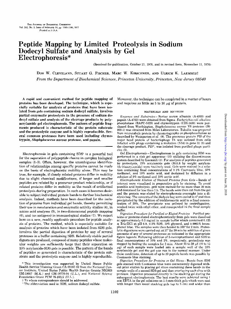
Reproducibility and Stability of Proteolysis Products- To test the reproducibility of the pattern of peptide bands pro- duced from a given protein by proteolysis in our SDScontain- ing buffer, Escherichia coli alkaline phosphatase was digested for various incubation times and at various enzyme concentra- tions with the Staphylococcus aureus V8 protease. (This pro- tease cleaves at the COOH-terminal side of aspartic and glu- tamic acid residues (11) and has proven to be eminently suita- ble for generating digests which contain many appropriately sized peptide fragments.) Fig. IA shows the digestion at fixed enzyme concentration for incubation times between 0 and 150 min. Alkaline phosphatase, whose apparent molecular weight on SDS gels is about 50,060, is labeled with the lettersAP. The Staphylococcus aureus protease generates a doublet, labeled E, which runs slightly faster than the alkaline phosphatase band. It can be seen that with increasing time of digestion there is a progressive disappearance (complete after 30 min) of the alkaline phosphatase band. Depending on the length of incubation, about 10 to 15 well resolved cleavage fragments can be observed. The apparent molecular weights of the frag- ments range from 6,006 to 30,660. Although some bands exist- ing at early times disappear upon further digestion and others are generated only at later times, the stability of many of the cleavage fragments is remarkable. Of the 10 fragments which can be seen following a 15-min incubation period, all but two can still be detected after 60 min, with two to three new fragments appearing during this time period.
The digestion of alkaline phosphatase using increasing Staphylococcus aureus protease concentrations, but at a fixed incubation time of 150 min, is shown in Fig. lB. The increase in the intensity of the protease doublet at higher enzyme concentrations is evident. It can also be seen that the same peptide fragments which were present in the time course experiment (Fig. LA) are again observable in this experiment. As expected, bands corresponding to larger peptides are lost at increasing protease levels, with most of the higher molecular weight fragments disappearing at enzyme concentrations above 20 yglml.
In a separate experiment (not shown), the peptide pattern produced by digestion of albumin with a constant concentra- tion of chymotrypsin was observed to be unaffected by 16-fold variation in albumin concentration between 0.16 mg/ml and
0 0.5 1.5 5 10 15 20 30 45 60 90 120 150 AP-
E--,/ . - J-
0 0.04O.l6 0.6 1.2 5 10 20 40 80 200
1
i
I
FIG. 1. Kinetic study of digestion of alkaline phosphatase. A., alkaline phosphatase (labeled AP) at a concentration of 0.33 mg/ml in sample buffer was digested at 37” with a final concentration of 25 pg/ml of S’tophylococc~s aureas protease (labeled E). The length of incubation was varied between 0 and 150 min as indicated in the figure. Twenty microliters were loaded per sample well. B, alkaline phosphatase at a concentration of 0.33 mg/ml in sample buffer was digested at 37” for 150 min with increasing concentration ofS. a~rea.s protease, as indicated in the figure in micrograms/ml. Twenty mi- croliters were loaded per sample well. Four micrograms of S. a~reus protease, incubated at 200 pg/ml in the absence of alkaline phospha- tase, were mn in the final slot on the right.
2.56 mglml. These results collectively demonstrate that repro- ducible cleavage fragments of suitable size are generated by proteolytic digestion of a substrate protein in our standard SDS-containing buffer.
Digestion of Bovine Serum Albumin, Tubulin, and Alka-
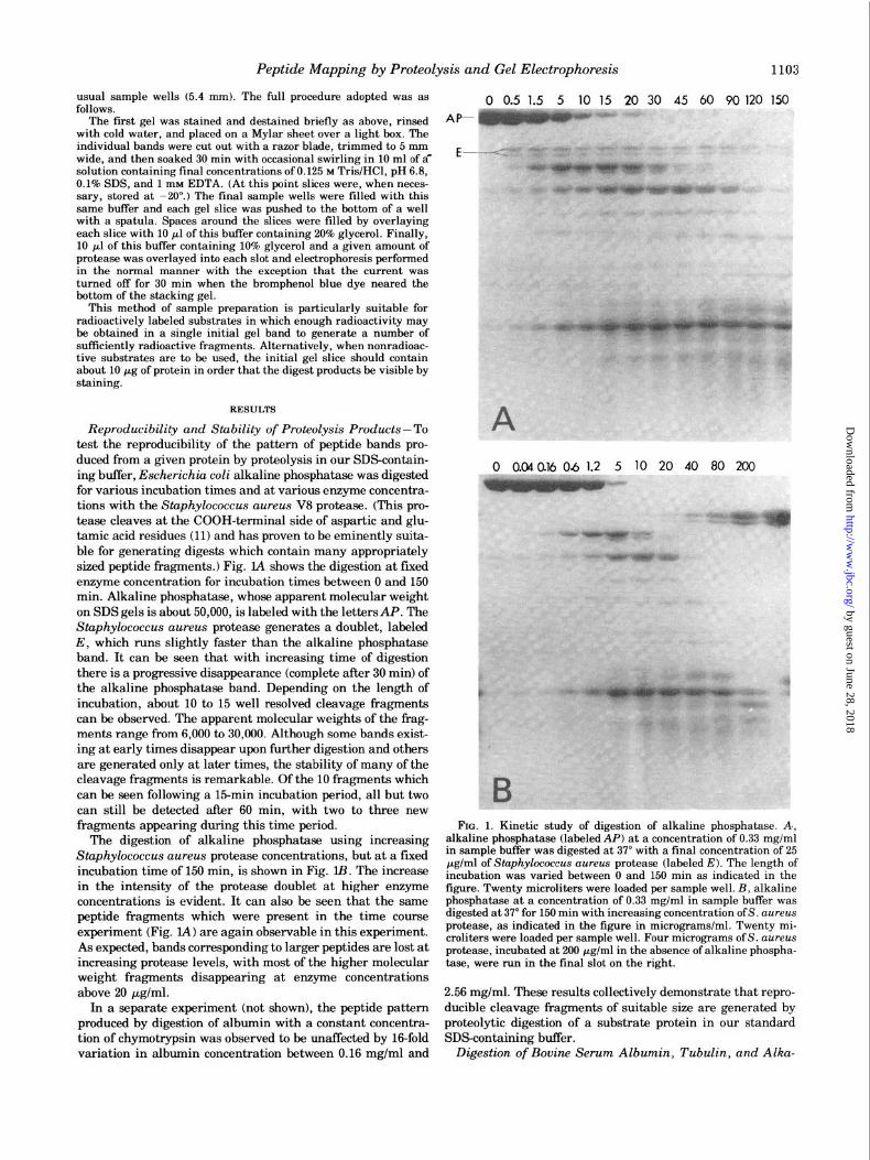
FIG. 2 (top). Peptide maps of albumin, tubulin, and alkaline tively, of papain. D toF, digestions of albumin, tubulin, and alkaline phosphatase. Albumin, tubulin, and alkaline phosphatase, each at a phosphatase with 133 pglml, 67 pg/ml, and 67 pg/ml of Stuphylococ- concentration of 0.67 mg/ml in sample buffer, were incubated at 37” cus aureus protease. G to I, digestions of albumin, tubulin, and for 30 min with concentrations of protease as indicated. Thirty alkaline phosphatase with 133 yglml, 67 yglml, and 67 pg/ml of microliters of sample were loaded per sample well of a 20% gel. A to chymotrypsin. J to L, undigested albumin, tubulin, and alkaline C, digestions of albumin, tubulin, and alkaline phosphatase with phosphatase. A total of 2.5 pg was loaded per sample well. M to 0, final concentrations of 33 pg/ml, 3.3 pg/ml, and 33 fig/ml, respec- highest total amounts of protease used in the digestions-l pg of
Peptide Mapping by Proteolysis and Gel Electrophoresis 1105
line Phosphatase with Different Enzymes- To assess the use- fulness of this technique in clearly distinguishing various proteins, the digestion patterns of three different substrate proteins produced with three different proteases were exam- ined. The conditions of the digestions were chosen in each case so that partial digests, which were insensitive to at least small changes in enzyme concentration or incubation time, were produced.
The peptide patterns of albumin, tubulin, and alkaline phosphatase which resulted from digestion with papain are shown in Fig. 2, A, B, and C, respectively. Although roughly 15 bands are observable for each substrate, the three patterns are strikingly distinct, with essentially no bands appearing in common between any pair. This specificity is further under- scored in Fig. 2, D, E, and F, where these same three sub- strates have been incubated with the Staphylococcus aureus protease and in Fig. 2, G, H, and I, in which the digestions have been effected with chymotrypsin. Not only are the band- ing patterns produced in these examples again unique to the protein substrate, but furthermore, by comparison of the pat- terns generated from a single substrate by the action of the three different proteases (e.g. the albumin patterns in Fig. 2, A, D, and G), it can be seen that the patterns produced from a particular substrate are unique for each of the proteases uti- lized as well.
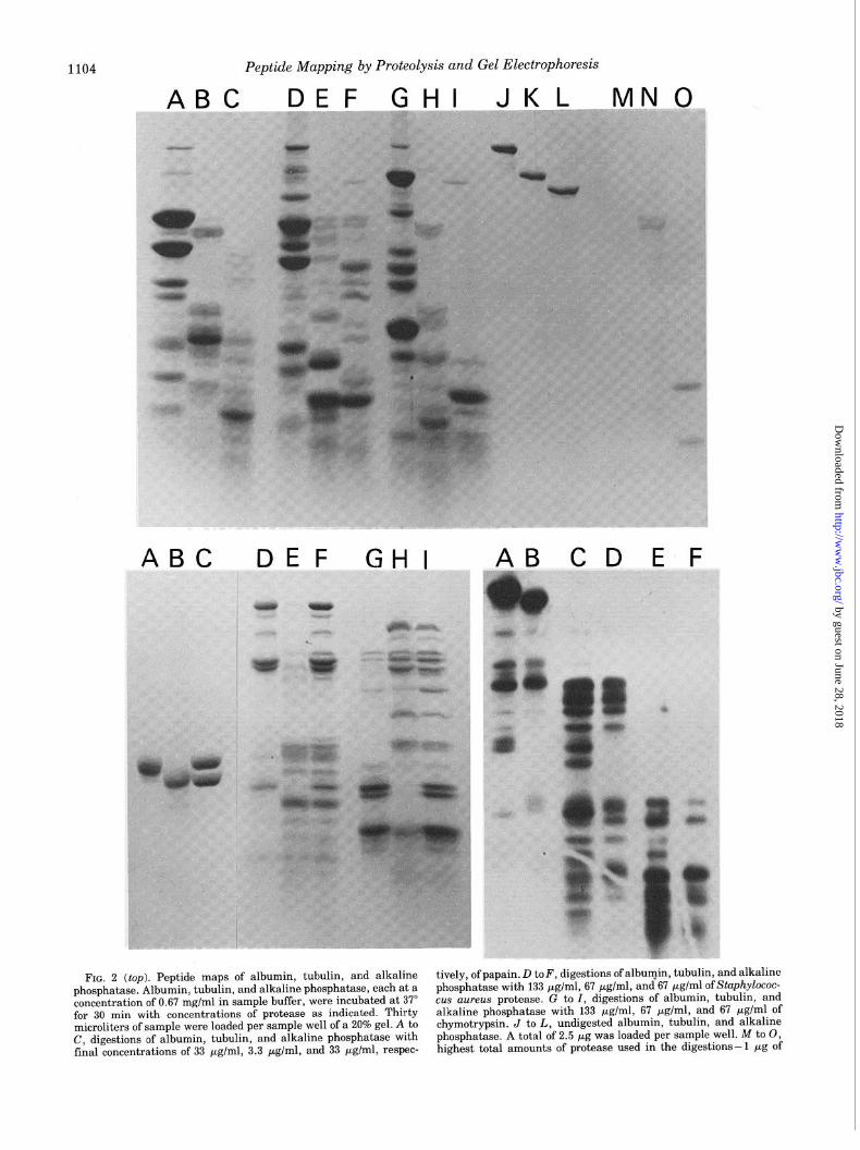
Comparison of Digestion Products of a- and pTubulin Eluted from SDS Gel -To illustrate the application of the present, technique of peptide mapping to proteins which have been obtained by electrophoretic elution from SDS gels, we chose to analyze the two polypeptide chains, LY and p, of tubulin. These two chains, which have similar amino acid compositions and related NH,-terminal sequences (12) and which exist together as a stable dimer under native conditions, may be separated under denaturing conditions on SDS gels into two distinct bands with respective apparent molecular weights of 55,000 and 53,000. In the present experiment, the a- and p-bands were cut from a preparative gel and eluted elec- trophoretically. Fig. 3, A and B show the re-electrophoresis of the separated a and p subunits, while Fig. 3C illustrates the equimolar presence of both bands in a gel slot loaded with a sample of the tubulin dimer. The banding patterns of the peptides generated by chymotryptic digestion of the isolated a- and /3-tubulins are given in Fig. 3, D and E. It is notable that the two patterns are completely distinct, with each pattern containing many peptides of mobility not present in the other. Fig. 3F shows the digestion products of the pure tubulin dimer produced with chymotrypsin. As expected, this pattern is seen
to be simply a composite of the peptide fragments observed in the digestions of the separated a- and /3-tubulins (Fig. 3, D and E). An identical experiment is shown in Fig. 3, G, H, and I, with the exception that digestions were performed with the Staphylococcus aureus protease.
Proteolysis during Re-electrophoresis- In order to avoid an elution step for those proteins whose study requires their isolation from SDS gels, we have developed a method in which a protein band of interest may be cut from a preparative gel and subsequently applied directly to a high per cent acrylam- ide gel in the presence of a proteolytic enzyme. This procedure generates banding patterns which are identical to those ob- tained from the same protein (prepared by elution from pre- parative gels or purification with classical biochemical tech- niques) after digestion in solution prior to loading onto the high per cent gel.
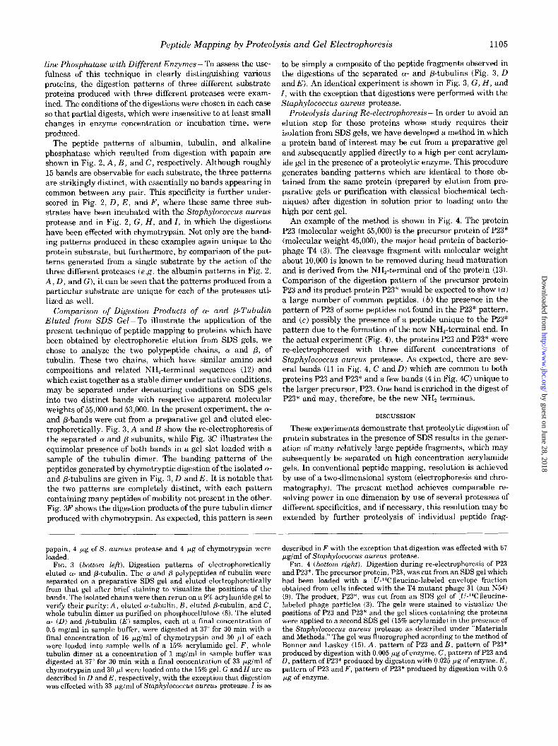
An example of the method is shown in Fig. 4. The protein P23 (molecular weight 55,000) is the precursor protein of P23* (molecular weight 45,000), the major head protein of bacterio- phage T4 (3). The cleavage fragment with molecular weight about 10,000 is known to be removed during head maturation and is derived from the NH,-terminal end of the protein (13). Comparison of the digestion pattern of the precursor protein P23 and its product protein P23* would be expected to show (a) a large number of common peptides, (b) the presence in the pattern of P23 of some peptides not found in the P23* pattern, and (c) possibly the presence of a peptide unique to the P23* pattern due to the formation of the new NH,-terminal end. In the actual experiment (Fig. 4), the proteins P23 and P23* were re-electrophoresed with three different concentrations of Staphylococcus aureus protease. As expected, there are sev- eral bands (11 in Fig. 4, C and D) which are common to both proteins P23 and P23* and a few bands (4 in Fig. 4C) unique to the larger precursor, P23. One band is enriched in the digest of P23* and may, therefore, be the new NH* terminus.
DISCUSSION
These experiments demonstrate that proteolytic digestion of protein substrates in the presence of SDS results in the gener- ation of many relatively large peptide fragments, which may subsequently be separated on high concentration acrylamide gels. In conventional peptide mapping, resolution is achieved by use of a two-dimensional system (electrophoresis and chro- matography). The present method achieves comparable re- solving power in one dimension by use of several proteases of different specificities, and if necessary, this resolution may be extended by further proteolysis of individual peptide frag-
papain, 4 pg of S. aureus protease and 4 pg of chymotrypsin were loaded.
FIG. 3 (bottom left). Digestion patterns of electrophoretically eluted a- and p-tubulin. The u and p polypeptides of tubulin were separated on a preparative SDS gel and eluted electrophoretically from that gel after brief staining to visualize the positions of the bands. The isolated chains were then rerun on a 9% acrylamide gel to verifv their nuritv: A, eluted a-tubulin, B, eluted B-tubulin, and C, whole tubulin di&er as purified on phosphocellulose (8). The eluted w (D) and &tubulin (E) samples, each at a final concentration of 0.5 mglml iA sample buffer, were digested at 37” for 30 min with a final concentration of 16 pg/ml of chymotrypsin and 30 ~1 of each were loaded into samnle wells of a 15% acrvlamide gel. F, whole tubulin dimer at a concentration of 1 mg/mi in sampie buffer was digested at 37” for 30 min with a final concentration of 33 pg/ml of chymotrypsin and 30 ~1 were loaded onto the 15% gel. G and H are as described in D and E, respectively, with the exception that digestion was effected with 33 pg/ml of Staphylococcus aureus protease. I is as
described inF with the exception that digestion was effected with 67 UK/ml of Staphvlococcus aurez~ protease.
-FIG. 4 (bo>to”m right). Digest& during re-electrophoresis of P23 and P23*. The precursor protein, P23, was cut from an SDS gel which ._._.. had been load-ed with a [U-‘4Clleucine-labeled envelope fraction obtained from cells infected with the T4 mutant phage 31 (am N54) (9). The product, P23*, was cut from an SDS gel of [U-14C]leucine- labeled phage particles (3). The gels were stained to visualize the positions of P23 and P23* and the gel slices containing the proteins were applied to a second SDS gel (15% acrylamide) in the presence of the Staphylococcus aureus protease as described under “Materials and Methods.” The gel was fluorographed according to the method of Bonner and Laskey (15). A, pattern of P23 and B, pattern of P23* produced by digestion with 0.005 pg of enzyme. C, pattern of P23 and D, pattern of P23* produced by digestion with 0.025 pg of enzyme. E, pattern of P23 and F, pattern of P23* produced by digestion with 0.5 pg of enzyme.
1106 Peptide Mapping by Proteolysis and Gel Electrophoresis
ments with additional enzymes. Indeed, the sensitivity of the method in identifying particular proteins has been clearly documented. Different protein substrates produce strikingly different patterns of digestion with a given protease, and similarly, unique patterns are produced from a single sub- strate by action of proteases of different specificities. In addi- tion, although observable differences in the peptide patterns of (Y- and p-tubulin were not unexpected, since the cyanogen bromide fragments of the two proteins are also distinct (12), the complete dissimilarity of the patterns generated does serve to illustrate the power of the technique in discriminating between two proteins which have similar amino acid composi- tions and which probably share a common evolutionary origin.
Reproducibility of the pattern of peptides is ensured by the marked insensitivity of the digestions to large variations in protease and substrate concentrations and in the length of incubation with the protease. In addition, the digests are unaffected by the manner of preparation and are reproducible as demonstrated in Fig. 3. The a- and p-tubulin bands were purified by SDS-gel electrophoresis, fixed and stained with Coomassie blue in acetic acid and methanol, electrophoreti- tally eluted from the gel, precipitated with trichloroacetic acid, and finally resuspended in SDS-containing sample buffer. Unfractionated tubulin was merely diluted into the sample buffer. Yet, the peptide fragments of the unfraction- ated tubulin are simply the sum of the fragments obtained with the (Y and p peptides. No extraneous bands were pro- duced. Numerous analyses of (Y- and p-tubulin, serum albu- min, and alkaline phosphatase have yielded reproducible pep- tide fragments. In addition to the protein digests reported here, the method has been used to show the similarity of actin from muscle and actin from cultured Chinese hamster ovary cells,” to identify histone Fl from physarum,” for identifica- tion of two adenovirus-specific DNA-binding proteins, a viral structural protein, and the adenovirus tumor antigen,4 to demonstrate the similarity in various peptides fromr protein involved in microtubule assembly,” and other applications as well. These studies demonstrate the utility of the method, though there may of course be applications for which it is not suitable. However, the large flexibility in the choice of proteo- lytic enzyme should widen the applicability of the method. Indeed, for some preparations, we have used successfully the following enzymes: chymotrypsin, StuphyZococcus aureus pro-
e B. Spiegelman, unpublished results. J S. Fischer, unpublished results. * A. Levinson and A. Levine, manuscript in preparation. s D. Cleveland, S. Hwo, and M. Kirschner, manuscript in prepara-
tion.
tease, papain, subtilisin, Streptomyces griseus protease, ficin, and elastase.
Samples to be analyzed may be prepared by classical bio- chemical purification techniques or by electrophoretic elution of single bands from acrylamide gels. More conveniently, a protein band in a gel slice may be analyzed without prior elution by direct application of the gel slice onto a resolving gel which is then overlayed with a proteolytic enzyme. The pat- tern of peptide fragments generated is identical for all three methods of sample preparation. As little as 5 to 10 pg of protein are required to generate a pattern which is observable by Coomassie blue staining. Of course, the use of radioactive protein substrates and analysis of the digestion patterns by autoradiography (14) or fluorography (15) reduces the amount of substrate needed.
The technique is quite versatile and simple to use. Several patterns can be obtained simultaneously in a matter of hours and can be displayed in the same slab gel, thereby allowing easy, unambiguous comparison. No special equipment beyond the standard slab gel electrophoresis apparatus is required. Furthermore, the proteases which have been found to produce suitable digests are all relatively inexpensive and commer- cially available.
1.
2. 3. 4. 5. 6.
7.
a.
9.
10. 11.
12.
13.
14.
15.
REFERENCES
Shapiro, A. L., Vinuela, E., and Ma&l, J. V. (1967) Biochem. Biophys. Res. Commun. 28, 815-820
Weber. K.. and Osborn. M. (1969)J. Biol. Chem. 244. 4406-4412 Laemmli, ‘U. K. (1970) ‘Nature 227, 680-685 Weber, K.. andKuter, D. J. (1971)J. Biol. Chem. 246,4504-4509 Kyte, j. (i971) J. B&i. Chem. 246, 4157-4165 Weber, K., and Osborn, M. (1975) in The Proteins (Neurath, H.,
and Hill, R., eds) 3rd Ed, Vol. 1, pp. 179-223, Academic Press, New York
Lazarides, E., and Weber, K. (1974) Proc. Natl. Acad. Sci. U. S. A. 71, 2268-2272
Weingarten, M. D., Lockwood, A. H., Hwo, S.-Y., and Kir- schner, M. W. (1975) Proc. Natl. Acad. Sci. U. S. A. 72, 1858- 1862
Laemmli. U. K.. Molbert. E.. Showe. M.. and Kellenbereer. E. (1970) j. Mol. ‘Biol. 49, b9-i13 ’
Studier. F. W. (1973) J. Mol. Biol. 79. 237-248 Homnard, J., and Drapeau, G. R. (1972) Proc. Natl. Acad. Sci.
17. S. A. 69, 3506-3509 Luduena, R. F., and Woodward, D. 0. (1973) Proc. Natl. Acad.
Sci. U. S. A. 70, 3594-3598 Celis. J. E.. Smith. J. D.. and Brenner. S. (1973) Nature New
Bioi. 241,‘130-132 ’ Fairbanks, G., Jr., Levinthal, C., and Reeder, R. H. (1965)
Biochem. Biophys. Res. Commun. 20, 393-399 Bonner, W. M., and Laskey, R. A. (1974) Eur. J. Biochem. 46,