NEW YORK STATE DEPARTMENT OF HEALTH DIVISION OF QUALITY AND PATIENT SAFETY CARDIAC SERVICES PROGRAM Percutaneous Coronary Interventions Report Form DOH-3331 Clarification Document August 2012 Instructions and Data Element Definitions 2012 Discharges Note: This document contains several clarifications pertaining to the 2012 PCIRS data reporting system. New text is presented as underlined . Any deleted text is noted as strike - through . CARDIAC SERVICES PROGRAM CONTACTS: One University Place, Suite 218 Rensselaer, NY 12144-3455 Phone: (518) 402-1016 Fax: (518) 402-6992 Kimberly S. Cozzens MA, Cardiac Initiatives Research Manager, [email protected]Cynthia L. Johnson, PCI and Special Projects Coordinator, [email protected]Erika Ihara, MA, Clinical Data Coordinator, [email protected]
Transcript
NEW YORK STATE DEPARTMENT OF HEALTH DIVISION OF QUALITY AND PATIENT SAFETY
CARDIAC SERVICES PROGRAM
Percutaneous Coronary Interventions Report
Form DOH-3331
Clarification Document August 2012
Instructions and Data Element Definitions 2012 Discharges
Note: This document contains several clarifications pertaining to the 2012 PCIRS data reporting system. New text is presented as underlined. Any deleted text is noted as strike-through.
CARDIAC SERVICES PROGRAM CONTACTS: One University Place, Suite 218 Rensselaer, NY 12144-3455 Phone: (518) 402-1016 Fax: (518) 402-6992 Kimberly S. Cozzens MA, Cardiac Initiatives Research Manager, [email protected] Cynthia L. Johnson, PCI and Special Projects Coordinator, [email protected] Erika Ihara, MA, Clinical Data Coordinator, [email protected]
Table of Contents Topic Page Revision Highlights and Coding Clarification …………………………. 6 PCIRS Data Reporting Policies ………………………………………... 8 ITEM-BY-ITEM INSTRUCTIONS PFI Number ………………………………………………………………. 11 Sequence Number ………………………………………………………. 11 I. Patient Information Patient Name …………………………………………………………….. 11 Medical Record Number ………………………………………………… 11 Social Security Number …………………………………………………. 11 Date of Birth ……………………………………………………………… 11 Sex ………………………………………………………………………… 12 Ethnicity …………………………………………………………………… 12 Race ………………………………………………………………………. 12 Residence Code …………………………………………………………. 13 Hospital Admission Date ………………………………………………... 13 Primary Payer ………………………………………………................... 13 Medicaid ………………………………………………............................ 14 PFI of Transferring Hospital …………………………………………….. 14 II. Procedural Information Hospital That Performed Diagnostic Cath ……………………………… 15 Primary Physician Performing PCI …………………………….. ……… 15 Date of PCI ………………………….……………………………………. 15 Time of First Interventional Device ……………………………………. 15 Diagnostic Cath During Same Lab Visit ………………………………. 16 Previous PCI This Admission …………………………………………… 16 PCI Prior to This Admission …………………………………………… 16 Follow-up PCI - Staged Procedure ………….…………………..……. 16 Contrast Volume ………………………...………………………………. 17 Access Site ………………………………………………………………. 17 Thrombolytics …………………………………………..………………… 17 III. Vessels Diseased and Lesion-Specific Information Vessels Diseased …………………………….…………………………. 18 Previous LIMA Use ……………………………………………………… 18
Form DOH-3331(01/12) --- 2012 Discharge Year 2
Table of Contents (continued) Topic Page III. Vessels Diseased and Lesion-Specific Information (continued) Lesion-Specific Information …………………………………………….. 19
Location…………………………….…………………………………. 19 Bypassed (A or V) …………………………………………………… 19 Bypass Stenosis …………………………….………………………. 19 % Pre-Op Stenosis …………………………….……………………. 20 IVUS …………………………………………………………………… 20 FFR ……………………………………………………………………. 20 Previous PCI …………………………….…………………………… 20 Device 1 and Device 2 ….…………………………………..………. 20 Stent 1 and Stent 2 ..………….…………………………………….. 21 Lesion Description………………….……………………………….. 21 % Post-Op Stenosis…………………………….……………………. 22 IV. Cardiac Presentation Cardiac Presentation ………….…………………………………… 23 Angina Classification Within 2 Weeks …………………………………. 25 Onset of Ischemic Symptoms ..…………………….…………………… 26 Arrival at Transferring Hospital ……….………………………………... 26 Arrival at PCI Hospital ………………………….……………………….. 26 Onset Time, Estimated …………………….…………………………. 27 New ST ↓ or T ↓ …… ..…………………….……………………..……… 27 TIMI < II …………………………….…………………………………….. 27 Ongoing Ischemia at Time of Procedure ……………………………… 27 Killip Class 2 or 3 ………………………………………………………… 27 V. Pre-Intervention Risk Factors PCI Status……………………….………………………………………. 28 Height …………………………….……………………………………….. 29 Weight …………………………….………………………………………. 29 Stress Test / Imaging Study Done …………………………………….. 29 Stress Test / Imaging Study Type …………………………………….. 29 Stress Test / Imaging Study Results ………………………………….. 30 Anti-Anginal Medication Within 2 Weeks ……………………………… 30 Ejection Fraction and Measure ………………………………………… 31 Creatinine …………………………….…………………………………… 31 Pre-Intervention Risk Factors (None) …………………………………. 32 Previous PCIs …………………………….……………………………… 32 Previous MI (most recent) ……………………………………………… 32
Form DOH-3331(01/12) --- 2012 Discharge Year 3
Table of Contents (continued) Topic Page V. Pre-Intervention Risk Factors (continued) Cerebrovascular Disease ………………………………………………. 33 TIA, Only Cerebrovascular Disease ….………………………………. 33 Peripheral Vascular Disease …………………………………………… 34 Neurologic State Anoxic Brain Injury Criteria ………………………………………… 35 Hemodynamic Instability at the Time of the Procedure Unstable …………………………….………………………………… 36 Shock …………………………….…………………………………… 37 Congestive Heart Failure, Current …………………………………….. 38 Congestive Heart Failure, Past ………………………………………… 38 BNP, Three Times Normal ………………………………………..…… 39 Malignant Ventricular Arrhythmia ……………………………………… 39 Chronic Lung Disease ………………………………………………..… 40 Diabetes ……….……………… ………………………………………… 41 Diabetes Therapy …….……….………………………………………… 41 Renal Failure, Dialysis ………………………………………………….. 42 Previous CABG Surgery ………………………………………………… 42 Emergency PCI Due to DX Cath Complication ………………………. 42 Stent Thrombosis …………………………….…………………………. 43 Any Previous Organ Transplant ……………………………………….. 43 Contraindication to Antiplatelet Therapy……………….……………… 43 VI. Major Events Following PCI None …………………………….………………………………………… 44 Stroke ………………………………………………….…………………. 44 Q-Wave MI …………………… …………………………………………. 44 Acute Occlusion in the Targeted Lesion ………………… …………… 44 Acute Occlusion in a Significant Side Branch ………………………… 45 A/V Injury at Cath Entry Site, Requiring Intervention ………………. 45 Renal Failure …………………………….………………………………. 45 Emergency Cardiac Surgery …………………………………………… 46 Stent Thrombosis …………………………….………………………….. 46 Emergency Return to the Cath Lab for PCI ………………………….. 46 Coronary Perforation ……………………………………………………. 46
Form DOH-3331(01/12) --- 2012 Discharge Year 4
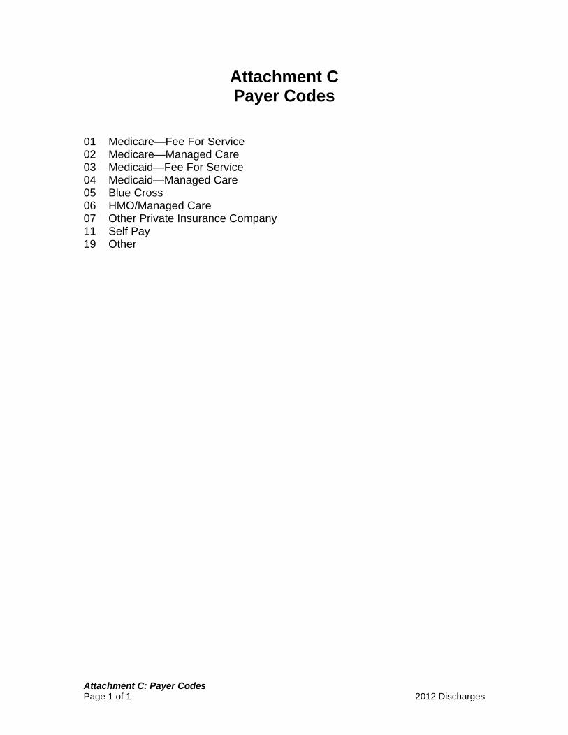
Table of Contents (continued) Topic Page VII. Discharge Information Additional Procedure Planned - Staged Procedure …..…………….. 47 Discharge Status Discharged Alive To. ………………………………………………… 47 Died In …………………………….…………………………………… 48 Hospital Discharge Date ………………………………………………… 48 30-Day Status …………………………………………………………… 48 Attachments A: PFI Numbers for Cardiac Diagnostic and Surgical Centers B: Residence Codes C: Payer Codes D: Codes for Location of Lesion E: Device and Stent List F: Stress Test Results Definitions and Clarification G: Guidelines for Requesting PCIRS Anoxic Encephalopathy Mortality Exclusion
Form DOH-3331(01/12) --- 2012 Discharge Year 5
Revision Highlights and Coding Clarification
Deleted Data Elements The following data elements will no longer be collected effective January 2012.
• Additional Procedure Using Contrast • Thrombolytics contraindicated • Acute MI Information: EKG Changes New ST Elevation • Acute MI Information: EKG Changes: New LBBB • Priority • Angina Type • Major Event - Stroke over 24 hours.
New Data Elements The following data elements have been added to the PCIRS data collection effective January 2012.
• Cardiac Presentation • PCI Status • TIA, only cerebrovascular risk • Diabetes Therapy
Revised Data Elements The following revisions are effective January 2012. Complete data element definitions are located in the main body of this document.
• PCI prior to this admission at this hospital is now collected as “PCI
prior to this admission” and should be reported if the prior PCI occurred at another hospital or this hospital.
• Date of PCI prior to this admission at this hospital is now collected as
“Date of PCI prior to this admission” and should be reported if the prior PCI occurred at another hospital or this hospital. Use 01 if the day and/or month of the previous PCI are unknown.
• Total Contrast Volume in 72 hours is now collected as “Contrast
Volume” in this lab visit. • Creatinine has been modified to indicate the reported creatinine level
closest to, but before, the start of the PCI.
• CCS Class has been modified to indicate that reporting should be based upon the highest grade of angina or chest pain within the past 2 weeks.
Form DOH-3331(01/12) --- 2012 Discharge Year 6
Revision Highlights and Coding Clarification (continued)
Revised Data Elements (continued)
• Renal Failure, Dialysis has a revised definition.
• Major Event Stroke 24 hrs or less is now called “Major Event - Stroke” and has a revised definition.
• Cerebrovascular Disease has a revised definition.
• COPD is now called “Chronic Lung Disease” and has a revised definition
and expanded response categories.
• Diabetes requiring Medication is now called “Diabetes” and has a revised definition.
• Contraindication to Aspirin and Plavix is now called “Contraindication
to Antiplatelet Therapy” and a clarification has been added indicating this should be reported when the patient has a contraindication to all antiplatelet therapy.
Form DOH-3331(01/12) --- 2012 Discharge Year 7
PCIRS Data Reporting Policies End of PCI, Generation of a New Form For purposes of determining a return to the cath lab, we use the term cath lab in the narrowest sense – that is, the PCI is considered finished when the patient leaves the actual room in which the procedure was performed. If a patient leaves the actual procedure room, but remains in a holding room, staging area or even an adjacent hallway and returns to a procedure room for another PCI, a new form should be generated. Hospice Policy Beginning with patients discharged on or after January 1, 2003, any patient that is discharged from the hospital after cardiac surgery or PCI to hospice care (inpatient or home with hospice care) and is still alive 30 days after the discharge from the hospital will be analyzed as a live discharge. All patients discharged to a hospice or home with hospice care should continue to be reported with Discharge Status – 12: Hospice. If a patient is still alive 30 days after discharge, whether in hospice or not, appropriate supporting documentation should be sent to Cardiac Services Program. Examples of appropriate documentation include: a dated progress note from the hospice service, evidence of a follow-up doctor’s visit 30 days after discharge, evidence of subsequent hospital admission 30 days after initial discharge. It will be the responsibility of the hospital (physician) to send documentation to the Department of Health’s Cardiac Services Program to support this change. Upon receipt, review, and verification of the documentation, Cardiac Services Program staff will change the discharge status from dead to alive for purposes of analysis. All documentation must be received before the final volume and mortality for a given year of data is confirmed by the hospital. Physician Assignment When multiple records exist for the same patient during a hospital admission, and two or more physicians were reported for those procedures, the case will be assigned for analysis to the physician performing the first PCI. However, the hospital may submit a letter from the CEO or Medical Director requesting that the case be assigned to the physician performing a later PCI. Cardiogenic Shock Cases Beginning with cases discharged January 1, 2006 and continuing for a period of at least two years, cases in pre-procedural Cardiogenic Shock will not be included in the publicly released reports and analyses. This applies only to cases that meet the NYS Cardiac Services Program definition of Cardiogenic Shock (risk factor #13).
Form DOH-3331(01/12) --- 2012 Discharge Year 8
PCIRS Data Reporting Policies (continued)
Cardiogenic Shock Cases (continued) Data for these cases must still be submitted electronically and will be subject to data verification activities. To ensure that the appropriate cases are identified as “Shock” cases, we will continue to require submission of medical record documentation of any case reported with this risk factor. If appropriate documentation is not provided by your center, the risk factor will be removed from the data and the case will be included in analysis. In addition, we anticipate that there will be increased requirements for medical record documentation for cases coded as “Hemodynamically Unstable” as well. It is strongly suggested that all appropriate staff closely review the definitions and documentation requirements for these two risk factors. Note: The above policy regarding cases in Shock will be continued for at least another year (2012 discharges). Anoxic Brain Injury Exclusion Beginning January 1, 2010 and continuing for a period of at least 3 years, patients with documented pre-procedural acute MI, cardiac arrest and anoxic/hypoxic brain injury who expire under certain conditions subsequent to PCI will be excluded from Department of Health analysis and public reporting. This policy is the result of ongoing discussions with NYS providers, careful deliberations among the New York State Cardiac Advisory Committee (CAC) members, and feedback provided through the 2007 and 2008 annual cause of death surveys. All PCI patients will continue to be reported to the PCIRS database. After quarterly reporting, the hospital will be provided the opportunity to indicate, through a written letter and medical record documentation, if any of the mortalities meet the criteria for death from anoxic brain injury. Please See Attachment G: Guidelines for Requesting PCIRS Anoxic Encephalopathy Mortality Exclusion
Form DOH-3331(01/12) --- 2012 Discharge Year 9
PCIRS Data Reporting Policies (continued)
Reporting Schedule PCIRS data is reported quarterly by discharge date. It is due to the Cardiac Services Program one month after the end of the quarter. The 2012 reporting schedule is as follows: Quarter 1 (1/1/12 – 3/31/12 Discharges) due on or before April 30, 2012 Quarter 2 (4/1/12 – 6/30/12 Discharges) due on or before July 31, 2012 Quarter 3 (7/1/12 – 9/30/12 Discharges) due on or before October 31, 2012 Quarter 4 (10/1/12 – 12/31/12 Discharges) due on or before January 31, 2013 Limited extensions to the above deadlines will be granted on a case by case basis when warranted by extenuating circumstances. They must be requested in writing prior to the required submission date.
Form DOH-3331(01/12) --- 2012 Discharge Year 10
Item-By-Item Instructions
PFI Number Variable Name: PFI The PFI Number is a Permanent Facility Identifier assigned by the Department of Health. Enter your facility's PFI Number as shown in Attachment A. Sequence Number Variable Name: SEQUENCE If your facility assigns a sequence number to each case on a chronological flow sheet or similar log, enter the sequence number here. The sequence number is not required for the Percutaneous Coronary Interventions Reporting System, but has been included on the form to assist facilities in identifying and tracking cases.
I. Patient Information Patient Name Variable Names: LASTNAME, FIRSTNAME Enter the patient’s last name followed by his/her first name. Medical Record Number Variable Name: MEDRECNO Enter the patient’s medical record number. Social Security Number Variable Name: SSNO Enter the patient's social security number as shown in the medical record. If the medical record does not contain the patient's social security number, leave this item blank. Date of Birth Variable Name: DOB Enter the patient's exact date of birth.
Form DOH-3331(01/12) --- 2012 Discharge Year 11
I. Patient Information (continued) Sex Variable Name: SEX Check the appropriate box for the patient’s sex at birth. Note: In the absence of any other information, it is reasonable to assume that the sex at birth is the same as at the time of admission. Ethnicity Variable Name: ETHNIC Check the appropriate box. Note: The term “Hispanic” refers to persons who trace their origin or descent to Mexico, Puerto Rico, Cuba, Central and South America or other Spanish cultures. Race Variable Names: RACE, RACESPEC Select the appropriate code below: 1. White. A person having origins in any of the original peoples of Europe, the Middle East, or North Africa. 2. Black or African American. A person having origins in any of the black racial groups of Africa. Terms such as "Haitian" or "Negro" can be used in addition to "Black or African American." 3. Native American / American Indian or Alaska Native. A person having origins in any of the original peoples of North and South America (including Central America), and who maintains tribal affiliation or community attachment. 4. Asian. A person having origins in any of the original peoples of the Far East, Southeast Asia, or the Indian subcontinent including, for example, Cambodia, China, India, Japan, Korea, Malaysia, Pakistan, the Philippine Islands, Thailand, and Vietnam. 5. Native Hawaiian or Other Pacific Islander. A person having origins in any of the original peoples of Hawaii, Guam, Samoa, or other Pacific Islands. 8. Other. Report for those responses that are not covered by an above category. Please provide the specific race for any case marked “Other.”
Form DOH-3331(01/12) --- 2012 Discharge Year 12
I. Patient Information (continued) Race (continued) Note: Please note that race should be based on the patient’s racial/ethnic origins, which is not necessarily the same as their country or place of origin. Indicate “multi-racial” by checking “8-Other” and providing details in the “specify” field. For White Hispanics, check "White"; for Black Hispanics, check "Black." Residence Code Variable Names: RESIDENC, STATE Enter the county code of the patient's principal residence, as shown in Attachment B. If the patient lives outside New York State, use code 99 and print the name of the state or country where the patient resides in the space provided. If you enter a valid NYS County Code then the “State or Country” field should be left blank. If the patient is from a foreign country, but is staying in the US during the pre-intervention and post-intervention time period, you must enter 99 and print the name of the country that the patient is from. Do not enter the residence code of where the patient is staying in the US. Hospital Admission Date Variable Name: ADMIDATE Enter the date that the patient was admitted to your hospital. Note: If the admission date is after the PCI date, then you must also report the date for “Arrival at PCI Hospital,” even if the patient did not have an MI. Primary Payer Variable Name: PRIMEPAY Enter the primary source of payment for this hospital stay as shown in Attachment C. Please note that Workers Compensation, Family Health Plus, and Other Federal Programs are reported as code “19 - Other.”
Form DOH-3331(01/12) --- 2012 Discharge Year 13
I. Patient Information (continued) Primary Payer (continued) Interpretation: For “Medicaid Pending” code Primary Payer as “11 - Self-Pay” and check the box for Medicaid. For patients in prison, code Primary Payer as “19 - Other”. Please note the difference between “07 - Other Private Insurance Company” and “19 - Other”. Code 07 refers to a Private Insurance Company (also referred to as “Commercial” insurance) that is not listed elsewhere. Code 19 is any other type of insurance that is not given a code of its own (e.g. Corrections). If the patient has Blue Cross and Medicare, code Medicare if there is no indication of which is primary. Report a PPO (Preferred Provider Organization) as Code 06 – HMO/Managed Care. If a patient has Medicare or Medicaid, but you do not know if it is Fee for Service or Managed Care, report Fee for Service. Medicaid Variable Name: MEDICAID Check this box if the patient has Medicaid that will provide payment for any portion of this hospital admission. If the patient’s primary payer is Medicaid, check this box in addition to entering “03” or “04” under Primary Payer. PFI of Transferring Hospital Variable Name: TRANS_PFI If the patient was transferred from another Acute Care Facility, enter the PFI of the transferring hospital. This element only needs to be completed for transfer patients. A listing of PFI for cardiac diagnostic centers in NYS is provided in Attachment A. If transferred from a Veterans Administration hospital in NYS, enter "8888"; if transferred from outside NYS, enter "9999". For patients transferred from another hospital in NYS, please see http://hospitals.nyhealth.gov for a complete listing of NYS hospitals, including PFI. Please note: PFI on the above website is listed without leading 0s. For purposes of cardiac reporting, PFI should always be four (4) numeric characters. For example, PFI “1” should be reported as “0001”.
Form DOH-3331(01/12) --- 2012 Discharge Year 14
II. Procedural Information Hospital That Performed Diagnostic Cath Variable Name: CATH_PFI If the angioplasty was preceded by a diagnostic catheterization, enter the name and PFI number of the hospital in the space provided. If the catheterization was at a cardiac diagnostic center in NYS, enter its PFI Number from Attachment A; if done at a Veterans Administration hospital in NYS, enter "8888"; if done outside NYS, enter "9999". If there was no diagnostic catheterization, leave this item blank. Note: If the patient does not have a diagnostic catheterization but is diagnosed via CT scan, do not report the Hospital that performed the CT scan here. Primary Physician Performing PCI Variable Name: PHYSNUM Enter the name and license number of the primary physician who performed the PCI. Note: Physician name is included on the paper version of the data collection form for abstractor convenience. Physician name is not part of the required PCIRS data structure. Date of PCI Variable Name: PCI_DATE Enter the date on which the PCI was performed. Time of First Interventional Device Variable Names: PCI_HR, PCI_MIN Report the earliest time of any of the following: Balloon inflation, stent deployment, treatment of lesion (e.g. AngioJet or other thrombectomy/aspiration device, laser, rotational atherectomy). Time should be reported using military time (e.g. 1:00 am is 01:00, and 1:00 pm is 13:00). Interpretation: In the case of an attempted PCI when no interventional device can be deployed, report the time that the guidewire leaves the catheter.
Form DOH-3331(01/12) --- 2012 Discharge Year 15
II. Procedural Information (continued) Diagnostic Cath During Same Lab Visit Variable Name: CATHSAME If a full diagnostic catheterization was performed during the same cath lab visit as the PCI, then check “Yes”. Otherwise check “No”. Interpretation: This does NOT include the case where there was a “quick look” done on the vessel to have the intervention. The diagnostic cath does not have to be every vessel, but should be a complete diagnostic of the area of interest. Previous PCI This Admission Variable Name: PCI_SAME, SAMEDATE For patients who have had a previous PCI during this admission, check “Yes”. Otherwise check “No”. Interpretation: If ”Yes,” it is very important to enter the date of this procedure. It is this date that aids in combining multiple procedures from the same hospital admission in the proper order. This becomes especially important when determining Emergency/Non-Emergency status, since certain risk factors are only “credited” if they occur prior to the first procedure in a hospital admission. PCI Prior to This Admission Variable Name: PCIPRIOR, PRIODATE For patients who have had a PCI prior to this admission, check “Yes” and report the date of this previous procedure. If only the month and year are known, use 01 for the day and write in the correct month and year. If only the year is known, write in 01 for both the month and the day then the correct year. If the year of the procedure is also unknown, enter the date as 01/01/1900. Follow-up PCI - Staged Procedure Variable Name: PART2 Use the following codes to indicate if the current procedure is in follow-up to a previous PCI or CABG as part of a staged treatment strategy.
0. No, not a staged follow-up to a previous procedure. 1. Yes, staged follow-up to a previous PCI 2. Yes, staged follow-up to a previous CABG
The follow-up PCI in a staged procedure would be a non-emergency PCI occurring after completion, but within 60 days, of an initial PCI or CABG with the
Form DOH-3331(01/12) --- 2012 Discharge Year 16
II. Procedural Information (continued) Follow-up PCI - Staged Procedure (continued) intervention at a different lesion location than the previous procedure. Typically the intervention is on a different vessel than was treated in the first procedure. Interpretation: Staging for these purposes DOES include a planned treatment strategy of PCI and CABG. The following scenario would NOT be considered a staged procedure: The first PCI was unsuccessful and the patient returns to the lab at a later point for another attempt. Contrast Volume Variable Name: CONTRAST Report the total contrast used (ml) for this lab visit. Access Site Variable Names: ACCESS_ARM, ACCESS_LEG Indicate if the access site was in the arm (radial or brachial) or the leg (femoral artery). Interpretation: Report the site through which access to the ascending aorta was successfully achieved. If access through one site was attempted but failed, do not report. If access was achieved through both sites, check both. Thrombolytics Variable Names: THROMLT3, THROM3_6, THROMGT6, Check the appropriate box to indicate if, and at what time interval, thrombolytics were administered.
Form DOH-3331(01/12) --- 2012 Discharge Year 17
III. Vessels Diseased and Lesion Specific Information Vessels Diseased Variable Names: LMT, PROX_LAD, MID_LAD, RCA, LCX For each diseased vessel, check the appropriate box to indicate the percent diameter stenosis. Include all vessels diseased, even branches. Interpretation: If the diseased segment of the native vessel is bypassed by an open artery or vein graft, do not code as diseased. This vessel is revascularized. Use the ranges listed below when the medical record describes the percent stenosis in the following ways: MILD = plaques to < 50% MODERATE = 50-69% SEVERE = > 70% If a vessel or branch is described as having “mild” stenosis then the vessel would not be coded as diseased, since we only code 50-100% stenosis. If the medical record reports the range “40-50% stenosis”, then do not code as diseased. If the medical record reports the range “60-70% stenosis”, then code 50-69%. The Ramus Intermediate can be coded as either the marginal or the diagonal depending on the origin of the vessel. Always take the highest stenosis reported for a vessel. If the medical record reports the proximal RCA with a 70% lesion and the distal RCA with a 50% you should code the RCA as 70-100%, since the proximal RCA has a 70% lesion. If the medical record only has documentation that states the LAD was stenosed, then code the mid LAD and not the proximal LAD. Disease of a major diagonal should be reported with mid/distal LAD, not with the proximal LAD. Previous LIMA Use Variable Name: LIMA_USE Choose one: 1 –LIMA used as a graft and remains patent to native coronary artery 2 –LIMA used as a graft but is no longer functional 3 –Never used – includes no previous CABG 4 –Unknown – the existence or condition of the LIMA graft is unknown
Form DOH-3331(01/12) --- 2012 Discharge Year 18
III. Vessels Diseased and Lesion Specific Information (continued)
Previous LIMA Use (continued) Interpretation: The graft would be considered “no longer functional” if there is angiographic stenosis of 70% or more or there is evidence of significant flow restriction documented by FFR or by stress test (with echo or nuclear to localize the ischemia). Lesion-Specific Information Variable Names: LES_LOC1 – LES_LOC7, BYPASS_1 – BYPASS_7, BPSTEN1 – BPSTEN7, PRESTEN1 – PRESTEN7, IVUS1 – IVUS7, FFR1 – FFR7, PREVPCI1- PREVPCI7, DEVICE_1- DEVICE_7, DEVSPEC1-DEVSPEC7, SECOND_1- SECOND_7, STENT1- STENT7, STENTB_1-STENTB_7, STNTSPC1-STNTSPC7, LESDESA1- LESDESA7, LESDESB1- LESDESB7, LESDESC1- LESDESC7 POSTSTEN1- POSTSTEN7. Complete one line for every lesion for which PCI was attempted (even if pre-stenosis is < 50%), and one line for each non-attempted lesion with diameter stenosis of 50% or more. If there are more than seven lesions, report the seven most significant. Stenting of a coronary artery aneurysm is reportable in PCIRS.
Enter the code indicating the location of the lesion, as shown in Location Attachment D. For lesions in a "sequential" graft going to two of the major coronary systems, complete a separate line for each coronary artery jeopardized (LAD, LCX, RCA). Interpretation: In the event of a long lesion that spans across two locations as defined in Attachment D, report this lesion as the more proximal location. For the ramus use '15' for an LAD derived ramus and '20' for an LCX derived ramus.
Bypassed (A or V)
If the lesion has been bypassed by a vein graft, enter "V." If the lesion has been bypassed by an artery graft, enter "A." If the lesion was not bypassed leave blank.
Bypass Stenosis
If the lesion has a vein or artery graft, use the following code to report the level of stenosis found in the graft: 1. > 70% 2. < 70% 3. Unknown
Form DOH-3331(01/12) --- 2012 Discharge Year 19
III. Vessels Diseased and Lesion Specific Information
Lesion-Specific Information (continued)
Enter the pre-PCI percent diameter reduction. Measurement with calipers is recommended. Note: Findings by IVUS are not acceptable. This should be the angiographic finding.
% Pre-Op Stenosis
IVUS For lesions with pre-PCI stenosis of 40-70% (determined by angiography), indicate
if prior to intervention there is a significant reduction in cross-sectional area as documented by IVUS. Significant reduction is defined as 6mm2 for the left main and 4mm2 for major epicardial vessels other than the left main. Report 1 for significant IVUS findings, 0 or Blank for not done or not significant. Significant results by optical coherence tomography (OCT) results may be reported here as a significant IVUS finding.
FFR For lesions with pre-PCI stenosis of 40-70% (as determined by angiography), indicate the fractional flow reserve if determined prior to intervention, if available. If FFR not done, leave blank.
Previous PCI
Use the following codes to indicate if the lesion is restenotic following a previously successful PCI. 0. No Previous PCI 1. No Restenosis 2. Restenosis, No Stent Previously Placed in the Vessel 3. Restenosis, Stent Previously Placed in the Vessel Interpretation: For the purposes of this data element, report the presence of thrombus as restenosis.
Device 1 and
From the PCI Devices list in Attachment E, indicate the device used. If the device used is not found in Attachment E, use Device Code “99 – Other” and specify the device used. If two different devices were used on the same lesion, complete Device 2 as well. Interpretation: In the event of a failed PCI attempt, when the guidewire is advanced but no device is used, report the Device Code “98 – Failed PCI, No Device Used.” If a Balloon and a Stent are both used, it is at the discretion of the physician if the Balloon is coded as the Device 1 or not coded at all. For purposes of analysis/ interpretation, the stent will be considered the primary or most important intervention Device Code “12 – Mechanical Thrombus Extraction” should be used to code Export Catheters or Extraction/Aspiration Devices when they are used independently of Distal Protection Devices. Report Coil Embolization with code “99- Other” when done in the same setting as PCI. If no other device is used, then it is not a PCIRS reportable case.
Device 2
Form DOH-3331(01/12) --- 2012 Discharge Year 20
III. Vessels Diseased and Lesion Specific Information (continued)
Lesion-Specific Information (continued) Stent 1 and Stent 2
From the Stent Code list in Attachment E, indicate the type of stent used. If the stent used is not found in Attachment E, use Stent Code “9 – Other” and specify the type of stent used. Interpretation: If two different stents were used on the same lesion, complete Stent 2 as well. If multiple stents of the same type were used in the lesion, then only report Stent 1. When two lesions are treated with a single stent, it should be reported as one lesion and reported on a single row in the lesion specific grid.
Lesion Description
Report all that apply (up to 3) 1. Small vessel (<2.5 mm diameter) 2. Long lesions (stenting ≥33 mm) 3. Bifurcation stenting 4. Heavily calcified and/or unyielding lesion 5. Tortuous and/or angled vessel obstructing stent delivery 6. Complex lesion – details not documented 7. Chronic Total Occlusion (CTO) 8. Dissection without prior significant disease 9. None of the above apply Interpretation: 2 – Long lesion should only be reported when the actual length of the lesion is documented to be > 33 mm. A note of “long lesion” should not be used as evidence for reporting this element. 4- Heavily calcified and/or unyielding lesion may be reported when a rotational atherectomy device is used, even if there is no specific notation of calcification. 6- Complex lesion, details not documented – should only be reported when there is a note of “complex lesion” and the documentation does not support coding any of the other lesion description codes. 7 - Chronic Total Occlusion (CTO) should be indicated for any CTO, even if it is not attempted. This is defined as: a vessel with 100% pre-procedure stenosis presumed to be 100% occluded for at least three months previous to this procedure. Note: This description should be reported if a lesion is described as a CTO even if there is no specific documentation with regard to timeframe of three months. 8 –Dissection without prior significant disease refers to intra-PCI dissections caused by the procedure which necessitate treatment. The pre-PCI stenosis for these lesions should be reported as the stenosis prior to the dissection occurring.
Form DOH-3331(01/12) --- 2012 Discharge Year 21
III. Vessels Diseased and Lesion Specific Information (continued)
Lesion-Specific Information (continued)
If a PCI was attempted on this lesion, enter the percent diameter of the stenosis immediately following the PCI.
% Post-Op Stenosis
Measurement with calipers is recommended. If PCI was not attempted, leave post-op stenosis blank. If the Medical Record says % Post-Stenosis was 0%, record it as 1% to indicate that it was actually a successful PCI and not left blank by mistake.
Form DOH-3331(01/12) --- 2012 Discharge Year 22
IV. Cardiac Presentation Complete this section for all patients. Cardiac Presentation Variable Name: CAD_PRSNT
Indicate the type of angina present prior to this procedure. 1 No Symptoms, No Angina 2 Symptoms Unlikely to be Ischemia Pain, pressure or discomfort in the chest, neck or arms not clearly exertional or not otherwise consistent with pain or discomfort of myocardial ischemic origin. This includes patients with non-cardiac pain (e.g., pulmonary embolism, musculoskeletal, or esophageal discomfort), or cardiac pain not caused by myocardial ischemia (e.g. acute pericarditis). 3 Stable Angina Angina without a change in frequency or pattern for the six weeks prior to this surgical intervention. Angina is controlled by rest and/or oral or transcutaneous medications. 4 Unstable Angina There are three principal presentations of unstable angina: a. Rest angina (occurring at rest and prolonged usually >20 minutes);
b. New-onset angina (within the past 2 months, of at least CCS Class III severity); or
c. Increasing angina (previously diagnosed angina that has become distinctly more frequent, longer in duration, or increased by 1 or more CCS Society class to at least CCS III severity). 5 Non-ST Elevation MI (Non-STEMI) Non-ST elevation myocardial infarction as documented in the medical record. Non-STEMIs are characterized by the presence of both criteria:
a. Cardiac biomarkers (creatinine kinase-myocardial band, Troponin T or I) exceed the upper limit of normal according to the individual hospital’s laboratory parameters with a clinical presentation which is consistent or suggestive of ischemia. ECG changes and/or ischemic symptoms may or may not be present. b. Absence of ECG changes diagnostic of a STEMI (see STEMI).
Form DOH-3331(01/12) --- 2012 Discharge Year 23
IV. Cardiac Presentation (continued) Cardiac Presentation (continued) 6 ST-Elevation MI (STEMI) or equivalent. The patient presented with a ST elevation myocardial infarction (STEMI) or its equivalent as documented in the medical record. STEMIs are characterized by the presence of both criteria:
a. ECG evidence of STEMI: New or presumed new ST-segment elevation or new left bundle branch block not documented to be resolved within 20 minutes. ST-segment elevation is defined by new or presumed new sustained ST-segment elevation at the J-point in two contiguous ECG leads with the cut-off points: ≥0.2 mV in men or ≥ 0.15mV in women in leads V2-V3 and/or ≥ 0.1 mV in other leads and lasting greater than or equal to 20 minutes. If no exact ST-elevation measurement is recorded in the medical chart, physician's written documentation of ST-elevation or Q waves is acceptable. If only one ECG is performed, then the assumption that the ST elevation persisted at least the required 20 minutes is acceptable. Left bundle branch block (LBBB) refers to new or presumed new LBBB on the initial ECG.
b. Cardiac biomarkers (creatinine kinase-myocardial band, Troponin T or I) exceed the upper limit of normal according to the individual hospital's laboratory parameters and a clinical presentation which is consistent or suggestive of ischemia
Note: For purposes of the Registry, ST elevation in the posterior chest leads (V7 through V9), or ST depression that is maximal in V1-3, without ST-segment elevation in other leads, demonstrating posterobasal myocardial infarction, is considered a STEMI equivalent.
Society of Thoracic Surgeons, Adult Cardiac Surgery Database, Version 2.73, used with permission. Clarification: Report Cardiac Presentation based on the worst status present within 7 days prior to this PCI or since the most recent PCI, whichever time period is shorter. Atypical symptoms (e.g. shortness of breath, upper abdominal pain, left arm pain) may be considered in identifying the Cardiac Presentation when they are documented as an anginal equivalent or evidence of myocardial ischemia. If these symptoms are not documented as an anginal equivalent, then report response category 2 - Symptoms Unlikely to be Ischemia.
Form DOH-3331(01/12) --- 2012 Discharge Year 24
IV. Cardiac Presentation (continued) Anginal Classification Within 2 weeks Variable Name: CCS_CLAS Indicate the patient’s anginal classification or symptom status within the past 2 weeks. The anginal classification or symptom status is classified as the highest grade of angina or chest pain by the Canadian Cardiovascular Angina Classification System (CCA).
1 CCA I Ordinary physical activity does not cause angina; for example
walking or climbing stairs, angina occurs with strenuous or rapid or prolonged exertion at work or recreation.
2 CCA II Slight limitation of ordinary activity; for example, angina occurs walking or stair climbing after meals, in cold, in wind, under emotional stress or only during the few hours after awakening, walking more than two blocks on the level or climbing more than one flight of ordinary stairs at a normal pace and in normal conditions.
3 CCA III Marked limitation of ordinary activity; for example, angina occurs walking one or two blocks on the level or climbing one flight of stairs in normal conditions and at a normal pace.
4 CCA IV Inability to carry on any physical activity without discomfort - angina syndrome may be present at rest.
8 No Symptoms, No Angina - The patient has no symptoms, no angina. Clarification: Report this data element reflective of the patient’s CCS Class within two weeks or since the most recent PCI, whichever time period is shorter. Atypical symptoms (e.g. shortness of breath, upper abdominal pain, left arm pain) may be considered in identifying the CCS class when they are documented as an anginal equivalent or evidence of myocardial ischemia. If these symptoms are not documented as an anginal equivalent, then report response category 8 - No Symptoms, No Angina.
Form DOH-3331(01/12) --- 2012 Discharge Year 25
IV. Cardiac Presentation (continued) NOTE: Data elements for MI patients The remaining items in this section are only reported for patients with an MI < 24 prior to the PCI, however, for patients with an admission date that is after the PCI date, you must complete the “Arrival at PCI Hospital” date, even if the patient did not have an MI. Onset of Ischemic Symptoms Variable Name: CHESTPDATE Report the date and time of the onset of chest pain or surrogate ischemic symptoms. This may be reported by the patient as pain, pressure, burning, heaviness or discomfort in the upper abdomen, shoulder, arm, jaw or upper back. This may also be accompanied by nausea and/or diaphoresis. Note: The time reported here should be the time of the onset of symptoms that brought the patient to the hospital or caused the patient to seek care. If the symptoms have stopped before the start of the procedure, you can still report the date and time that they began. If the exact symptom onset time is not specified in the medical record, it may be recorded as 0700 for morning, 1200 for lunchtime, 1500 for afternoon, 1800 for dinnertime, 2200 for evening and 0300 if awakened from sleep. Arrival at Transferring Hospital Variable Name: TRANARRDATE Only for patients that are transferred from another acute care facility (with the pre-intervention risk factor MI < 24 hours), enter the date and time of arrival at the transferring institution. Arrival at PCI Hospital Variable Name: PCIARRDATE Enter the date and time the patient arrives in the PCI hospital. Interpretation: If the patient presents first to another center (for example a community hospital), the time reported should be when the patient reaches the hospital that is going to perform the PCI. When an MI develops in the PCI hospital, code the date and time documented by the nurses’ notes as the start of chest pain or an equivalent cardiac symptom (jaw pain, shortness of breath, etc). Also report this information when the patient’s admission date is after the PCI date.
Form DOH-3331(01/12) --- 2012 Discharge Year 26
IV. Cardiac Presentation (continued) Onset Time, Estimated Variable Name: EST_ONSET Indicate if the symptom onset time was estimated. New ST ↓ or T ↓ Variable Name: STORTDEP New ischemic changes on EKG appearing as ST depression, T-Wave inversion, or both. TIMI < II Variable Name: TIMILTII Evidence of TIMI flow < II with either total vessel occlusion or a high-grade lesion.
Ongoing Ischemia at Time of Procedure Variable Name: ONGOINGISCH
Check this box if the patient is experiencing chest pain and acute ST or T-Wave changes at the start of the PCI.
Killip Class 2 or 3 Variable Name: KILLIP23 Indicate severe heart failure in the acute MI patient as evidenced by any of the following:
• Documentation of Killip Class 2 or 3 • NYHA functional classification IV- symptoms at rest • Symptoms are dyspnea and there may be note of orthopnea and
paroxysmal nocturnal dyspnea (PND) . NOTE: If the patient requires oxygen to control dyspnea and then the chart notes "no longer short of breath or no longer dyspneic," this should still be considered evidence of dyspnea.
• Physical examination/ clinical evidence of fluid overload, and documentation of rales, crackles or pulmonary edema.
NOTE: A description of the rales as "mild, minimal or bibasilar" or rales which "clear with deep breathing" is not sufficient. Notation of jugular venous distension ( JVD), hepatic congestion, ascites and/or peripheral edema, chart notes of "grossly edematous or fluid overloaded" are not sufficient in the absence of clear statement about the pulmonary findings. In this case, it is reasonable to look elsewhere in the chart for evidence of pulmonary fluid overload (e.g. the anesthesiologist notes on intubation that there is "pink, frothy sputum" or notation of "not moving any air" or even an x-ray finding).
Form DOH-3331(01/12) --- 2012 Discharge Year 27
V. Pre-Intervention Risk Factors
PCI Status Variable Name: PCI_STAT Check the most appropriate box for the reason the PCI is being performed.
1 STEMI, Immediate - Check if patient is being treated for STEMI (or STEMI equivalent) within 12 hours of symptom onset.
2 STEMI, >12 hrs, Symptomatic - Check if patient is being treated for
STEMI (or STEMI equivalent) more than 12 hours from symptom onset and at the time of the procedure has symptoms of severe heart failure, persistent ischemic symptoms, or hemodynamic or electrical instability.
3 STEMI, >12 hrs, Asymptomatic - Check if patient is being treated for
STEMI (or STEMI equivalent) more than 12 hours from symptom onset and is asymptomatic; without hemodynamic instability, electrical instability, persistent or recurrent ischemia, and symptoms of heart failure.
4 STEMI, successful lytics - Check if patients is stable after presumed
successful treatment with full-dose thrombolytics. 5 STEMI, failed lytics - Check if patient patient is being treated after failed
thomboltic therapy. 6 NSTEMI or UA, high risk - Check for patients with unstable angina or
NSTEMI who have high risk features for short-term risk of death or nonfatal MI. High risk features includes at least one of the following:
o History - Accelerating tempo of ischemic symptoms in preceding 48 hrs o Character of Pain - ongoing prolonged (longer than 20 minutes) rest pain o Clinical Findings:
Pulmonary edema, most likely due to ischemia; New or worsening mitral regurgitation murmur; S3 or new/worsening rales; Hypotension, bradycardia, tachycardia; Age > 75 years
o ECG: Angina at rest with transient ST-segment changes greater than
0.5 mm; Bundle-branch block, new or presumed new; Sustained ventricular tachycardia
o Cardiac markers - elevated cardiac troponin T, troponin I, or creatinine kinase-MB (eg, troponin T or I greater than 0.1 ng per mL)
7 None of the above - Check here if the patient fits into none of the above
categories (ie no STEMI, no NSTEMI, no high risk Unstable Angina).
Form DOH-3331(01/12) --- 2012 Discharge Year 28
V. Pre-Intervention Risk Factors (continued)
PCI Status (Cont) Clarification: Report PCI Status based on the reason for performing the PCI that is being reported on the current form. That is, “why is this PCI being performed?” If this PCI is the second part of a staged procedure report 7 - none of the above. Staged cases will be identified using the two PCIRS questions on staging. Atypical symptoms (e.g. shortness of breath, upper abdominal pain, left arm pain) may be considered in identifying the PCI Status when they are documented as an anginal equivalent or evidence of myocardial ischemia.
Height Variable Name: HEIGHT Enter the patient’s height in centimeters (cm).
Weight Variable Name: WEIGHT Enter the weight of the patient, in kilograms (kg), closest to the date of the procedure.
Stress Test / Imaging Study Done Variable Name: STRS_DONE Use the codes below to indicate if a stress test was performed prior to this procedure but within 6 months.
1. Yes 2. No 9. Unknown
Stress Test / Imaging Study Type Variable Name: STRS_TYP Use the codes below to indicate the type of stress test / imaging study performed
1. Standard Exercise Stress Test – without imaging 2. Stress Echocardiogram 3. Stress Testing with single photon emission computed tomography
(SPECT) myocardial perfusion imaging (MPI) 4. Stress Testing with cardiac magnetic resonance (CMR) 9. Not Done / Unknown
If more than one type of stress test was performed within the past 6 months, report on the most recent test.
Form DOH-3331(01/12) --- 2012 Discharge Year 29
V. Pre-Intervention Risk Factors (continued)
Stress Test / Imaging Study Results Variable Name: STRS_RES Use the codes below to indicate the stress test results. Definitions and clarification can be found Attachment F: Stress Test Results.
Note: Inclusion of stress test reports in the medical record is encouraged to allow for accurate and complete reporting of these data elements. Anti-Anginal Medication Within 2 Weeks Variable names: MED_BB, MED_CA, MED_NIT, MED_RAN, MED_OTH Indicate if the patient was taking any of the following agents to treat anginal symptoms within the past two weeks. Check all that apply.
Beta-Blockers Calcium Channel Blockers Long Acting Nitrates Ranolazine Other
Clarification: Do not report if the patient was given sublingual, IV, or short acting formula of the medications. Do not report if the patient has been prescribed the medication but is known to be not taking it. Report if the patient was started on an oral form of the medication after admission but prior to this procedure. Report if this medication was prescribed for this patient, but you are unsure it has been prescribed specifically to treat anginal symptoms. Nitro paste and nitro patch are considered Long Acting Nitrates. “Other” excludes short acting anti-anginal medications such as nitroglycerin sublingual tablets or spray that is used to relieve an acute episode of chest pain.
Form DOH-3331(01/12) --- 2012 Discharge Year 30
V. Pre-Intervention Risk Factors (continued) Ejection Fraction and Measure Variable Names: EJEC_FRA, MEASURE Record the ejection fraction taken closest to (but before) the intervention. If a pre-intervention ejection fraction is not available, it is acceptable to report the ejection fraction as measured after intervention but within 1 day. If an ejection fraction is unavailable, enter “0” and enter “9 - Unknown” for the measure. Note: Intraoperative direct observation of the heart is NOT an adequate basis for a visual estimate of the ejection fraction. Indicate how the Ejection Fraction was measured using one of the following:
1. LV Angiogram 2. Echocardiogram 3. Radionuclide Studies 4. Transesophageal Echocardiogram (TEE), this includes intra-operative 8. Other 9. Unknown
Interpretation: An ejection fraction that is described in the medical record as “Normal” should be considered 55%. An EF measured up to one year prior to the PCI may be used if there is not a more recent value and if there was no change in clinical condition that would indicate the value was likely to change in that time period. Any cases with a missing or unusual ejection fraction will be sent back to the centers during quarterly and/or annual data validation to verify accuracy of this data element.
Creatinine Variable Name: CREATININE Enter the patient’s creatinine level (mg/dL) closest to, but prior to the intervention. Interpretation: If no Pre-PCI creatinine values are available from the current hospital stay, it is acceptable to use values found during Pre-Admission Testing (up to 2 weeks prior to the intervention). If the patient is transferred, the creatinine can come from the transferring hospital. A creatinine value from up to one month prior to arrival may be reported here.
Form DOH-3331(01/12) --- 2012 Discharge Year 31
V. Pre-Intervention Risk Factors (continued) 0. None Variable Name: NORISK None of the pre-intervention risk factors listed below are present.
1-3. Previous PCIs Variable Names: PREV_PR1, PREV_PR2, PREV_PR3 If the patient had one or more previous PCI, check the appropriate box to indicate the number of previous PCIs. Include any interventions that occurred prior to this one during the current admission. If there was a previous procedure this admission, please be sure that the date of the most recent PCI is indicated for “Previous PCI This Admission” on the form. 4-7. Previous MI (most recent) Variable Names: PREMILT6, PREMI611, PRMI1223, PREMIDAY If the patient had one or more myocardial infarctions before PCI, report the length of time since the most recent MI. The timing should be from the onset of symptoms that prompted the patient to seek medical care to the time of first interventional device. The diagnosis of Acute Coronary Syndrome (ACS) in the medical record is not sufficient to code risk factors 4 – 7. There must be documentation of a myocardial infarction. If less than 6 hours, check box “4”. If >6 - <12 hours, check box “5”. If >12 - <24 hours, check box “6”. If 24 hours or more, enter the number of days in the space provided next to “7”. If 21 days or more, enter "21".
Form DOH-3331(01/12) --- 2012 Discharge Year 32
V. Pre-Intervention Risk Factors (continued)
9. Cerebrovascular Disease Variable Name: CEREBRO Indicate whether the patient has cerebrovascular disease, documented by any one of the following:
• CVA (symptoms > 24 hrs after onset, presumed to be from vascular etiology);
• TIA (recovery within 24 hrs); • Non-invasive carotid test with > 79% diameter occlusion.; or • Prior carotid surgery or stenting or prior cerebral aneurysm
clipping or coil. Does not include neurological disease processes such as metabolic and/or anoxic ischemic encephalopathy.
Society of Thoracic Surgeons, Adult Cardiac Surgery Database, Version 2.73, used with permission. 9a. TIA, Only Cerebrovascular Risk Variable Name: TIA Indicate whether the patient has a history of a Transient Ischemic Attack (TIA) as the only qualifying feature of “Risk Factor #9 - Cerebrovascular disease.” Patient has a history of loss of neurological function that was abrupt in onset but with complete return of function within 24 hours. Patient meets no other elements of the Cerebrovascular disease risk factor.
Interpretation: This element can only be reported if Risk Factor #9 - Cerebrovascular Disease is also reported. TIA should only be reported when the patient meets no other criteria included in the Cerebrovascular Disease definition. For example, if the patient has a history of CVA and TIA, report only #9 - Cerebrovascular Disease.
Form DOH-3331(01/12) --- 2012 Discharge Year 33
V. Pre-Intervention Risk Factors (continued) 10. Peripheral Vascular Disease Variable Name: PERIPH Angiographic demonstration of at least 50% narrowing in a major aortoiliac or femoral/popliteal vessel, previous surgery for such disease, absent femoral or pedal pulses, or the inability to insert a catheter or intra-aortic balloon due to iliac aneurysm or obstruction of the aortoiliac or femoral arteries. Ankle-Brachial Index < 0.9 is also acceptable documentation. Examples:
Peripheral Vascular Disease Code Do Not Code 1. Tortuosity of the vessel alone X 2. Tortuosity of the vessel with an inability to insert a Catheter X
3. Abdominal aortic aneurysm (AAA) X 4. Aneurysm in the ascending or descending aorta X 5. Absence of femoral pulse on either the right or the left X 6. Diminished femoral pulse on either right or left or both X 7. Claudication X 8. A negative popliteal pulse alone (1+1- or 1-1+) X 9. Palpable dorsalis pedis and posterior tibial pulses X 10. If pulses are non-palpable, but are dopplerable X 11. Inability to insert a catheter or IABP in femoral Arteries X
12. Amputated toes, necrotic toes, gangrene of the foot in the absence of other acceptable criteria X
13. Renal artery with significant stenosis X 14. Subclavian artery with significant stenosis X
Form DOH-3331(01/12) --- 2012 Discharge Year 34
V. Pre-Intervention Risk Factors (continued)
Pre- PCI Neurologic State 38. Anoxic Brain Injury Criteria Variable Name: NEUROST Indicate if the patient met all of the following criteria prior to PCI:
1. AMI - PCI is done for Acute Myocardial Infarction; 2. CARDIAC ARREST- Documented cardiac arrest has occurred as part of initial presentation for the AMI and before the patient is brought to the cardiac catheterization laboratory (typically out-of-hospital cardiac arrest); 3.COMA - The patient had normal consciousness before the cardiac arrest, but becomes comatose, broadly defined as the failure to exhibit adequate responsiveness to external stimuli with the understanding that early after cardiac arrest this can be due to multiple factors and not just prolonged hypoxia. There is no need to “prove” anoxic/hypoxic encephalopathy at this time and indeed it cannot be “proved”.
Additional documentation will be requested for all cases reported with this risk factor. Mortalities that also meet additional post-PCI criteria upon review of documentation will be excluded from analysis. Please see Attachment G: Guidelines for Requesting PCIRS Anoxic Encephalopathy Mortality Exclusion for post-PCI criteria required for exclusion of mortalities. Important Note: Reporting this risk factor does not automatically mean that a case will be excluded from analysis. The information is collected here to serve as a screening tool and trigger for the collection of additional information.
Form DOH-3331(01/12) --- 2012 Discharge Year 35
V. Pre-Intervention Risk Factors (continued)
Hemodynamic Instability at Time of Procedure 12. Unstable Variable Name: UNSTABLE The patient requires pharmacologic or mechanical support to maintain blood pressure or cardiac index. Interpretation: Key elements for documentation of Unstable include Pre-PCI evidence of the following: 1. Evidence of hypotension or low cardiac index and 2. Administration of mechanical or pharmacological support. – The procedure itself does not constitute support. – Fluid replacement alone does not constitute support. – IABP constitutes support only when documented that it was placed for
hemodynamics. Pain control, anatomy, or undocumented indication for IABP do not support coding Unstable.
When coding Unstable, be careful of timing. It needs to be just prior to the commencement of the PCI. Once the guide-wire has left the catheter any instability after that would not constitute the patient being coded Unstable. Unstable cannot be coded with Shock.
Form DOH-3331(01/12) --- 2012 Discharge Year 36
V. Pre-Intervention Risk Factors (continued) 13. Shock Variable Name: SHOCK Acute hypotension (systolic blood pressure < 80 mmHg) or low cardiac index (< 2.0 liters/min/m2) despite pharmacologic or mechanical support. Interpretation: Key elements for the documentation of Shock include evidence of all three of three prior to PCI: 1. Documented acute hypotension (systolic blood pressure < 80 mmHg) or low
cardiac index (< 2.0 liters/min/m2), and 2. Mechanical or pharmacological support, and 3. Persistent acute hypotension (systolic blood pressure < 80 mmHg) or low
cardiac index (< 2.0 liters/min/m2) while receiving mechanical or pharmacological support.
Ongoing resuscitation warrants coding Shock. If the patient has an IABP, the non-augmented blood pressure should be < 80 mmHg to code Shock. If the patient is Ventricular Assist Device (VAD) dependent then Shock can be coded. The type of VAD (Right, Left, Bi) is not important. When coding Shock, be careful of timing. It needs to be just prior to the commencement of the PCI. Once the guide-wire has left the catheter any factors that would constitute the patient being coded Shock would not matter. Shock cannot be coded with Unstable. Clarification: The intent of this data element is to capture patients with pre-procedural cardiogenic shock, whose hemodynamics cannot be stabilized with pharmacologic or mechanical support. Patients whose hemodynamics are maintained (SBP > 80 or CI ≥2.0) by pharmacological or mechanical support should be coded as Unstable, not as Shock.
Form DOH-3331(01/12) --- 2012 Discharge Year 37
V. Pre-Intervention Risk Factors (continued)
18. Congestive Heart Failure, Current Variable Name: CHF_CURRENT Within 2 weeks prior to the procedure, the patient has a clinical diagnosis of CHF, and symptoms requiring treatment for CHF. Note: Physician diagnosis of CHF may be based on one of the following:
• Paroxysmal nocturnal dyspnea (PND) • Dyspnea on exertion (DOE) due to heart failure • Chest X-Ray showing pulmonary congestion
Documentation must include the presence of a diagnosis of CHF, evidence of symptoms, and treatment for CHF.
19. Congestive Heart Failure, Past Variable Name: CHF_PAST Between 2 weeks and 6 months prior to the procedure, the patient has a clinical diagnosis/ past medical history of CHF and ongoing treatment for CHF. Note: Physician diagnosis of CHF may be based on one of the following:
• Paroxysmal nocturnal dyspnea (PND) • Dyspnea on exertion (DOE) due to heart failure • Chest X-Ray showing pulmonary congestion
Documentation must include a diagnosis of CHF and evidence of treatment for CHF. Patient’s clinical status may be compensated. It is acceptable to report both Congestive Heart Failure Current and Past.
Form DOH-3331(01/12) --- 2012 Discharge Year 38
V. Pre-Intervention Risk Factors (continued) 37. BNP, Three Times Normal Variable name: BNP3X Report if prior to PCI but within this admission, the BNP was at least three times the lab’s upper limit of normal value. 20. Malignant Ventricular Arrhythmia Variable Name: MAL_VENT Recent (within the past 14 days) sustained ventricular tachycardia requiring electrical defibrillation or conversion with intravenous antiarrhythmic agents or ventricular fibrillation requiring electrical defibrillation. Excludes V-Tach or V-Fib occurring within 6 hours of the diagnosis of a myocardial infarction and responding well to treatment. Interpretation: Sustained arrhythmia is that which continues until something is done to stop it; it does not resolve on its own. If a patient is experiencing V-Tach or V-Fib that otherwise meets the criteria, but is within 6 hours of an MI, you may still code this risk factor, IF the arrhythmia is not responding well to treatment. That is, if it continues despite electrical defibrillation or conversion with intravenous anti-arrhythmic agents. If the patient has an AICD that is documented to have fired then CODE, unless the patient has had an MI within the last 6 hours. Regular oral medication for a ventricular arrhythmia is NOT sufficient reason to code the risk factor.
Form DOH-3331(01/12) --- 2012 Discharge Year 39
V. Pre-Intervention Risk Factors (continued) 21. Chronic Lung Disease Variable name: COPD Indicate whether the patient has chronic lung disease, and the severity level according to the following classification:
1 No 2 Mild - FEV1 60% to 75% of predicted, and/or on chronic inhaled or oral
bronchodilator therapy. 3 Moderate - FEV1 50% to 59% of predicted, and/or on chronic steroid
therapy aimed at lung disease. 4 Severe - FEV1 <50% predicted, and/or Room Air pO2 < 60 or Room Air
pCO2 > 50. Interpretation: The diagnosis of chronic lung disease is not based solely on the fact that a person has or currently is smoking, or is on home oxygen. Diagnostic testing and or pharmacological criteria must be met. Chest x-ray is not included in the data specs for inclusion as chronic lung disease and should not be coded as “Yes.” A history of chronic inhalation reactive disease (asbestosis, mesothelioma, black lung disease or pneumoconiosis) qualifies as chronic lung disease. Radiation induced pneumonitis or radiation fibrosis also qualifies as chronic lung disease. A history of atelectasis is a transient condition and does not qualify. Chronic lung disease can include patients with chronic obstructive pulmonary disease, chronic bronchitis, or emphysema. It can also include a patient who is currently being chronically treated with inhaled or oral pharmacological therapy (e.g., beta-adrenergic agonist, anti-inflammatory agent, leukotriene receptor antagonist, or steroid). Patients with asthma or seasonal allergies are not considered to have chronic lung disease.
Society of Thoracic Surgeons, Adult Cardiac Surgery Database, Version 2.73, used with permission.
Form DOH-3331(01/12) --- 2012 Discharge Year 40
V. Pre-Intervention Risk Factors (continued) 24. Diabetes Variable Name: DIABETES Indicate whether patient has a history of diabetes diagnosed and/or treated by a physician. Interpretation: The definition below is informational and data coordinator is not expected to diagnose diabetes. The American Diabetes Association criteria include documentation of the following: 1. A1c >=6.5%; or 2. Fasting plasma glucose >=126 mg/dl (7.0 mmol/l); or 3. Two-hour plasma glucose >=200 mg/dl (11.1 mmol/l) during an oral glucose tolerance test; or 4. In a patient with classic symptoms of hyperglycemia or hyperglycemic crisis, a random plasma glucose >=200 mg/dl (11.1 mmol/l) It does not include gestational diabetes. Society of Thoracic Surgeons, Adult Cardiac Surgery Database, Version 2.73, used with permission. 24a. Diabetes Therapy Variable Name: DM_TRT Indicate the control method the patient presented with on admission. Patients placed on a preprocedure diabetic pathway of insulin drip at admission but were previously controlled by diet or oral method are not coded as insulin treated. Choose the most aggressive therapy used prior to admission.
1 No treatment for diabetes 2 Diet treatment only 3 Oral agent treatment (includes oral agent with/without diet treatment) 4 Insulin treatment (includes any combination with insulin) 5 Other adjunctive therapy
Report this element for all cases where “Risk Factor #24 - Diabetes” is also reported. If the patient does not qualify for “Risk Factor #24 - Diabetes,” then leave the field blank or enter 0. Society of Thoracic Surgeons, Adult Cardiac Surgery Database, Version 2.73, used with permission.
Form DOH-3331(01/12) --- 2012 Discharge Year 41
V. Pre-Intervention Risk Factors (continued)
24. Renal Failure, Dialysis Variable Name: REN_DIAL Indicate whether the patient is currently undergoing dialysis. Interpretation: Includes any form of peritoneal or hemodialysis patient is receiving at the time of admission. Also, may include Continuous Veno-Venous Hemofiltration (CVVH, CVVH-D), and Continuous Renal Replacement Therapy (CRRT) as dialysis. Code ”No” for renal dialysis if ultrafiltration is the only documentation found in the record since this is for volume management
Society of Thoracic Surgeons, Adult Cardiac Surgery Database, Version 2.73, used with permission. Interpretation: A single dialysis treatment does not constitute coding this risk factor. 28. Previous CABG Surgery Variable Name: PREVSURG Previous coronary artery bypass graft (CABG) surgery. Interpretation: This risk factor may be reported if the CABG was during this admission, but before PCI, or in a previous admission. If the patient has an “A” or “V” coded in the lesion specific section and this risk factor is not reported, the case will be returned for validation. 32. Emergency PCI Due to DX Cath Complication Variable Name: EME_PTCA Catheterization related dissection or obstruction of coronary artery during diagnostic catheterization, requiring immediate, unplanned angioplasty to treat closure or threatened closure of the vessel.
Form DOH-3331(01/12) --- 2012 Discharge Year 42
V. Pre-Intervention Risk Factors (continued) 34. Stent Thrombosis Variable Name: STETHROM Formation of a blood clot/thrombus in the stented segment of an artery and/or adjacent area. This usually results in an acute occlusion, chest pain or development of an acute MI. Patient must be currently affected by stent thrombosis as evidenced by AMI, ACS, or clinical angina to code this risk factor. Interpretation: An occlusion alone, in-stent restenosis, or plaque build-up does not constitute coding. The thrombus needs to be in or around the area that is stented for the risk factor to be coded. 35. Any Previous Organ Transplant Variable Name: ORGAN_TRANS The patient has had any organ transplant prior to the PCI. This includes, but is not limited to: heart, lung, kidney, and liver transplants. Interpretation: Also code for bone marrow transplant. Do not code for corneal transplant or skin transplant (grafting). 36. Contraindication to Antiplatelet Therapy Variable Name: BLEEDRSK Report if any of the following apply: • Hereditary or acquired bleeding disorders or conditions associated with
increased bleeding risk • Allergic or idiosyncratic reactions to Aspirin, Plavix and other antiplatelet
drugs • Anticipated need for an operation or procedure which would require cessation
of the medications in a way that would unacceptably increase stent thrombosis risk.
Do not report for reasons such as “inability to afford medications” or “expectation of non-compliance”. Interpretation: Should be reported when the patient has a contraindication to all antiplatelet therapy.
Form DOH-3331(01/12) --- 2012 Discharge Year 43
VI. Major Events Following PCI
Check to be sure that all of the listed major events occurred during or after the intervention. Check at least one box in this section. Please Note: A documented pre-intervention condition that persists post-intervention with no increase in severity is not a reportable major event. All major events are only reported if they occur during or after PCI, but before hospital discharge. 0. None Variable Name: NO_COMPS Check if none of the Major Events listed below occurred during or after PCI, but before hospital discharge. 1. Stroke Variable Name: STROKE Indicate whether the patient has a post-PCI stroke (i.e., any confirmed neurological deficit of abrupt onset caused by a disturbance in blood supply to the brain) that did not resolve within 24 hours. Society of Thoracic Surgeons, Adult Cardiac Surgery Database, Version 2.73, used with permission. 2. Q- Wave MI Variable Name: TRANS_MI New Q waves and a rise in cardiac enzyme (CK) to at least 2.5 times the normal range, occurring within 24 hours after PCI. 7A. Acute Occlusion in the Targeted Lesion Variable Name: OCC_TL Acute occlusion, complete or partial, in the targeted lesion resulting in reduction of flow through the dilated artery. Usually caused by thrombosis, intimal flap, or dissection. An occlusion which is reopened before the patient leaves the catheterization laboratory and stays open should not be reported. An occlusion requiring the patient’s return to the catheterization laboratory should be reported even if the vessel is then reopened. If the acute occlusion is caused by a stent thrombosis, only code the stent thrombosis.
Form DOH-3331(01/12) --- 2012 Discharge Year 44
VI. Major Events Following PCI (continued)
7B. Acute Occlusion in a Significant Side Branch Variable Name: OCC_SSB Acute occlusion, complete or partial, in a significant side branch resulting in reduction of flow. This should include any occlusion in any location within the significant proximal or distal branches of the targeted or treated vessel. Usually caused by thrombosis, intimal flap, or dissection. An occlusion, which is re-opened before the patient leaves the catheterization laboratory and stays open, should not be reported. An occlusion requiring the patient’s return to the catheterization laboratory should be reported even if the vessel is then reopened. 8. A/V Injury at Cath Entry Site, Requiring Intervention Variable Name: AV_INJUR Arterial or Venous injury requiring intervention, including, but not limited to:
Those requiring femoral or brachial embolectomy Evacuation of a hematoma Repair of false aneurysm, example: ultrasound guided compressions Closure of arterial-venous fistula Thrombin injection Transfusion with no other intervention does not require coding the major event.
10. Renal Failure Variable Name: RENALFAI Temporary or permanent renal dialysis of any type before hospital discharge. Do not code this item if “Risk Factor #24 -Renal Failure, Dialysis” is reported. Interpretation: For renal failure, initiation of dialysis is always a major event, regardless of the Pre-PCI creatinine or expectation of future need for dialysis.
Form DOH-3331(01/12) --- 2012 Discharge Year 45
VI. Major Events Following PCI (continued)
14. Emergency Cardiac Surgery Variable Name: EMESURG The patient requires cardiac surgery on an emergency basis due to a complication of PCI. Interpretation: This major event should be reported for any cardiac surgery, not just those reportable in the NYS Cardiac Surgery Reporting System (CSRS). This includes cardiac surgery that does not take place in the operating room. Examples of reportable surgeries include but are not limited to: CABG, cardiac massage and cardiac explorations. 17. Stent Thrombosis Variable Name: ST_THROM Formation of a blood clot in the stented segment of the artery and/or adjacent area. This usually results in an acute occlusion, chest pain, or development of an acute MI. Interpretation: An occlusion alone or plaque build-up does not constitute coding. The thrombus needs to be in or around the area that is stented for the major event to be coded. Report only if stent thrombosis occurs before hospital discharge. 18. Emergency Return to the Cath Lab for PCI Variable Name: ER_CATH The patient is taken to the Cath Lab for PCI on an emergency basis due to a complication of a previous PCI. 19. Coronary Perforation Variable Name: CORN_PERF Indicate if there was a coronary perforation during this lab visit. Type III – extravasation through a frank (1 mm) perforation Do not code if the perforation is repaired during the same lab visit as the PCI. If the perforation requires emergency cardiac surgery then the Major Event #14- Emergency Cardiac Surgery should also be coded.
Form DOH-3331(01/12) --- 2012 Discharge Year 46
VII. Discharge Information
Additional Procedure Planned - Staged Procedure Variable Name: STAGE_PLAN Use the following codes to indicate if, at the end of this procedure, it is expected that another procedure (PCI or CABG) will be performed within 60 days on a different lesion location in a non-emergency setting. 0. No additional procedure planned as staged treatment strategy 1. Yes, additional PCI planned as part of staged treatment strategy 2. Yes, CABG planned as part of staged treatment strategy. Interpretation: Report “No” if at the end of this procedure there is a plan to wait for clinical or laboratory evidence to decide if another procedure is necessary. Report “No” if this procedure was a failed attempt and the plan is to “try again” at a later time. Discharged Alive To Variable Name: STATUS, STAT_SPE Check the appropriate box. Hospice discharge (including home with hospice), should be reported as code “12”. For purposes of analysis this is considered an in-hospital mortality unless the hospital provides documentation that 30 days after discharge the patient was still alive (even if still in hospice). Use code “11- Home” for patients who arrive from and are discharged to prison or correctional facility. If the patient is discharged to sub-acute rehab that is in a skilled nursing facility then the discharge status would be “14”. If it is unknown where the sub-acute rehab facility is located then the discharge status would be “19”. Use code “14” for patients who arrive from and are discharged to a skilled nursing home. Use code “15” for patients discharged to an in-patient physical medicine and rehabilitation unit. Use “19–Other” for a live discharge status not otherwise specified (e.g. AMA). Any discharge status “19” that does not specify where the patient was discharged to will be sent back to the hospital for completion.
Form DOH-3331(01/12) --- 2012 Discharge Year 47
Form DOH-3331(01/12) --- 2012 Discharge Year 48
VII. Discharge Information (continued)
Died In Variable Name: STATUS, STAT_SPE Check the appropriate box. If “8 – Elsewhere in Hospital” is checked, specify where the patient died. Hospital Discharge Date Variable Name: DISDATE Enter the date the patient was discharged from the hospital. If the patient died in the hospital, the hospital discharge date is the date of death. 30-Day Status Variable Name: THIRTYDAY Report the patient’s status at 30 days post-procedure using the appropriate code. Live (1); Dead (2); Unknown (9)
This data element is intended as a tool to assist in tracking post-discharge outcomes. It is not required for data reporting.
Attachment A PFI Numbers for Cardiac Diagnostic and Surgical Centers
PFI Facility ALBANY AREA 0001 Albany Medical Center Hospital 0135 Champlain Valley Physicians Hospital Medical Center 0829 Ellis Hospital 1005 Glens Falls Hospital 0746 Mary Imogene Bassett Hospital 0755 Rensselaer Regional Heart Institute – St. Mary’s 0756 Rensselaer Regional Heart Institute – Samaritan 0818 Saratoga Hospital 0005 St. Peter's Hospital BUFFALO AREA 0207 Buffalo General Hospital 0210 Erie County Medical Center 0213 Mercy Hospital of Buffalo 0215 Millard Fillmore Hospital – Gates 0066 Olean General Hospital 0103 Women's Christian Association Hospital ROCHESTER AREA 0116 Arnot Ogden Medical Center 0471 Unity Hospital of Rochester 0411 Rochester General Hospital 0413 Strong Memorial Hospital SYRACUSE AREA 0977 Cayuga Medical Center at Ithaca 0628 Upstate University Hospital at Community General 0636 Crouse Hospital 0599 Faxton-St. Luke's Healthcare, St. Luke’s Division 0598 St. Elizabeth Medical Center 0630 St. Joseph's Hospital Health Center 0058 United Health Services Hospital, Inc.-Wilson Medical Center 0635 University Hospital SUNY Health Science Center (Upstate)
Attachment A: PFI Numbers for Cardiac Diagnostic and Surgical Centers Page 1 of 3 2012 Discharges
PFI Facility NEW ROCHELLE AREA 0989 Benedictine Hospital 0885 Brookhaven Memorial Hospital Miedcal Center 0779 Good Samaritan Hospital of Suffern 0925 Good Samaritan Hospital Medical Center-West Islip 0913 Huntington Hospital 0513 Mercy Medical Center 0528 Nassau University Medical Center 0541 North Shore University Hospital 0686 Orange Regional Medical Center 1072 Sound Shore Medical Center of Westchester 0527 South Nassau Communities Hospital 0924 Southside Hospital 0943 St. Catherine of Siena Medical Center 0563 St. Francis Hospital (aka St. Francis Hospital The Heart Center, Roslyn) 0180 St. Francis Hospital (aka St. Francis Hospital & Health Ctrs, Poughkeepsie)
0694 St. Luke’s Cornwall Hospital/Newburgh 0245 University Hospital - Stony Brook 0990 Kingston Hospital 0181 Vassar Brothers Medical Center 1139 Westchester Medical Center 1045 White Plains Hospital Center 0511 Winthrop University Hospital NY CITY AREA 1438 Bellevue Hospital Center 1439 Beth Israel Medical Center / Petrie Campus 1164 Bronx-Lebanon Hospital Center-Fulton Division 1178 Bronx-Lebanon Hospital Center-Concourse Division 1286 Brookdale Hospital Medical Center 1288 Brooklyn Hospital Center-Downtown 1626 Elmhurst Hospital Center 1294 Coney Island Hospital 1445 Harlem Hospital Center 1300 Interfaith Med Ctr, Jewish Hospital Med Ctr of Brooklyn Division 1165 Jacobi Medical Center 1629 Jamaica Hospital Medical Center 1301 King’s County Hospital Center 1450 Lenox Hill Hospital 1302 SUNY Downstate Medical Center @ Long Island College Hospital 1630 Long Island Jewish Medical Center 1304 Lutheran Medical Center
1305 Maimonides Medical Center
Attachment A: PFI Numbers for Cardiac Diagnostic and Surgical Centers Page 2 of 3 2012 Discharges
Attachment A: PFI Numbers for Cardiac Diagnostic and Surgical Centers Page 3 of 3 2012 Discharges
PFI Facility NY CITY AREA (CONT.) 3058 Montefiore Medical Center-Jack D. Weiler Hospital of A. Einstein College Division 1169 Montefiore Medical Center-Henry and Lucy Moses Division 1456 Mount Sinai Hospital 1637 NY Hospital Medical Center of Queens 1306 NY Methodist Hospital 1464 NY Presbyterian-Columbia Presbyterian Center 1458 NY Presbyterian-NY Weill Cornell Center 1463 NYU Medical Center 1176 St. Barnabas Hospital 1466 St. Luke's Roosevelt Hospital Center-Roosevelt Hospital Division 1469 St. Luke's Roosevelt Hospital-St. Luke's Hospital Division 1740 Staten Island University Hospital-North 1738 Richmond University Medical Center 1320 University Hospital of Brooklyn 1318 Wyckoff Heights Medical Center 8888 Catheterization Laboratory at a Veterans Administration Hospital in New
York. (for use in this reporting system; not an official Permanent Facility Identifier)
9999 Catheterization Laboratory Outside New York State (for use in this
reporting system; not an official Permanent Facility Identifier) A complete listing of NYS hospitals, including their PFI can be found at: http://hospitals.nyhealth.gov/ .
Attachment B: Residence Codes Page 1 of 1 2012 Discharges
Attachment C Payer Codes
01 Medicare—Fee For Service 02 Medicare—Managed Care 03 Medicaid—Fee For Service 04 Medicaid—Managed Care 05 Blue Cross 06 HMO/Managed Care 07 Other Private Insurance Company 11 Self Pay 19 Other
Attachment C: Payer Codes Page 1 of 1 2012 Discharges
Attachment D Codes for Location of Lesion
Use the list and diagram below to find the code for location of lesion.
10. Ac Marg 11. LMCA 12. Prox LAD * 13. Mid LAD 14. Dist LAD 15. 1st Diag or
Intermediate Branch 16. 2nd Diag 17. 1st Septal 18. Prox CX 19. Dist CX 20. 1st Ob Marginal 21. 2nd Ob Marginal 22. 3rd Ob Marginal 23. L A V 24. 1st LPL 25. 2nd LPL 26. 3rd LPL 27. LPDA
41. Vein Graft to LMCA 42. Artery Graft to LMCA
51. Vein Graft to LAD 52. Artery Graft to LAD
61. Vein Graft to LCX 62. Artery Graft to LCX
71. Vein Graft to RCA 72. Artery Graft to RCA
88. PTMR
* Code 12 refers to the region before the origin of the major septal artery.
Attachment D: Codes for Location of Lesion Page 1 of 1 2012 Discharges
Attachment E Device and Stent List
Use the following values to code procedures and/or devices used during the intervention. Device Codes: 0 Lesion Not Attempted or No Device Used 1 Balloon 3 Rotational Atherectomy 4 Protective Devices (Including Filter Wires) 5 Cutting Balloon 11 Angiojet 12 Mechanical Thrombus Extraction 98 Failed PCI – No Device Used 99 Other (Specify) Stents: 0 No Stent Used 1 Un-Coated Stent 2 Covered Stent 4 Paclitaxel Coated Stent (Taxus) 6 Sirolimus Coated Stent (Cypher) 7 Zotarolimus Coated Stent (Endeavor) 8 Everolimus Coated Stent (Xience) 9 Other Coated Stent (Specify)
Attachment E: Device and Stent List Page 1 of 1 2012 Discharges
Attachment F – Stress Test Results Definition and Clarification
Use the codes and descriptions below to indicate the stress test results based on the type of performed. Standard Exercise Stress Test 1. Negative: A stress test is negative when the electrocardiogram (ECG) is normal or not suggestive of ischemia. ECGs are not suggestive of ischemia when there is <1 mm of horizontal or downsloping ST-segment depression or elevation for >= 60 - 80 milliseconds after the end of the QRS complex, either during or after exercise.
Positive: A stress test is positive when the electrocardiogram (ECG) suggests ischemia. ECGs suggestive of ischemia can be described as having >= 1 mm of horizontal or downsloping ST-segment depression or elevation for >=60-80 milliseconds after the end of the QRS complex, either during or after exercise. It is also suggestive of ischemia if the patient had symptoms of ischemia (i.e. chest pain), arrhythmias, and/or a fall in blood pressure during or immediately after the procedure. If more than one study was performed with conflicting results and one study suggested coronary artery disease, code positive. 2. Positive, Low Risk: Low-risk treadmill score (score >=5) 3. Positive Intermediate Risk: Intermediate risk treadmill score (-11 <score<5). 4. Positive, High Risk: High risk treadmill score (score <= - 11). 5. Positive, Risk Unknown: Positive as above, but risk is unknown. Stress Echo Imaging Results 1. Negative: The imaging study was normal. There was no change in wall motion during the procedure.
Positive: The imaging study was abnormal. There were changes that reflected wall motion abnormalities during the procedure. 2. Positive Low Risk: (any of the following)
a. Low-risk treadmill score (score >=5). b. Normal stress echocardiographic wall motion or no change of limiting resting wall motion abnormalities during stress.*
*Although the published data are limited, patients with these findings will probably not be at low risk in the presence of either a high-risk treadmill score or severe resting left ventricular dysfunction (LVEF <35%).
Attachment F: Stress Test Results Definition and Clarification Page 1 of 3 2012 Discharges
Stress Echo Imaging Results (continued) 3. Positive Intermediate Risk: (any of the following)
a. Mild/moderate resting left ventricular dysfunction (LVEF =35% to 49%) b. Intermediate-risk treadmill score (-11 <score<5). c. Limited stress echocardiographic ischemia with a wall motion abnormality only at higher doses of dobutamine involving less than or equal to two segments
4. Positive, High Risk: (any of the following) a. Severe resting left ventricular dysfunction (LVEF <35%). b. High-risk treadmill score (score <= -11). c. Severe exercise left ventricular dysfunction (exercise LVEF <35%) d. Echocardiographic wall motion abnormality (involving greater than two segments) developing at low dose of dobutamine (<=10 mg/kg/min) or at a low heart rate (<120 beats/min). e. Stress echocardiographic evidence of extensive ischemia.
5. Positive, Risk Unknown: Positive as above, but risk is unknown. SPECT MPI Imaging Results and Stress Test With CMR : 1. Negative: The results of the imaging study revealed no myocardial perfusion defects.
Positive: The result of the imaging study revealed one or more stress-induced myocardial perfusion defects. 2. Positive, Low Risk: (any of the following)
a. Low-risk treadmill score (score >=5). b. Normal or small myocardial perfusion defect at rest or with stress.*
*Although the published data are limited, patients with these findings will probably not be at low risk in the presence of either a high-risk treadmill score or severe resting left ventricular dysfunction (LVEF <35%). 3. Positive, Intermediate Risk: (any of the following)
a. Mild/moderate resting left ventricular dysfunction (LVEF=35% to 49%). b. Intermediate-risk treadmill score (-11 < score <5) c. Stress-induced moderate perfusion defect without LV dilation or increased lung intake (thallium-201)
4. Positive, High Risk: (any of the following) a. Severe resting left ventricular dysfunction (LVEF <35%) b. High-risk treadmill score (score <=-11) c. Severe exercise left ventricular dysfunction (exercise LVEF <35%) d. Stress-induced large perfusion defect (particularly if anterior) e. Stress-induced multiple perfusion defects of moderate size f. Large, fixed perfusion defect with LV dilation or increased lung update (thallium-201) g. Stress-induced moderate perfusion defect with LV dilation or increased lung uptake (thallium-201)
5. Positive, Risk Unknown: Positive as above, but risk is unknown.
Attachment F: Stress Test Results Definition and Clarification Page 2 of 3 2012 Discharges
Attachment F: Stress Test Results Definition and Clarification Page 3 of 3 2012 Discharges
For All Test Types: 6. Indeterminate: The results of the study were indeterminate or un-interpretable. They cannot be considered positive or negative. 7. Unavailable: The results of the study were not available. 9. Not Done / Unknown: No stress test/imaging study was performed within the past 6 months or it is not known if a stress test/imaging study was performed in the past 6 months.
Attachment G
Guidelines for Requesting PCIRS Anoxic Encephalopathy Mortality Exclusion
A. Criteria present before the PCI is performed 1. AMI: PCI is done for Acute Myocardial Infarction; 2. CARDIAC ARREST: Documented cardiac arrest has occurred as part of initial presentation for the AMI and before the patient is brought to the cardiac catheterization laboratory (typically out-of- hospital cardiac arrest); 3.COMA: The patient had normal consciousness before the cardiac arrest, but becomes comatose, broadly defined as the failure to exhibit adequate responsiveness to external stimuli with the understanding that early after cardiac arrest this can be due to multiple factors and not just prolonged hypoxia. There is no need to “prove” anoxic/hypoxic encephalopathy at this time and indeed it cannot be “proved”; B. Criteria involving the procedure 1. NO IN-LAB DEATH: The patient survives the procedure, even if emergency surgery is done. C. Criteria present after the procedure 1. RESPIRATOR DEPENDENT: The patient is dependent on mechanical respiratory support up to the time it is withdrawn. (This does not necessarily mean the ET tube is removed.) 2. CONSULTATION by Neurology or Critical Care (not a PCI physician) documenting the presence of anoxic/hypoxic encephalopathy: a. the neurological status - physical findings consistent with Glasgow Coma Score (GCS) < 5 or wording generally to that effect (see Table 1). Exclusion may be appropriate despite GCS of 5 or greater in some circumstances. Providers are encouraged to use clinical judgment in submitting a request for exclusion for patients with GCS ≥ 5. The exclusion decision will be made based upon the totality of the documentation provided;
b. Tests supporting the consultant’s diagnosis of persistent anoxic/hypoxic encephalopathy such as EEG’s, apnea tests, etc. These tests, while not required, will be particularly useful for exclusion requests for cases where death occurs within 72 hours of the PCI;
c. Specific language by the consultant stating that the neurological condition is due primarily to anoxic/hypoxic encephalopathy, indicating clearly that prognosis is poor for recovery from the coma, and that the consultant supports the withdrawal of care;
Attachment G: Guidelines for Requesting PCIRS Anoxic Encephalopathy Mortality Exclusion Page 1 of 2 2012 Discharges
Attachment G: Guidelines for Requesting PCIRS Anoxic Encephalopathy Mortality Exclusion Page 2 of 2 2012 Discharges
d. Documentation specifying the people involved in the decision to withdraw care (viz. physicians, family, proxies);
e. Documentation of the cardiac status as being hemodynamically stable (no shock or unstable hemodynamics, i.e. SPB> 90 mmHg), without reinfarction or repeat cardiac arrest while still being supported;
f. A timed and dated note in the chart to indicate that withdrawal is planned before it is actually carried out. This should be clearly written before the support is withdrawn. A reading of the chart which can be interpreted to indicate a planned withdrawal will be accepted if it is then seen to have been carried out only after appropriate consultation notes and documentation (see sections C.2.a-e above) are present. The specific cause of death is not important once withdrawal of support has occurred but it is encouraged to have the death certificate note anoxic/hypoxic encephalopathy or terms to that effect (“coma”, loosely “brain death”) as at least a contributing cause of death;
g. The patient has survived a minimum of 72 hours after the index PCI unless it can be documented that the patient meets generally accepted criteria for brain death prior to 72 hours after the index PCI. The following features will make a case ineligible for exclusion: A. Before the procedure:
No cardiac arrest has occurred, regardless of the cause of coma. B. During the procedure:
In-lab death occurs for any reason. C. After the procedure:
Death before planned withdrawal of support as described above.