Case Report Veterinarni Medicina, 58, 2013: (11): 594–598 594 Pericardial mesothelioma in a German Shepherd dog: a case report S. Ceribasi 1 , M. Ozkaraca 2 , A.O. Ceribasi 1 , H. Ozer 1 1 Faculty of Veterinary Medicine, University of Firat, Elazig, Turkey 2 Veterinary Control Institute, Elazig, Turkey ABSTRACT: In this case report, a diagnosis of pericardial mesothelioma in a four year old male German shepherd dog is described. The dog, which had anorexia, bloody diarrhoea, dehydration and depression and, died on day 10 of therapy, was systematically necropsied. At necropsy, approximately 1.5 litres of cloudy and bloody exudate were detected in the thoracic cavity. The parietal lamina of the pericardium was covered with multilobular nodular masses related with each other, 1 to 5 cm in diameter and grey-yellow in colour. There were proliferations charac- terised with grey-yellow colour and approximately 1 to 5 mm in length on visceral pleura. The presence of abscess foci with liquefied-centrum was observed when examining a section surface of the lung. The appearance of the oesophageal serosa, thoracic aorta and the thoracic section of the diaphragm were similar to pleura. A thickening was microscopically determined in the parietal lamina of the pleura and pericardium due to papillary proliferations consisting of cells similar to cubic or cylindrical epithelium. Severe lymphocyte and plasma cell infiltrations were observed in the pleural sections next to the lung. Neoplastic cells had nuclei with large eosinophilic granular cyto- plasms and large vesicular and single nucleoli. Some neoplastic cells were determined to include intracytoplasmic vacuoles. The neoplastic cells contained some mitotic figures. It was observed that some tumour cells contributed to giant cell formation through integration. In periodic acid Schiff-haematoxylin (PAS-H) examinations it was determined that the pleural basal membrane maintained its integrity. Immunohistochemically, the tumour gave a weak positive reaction with anti-pancytokeratin staining while giving intense reaction with anti-vimentin staining. Keywords: dog; mesothelioma; pericardium Mesothelioma is a rare tumour which either covers surfaces including the pericardium, pleura and peritoneum, or originates from the mesothelial cells in the tunica vaginalis of the testes (Head et al. 2002; Caswell and Wiliams 2007). The prevalence of this tumour has been reported as one in every 1000 dogs (Garrett 2007). Although the tumour is mostly observed in dogs aged four to thirteen years old (Head et al. 2002), juvenile mesothelioma and epitheloid mesothelioma in a nine-month puppy have previously been described (Kim et al. 2002; Vural et al. 2007). The tumour is observed in males more frequently than females and no breed predis- position has been reported up to now (Ledecka et al. 2010). The most prominent and reliable clini- cal finding in mesothelioma is respiratory distress caused by pleural exudation or abdominal disten- tion due to peritoneal fluid accumulation. Also typical are excess effusions in cavities caused by exudation and infiltration on tumourous surfaces or lymphatic vessels under the pressure of tumour- ous tissues (Harbison and Godleski 1983; Ogilvie and Moore 2006). Classical mesotheliomas are dif- fuse nodular multifocal masses that cover the body cavities (Head et al. 2002). The primary sites of tumour development in dogs as well as in humans have been reported to be the pleura, followed by the pericardium and the peritoneum (Garrett 2007). Histologically, three main types of mesothelioma have been defined: epitheloid, sarcomatoid and bi- phasic or mixed in domestic animals (Head et al. 2002; Caswell and Wiliams 2007). In this report we describe a case of epitheloid- type pericardial mesothelioma with macroscopic, microscopic and immunohistochemical findings in a German shepherd used as a security tracking dog.
Transcript
Case Report Veterinarni Medicina, 58, 2013: (11): 594–598
594
Pericardial mesothelioma in a German Shepherd dog: a case report
S. Ceribasi1, M. Ozkaraca2, A.O. Ceribasi1, H. Ozer1
1Faculty of Veterinary Medicine, University of Firat, Elazig, Turkey2Veterinary Control Institute, Elazig, Turkey
ABSTRACT: In this case report, a diagnosis of pericardial mesothelioma in a four year old male German shepherd dog is described. The dog, which had anorexia, bloody diarrhoea, dehydration and depression and, died on day 10 of therapy, was systematically necropsied. At necropsy, approximately 1.5 litres of cloudy and bloody exudate were detected in the thoracic cavity. The parietal lamina of the pericardium was covered with multilobular nodular masses related with each other, 1 to 5 cm in diameter and grey-yellow in colour. There were proliferations charac-terised with grey-yellow colour and approximately 1 to 5 mm in length on visceral pleura. The presence of abscess foci with liquefied-centrum was observed when examining a section surface of the lung. The appearance of the oesophageal serosa, thoracic aorta and the thoracic section of the diaphragm were similar to pleura. A thickening was microscopically determined in the parietal lamina of the pleura and pericardium due to papillary proliferations consisting of cells similar to cubic or cylindrical epithelium. Severe lymphocyte and plasma cell infiltrations were observed in the pleural sections next to the lung. Neoplastic cells had nuclei with large eosinophilic granular cyto-plasms and large vesicular and single nucleoli. Some neoplastic cells were determined to include intracytoplasmic vacuoles. The neoplastic cells contained some mitotic figures. It was observed that some tumour cells contributed to giant cell formation through integration. In periodic acid Schiff-haematoxylin (PAS-H) examinations it was determined that the pleural basal membrane maintained its integrity. Immunohistochemically, the tumour gave a weak positive reaction with anti-pancytokeratin staining while giving intense reaction with anti-vimentin staining.
Keywords: dog; mesothelioma; pericardium
Mesothelioma is a rare tumour which either covers surfaces including the pericardium, pleura and peritoneum, or originates from the mesothelial cells in the tunica vaginalis of the testes (Head et al. 2002; Caswell and Wiliams 2007). The prevalence of this tumour has been reported as one in every 1000 dogs (Garrett 2007). Although the tumour is mostly observed in dogs aged four to thirteen years old (Head et al. 2002), juvenile mesothelioma and epitheloid mesothelioma in a nine-month puppy have previously been described (Kim et al. 2002; Vural et al. 2007). The tumour is observed in males more frequently than females and no breed predis-position has been reported up to now (Ledecka et al. 2010). The most prominent and reliable clini-cal finding in mesothelioma is respiratory distress caused by pleural exudation or abdominal disten-tion due to peritoneal fluid accumulation. Also
typical are excess effusions in cavities caused by exudation and infiltration on tumourous surfaces or lymphatic vessels under the pressure of tumour-ous tissues (Harbison and Godleski 1983; Ogilvie and Moore 2006). Classical mesotheliomas are dif-fuse nodular multifocal masses that cover the body cavities (Head et al. 2002). The primary sites of tumour development in dogs as well as in humans have been reported to be the pleura, followed by the pericardium and the peritoneum (Garrett 2007). Histologically, three main types of mesothelioma have been defined: epitheloid, sarcomatoid and bi-phasic or mixed in domestic animals (Head et al. 2002; Caswell and Wiliams 2007).
In this report we describe a case of epitheloid-type pericardial mesothelioma with macroscopic, microscopic and immunohistochemical findings in a German shepherd used as a security tracking dog.
Veterinarni Medicina, 58, 2013 (11): 594–598 Case Report
595
Case description
A four-year-old male German shepherd dog, which had died on the tenth day of antibiotic and vitamin treatment because of anorexia, bloody diarrhoea, dehydration and depression, was ad-mitted to the pathology department for diagnosis. Following necropsy, collected tissue samples were fixed in 10% neutral buffered formalin, embedded in paraffin, and cross sections were stained with haematoxylin and eosin (HE) and periodic acid Schiff-haematoxylin (PAS-H). The sections were also immunohistochemically stained utilising the avidin-biotin-peroxidase complex method for pan-cytokeratin and vimentin antibodies (Bourne 1985). Mouse anti-human vimentin and pancytokeratin kits (Dako Corp, Carpinteria, CA) that were used as primary antibodies were diluted to 1 : 100. Skin sections were used as positive and negative controls for vimentin and pan-cytokeratin. The epidermal layer was the positive control for pancytokeratin, while the dermal layer was the positive control for vimentin. Immune complexes were stained with di-aminobenzidine tetrahydrochloride, and counter-stained with Mayer’s haematoxylin (MH).
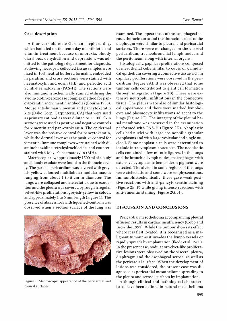
Macroscopically, approximately 1500 ml of cloudy and bloody exudate were found in the thoracic cavi-ty. The parietal pericardium was covered with grey-ish-yellow coloured multilobular nodular masses ranging from about 1 to 5 cm in diameter. The lungs were collapsed and atelectatic due to exuda-tion and the pleura was covered by rough irregular velvet-like proliferations, greyish-yellow in colour, and approximately 1 to 5 mm length (Figure 1). The presence of abscess foci with liquefied-centrum was observed when a section surface of the lung was
examined. The appearances of the oesophageal se-rosa, thoracic aorta and the thoracic surface of the diaphragm were similar to pleural and pericardial surfaces. There were no changes on the visceral pericardium, tracheobronchial lymph nodes and the peritoneum along with internal organs.
Histologically, papillary proliferations composed of mesothelial cells similar to cubic or cylindri-cal epithelium covering a connective tissue rich in capillary proliferations were observed in the peri-cardium (Figure 2A). It was observed that some tumour cells contributed to giant cell formation through integration (Figure 2B). There were ex-tensive neutrophil infiltrations in the connective tissue. The pleura were also of similar histologi-cal appearance and there were marked lympho-cyte and plasmocyte infiltrations adjacent to the lungs (Figure 2C). The integrity of the pleural ba-sal membrane was preserved in the examination performed with PAS-H (Figure 2D). Neoplastic cells had nuclei with large eosinophilic granular cytoplasms and with large vesicular and single nu-cleoli. Some neoplastic cells were determined to include intracytoplasmic vacuoles. The neoplastic cells contained a few mitotic figures. In the lungs and the bronchial lymph nodes, macrophages with extensive cytoplasmic hemosiderin pigment were detected. The alveoli in some regions of the lungs were atelectatic and some were emphysematous. Immunohistochemically, these gave weak posi-tive reactions with anti-pancytokeratin staining (Figure 2E, F) while giving intense reactions with anti-vimentin staining (Figure 2G, H).
DISCUSSION AND CONCLUSIONS
Pericardial mesothelioma accompanying pleural effusion results in cardiac insufficiency (Cobb and Brownlie 1992). While the tumour shows its effect where it is first located, it is recognised as a ma-lignant tumour as it invades the lymph vessels or rapidly spreads by implantation (Ikede et al. 1980). In the present case, nodular or velvet-like prolifera-tive lesions were observed on the visceral pleura, diaphragm and the esophageal serosa, as well as the pericardial surface. When the development of lesions was considered, the present case was di-agnosed as pericardial mesothelioma spreading to the pleura and serosal surfaces by implantation.
Although clinical and pathological character-istics have been defined in natural mesothelioma
Figure 1. Macroscopic appearance of the pericardial and pleural surfaces
Case Report Veterinarni Medicina, 58, 2013: (11): 594–598
596
cases in dogs, the etiological and epidemiological factors that cause these tumours are not yet un-derstood. One of the etiological factors of meso-thelioma is contact with chemical substances such as asbestos (primarily), iron, and silicate (Harbison and Godleski 1983; Head et al. 2002; Caswell and Wiliams 2007). According to the reports of dog owners, high rates of asbestos were detected in the
lungs of dogs belonging to people in contact with asbestos in their professional life (Glickmann et al. 1983). It has been stated that pesticides are im-portant etiological factors in the development of mesothelioma (Ogilvie and Moore 2006). However, genetic factors and some viruses have also been reported to lead to tumour development (Cacciotti et al. 2001). Asbestos particles taken in through the
Figure 2. A. Histological appearance of the pericardium, HE, bar = 200 µm. B. Giant cell formations in the tumour cells (arrow heads), HE, bar = 50 µm. C. Mononuclear cell infiltrations in the border of the pleura (arrow heads), PAS-H, bar = 200 µm. D. Appearance of pleural basement membrane (arrow heads), PAS-H, bar = 50 µm. E. Weak immu-nohistochemical reaction with anti-pancytokeratin antibody in the neoplastic cells, MH, bar = 200 µm. F. Higher magnification anti-pancytokeratin staining in the neoplastic mesothelial cells, MH, bar = 50 µm. G. Intense immuno-histochemical reaction with anti-vimentin antibody in the tumour cells, MH, bar = 200 µm. H. Higher magnification anti-vimentin staining in the neoplastic cells, MH, bar = 50 µm
Veterinarni Medicina, 58, 2013 (11): 594–598 Case Report
597
respiratory tract in humans spread to the pleura through the lymphatic system and cause chromo-somal defects in mitotic mesothelial cells. Genetic mutations in mesothelial cells have been reported to occur by inactivation or loss of tumour suppres-sor genes (Jaurand and Fleury-Feith 2005). In this case, the animal was a tracking dog; the tumour may have developed as a result of lung exposure to asbestos and other chemical substances through the respiratory tract.
Mainly three types of mesotheliomas have been histologically defined in domestic animals: epi-theloid, sarcomatoid and biphasic or mixed. The most common form is the epitheloid type, because of the similarity of the tumour cells to epithelial cells and papillary formations (Caswell and Wiliams 2007). The sarcomatoid type, which is similar in appearance to a fibrosarcoma has been described less frequently (Head et al. 2002; Kapakin et al. 2012). Biphasic or mixed type denotes mesothe-liomas that carry both characteristics of the epith-eloid and sarcomatoid types (Sevcikova et al. 2000). Mesotheliomas show malignant characteristics at low rates, invade the tissues at minimal degrees, and rarely metastasise to lymph nodes and remote tissues (Head et al. 2002; Caswell and Wiliams 2007). The visceral metastasis of mesothelioma has been reported to be rare (Kim et al. 2002), but metastasis to the accessory lobe of the right lung was detected in a dog with pericardial mesothe-lioma (Ledecka et al. 2010). The differential diag-nosis of mesotheliomas generally depends on its basic histological characteristics. In the differen-tiation of epitheloid mesotheliomas, carcinomas and other epithelium-originated tumours, and in the differentiation of sarcomatoid mesothelioma, sarcoma and other spindle cell tumours, and in the differentiation of mixed type, other mixed tumours such as synovial-originating tumours should be considered (Head et al. 2002; Caswell and Wiliams 2007). Immunohistochemistry is useful in differen-tiating mesothelioma. Vimentin-positivity is used to differentiate mesothelioma from pulmonary carcinoma by vimentin negativity; pancytokera-tin-positivity in sarcomatoid type mesotheliomas is generally used to differentiate it from cytokera-tin-negative sarcomas (Kapakin et al. 2012). Both positive pancytokeratin and vimentin staining are constant features of mesotheliomas which may dif-ferentiate as mesothelial cells distinct from epithe-lial and mesenchymal cells (Attanoos et al. 2000). In the present case, the tumour cells were positive
for the epithelial marker pancytokeratin and the mesenchymal marker vimentin.
In conclusion, in this case, a diagnosis of peri-cardial epitheloid mesothelioma was made based on the macroscopic appearance and distribution of the tumour and the histopathological and im-munohistochemical characteristics.
REFERENCES
Attanoos RL, Dojcinov SD, Webb R, Gibbs AR (2000): Anti-mesothelial markers in sarcomatoid mesotheli-oma and other spindle cell neoplasms. Histopathology 37, 224–231.
Bourne JA (ed.) (1985): Handbook of Immunoperoxidase Staining Methods. Immunohistochemistry Labora-tory, DAKO Corporation, USA. 17–34.
Cacciotti P, Libener R, Betta P, Martini F, Porta C, Pro-copio A, Strizzi L, Penengo L, Tognon M, Mutti L, Gaudino G (2001): SV40 replication in human meso-thelial cells induces HGF/Met receptor activation: a model for viral-related carcinogenesis of human ma-lignant mesotelioma. Proceedings of the National Academy of Sciences of the United States of America 98, 12032–12037.
Caswell J, Wiliams K (2007): Respiratory system. In: Maxie M (ed.): Jubb, Kennedy, and Palmer’s Pathology of Domestic Animals. 5th ed. Elsevier, New York. 523–655.
Cobb MA, Brownlie SE (1992): Intrapericardial neopla-sia in 14 dogs. Journal of Small Animal Practise 33, 309–311.
Garrett LD (2007): Mesothelioma. In: Withrow SJ, Vail DM (eds.): Small Animal Clinical Oncology. 4th ed. Saunders Elsevier. 847 pp.
Glickmann LT, Domanski LM, Magure TG, Dubielzig RR, Churg A (1983): Mesothelioma in pet dogs associ-ated with exposure of the owners to asbestos. Envi-ronmental Research 32, 305–313.
Head KW, Else RW, Dubielzig RR (2002): Tumours of the alimentary tract. In: Meuten DJ (ed.): Tumours in Domestic Animals. 4th ed. Iowa State Press, Iowa. 401–481.
Ikede BO, Zubaidy A, Gill CW (1980): Pericardial mes-othelioma with cardiac tamponade in a dog. Veterinary Pathology 17, 496–499.
Case Report Veterinarni Medicina, 58, 2013: (11): 594–598
598
Kapakin Terim KA, Haziroglu R, Gursan N, Yucel G (2012): Mesothelioma in a dog. Veterinary Journal of Ankara University 59, 151–153.
Kim JH, Choi YK, Yoon HY, Kweon OK, Kim DY (2002): Juvenile malignant mesotelioma in a dog. Journal of Veterinary Medical Science 64, 269–271.
Ledecka K, Sevcikova Z, Mihaly M, Hajurka J, Pavuk V, Hluchy M, Skurkova L, Lackova M, Ledecky V (2010): Mesothelioma of the pericardium in a Bernese meso-thelioma of the pericardium in a Bernese Mountain dog. Veterinarski Arhiv 80, 797–806.
Ogilvie GK, Moore AS (2006): Managing the Canine Can-cer Patient: A Practical Guide to Compassionate Care. Veterinary Learning Systems Book. Trenton, NJ. 733.
Sevcikova Z, Kolodzieyski M, Levkut M, Ledecky V, Sevcik A (2000): Malignant and biphasic peritoneal mesothelioma in dogs. Indian Veterinary Journal 77, 852–855.
Vural SA, Ozyildiz Z, Ozsoy SY (2007): Pleural meso-thelioma in a nine month-old dog. Irish Veterinary Journal 1, 30–33.
Received: 2012–11–06Accepted after revision: 2013–11–20
Corresponding Author:
Songul Ceribasi, Firat University, Faculty of Veterinary Medicine, Department of Pathology, 23119 Elazig, TurkeyE-mail: [email protected]


![Genomic Landscape of Malignant Mesotheliomas · 8/9/2016 · Pericardial mesothelioma was the least common subtype (5% [2/42]), and 14% (6/42) of mesothelioma samples had unknown](https://static.documents.pub/doc/80x56/60aba46dd6b2bd17077b70b6/genomic-landscape-of-malignant-mesotheliomas-892016-pericardial-mesothelioma.jpg)
![Mesothelioma lawyers ] mesothelioma attorneys](https://static.documents.pub/doc/80x56/5497f892ac795959288b5644/mesothelioma-lawyers-mesothelioma-attorneys.jpg)


