Page 1
Chapter 4
Periodic paralysis
DOREEN FIALHO AND MICHAEL G. HANNA*
Institute of Neurology, London, UK
s0010 4.1. Introduction
p0010 Periodic paralysis is a disorder of skeletal muscle in
which patients experience attacks of muscle weakness
of variable duration and severity. The attacks can last
from a few minutes to several days. The weakness in an
attack can be generalized or focal. Early in the natural his-
tory of the disease muscle strength returns to normal after
an attack, but later significant fixed muscle weakness
often develops. The variability of the symptoms often
leads to delays in accurate diagnosis and treatment.
p0020 Although the clinical phenotype of periodic paralysis
has been recognized for many years, it is only in recent
times that the underlying pathophysiology has been
deduced at a molecular genetic level. In all forms of this
disorder, electrophysiological examination during an
attack reveals that the skeletal muscle fiber membrane
is in a partially depolarized and inexcitable state. Mus-
cle membrane excitability depends on the coordinated
interplay of key voltage-gated ion channels. It is now
known that in both genetic and acquired forms of peri-
odic paralysis dysfunction of these key membrane-
bound ion channels underlies the pathophysiology and
explains the altered muscle excitability. Periodic paraly-
sis was one of the first neurological channelopathies to
be characterized at a genetic and cellular level. To a
certain extent the current detailed molecular knowledge
about periodic paralysis represents a paradigm for our
understanding of subsequently discovered muscle and
brain channelopathies.
p0030 Historically, periodic paralysis was classified
according to serum potassium abnormalities during
attacks into hypo- and hyperkalemic periodic paralysis
(hypoPP and hyperPP). This classification depending
on serum potassium is still of use clinically but has
now been supplemented by the newer molecular genetic
classification which we describe here.
p0040In this chapter we provide a detailed review of current
knowledge regarding clinical features, investigations,
treatment, genetics and molecular pathophysiology of
the periodic paralyses.
s00204.2. Clinical features
s00304.2.1. Familial hypokalemic periodic paralysis
(hypoPP)
p0050Most of the early original publications on periodic paral-
ysis were probably describing hypoPP, as this is the com-
monest form of periodic paralysis. Talbott published an
extensive review of the literature on periodic paralysis
in 1941 (Talbott, 1941). This paper summarized many
of the characteristic features of periodic paralysis includ-
ing age of onset, male predilection, development of
fixed weakness and provoking factors. Talbott cites
Musgrave’s interesting observation from 1727 of a 21-
year-oldwomanwho presentedwith attacks ofweakness,
and suggests this may be the first description of periodic
paralysis (Musgrave, 1727). However, some of the fea-
tures in Musgrave’s original case were atypical, includ-
ing loss of speech and attacks always occurring on the
same day of the week. From the beginning of the 19th
century a number of reports started to appear describing
cases of sporadic periodic paralysis and the first familial
case of an affected father and son was reported by
Shakhnowitsch in 1882. Early hypotheses on the patho-
genesis of periodic paralysis included the theory of
muscle ischemia as the underlying pathology (Westphal,
1885, Holtzapple, 1905, Schmidt, 1919, Mankowsky,
1929). Goldflam (Goldflam, 1890) and others (Crafts,
*Correspondence to: Dr. M.G. Hanna, Centre for Neuromuscular Disease, National Hospital for Neurology and Neurosurgery,
University College London Foundation NHS Trust, and Department of Molecular Neuroscience, Institute of Neurology,
University College London, Queen Square, London, WC1N 3BG, UK. E-mail: [email protected] , Tel: þ44-(0)207-
837-3611, Fax: þ44-(0)207-6921-2085.
Handbook of Clinical Neurology, Vol. 86 (3rd series)MyopathiesF. L. Mastaglia, D. Hilton-Jones, Editors# 2007 Elsevier B.V. All rights reserved
Mastalgia, 0-444-51899-1
10004
Page 2
1900, Singer and Goodbody, 1901) suggested that an
autotoxin was responsible. Hartwig (1874) was the first
to describe electrical inexcitability of muscles during an
attack of paralysis. Indeed, Hartwig was so surprised by
the lack of response to electrical stimulation that he initi-
ally thought that his apparatus was malfunctioning. Bie-
mond and Daniels (1934) provided the first report of
low potassium levels during a spontaneous attack. This
was confirmed in another case a year later when Walker
(1935) reported convincing evidence that there was a
50% decrease of serum potassium during an attack.
p0060 It is now known that hypoPP is the most common
form of familial periodic paralysis with a prevalence
of 0.4–1:100 000 in Europe (Kantola and Tarssanen,
1992, Fontaine, 1994). The inheritance is autosomal
dominant with reduced penetrance in women giving a
male:female ratio of ~3:1 (Elbaz et al., 1995).
p0070 There are currently three genes implicated in familial
hypoPP including CACNA1S, SCN4A and KCNJ2.Mutations in the voltage-gated calcium channel gene
CACNA1S account for the majority of cases (~70%;
Fouad et al., 1997, Miller et al., 2004). In less than
10% of cases mutations in the voltage-gated sodium
channel gene SCN4A are reported (Bulman et al.,
1999, Davies et al., 2001, Sternberg et al., 2001, Miller
et al., 2004). Mutations in KCNJ2 encoding an inward-
rectifying potassium channel can cause Andersen–
Tawil syndrome (Plaster et al., 2001). Since this con-
dition is distinct and can present with both hypo- and
hyperkalemic periodic paralysis it will be discussed sep-
arately. A mutation in KCNE3 reported as pathogenic inhypoPP was later found to be a benign polymorphism
(Abbott et al., 2001, Sternberg et al., 2003, Jurkat-Rott
and Lehmann-Horn, 2004).
p0080Hypokalemic periodic paralysis generally presents
later than hyperkalemic paralysis, usually between the
ages of 5 and 20, typically in the teenage years (Fouad
et al., 1997, Miller et al., 2004; see Table 4.1). However,
onset over the age of 20 has been reported (Miller et al.,
2004). Attacks tend to last from several hours up to 2–3
days. It is often difficult for patients to give a precise
estimate of attack duration as both onset and resolution
tend to be gradual. A sudden onset of weakness leading
to a collapse would argue against a diagnosis of periodic
paralysis. It is generally considered that hypoPP attacks
are longer and more severe than in hyperPP. Although
this is our experience, a recent retrospective study did
not confirm this. It is possible the use of medication
by patients in the study may have influenced attack
duration (Miller et al., 2004).
p0090In a typical hypoPP episode the patient wakes in the
night or in the morning with generalized severe weak-
ness being “unable to move”. Often intake of a carbohy-
drate-rich meal or strenuous exercise the preceding day
or night can be identified as a triggering factor. Focal
episodes of weakness may be triggered by exercise only
involving one limb but are more common in hyperPP.
Tendon reflexes are diminished or absent. Even in a
severe attack cranial muscles are spared so that speech
and eye opening remain intact. Impairment of speech,
visual symptoms or alterations in consciousness are
not expected and should trigger consideration of other
diagnostic possibilities. Respiratory muscles are mostly
spared but a reduction in vital capacity and consequent
t0010 Table 4.1
Clinical features of hyperkalemic periodic paralysis and hypokalemic periodic paralysis
Hyperkalemic periodic paralysis Hypokalemic periodic paralysis
Onset of symptoms First decade Second decade
Triggers Rest after exercise, cold, fasting,
potassium-rich food
Rest after exercise, carbohydrate load
Time of attack Any time of the day Typically when waking up in the morning
Duration of attack Minutes to hours Hours to days
Severity of attack Mild to moderate, may be focal Moderate to severeAdditional symptoms Myotonia or paramyotonia
Serum potassium Usually high, may be normal Low
Interictal
electromyography
Myotonic discharges in some, positive
McManis test
Never myotonic discharges, positive McManis
test
Treatment Acetazolamide, dichlorphenamide, thiazide,
beta-agonist
Acetazolamide, dichlorphenamide, potassium
supplementation, potassium-sparing diuretics
Gene/ion channel SCN4A: Nav1.4 (sodium channel subunit),
KCNJ2: Kir2.1 (potassium channel subunit)
CACNA1S: Cav1.1 (calcium channel subunit),
SCN4A: Nav1.4 (sodium channel subunit),
KCNJ2: Kir2.1 (potassium channel subunit)
78 D. FIALHO AND M. G. HANNA
Mastalgia, 0-444-51899-1
10004
Page 3
respiratory failure has rarely been reported to occur in
severe attacks (Ziegler and McQuarrie, 1952, Rowley
and Kliman, 1960, Resnick and Engel, 1967). Strength
gradually improves over the course of the next day or
two although some patients indicate that it takes up to
a week to recover. Even when the patient is not com-
plaining of clear clinical attacks careful quantitative
strength measurement has suggested that there is diurnal
variation of muscle power, being lowest in the early
hours of the morning and highest in the afternoon and
evening (Engel et al., 1965). Attacks often become less
frequent and severe in later life and in common with
hyperPP a permanent myopathy may develop (Biemond
and Daniels, 1934). Interestingly fixed weakness has
been described to occur even in patients without a
strong history of frequent paralytic attacks (Sternberg
et al., 2001). For example, in some females the late-
onset myopathy may be the only manifestation without
any clinically evident paralytic attacks (Links et al.,
1990). A study of a large kindred with hypoPP showed
that nearly all subjects over the age of 50 years had evi-
dence of fixed muscle weakness (Links et al., 1994). It
remains unproven whether active treatment to reduce
the frequency of paralytic attacks might reduce the
development of fixed weakness later.
p0100 A useful feature to distinguish between hypo- and
hyperkalemic periodic paralysis clinically is the absence
of (true) myotonia in hypoPP. The only exception to
this rule so far is the SCN4A mutation P1158S which
has been described in a Japanese kindred causing myo-
tonia and cold-induced hypoPP (Sugiura et al., 2000).
Previously in the literature only a single case was
reported with myotonia and periodic paralysis where
the potassium level was low (1.9 mEq/l) during the
attack. However the patient was from a family with typ-
ical myotonic dystrophy and the precise diagnosis is
unclear (Leyburn and Walton, 1960). There are a hand-
ful of other reports of apparent clinical myotonia (most-
ly myotonic lid lag) in association with hypokalemic
periodic paralysis (Odor et al., 1967, Resnick et al.,
1967, Griggs et al., 1970). Here the explanation may
be that the lid lag was not due to true electrical myoto-
nia, which explains why no EMG myotonia could be
demonstrated in any of these patients. Although lid lag
is a sensitive marker of myotonia it does not appear to
be very specific as it has been found even in healthy
volunteers (Odor et al., 1967) and should therefore be
interpreted with caution.
p0110 Anumber of factorsmay induce or exacerbate attacks.
These include ingestion of carbohydrates, administration
of insulin and epinephrine injections (Ziegler and
McQuarrie, 1952, Rowley and Kliman, 1960, Engel
et al., 1965). Stress and excitement and exposure to cold
are also often listed by patients as triggers (Miller et al.,
2004). Menstruation and pregnancy have been reported
to cause an increase in frequency and severity of attacks
(Bender, 1936, Links et al., 1994).
p0120Although serum potassium levels are often reduced,
especially at the beginning of an attack, they may not
be below the normal range. The original studies of peri-
odic paralysis in the early 20th century reported a num-
ber of other electrolyte changes (for review see Talbott
1941), including a decrease in serum phosphate in par-
allel with potassium and reduced urinary excretion of
sodium, potassium, chloride and water. Serum creatine
kinase (CK) may be normal or slightly elevated in
between attacks. During paralytic attacks there can be
a moderate rise in CK (De Keyser et al., 1987).
p0130Electrogardiogram (ECG) changes have been
observed with very low potassium including prominent
U waves, flattening of T waves and ST depression. Inter-
ictal ECG is usually normal although affected members
of a kindred with hypokalemic periodic paralysis carry-
ing the R528H CACNA1Smutation were reported to suf-
fer from cardiac arrhythmias (Fouad et al., 1997). The
presence of prominent U waves, frequent ventricular
ectopic beats or arrhythmias should alert the clinician
to the possibility of Andersen–Tawil syndrome (ATS)
(see later section). Familial hypokalemic periodic paraly-
sis is not associated with clinical or echocardiographic
evidence of cardiomyopathy (Schipperheyn et al., 1978).
s00404.2.2. Familial hyperkalemic periodic paralysis
(hyperPP)
p0140In the early 1950s the Swedish pediatric neurologist
Gamstorp recognized a new form of periodic paralysis
associated with an elevated serum potassium. In her the-
sis in 1956 she coined the term “adynamia episodica
hereditaria” (Gamstorp, 1956) but later it was referred
to as hyperkalemic periodic paralysis.
p0150Familial hyperPP is due to mutations in SCN4Aencoding the a-subunit of the skeletal muscle voltage-
gated sodium channel Nav1.4. The clinical presentation
of hyperPP includes attacks of limb weakness lasting
minutes to hours. In contrast to hypoPP the attacks fre-
quently happen during daytime but nocturnal attacks
may occur (Gamstorp, 1956, Layzer et al., 1967). From
a clinical diagnostic perspective, frequent short daytime
attacks favor a diagnosis of hyperPP and nocturnal pro-
longed attacks may slightly favor hypoPP. The onset of
symptoms is typically within the first decade and attacks
tend to becomemilder and less frequent with age. A per-
sistent mildmyopathymay develop later in the course of
the disease and reports indicate that this is independent
of the number of attacks (Saunders et al., 1968, Bradley
et al., 1990, Ptacek et al., 1991a).
PERIODIC PARALYSIS 79
Mastalgia, 0-444-51899-1
10004
Page 4
p0160 The rise of potassium during attacks may be subtle
and transient, frequently not exceeding the normal range
and can therefore be easily missed (Plassart et al.,
1994). For many years normokalemic periodic paralysis
was considered to be a distinct disorder based on
descriptions of a limited number of families (Poskanzer
and Kerr, 1961, Meyers et al., 1972, Danowski et al.,
1975). However, the status of normokalemic PP as a
distinct entity now looks uncertain. We had the opportu-
nity to analyze the original 1961 family from the north-
east of England and showed that they harbored the
common M1592V hyperPP SCN4A mutation (Chinnery
et al., 2002). It seems likely that normokalemic periodic
paralysis should be considered a variant of hyperPP.
p0170 HyperPP, potassium aggravated myotonia (PAM)
and paramyotonia congenita (PMC) are allelic sodium
channel disorders and their phenotypes overlap to vary-
ing degrees (Layzer et al., 1967, de Silva et al., 1990).
In hyperPP and paramyotonia congenita women may
be less severely affected (Layzer et al., 1967).
p0180 Many patients who have both periodic paralysis and
myotonia find it difficult to distinguish between stiff-
ness and weakness and attacks are often initially domi-
nated by stiffness leading to paralysis later. EMG
myotonia can be demonstrated in at least 50% of
patients with the two most common SCN4A mutations
T704M and M1592V (Plassart et al., 1994, Miller
et al., 2004, Fournier et al., 2004) but myotonia on
examination is detected in a smaller percentage (Plas-
sart et al., 1994). Interestingly myotonic symptoms are
frequently experienced and easily elicited in the cranial
musculature (myotonic lid lag, eye closure myotonia)
which is not usually involved in the paralytic attack.
Consciousness is preserved and respiratory and cranial
musculature is usually spared. A number of factors have
been identified that can trigger or exacerbate attacks.
These include rest following exercise, fasting, cold,
stress, intercurrent infection and anesthesia. Hormonal
changes may also play a role as menstruation, oral con-
traception and pregnancy have been associated with an
increase in symptoms (Layzer et al., 1967, Ptacek
et al., 1993, Kim et al., 2001).
s0050 4.2.3. Andersen–Tawil syndrome (ATS)
p0190 Andersen–Tawil syndrome first fully described by
Andersen et al. (1971) is characterized by a triad of
periodic paralysis, ventricular arrhythmia and distinc-
tive physical features. Many patients do not have all
of these features and there can be marked intrafamilial
variation and evidence of incomplete penetrance
(Plaster et al., 2001). It is the rarest form of periodic
paralysis and no reliable data exist on prevalence.
Mutations in KCNJ2 encoding the inward-rectifying
potassium channel Kir2.1 have been identified in
about two-thirds of kindreds with ATS (Plaster et al.,
2001, Tristani-Firouzi et al., 2002). Up to 20% of indi-
viduals carrying pathogenic mutations may not exhibit
any phenotypic features (Andelfinger et al., 2002,
Tristani-Firouzi et al., 2002, Donaldson et al., 2003).
De novo mutations are frequent (Donaldson et al.,
2003).
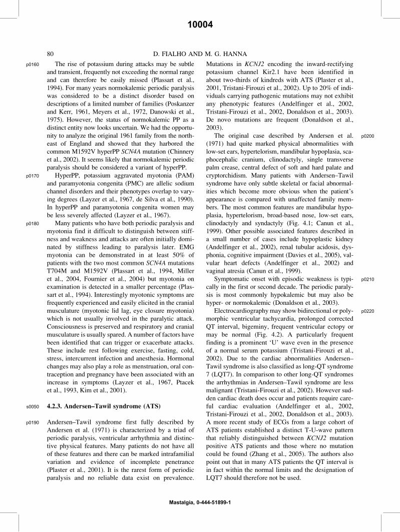
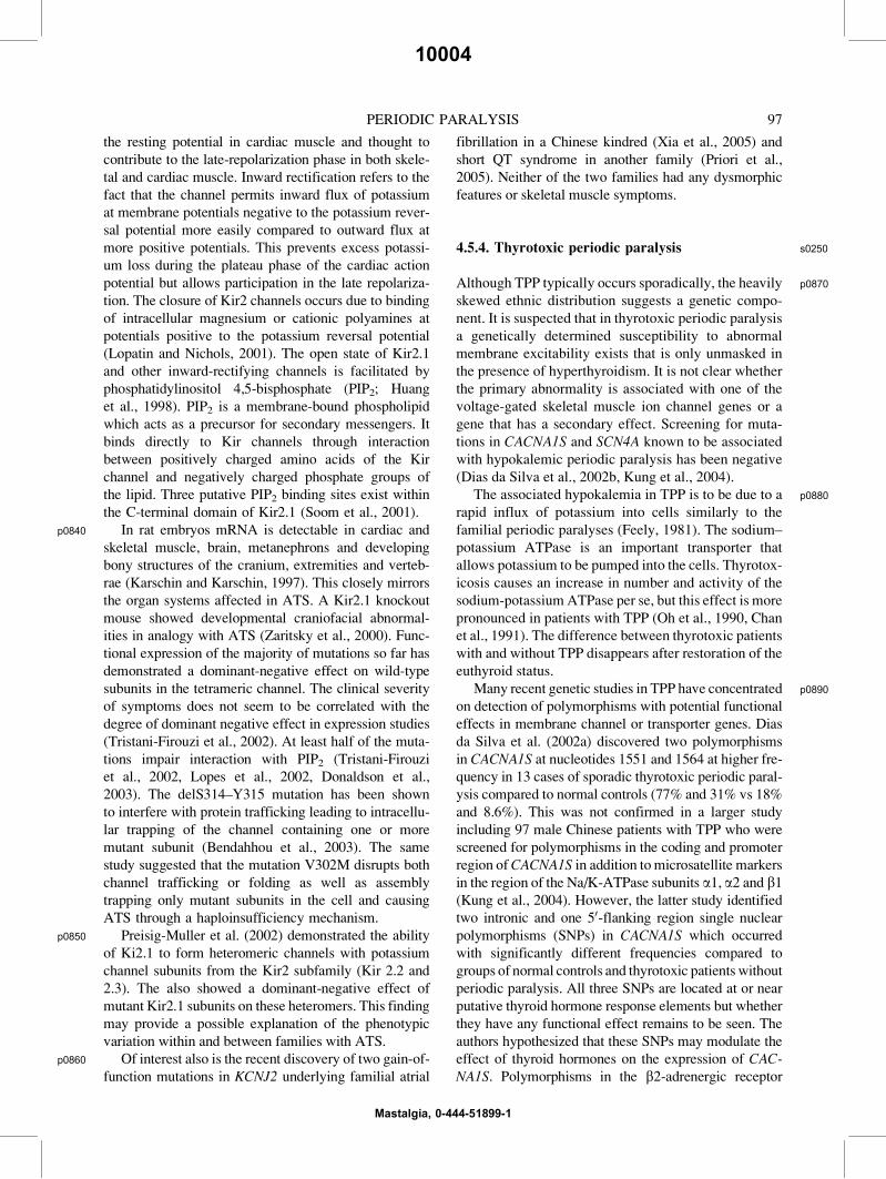
p0200The original case described by Andersen et al.
(1971) had quite marked physical abnormalities with
low-set ears, hypertelorism, mandibular hypoplasia, sca-
phocephalic cranium, clinodactyly, single transverse
palm crease, central defect of soft and hard palate and
cryptorchidism. Many patients with Andersen–Tawil
syndrome have only subtle skeletal or facial abnormal-
ities which become more obvious when the patient’s
appearance is compared with unaffected family mem-
bers. The most common features are mandibular hypo-
plasia, hypertelorism, broad-based nose, low-set ears,
clinodactyly and syndactyly (Fig. 4.1; Canun et al.,
1999). Other possible associated features described in
a small number of cases include hypoplastic kidney
(Andelfinger et al., 2002), renal tubular acidosis, dys-
phonia, cognitive impairment (Davies et al., 2005), val-
vular heart defects (Andelfinger et al., 2002) and
vaginal atresia (Canun et al., 1999).
p0210Symptomatic onset with episodic weakness is typi-
cally in the first or second decade. The periodic paraly-
sis is most commonly hypokalemic but may also be
hyper- or normokalemic (Donaldson et al., 2003).
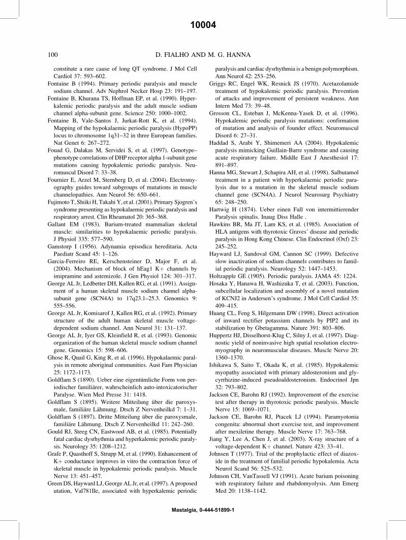
p0220Electrocardiographymay show bidirectional or poly-
morphic ventricular tachycardia, prolonged corrected
QT interval, bigeminy, frequent ventricular ectopy or
may be normal (Fig. 4.2). A particularly frequent
finding is a prominent ‘U’ wave even in the presence
of a normal serum potassium (Tristani-Firouzi et al.,
2002). Due to the cardiac abnormalities Andersen–
Tawil syndrome is also classified as long-QT syndrome
7 (LQT7). In comparison to other long-QT syndromes
the arrhythmias in Andersen–Tawil syndrome are less
malignant (Tristani-Firouzi et al., 2002). However sud-
den cardiac death does occur and patients require care-
ful cardiac evaluation (Andelfinger et al., 2002,
Tristani-Firouzi et al., 2002, Donaldson et al., 2003).
A more recent study of ECGs from a large cohort of
ATS patients established a distinct T-U-wave pattern
that reliably distinguished between KCNJ2 mutation
positive ATS patients and those where no mutation
could be found (Zhang et al., 2005). The authors also
point out that in many ATS patients the QT interval is
in fact within the normal limits and the designation of
LQT7 should therefore not be used.
80 D. FIALHO AND M. G. HANNA
Mastalgia, 0-444-51899-1
10004
Page 5
s0060 4.2.4. Thyrotoxic periodic paralysis (TPP)
p0230 The occurrence of periodic paralysis in association with
hyperthyroidism was reported as early as 1902
(Rosenfeld, 1902). This form of periodic paralysis is
more common in Asia, particularly China, Korea and
Japan, where more than 10% of male thyrotoxic patients
may be affected (Chen et al., 1965, McFadzean and
Yeung, 1967, Ober, 1992, Kung et al., 2004). The over-
all incidence in thyrotoxic patients from these popula-
tions is approximately 2% (McFadzean and Yeung,
1967) while the incidence in Caucasians has been
estimated at only 0.1–0.2% (Kelley et al., 1989). Due
to migration, cases of (TPP) are now increasingly seen
in the Western world (Ober, 1992). It is also recognized
in Caucasians (Linder, 1955), native American Indians
(Conway et al., 1974), Blacks (Kilpatrick et al., 1994),
Aborigines (Ghose et al., 1996) and Maoris (Wild,
2004). The male-to-female predominance is much more
marked in TPP (between 20:1 and 76:1) (Okinaka et al.,
1957, McFadzean and Yeung, 1967) compared to
hypoPP (3:1; Elbaz et al., 1995). This is even more sig-
nificant given that the prevalence of thyrotoxicosis is so
much higher in females.
p0240Most cases of TPP are sporadic but a few familial
cases have been described (Kufs et al., 1989, Dias da
Silva et al., 2002a). The onset of symptoms is most
frequently between the second and fourth decade in
parallel to the highest incidence of hyperthyroidism.
A significant proportion of patients have only subtle clin-
ical signs of hyperthyroidism (McFadzean and Yeung,
1967, Kelley et al., 1989). Autoimmune thyrotoxicosis
(Graves’ disease) is the most common underlying dis-
order but TPP may be caused by any form of hyper-
thyroidism in susceptible patients including excessive
administration of thyroid hormone replacement.
p0250Thyrotoxic periodic paralysis bears phenotypic
resemblance to familial hypokalemic periodic paralysis.
It is associated with low serum potassium during attacks,
may be triggered by glucose/insulin administration and
f0010
Fig. 4.1. Patient with ATS.
PERIODIC PARALYSIS 81
Mastalgia, 0-444-51899-1
10004
Page 6
may also be triggered by rest following exercise. Focal
weakness can develop in more strenuously exercised
muscles and attacks typically occur at night or on waken-
ing in the morning (McFadzean and Yeung, 1967). Rare
cases with associated normo- or hyperkalemia have been
reported, although this was prior to the availability of
DNA testing for familial periodic paralysis (Adachihara
and Takagi, 1974, Mehta et al., 1990). The respiratory
and cranial musculature tend to be spared. Morbidity
and mortality is low but significant arrhythmias asso-
ciated with severe hypokalemia have been reported
(McFadzean and Yeung, 1967, Fisher, 1982).
s0070 4.2.5. Secondary periodic paralysis
p0260 A number of secondary causes of periodic paralysis
should to be considered when evaluating a patient with
periodic paralysis. Both hypo- and hyperkalemia of
any origin can result in muscle weakness or paralysis.
Usually the patient remains weak until the underlying
cause of potassium alteration is identified and treated.
Occasionally patients with a secondary cause may
present with intermittent attacks of weakness and this
may make the distinction with sporadic genetic periodic
paralysis more difficult. In general the electrolyte dis-
turbance tends to be more severe than seen in the famil-
ial forms of periodic paralysis. Usually potassium levels
have to decline to <3mmol/l or rise to>7mmol/l before
significant muscle symptoms are experienced. With the
exception of barium poisoning and insulin excess there
is a loss or excess of total body potassium in secondary
periodic paralysis rather than a shift between intra-
and extracellular space as is the case in the familial
forms and in TPP. Metabolic abnormalities often persist
between attacks and this gives an important clue to
the underlying diagnosis. The treatment is aimed at
correcting the primary abnormality.
p0270A number of conditions mainly causing urinary or
gastrointestinal potassium loss leading to hypokalemia
have been reported in association with episodic weak-
ness (Table 4.2). With severe hypokalemia there is an
associated risk of significant arrhythmias, paralytic ile-
us and rhabdomyolysis in addition to respiratory fail-
ure secondary to muscle paralysis (Weiss-Guillet
et al., 2003). The presentation of patients with muscle
A
B
f0020Fig. 4.2. ECG traces from patients with ATS. (A) Frequent polymorphic ventricular ectopy with bidirectional ventricular
ectopics detectable in the lateral chest leads. QTc interval borderline prolonged. (B) Prominent U-wave.
82 D. FIALHO AND M. G. HANNA
Mastalgia, 0-444-51899-1
10004
Page 7
paralysis secondary to hyperkalemia is much less
common than hypokalemia (Evers et al., 1998). Most
cases of secondary hyperPP are due to potassium-
sparing diuretics (spironolactone) often on a back-
ground of renal impairment.
p0280 There have been many case reports of primary and
secondary renal tubular acidosis (RTA) associated
with hypoPP (Koul et al., 1993, Bresolin et al.,
2005). Renal tubular acidosis probably due to autoim-
mune tubulointerstitial nephritis may occur in Sjog-
ren’s syndrome and an association with periodic
paralysis has been described (Raskin et al., 1981). In
some of these cases the muscle symptoms were the
presenting complaints (Soy et al., 2005), even leading
to respiratory arrest (Poux et al., 1992, Fujimoto et al.,
2001). Habitual toluene inhalation (glue sniffing) can
also cause RTA and may present with paralysis
(Bennett and Forman, 1980).
p0290The first cases of barium poisoningwere referred to as
Pa Ping disease due to endemic periodic paralysis in the
Pa Ping area of the Szechwan province of China caused
by ingestion of table salt contaminated by barium (Allen,
1943). Accidental ingestion of barium salts used as rat
poison, industrial accidents, suicidal attempts and admin-
istration of barium carbonate instead of the insoluble
sulphate in radiodiagnosis have been reported (Lewi
and Bar-Khayim, 1964, Berning, 1975, Layzer, 1982,
Shankle and Keane, 1988, Ahlawat and Sachdev, 1999).
Manifestations of toxicity include hemorrhagic gastroen-
teritis with vomiting, colic and diarrhea, hypertension,
cardiac arrhythmias, muscle twitching, seizures, hypoka-
lemia and muscle paralysis (Johnson and VanTassell,
1991). The hypokalemia in barium poisoning occurs
due to a shift of potassium from the extracellular to intra-
cellular compartments. Barium competitively blocks
potassium channels causing reduction in potassium
t0020 Table 4.2
Causes of secondary periodic paralysis
Conditions leading to hyperkalemia Conditions leading to hypokalemia
Endocrine Addison’s disease (Pollen and Williams,
1960)
Hyperaldosteronism (primary/secondary) (Conn et al.,
1964, Ishikawa et al., 1985, Ma et al., 1986)
Hypoaldosteronism and hyporeninaemia
(Daughaday and Rendleman, 1967)
Cushing’s disease/syndrome
Gordon’s syndrome:
pseudohypoaldosteronism type II (Pasman
et al., 1989)
Hyperreninism (Umeki et al., 1986)
17a-hydroxylase deficiency (CYP17) (Yazaki et al.,1982)
Hyperinsulinemia
Renal Chronic renal failure (Cumberbatch and
Hampton, 1999)
Bartter’s syndrome (Shiah et al., 1994)
Liddle syndrome
Gitelman syndrome (Lin et al., 2003)
Distal tubular acidosis type 1 and 2 þ/� Sjogren’s
syndrome (Owen and Verner, 1960, Raskin et al.,
1981)
Gastro-intestinal Severe diarrhea and vomiting (Ortuno et al., 2002,
Haddad et al., 2004)
Ileostomy
Uterosigmoidostomy (Angeloni and Scott, 1960,
Sataline and Simonelli, 1961)
Villous adenoma (Keyloun and Grace, 1967)
Drugs/Toxins Potassium load (Muensterer, 2003) Licorice (Cumming et al., 1980, Ishikawa et al., 1985)
Potassium-sparing diuretics (Udezue and
Harrold, 1980)
Laxative abuse (Basser, 1979)
High-dose angiontensin-converting (ACE)
inhibitor (Dutta et al., 2001)
Potassium-wasting diuretics (Cohen, 1959)
Amphotericin B (McChesney and Marquardt, 1964)
Barium poisoning (Lewi and Bar-Khayim, 1964)
Toluene exposure (Bennett and Forman, 1980)
Cocaine (Nalluri et al., 2000, Lajara-Nanson, 2002)
Gossypol (Wang and Chen, 1991, Waites et al., 1998)
PERIODIC PARALYSIS 83
Mastalgia, 0-444-51899-1
10004
Page 8
permeability leading to membrane depolarization and
finally inexcitability (Sperelakis et al., 1967, Gallant,
1983). The potassium channels affected include the
inward-rectifying channel Kir2.1 which is mutated in
the familial periodic paralysis Andersen–Tawil syn-
drome (Schram et al., 2003). The main treatment consists
of oral or intravenous potassium which displaces barium
and allows it to be excreted.
s0080 4.2.6. Differential diagnosis
p0300 Other neuromuscular disorders should also be consid-
ered in the differential diagnosis of episodic weakness.
The difference between myasthenia and periodic paral-
ysis appears straight forward at first glance. Attacks of
weakness are more distinct in PP versus a more long-
term fluctuation of muscle strength in myasthenia. Gen-
tle exercise helps to lessen or abort PP attacks while
exertion worsens symptoms in myasthenia. The distri-
bution of muscles affected is different (bulbar and
extraocular muscles frequently affected in myasthenia
and spared in PP). Investigations (neuromuscular junc-
tion transmission deficit on repetitive nerve stimulation
and single fiber EMG, acetylcholine receptor antibo-
dies, genetic testing) should also easily distinguish
between these two disorders. However, diagnostic diffi-
culty may sometimes arise when distinguishing between
the limb girdle presentation of myaesthenia and period-
ic paralysis. In this context it is interesting to note the
discovery of a mutation in SCN4A leading to loss of
sodium channel Nav1.4 function in a patient with
attacks of bulbar and respiratory paralysis associated
with ptosis and a neuromuscular junction transmission
deficit on neurophysiological investigations (Tsujino
et al., 2003). This finding indicates that an overlap
between periodic paralysis and myasthenia gravis may
occur at a molecular level. Of interest is also an Austra-
lian family with episodic weakness affecting extraocu-
lar, facial, trunk and limb muscles lasting weeks to
months (Ryan et al., 1999). The disorder has been
linked to the X chromosome but the gene involved
has not been identified. Patients with both myotonia
congenita and paramyotonia/hyperPP can experience
intermittent weakness. In myotonia congenita this is
termed transient weakness and presents with brief loss
of muscle strength at initiation of movement particular-
ly after a period of rest. Attacks of weakness in patients
with hyperPP and paramyotonia congenita are usually
more profound and of longer duration. Most other disor-
ders causing acute or subacute muscle weakness (e.g.,
McArdle’s disease, Guillain-Barre syndrome, acute
intermittent porphyria) are normally straightforward to
exclude by appropriate history, clinical examination
and investigations.
s00904.3. Examination and investigations
s01004.3.1. General examination and laboratory
investigations
p0310General examination of patients between attacks is
often normal. Muscle strength testing may reveal evi-
dence of persistent proximal weakness. Patients with
hyperPP may show signs of action and percussion myo-
tonia. Lid lag often proves to be the most sensitive indi-
cator of myotonia but it can also be seen in healthy
volunteers. Patients with periodic paralysis and myoto-
nia may also exhibit a degree of muscle hypertrophy
(McArdle, 1962, Layzer et al., 1967). Attention should
be paid to detect any subtle dysmorphic features which
may indicate ATS.
p0320Laboratory investigations are directed to establish
potassium levels during attacks (ideally soon after the
onset of attack) and exclude secondary causes of peri-
odic paralysis. All patients with hypokalemic periodic
paralysis should have their thyroid function checked to
exclude the possibility of TPP. Routine 12-lead electro-
cardiography (ECG) should be undertaken in all PP
cases since the cranioskeletal features of ATS may be
subtle. There is also a risk of cardiac arrhythmias during
severe attacks when potassium levels are excessively
deranged. Patients with suspected ATS should undergo
more thorough cardiological work-up including pro-
longed ECG recordings, echocardiography and exercise
testing.
p0330In the past patients were often subjected to a range of
provocative tests, many of which have now been super-
seded by the availability of genetic analysis and
specialized neurophysiological investigations. The prin-
ciple aim was to induce a clinical focal or generalized
attack of paralysis. For hyperPP administration of potas-
sium (orally or intravenously), cooling of limbs and
exercise, or a combination has been used. In cases of
suspected hypoPP a glucose load with or without addi-
tional insulin was the preferred method of inducing
attacks. The glucose-insulin test needs to be interpreted
with caution as apparent weakness (although without
change in reflexes) has also been induced in patients
with hyperkalemic periodic paralysis (Layzer et al.,
1967). Cardiac monitoring and frequent testing of the
serum potassium and glucose level are essential. Another
provocative test involved intra-arterial epinephrine
together with EMG monitoring.
s01104.3.2. Genetic testing
p0340DNA testing is now a major diagnostic tool in familial
periodic paralysis. However, even with extensive DNA
sequencing of the ion channel genes known to be
84 D. FIALHO AND M. G. HANNA
Mastalgia, 0-444-51899-1
10004
Page 9
involved in periodic paralysis, mutations are not detected
in one-third of patients with either hyper- or hypokalemic
periodic paralysis (Miller et al., 2004). Both CACNA1Sand SCN4A are large genes containing 44 and 25 exons
respectively. The genetic testing generally available in
DNA diagnostic-service laboratories often only encom-
passes gene regions containing common mutations. It is
therefore important to note that a negative genetic result
from such a laboratory reduces the likelihood but does
not exclude a diagnosis of familial periodic paralysis.
The potassium channel gene KCNJ2 mutated in ATS is
a relatively small single exon gene and direct sequencing
analysis of thewhole gene ismore feasible in the diagnos-
tic laboratory setting. In ATS more than 30 mutations
have been identified (Table 4.3) but approximately 30%
of kindreds do not harbor mutations in KCNJ2. Thiscould be partly because there may be undetected muta-
tions in the promoter or intronic regions of the KCNJ2gene (Tristani-Firouzi et al., 2002).
p0350 In patients with clear evidence of hypoPP, analysis
for the known mutations in CACNA1S should be
undertaken first. Mutations have so far only been
described at residues 528 (R528H and R528G) and
1239 (R1239G and R1239H) and testing is therefore
relatively straightforward. The R528H or R1239H
mutations are each found in 40–50% of genotyped
hypoPP, patients while the R1239G mutation is much
rarer (Ptacek et al., 1994, Elbaz et al., 1995, Fouad
et al., 1997, Davies et al., 2001, Sternberg et al., 2001,
Miller et al., 2004). The R528G mutation has only been
reported in a single Chinese kindred (Wang et al., 2005).
Less commonly (<10%) changes are found in SCN4A in
hypoPP and exon 12 appears to be a hotspot (Bulman
et al., 1999, Davies et al., 2001, Sternberg et al., 2001,
Miller et al., 2004). Testing ofKCNJ2may also be help-
ful even in the absence of cardiac or distinctive physical
features as some patients only present with one of the
three typical features of ATS.
p0360DNA of patients with definite hyperkalemic periodic
paralysis and/or with evidence of myotonia should be
analysed for mutations in SCN4A. The two most com-
monly occurring mutations are T704M and M1592V
(Rojas et al., 1991, Ptacek et al., 1991a) accounting for
30–70% and 15–30% respectively of all genotyped
patients with hyperPP depending on the population
(Plassart et al., 1994, Miller et al., 2004). There are a
number of other mutations (Table 4.4). Patients with
Andersen syndrome may less commonly suffer from
t0030 Table 4.3
KCNJ2 mutations in Andersen–Tawil syndrome
Amino acid
change Functional domain References Functional effect
R67W N-terminal Andelfinger et al., 2002 (genþfunct),
Donaldson et al., 2003 (gen)Strong dominant-negative effect, affinity
to PIP2 affectedY68D N-terminal Davies et al., 2005 (gen)
D71N N-terminal Donaldson et al., 2003 (gen)
D71V N-terminal Plaster et al., 2001 (genþfunct), Lange
et al., 2003 (funct), Bendahhou et al.,
2003 (funct)
Equivalent to D74Y mutation in
Bartter’s syndrome; strong dominant-
negative effectT74A N-terminal Zhang et al., 2005 (gen)
T75A N-terminal Fodstad et al., 2004 (gen þfunct) No clear dominant-negative effectT75R N-terminal Donaldson et al., 2003 (gen)
T75M N-terminal Davies et al., 2005 (genþfunct) Dominant-negative effect
D78G N-terminal Davies et al., 2005 (genþfunct) Dominant-negative effect
R82Q M1 Davies et al., 2005 (genþfunct) Dominant-negative effect
Del 95–98 M1 Plaster et al., 2001 (gen), Tristani-Firouzi
et al., 2002 (genþfunct), Lange et al.,
2003 (funct), Bendahhou et al. 2003
(funct)
Dominant-negative effect
C101R M1 Chun et al., 2004 (genþfunct)
V123G Extra-cellular loop Davies et al., 2005 (gen)
S136F P Plaster et al., 2001 (gen), Tristani-Firouzi
et al., 2002 (genþfunct); Lange et al.,
2003 (funct), Bendahhou et al., 2003
(funct)
Dominant-negative effect
(continued)
PERIODIC PARALYSIS 85
Mastalgia, 0-444-51899-1
10004
Page 10
Table 4.3
(Continued)
Amino acidchange Functional domain References Functional effect
G144S P Plaster et al., 2001 (gen), Tristani-Firouzi
et al., 2002 (genþfunct), Lange et al.,
2003 (funct), Bendahhou et al., 2003
(funct)
First G of GYG motif; weak dominant-
negative effect
G146D P Donaldson et al., 2003 (gen) Second G of GYG motif
C154F Extra-cellular loop Bendahhou et al., 2005 (genþfunct) Dominant-negative effect
Del 163–164 M2 Fodstad et al., 2004 (genþfunct) No clear dominant-negative effect
P186L C-terminal Tristani-Firouzi et al., 2002
(genþfunct)
Alters PKKKR m otif (AA 186–9)
implicated in PIP2 binding
R189I C-terminal Donaldson et al., 2003 (gen) Affinity to PIP2T192A C-terminal Ai et al., 2002 (genþfunct) Region 175–206 binding of PIP2; also
region necessary for multimerization,
only minimal dominant-negative
effect
G215D C-terminal Hosaka et al., 2003 (genþfunct) Dominant-negative effect
N216H C-terminal Tristani-Firouzi et al., 2002
(genþfunct), Bendahhou et al., 2003
(funct)
Region 207–246 thought to be involved
in PIP2 interaction; weak dominant-
negative effect
L217P C-terminal Davies et al., 2005 (genþfunct) Dominant-negative effect
R218W C-terminal Plaster et al., 2001 (genþfunct), Donaldson
et al., 2003 (gen), Lange et al., 2003
(funct)
Affinity to PIP2; weak dominant-
negative effect
R218Q C-terminal Plaster et al., 2001 (gen), Tristani-Firouzi
et al., 2002 (genþfunct), Lopes et al.,
2002 (funct), Bendahhou et al., 2003
(funct)
Dominant-negative effect, decreases
PIP2 binding
G300D C-terminal Donaldson et al., 2003 (gen), Davies et al.,
2005 (genþfunct)
Dominant-negative effect, affinity to
PIP2 probably through allosteric
interaction
G300V C-terminal Plaster et al., 2001 (gen), Tristani-Firouzi
et al., 2002 (genþfunct), Lopes et al.,
2002 (funct), Lange et al., 2003 (funct),
Bendahhou et al., 2003 (funct)
Weak dominant-negative effect,
decreases PIP2 binding
V302M C-terminal Tristani-Firouzi et al., 2002 (genþfunct),
Bendahhou et al., 2003 (funct)
Affects trafficking and/or assembly,
mutant channels don’t reach
membrane; effect through
haploinsufficiency
E303K C-terminal Plaster et al., 2001 (gen), Tristani-Firouzi
et al., 2002 (genþfunct), Lopes et al.,
2002 (funct), Lange et al., 2003 (funct),
Bendahhou et al., 2003 (funct)
Strong dominant-negative, decreases
PIP2 binding
T309I C-terminal Bendahhou et al., 2005 (genþfunct) Dominant negative
R312C C-terminal Donaldson et al., 2003 (gen) Affinity to PIP2Del 314–315 C-terminal Plaster et al., 2001 (gen), Tristani-Firouzi
et al., 2002 (genþfunct), Lange et al.,
2003 (funct), Bendahhou et al., 2003
(funct)
Strong dominant-negative, trafficking of
channels containing mutant subunits
impaired
gen: genetic; funct: functional; del: deletion; PIP2: phosphatidylinositol 4,5-bisphosphate
86 D. FIALHO AND M. G. HANNA
Mastalgia, 0-444-51899-1
10004
Page 11
t0040 Table 4.4
SCN4A mutation causing periodic paralysis and/or myotonia
Amino acid
change
Domain/
segment Exon Phenotype References Functional effect; comments
L266V DI/S5 6 Cold-aggravated myotonia Wu et al., 2001 Au1(genþfunct) Impaired fast inactivation
V445M DI/S6 9 Myotonia Rosenfeld et al., 1997 Au2; 3(gen), Takahashi and Cannon,
1999(funct)
Impaired fast inactivation, enhanced slow
inactivation
R669H DII/S4 12 HypoPP Bulman et al. 1999 (gen), Struyck et al., 2000 (funct),
Kuzmenkin et al., 2002 (funct)
Enhanced fast and slow inactivation
R672G DII/S4 12 HypoPP Jurkat-Rott et al., 2000b (genþfunct), Sternberg et al.,
2001 (gen), Kuzmenkin et al., 2002 (funct)
Enhanced fast and slow inactivation
R672S HypoPP Bendahhou et al., 2001 (genþfunct), Sternberg et al.,
2001 (gen), Davies et al., 2001 (gen)
Enhanced fast and slow inactivation
R672H HypoPP Jurkat-Rott et al., 2000b (genþfunct), Sternberg et al.,
2001 (gen), Kuzmenkin et al., 2002 (funct)Enhanced fast inactivation
R672C HypoPP Kim et al., 2004 (gen), Miller et al., 2004 (gen)
R675G DII/S4 13 PP Vicart et al., 2004 (gen)
R675W PP Vicart et al., 2004 (gen)
R675Q PP Vicart et al., 2004 (gen)
L689V DII/S4–5 12 PP Miller et al., 2004 (gen)
L689I HyperPP Bendahhou et al., 2002 Au4(genþfunct) Impaired slow inactivation, enhanced activation
I693T DII/S4–5 13 PMC; PP Plassart et al., 1996 Au5(gen), Hayward et al., 1999(funct) Impaired slow activation
T704M DII/S5 13 HyperPP Ptacek et al., 1991a (gen), Cannon and Strittmatter,
1993b Au6(funct), Hayward et al., 1999
Impaired slow inactivation
V781I DII/S6 13 HyperPP Baquero et al., 1995 (gen), Miller et al., 2004 (gen),
Green et al., 1997(funct)
?benign polymorphism
S804F DII-III 14 Myotonia McClatchey et al., 1992a Au7; 8(gen), Green et al., 1998
(funct)
Impaired fast inactivation
A1156T DIII/S4–5 19 HyperPP McClatchey et al., 1992a (gen), Yang et al., 1994 Au9,
Hayward et al., 1999 (funct)
Impaired fast inactivation
P1158S DIII/S4–5 19 Cold-induced hypoPP þheat-induced myotonia
Sugiura et al., 2000 (gen), 2003 (funct) Temperature-dependent shift of voltage
dependence
I1160V DIII/S4–5 19 PAM Richmond et al., 1997b (genþfunct) Impaired fast inactivation
V1293I DIII/S6 21 PMC Koch et al., 1995 Au10(gen), Green et al., 1998 (funct) Impaired fast inactivation and enhanced
activation
G1306V DIII-IV 22 PMC McClatchey et al., 1992b (gen), Mitrovic et al., 1995 Au11(funct)
Impaired fast inactivation
(continued)
10004
87
Page 12
Table 4.4
(Continued)
Amino acidchange
Domain/ seg-ment Exon Phenotype References Functional effect; comments
G1306E Myotonia Lerche et al., 1993 Au12(gen), Mitrovic et al., 1995 (funct) Impaired fast inactivation and enhanced
activation
G1306A Myotonia Lerche et al., 1993 (gen), Mitrovic et al., 1995 (funct) Impaired fast inactivation
T1313M DIII-IV 22 PMC McClatchey et al., 1992b (gen), Richmond et al., 1997a
(funct)
Impaired fast inactivation
M1360V DIV/S1 23 HyperPP Lehmann-Horn et al., 1993 Au13; 14(gen), Wagner et al., 1997
(genþfunct)
Impaired inactivation
I1363T DIV/S1 23 ? Miller et al., 2004 (gen)
M1370V DIV/S1 23 HyperPP and PMC Okuda et al., 2001 Au15(gen)
L1433R DIV/S3 24 PMC; hyperPP Ptacek et al., 1993 (gen), Yang et al., 1994 (funct) Impaired inactivation
V1442E DIV/S3–4 24 Myasthenic syndrome Tsujino et al., 2003 (genþfunct) Enhanced fast inactivation, found together with
S246L (possible benign polymorphism)
R1448C DIV/S4 24 PMC; PMC þ hyperPP Ptacek et al., 1992 Au16; 17(gen), Chahine et al., 1994 (funct),
Richmond et al., 1997a (funct)
Impaired fast inactivation
R1448S PMC Bendahhou et al., 1999a (genþfunct) Impaired fast inactivation
R1448P PMC Wang et al., 1995 (gen), Mitrovic et al., 1999 (funct) Impaired inactivation
R1448H PMCþPP Ptacek et al., 1992 (gen), Chahine et al., 1994 (funct) Impaired fast inactivationG1456E DIV/S4 24 PMC Sasaki et al., 1999 (gen)
V1458F DIV/S4 24 PMC Lehmann-Horn et al., 1993 (gen)
F1473S DIV/S4–5 24 PMC Fleischhauer et al., 1998 Au18(genþfunct) Impaired fast inactivation
F1490L þM14931
DIV/S5 24 HyperPP Bendahhou et al., 2000 (genþfunct) Enhanced slow activation
I1495F DIV/S5 24 HyperPP Bendahhou et al., 1999b (genþfunct) Impaired fast inactivation, enhanced activation
and enhanced slow inactivation
V1589M DIV/S6 24 Myotonia Heine et al., 1993 Au19(gen), Mitrovic et al., 1994 (funct) Impaired fast inactivation
M1592V DIV/S6 24 HyperPP Rojas et al., 1991 (gen), Cannon and Strittmatter,
1993b (funct), Hayward et al., 1999 (funct)
Impaired slow activation
E1702K C-terminal 24 PMC Miller et al., 2004 (gen)
F1705I C-terminal 24 PMC Wu et al., 2005 (genþfunct) Impaired fast inactivation
gen: genetic; funct: functional, PMC: paramyotonia congenita
10004
88
Page 13
hyperkalemic periodic paralysis (without myotonia) and
testing of KCNJ2 may be indicated in selected cases.
p0370 In patients where the clinical data is insufficient to
decide whether the patient is suffering from hypo- or
hyperkalemic periodic paralysis testing for the common
mutations in both SCN4A and CACNA1S is a reasonablestrategy.
s0120 4.3.3. Neurophysiological examination
p0380 Routine nerve conduction studies between attacks are
normal. EMG may show myopathic changes, particular-
ly in those patients who have developed fixed weak-
ness. In patients with hyperPP evidence of
sarcolemmal hyperexcitability in the form of myotonic
discharges, increased insertional activity and spontane-
ous fibrillation and positive sharp waves may be found.
Myotonic discharges can be present even in the absence
of clinical symptoms or signs of (para)myotonia but the
degree of abnormality tends to correlate with the clini-
cal picture. The presence of myotonic discharges has
important implications as they are not seen in hypokale-
mic periodic paralysis regardless of the underlying
genetic defect (CACNA1S, SCN4A or KCNJ2) (Fournieret al., 2004). The detection of myotonia is therefore
helpful in directing gene analysis to SCN4A.
p0390 During an attack the compound motor action poten-
tial (CMAP) amplitude and area are reduced. Needle
EMG shows fibrillation potentials and positive sharp
waves, a decrease in insertional activity, and there is
an increased proportion of polyphasic motor unit poten-
tials (Engel et al., 1965). With severe paralysis the
muscle may become completely inexcitable.
p0400 More specific tests include the use of provocation
such as exercise, rest and cold, all in combination with
EMG or CMAP monitoring.
p0410 McManis et al. introduced the long exercise test in
1986 (McManis et al., 1986). This involves sustained
maximal isometric exercise for 2–5 min (with a short rest
period every 15–30 s) in one of the small hand muscles
(typically abductor digiti minimi; ADM) with CMAP
monitoring every 1–2 minutes during and after the exer-
cise for approximately 30–40 minutes or until no further
decrement occurs. The authors observed a significant
delayedCMAPamplitude decline in 75%of patientswith
clinically definite or possible familial periodic paralysis
with positive family history using a cutoff point of 40%
CMAP decrement. In this study the decline was greater
and more frequently seen in patients with hyperPP
compared to hypoPP. When familial and secondary
causes of periodic paralysis are considered together the
long exercise test has been found highly specific
(97.8%) in one study (Kuntzer et al., 2000). Prior to the
availability of genetic testing McManis et al. (1986)
found a sensitivity of approximately 73% for the long
exercise test (including acquired and familial periodic
paralysis). Kuntzer et al. (2000) quoted a sensitivity
of 81% for periodic paralysis caused by sodium- or
calcium-channel mutations. In a study of two families
with hypoPP the long exercise test only identified 55%
of subjects who where found to carry the CACNA1Smutation R528H (Tengan et al., 2004). All subjects who
were mutation positive but had a negative exercise test
were either asymptomatic carriers or had not had an
attack of paralysis in the year prior to the examination.
This indicates that the exercise test reflects disease activ-
ity, which needs to be taken into account when assessing
patients. Patients with frequent or recent attacks of paral-
ysis and a normal exercise test are unlikely to suffer from
periodic paralysis. With less recent attacks a negative
exercise test has tobe interpretedwith caution. InhyperPP
theCMAPdecrement in response toexercisemaybecome
more profound after cooling. Successful treatment, such
as with mexiletine, can lead to an improvement in the
neurophysiological abnormality (Kim et al., 2001). In
thyrotoxic periodic paralysis the exercise test normalizes
after correction of the hyperthyroidism (Jackson and
Barohn, 1992).
p0420Simple limb immobilization can lead to a decline in
CMAP in affected patients. The effect seems to be slight-
ly delayed compared to post-exercise measurements but
the percentage decline after 1 hour was not significantly
different in a group of three patients (Subramony and
Wee, 1986). This phenomenon may also explain why it
is impossible at times to obtain a stable baseline CMAP
in some patients (McManis et al., 1986).
p0430The short exercise test was originally described by
Streib and colleagues (1982) investigating patients with
myotonia. The technique involves a short period (10 s)
of isometric contraction of one of the small hand mus-
cles followed by CMAP monitoring every 10 s usually
up to one minute. In normal individuals a transient
small increase in CMAP amplitude may be observed
(Streib et al., 1982, Fournier et al., 2004). The short
exercise test has been found helpful in the evaluation
of patients with myotonia congenita where a transient
decrease in CMAP amplitude mirrors the transient
weakness elicited clinically (Streib et al., 1982, Fournier
et al., 2004). In paramyotonia congenita there is a
decrease in CMAP following exercise which is exacer-
bated or may only become apparent after cooling (Streib
et al., 1983, Jackson et al., 1994). Not many reports exist
on the use of the short exercise test in periodic paralysis.
Fournier et al. (2004) tested six patients with hyperkale-
mic periodic paralysis with the common T704M SCN4Amutation and found a more pronounced and sustained
CMAP increase compared to normal controls (23%�3% vs 5�1%). Further increase in CMAP amplitude
Mastalgia, 0-444-51899-1
10004
PERIODIC PARALYSIS 89
Page 14
was seen with repeated short exercise test (þ64%�11%). This correlates well with the experience of
patients that light activity may improve or even abort
an attack of paralysis. In the same study patients with
paramyotonia congenita (SCN4A mutations T1313M
and R1448C) showed a moderate decrease in CMAP
amplitude which in contrast to patients with myotonia
congenita persisted for at least one minute and worsened
with repeated exercise. Patients with hypokalemic peri-
odic paralysis (13 with CACNA1S mutation and 2 with
SCN4A mutation) showed no abnormalities in the short
exercise test. In a different study no changes were
demonstrated in two subjects with ATS (Bendahhou
et al., 2005).
p0440 Exposure to cold may trigger attacks of weakness in
patients with hyperPP, typically in those who suffer
with an overlap of paramyotonia and periodic paralysis.
This phenomenon is exploited in the cooling test. Dif-
ferent methods of limb cooling have been applied.
Bathing the hand or forearm in ice water is the quickest
way but can be uncomfortable. It is important to note
that the aim is to reduce the muscle temperature which
is usually only indirectly measured through surface tem-
perature. Using a cold water bath which is kept at a con-
stant temperature may achieve more even cooling with
less discomfort to the patient but takes much longer
than the ice-bath method. In normal subjects CMAP
amplitude and duration increases with lower tempera-
tures. In general the cooling test is most helpful in
patients with paramyotonia congenita where a signifi-
cant drop in CMAP amplitude or EMG signal or com-
plete electrical silence may be observed. Similar
findings can be seen in some subjects with hyperPP par-
ticularly those who have additional signs or symptoms
of myotonia (de Silva et al., 1990, Kim et al., 2001).
In addition the CMAP amplitude decrement seen during
the long exercise test may be exacerbated by cold
exposure (Kim et al., 2001).
p0450 A reduction of average muscle fiber conduction
velocity (MFVC) between attacks in familial hypoPP
was found by Troni et al. (1983) using needle EMG
and direct muscle stimulation. Similar changes were
later seen in familial and sporadic hypoPP utilizing
high-resolution surface EMG signals (Zwarts et al.,
1988, Brouwer et al., 1992, Cruz-Martinez and Arpa,
1997). This technique is less invasive and involves the
estimation of MFVC computed from the delay between
surface EMG signals detected from at least two different
muscle locations along the fiber direction during volun-
tary contraction. Although initially considered
promising as a non-invasive test, a major disadvantage
has been the poor reproducibility (Rainoldi et al.,
2001). Reproducibility can be improved by recording
from multiple channels using a linear electrode array
(Farina et al., 2004). Abnormalities in MFVC are not
specific for muscle channelopathies but can be detected
in other neuromuscular disorders (van der Hoeven et al.,
1993, Huppertz et al., 1997). These factors, together
with the need for specialist equipment, have prevented
this technique from becoming widely accepted as a
major diagnostic tool in clinical practice.
s01304.3.4. Histopathology
p0460Muscle biopsy is not usually indicated in making the
diagnosis of periodic paralysis. Commonly observed
changes in muscle biopsies include vacuolar changes
and tubular aggregates. Histopathological features gener-
ally do not distinguish between the subtypes of periodic
paralysis. Occasionally, a biopsy with typical changes
may be helpful in patients who are evaluated with promi-
nent myopathy in the absence of paralytic attacks. The
changes appear to be more closely related to the degree
of fixed weakness rather than the number of attacks. His-
topathological abnormalities including glycogen accu-
mulation have been reported in the absence of paralytic
attacks or clinical myopathy (Buruma and Bots, 1978).
p0470Vacuolization of muscle fibers in familial periodic
paralysis first discovered by Goldflam (1895, 1897)
has been shown repeatedly in cases with the hypo- and
hyperkalemic variants of the disorder. Studies on histo-
pathological and ultrastructural abnormalities prior to
1970 where extensively reviewed by Engel, who also
summarized his own observations (Engel, 1970). The
vacuoles are usually empty but at times contain granular
material with an affinity for glycogen staining. Periodic
acid-Schiff (PAS)-positive material occasionally fills
the entire vacuole but is more frequently located in
one of the vacuolar compartments or in small subsar-
colemmal or intermyofibrillar spaces. Regions with
increased acid phosphatase activity may be seen asso-
ciated with vacuoles. The same regions often also
show NADH dehydrogenase and cytochrome oxidase
activity. Engel studied the development of vacuoles in
detail and concluded that they originated from proli-
ferated T tubules and dilated sarcoplasmic reticulum
components.
p0480Tubular aggregates consisting of subsarcolemmal
proliferations of longitudinal components of the sarco-
plasmic reticulum are another feature described in peri-
odic paralysis (Engel, 1970). They may be particularly
frequent finding in Andersen–Tawil syndrome (Tawil
et al., 1994). However, tubular aggregates can be a non-
specific feature seen in a number of other neuromuscular
disorders (Morgan-Hughes, 1998).
p0490Many other non-specific findings, including variation
in fiber diameter, excess of internal nuclei and regional
rarefaction, have been described (Engel, 1970).
90 D. FIALHO AND M. G. HANNA
Mastalgia, 0-444-51899-1
10004
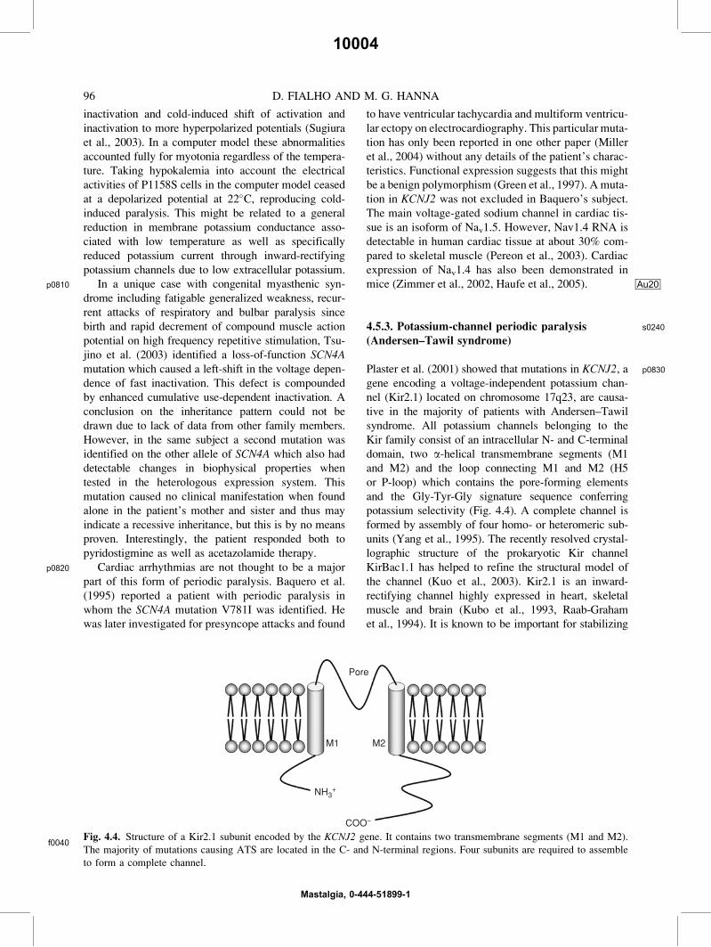
Page 15
s0140 4. Treatment
s0150 4.1. Treatment of familial periodic paralysis
s0160 4.1.1. Lifestyle and dietary advice
p0500 Simple advice on lifestyle changes to avoid recognized
triggering factors can be helpful and should be given
to all patients. In all patients with periodic paralysis
excessive exertion, particularly when followed by a
long period of rest, such as sleep overnight, should be
avoided. During an attack gentle physical activity can
be helpful in aborting symptoms. Many patients benefit
from “warming down” after exercise. Dietary advice
includes regular meals (to prevent fasting) and avoid-
ance of potassium-rich foods (banana, melon and a
number of other fruits) in hyperPP. Ingestion of carbo-
hydrate-containing drinks or snacks may abort attacks
in hyperPP while patients with hypoPP should avoid
large carbohydrate-rich meals, particularly late in the
evening.
s0170 3.4.1.2. Medication options
p0510 Potassium chloride can be used in the treatment of an
acute attack in hypoPP. Oral preparations are preferable
as there is a higher risk of rebound hyperkalemia with
intravenous administration. Regular use may reduce
the frequency of attacks. Agents that reduce urinary
potassium loss such as spironolactone (100 mg/day) or
triamterene (150 mg/day) can also improve symptoms
in hypoPP.
p0520 Patients with hyperPP may benefit from treatment
to prevent hyperkalemia including thiazide diuretics
(McArdle, 1962) and inhaled b-agonists (Wang and
Clausen, 1976, Bendheim et al., 1985, Hanna et al.,
1998).
p0530 Inhibitors of carbonic acid anhydrase (acetazol-
amide, dichlorphenamide) are useful in both hypoPP
and hyperPP (McArdle, 1962, Resnick et al., 1968).
Studies in hypoPP suggest that interictal low-grade
weakness may also improve (Griggs et al., 1970,
Dalakas and Engel, 1983). However, at present none
of the treatments used in periodic paralysis have been
proven to prevent the progressivemyopathy seen in both
hypoPP and hyperPP. The exact mechanism underlying
the beneficial effect of carbonic anhydrase inhibitors
remains unclear. One of several possibilities includes
acidification of the channel microenvironment. The
channel defect may be alleviated by a reduction in the
muscle pH as shown in expression studies for some
mutations (Kuzmenkin et al., 2002). A similar mecha-
nism may explain why gentle exercise (known to cause
transient hyperkalemia) can improve symptoms during
a mild attack. In vitro studies also show that carbonic
anhydrase inhibitor improve weakness in Kþ-deficient
rats (an animal model for hypoPP) through activation
of calcium-activated potassium channels rather than
direct inhibition of carbonic anhydrase (Tricarico
et al., 2000, 2004).
p0540Acetazolamide has been evaluated in a number of
case studies although evidence from a randomized dou-
ble-blind placebo-controlled trial is lacking. The dosage
should be started low at 62.5 or 125 mg daily and
increased gradually until a satisfactory response is
achieved but usually not higher than 1000 mg/day given
in two or three divided doses. Distal paresthesiae, head-
aches and occasionally mood disturbance including
depression can be experienced. An important long-term
complication is the development of renal calculi in 10–
20% of patients (Tawil et al., 1993). Therefore, all
patients should undergo baseline and yearly follow-up
renal imaging to enable early detection and treatment
of nephrolithiasis. Regular intake of citrus drinks
reduces the development of renal calculi.
p0550The efficacy of dichlorphenamide (50–300 mg/day)
was demonstrated in a double-blind placebo-controlled
crossover trial (Tawil et al., 2000). Despite the limita-
tions of this study such as the dropout rate and unblind-
ing of patients and investigators, the effectiveness of
dichlorphenamide to prevent or reduce the severity and
frequency of attacks in both hyperPP and hypoPP was
clearly shown. Side-effects and consequent precautions
are similar to acetazolamide.
p0560Some reports suggest that acetazolamide can exacer-
bate symptoms in patients with hypoPP due to sodium
channel mutations (Bendahhou et al., 2001, Sternberg
et al., 2001) but others report benefit (Kuzmenkin
et al., 2002, Kim et al., 2004). Treatment-induced
worsening with carbonic anhydrase inhibitors can also
occur with othermutations and patient should bewarned
and monitored accordingly.
p0570Patients with hyperPP and myotonia may also
benefit from antimyotonic agents such as mexiletine
(200–600 mg/day in two or three divided doses). Due
to its cardiac side-effects mexiletine should be moni-
tored with baseline and follow-up ECGs.
p0580Potassium-channel openers have been investigated
as potential treatment agents in hypoPP. Theoretically,
by increasing potassium conductance, the muscle
membrane could be repolarized and attacks prevented.
Diazoxide, cromakalim and pinacidil, drugs with an
antihypertensive vasodilator effect, are known to direct-
ly activate ATP-sensitive potassium channels. Diazox-
ide was initially effective in preventing attacks in
patients with hypoPP but became ineffective after a
few months (Johnsen, 1977). In vitro studies in human
hypoPP muscle fibers showed that cromakalim did
repolarize the muscle membrane and restore twitch
force (Grafe et al., 1990). Ligtenberg et al. (1996) found
PERIODIC PARALYSIS 91
Mastalgia, 0-444-51899-1
10004
Page 16
some increase in muscle strength following carbohy-
drate challenge in two out of four hypoPP patients after
using pinacidil. Clinically the use of K-ATP openers has
been limited due to severe side-effects including hypo-
tension and hyperglycaemia. Nevertheless more selec-
tive channel modulators may improve management in
the future.
s0180 4.4.2. Periodic paralysis and anesthesia
p0590 There are case reports of patients with periodic paraly-
sis having episodes of malignant hyperthermia (Paasuke
and Brownell, 1986, Lambert et al., 1994, Rajabally and
El Lahawi, 2002). In one of these patients a mutation in
the ryanodine receptor has been identified (Marchant
et al., 2004). Whether another unidentified mutation in
a voltage-gated channel is responsible for the periodic
paralysis in this particular case is uncertain. From a
practical point of view it is advisable to avoid volatile
anesthetics although there is no definite evidence of
an increased risk of malignant hyperthermia in this
patient group. The more frequent anesthetic complica-
tion is an attack of paralysis following an intervention
(Fouad et al., 1997). This is not unexpected given the
known trigger factors (stress, immobility, cold, exertion
during labor) in addition to anesthetic drugs. The man-
agement plan should take these factors into account
(avoidance or minimization of pain, carbohydrate loads
in hypoPP, fasting and cold in hyperPP, sympathomi-
metics, prolonged labor, etc.). Non-depolarizing muscle
relaxants, propofol, and regional anesthesia have been
found to be relatively safe (Aarons et al., 1989, Ashwood
et al., 1992, Cone and Sansome, 1992, Weller et al.,
2002).
s0190 4.4.3. Treatment of Andersen–Tawil syndrome
p0600 Treatment of ATS presents a particular problem as mus-
cle and cardiac symptoms often occur independently
and treatment of onemay exacerbate the other. Carbonic
anhydrase inhibitors appear to be beneficial and are
probably the first line treatment for the muscle symp-
toms. A single report suggested efficacy of terbutaline,
a ß2-agonist, reducing the frequency of paralytic attacks
(Djurhuus et al., 1998). The same patient had also
responded to potassium and spironolactone. It is curious
that a b2-agonist, usually helpful in hyperPP, and medi-
cation often given in hypoPP, should be beneficial in the
same patient. The lack of evidence from randomized
controlled trials in this rare condition is unlikely to
change soon.
p0610 The management of cardiac arrhythmias can range
between simple monitoring to necessity of pacemaker
or implantable cardioverter defibrillator. Case reports
exist on the successful use of amiodarone (Junker et al.,
2002) and imipramine (Gould et al., 1985, Tawil et al.,
1994). Imipramine does not interact with Kir2.1 channels
(Kobayashi et al., 2004) but it has inhibitory effects on
many other cardiac potassium, sodium and calcium chan-
nels (Garcia-Ferreiro et al., 2004). Beta-blockers have
been tried (Sansone et al., 1997). Verapamil has been
found beneficial in one patient (Kannankeril et al.,
2004) but worsened muscle symptoms in another
(Sansone et al., 1997).
s02004.4.4. Treatment of thyrotoxic periodic paralysis
p0620Effective treatment of TPP requires the correction of the
endocrine abnormality. Once the patient becomes
euthyroid the paralytic attacks cease and neurophysio-
logical abnormalities disappear (Jackson and Barohn,
1992). The underlying susceptibility however remains
and excessive thyroid supplementation may induce
recurrence of attacks. Correcting thyrotoxicosis can
sometimes take weeks or months during which time pre-
vention and treatment of acute attacks may be desirable
in severely affected patients.
p0630In contrast to the familial periodic paralyses no con-
vincing benefit from carbonic anhydrase inhibitors has
been described in TPP (Norris, et al., 1971, Yeung and
Tse, 1974). Most centers use potassium supplementa-
tion, a beta-blocker, or a combination to treat acute
attacks. Lu et al. (2004) conducted a small study com-
paring intravenous potassium chloride in 20 patients
with no potassium chloride administration in 12
patients. Patients in the untreated group all recovered
spontaneously but took twice as long as the treated
cohort (13.5�7.5 vs 6.3�3.8 hours, p<0.01). However,in 40% of patients receiving potassium rebound hyper-
kalemia developed with Kþ>5.5 mmol/l. Intravenous
potassium chloride for the acute treatment of paralysis
in TPP should therefore probably be reserved for severe
cases with associated cardiac arrhythmias where rapid
normalization of serum potassium level is required. In
other cases oral potassium supplement or simple moni-
toring with no potassium supplementation may suffice.
p0640Beta-blockers can be used both in acute attacks as
well as a preventive measure. It has been postulated that
hyperadrenergia during thyrotoxicosis contributes to the
muscle weakness. Indeed, a 6-day course of propranolol
(40 mg four times daily) prevented or lessened the
severity of paralysis induced by a high carbohydrate
diet in five out of seven patients with TPP (Yeung
and Tse, 1974). Oral propranolol without potassium
supplementation has been found by other authors to be
beneficial (Conway et al., 1974, Lin and Lin, 2001).
Intravenous propranolol together with potassium sup-
plementation has also been described (Payne et al.,
92 D. FIALHO AND M. G. HANNA
Mastalgia, 0-444-51899-1
10004
Page 17
1979, Shayne and Hart, 1994, Birkhahn et al., 2000).
Again, rebound hyperkalemia with cardiac arrhythmias
was observed.
s0210 4.5. Genetic and in vitro electrophysiologicalcharacteristics
s0220 4.5.1. Calcium channel periodic paralysis
p0650 Missense mutations in the pore-forming a-subunit ofthe dihydropyridine-sensitive (L-type) calcium channel
Cav1.1 of skeletal muscle are the main cause of familial
hypokalemic periodic paralysis. In 1994, in a genome-
wide search in three affected European families,
Fontaine et al. (1994) discovered linkage to chromosome
1q31–q32. They also established that the CACNA1Sgene mapped to the same region and cosegregated with
the disease with no recombinants in two families. The
first mutations were identified by Ptacek et al. (1994)
and Jurkat-Rott et al. (1994). A founder effect has not
been established (Elbaz et al., 1995, Grosson et al.,
1996).
p0660 The Cav1.1 gene spans about 73 kb, and consists of
44 exons (Drouet et al., 1993). Similarly to other volt-
age-gated sodium and calcium channels, Cav1.1 is made
up of the main pore-forming a-subunit which is asso-
ciated with accessory units (a2, d, b and g). Within the
a-subunit four homologous domains can be distin-
guished (DI–IV). Each domain correlates to a single
subunit of the voltage-gated potassium channel, which
requires four subunits to assemble a complete pore-
forming channel. Evolutionarily, the a-subunit of thecalcium and sodium channels developed through gene
duplication from these potassium channels. Each
domain of Cav1.1 is made up of six transmembrane seg-
ments. The fourth transmembrane segment (S4) con-
tains regularly-spaced positively charged amino acids
and functions as the voltage sensor. This segment is
thought to move outward upon depolarization and chan-
nel openings (Mannuzzu et al., 1996, Yang et al., 1996).
Other important structures are the loops between seg-
ments five and six of each domain which re-enter the
membrane and come together to provide the lining of
the pore and determine the ion selectivity. In skeletal
muscle conformational changes of Cav1.1 have been
shown to activate the ryanodine receptor, facilitating
calcium release from the sarcoplasmic reticulum, thus
mediating excitation-contraction coupling.
p0670 Some controversy exists regarding the precise sub-
unit topology and voltage sensor movement, following
the crystallization of a bacterial voltage-gated potassi-
um channel (Jiang et al., 2003). Two main models for
the voltage sensor movement exist (Ahern and Horn,
2004). In the conventional model, which seems to be
more in keeping with most of the experimental data
obtained so far, S4 moves in a helical screw or in a heli-
cal twist pattern inside the densely packed channel pro-
tein. However, the “paddle” model assumes that the
S4-charged helical segment and portions of S3 form a
paddle that lies at the periphery of the channel, parallel
to the intracellular membrane–water interface. During
depolarization, the paddle-like motif moves across the
membrane toward the extracellular side, thus triggering
channel opening.
p0680All four mutations identified in CACNA1S causing
periodic paralysis occur at positively charged arginines
in the voltage-sensing region of the channel. Interesting-
ly, the sodium channel mutations identified causing
hypoPP also affect positively charged arginines in the
voltage sensing region of SCN4A. Two other changes in
CACNA1S have been identified in a few families causing
malignant hyperthermia. These mutations (R1086H and
R1086C) occur in the loop connecting domains III and
IV (Monnier et al., 1997, Jurkat-Rott et al., 2000a).
p0690The exact mechanism through which mutations in
CACNA1S cause periodic paralysis is unknown. The
channel does not contribute on its own to membrane
excitability. Expression studies of mutant channels as
well as primary cultures of affected muscle have shown
only moderate functional changes. These range from
reduced current density, slowing in activation rate to
enhanced rate of closing (Lapie et al., 1996, Jurkat-Rott
et al., 1998, Morrill and Cannon, 1999). The effect of
these changes is a reduction in calcium influx into the
muscle. It has been suggested that an indirect effect on
other channels is responsible for the clinical presenta-
tion. In keeping with this, patch recordings from fibers
with the R528H mutation showed a loss of potassium
conductance of an ATP-sensitive Kþ channel (Tricarico
et al., 1999). Ruff (1999) also reported an insulin-
induced reduction in potassium currents. How this is
linked to the calcium channel remains unclear. One
hypothesis for the pathogenesis of hypoPP is that a dis-
ruption of the calcium homeostasis due to mutant
Cav1.1 channels alters the transcription, expression or
regulation of other ion channels including potassium
channels. A reduced potassium current in turn could
then explain the depolarized resting potential and the
intracellular trapping of potassium during attacks.
p0700Even at baseline the resting potential in hypoPP
muscle is depolarized by 5–10 mV compared to normal
(Rudel et al., 1984, Ruff, 1999). Hypokalemia in
hypoPP results from the physiological effect of glucose
intake and the release of insulin which in turn stimu-
lates the sodium–potassium pump and shifts potassium
from the extracellular to the intracellular space. In nor-
mal muscle fibers this leads to hyperpolarization.
In contrast, in hypoPP muscle fibers hypokalemia
PERIODIC PARALYSIS 93
Mastalgia, 0-444-51899-1
10004
Page 18
causes depolarization and induces an attack of paralysis
(Rudel et al., 1984, Minaker et al., 1988).
s0230 4.5.2. Sodium-channel periodic paralysis
p0710 Clinically the sodium channelopathies of skeletal mus-
cle can be divided into three main allelic disorders:
hyperkalemic periodic paralysis, paramyotonia conge-
nita and potassium-aggravated myotonia. Patients with
sodiumchannel hyperPPmay also complain of symptoms
suggestive of paramyotonia congenita or potassium-
aggravated myotonia as these conditions frequently
overlap (Sasaki et al., 1999).
p0720 The pioneering work on muscle specimens from
myotonic goat by Bryant and colleagues (Bryant,
1962, Lipicky and Bryant, 1966) identified the loss of
resting chloride conductance as the primary underlying
defect, which was later confirmed in myotonia conge-
nita in humans (Lipicky and Bryant, 1973). In the early
1980s Lehmann-Horn and colleagues undertook a series
of in-vitro electrophysiological studies on human inter-
costals muscle fibers to see whether patients with both
myotonia and periodic paralysis also had a chloride-
channel defect (Lehmann-Horn et al., 1981, 1983).
Unlike in muscle with myotonia congenita, chloride
conductance was normal but they identified an anoma-
lous persistent inward cation current. This current was
blocked by tetrodotoxin which implicated the voltage-
gated skeletal-muscle sodium channel. An isoform of
the a-subunit of this channel was first cloned from rat
by Trimmer et al. (1989). The human gene SCN4Amaps
to 17q23–q24, spans 35 kb, contains 25 exons and codes
for a 1836-amino-acid protein (George et al., 1991,
1992, 1993). Linkage for hyperkalemic periodic paraly-
sis to SCN4A was found in 1990 by Fontaine et al.
(1990). This was confirmed by Ptacek et al. (1991b)
and Koch et al. (1991a). Several groups found linkage
of paramyotonia congenita to SCN4A establishing the
fact that these are allelic disorders (Ebers et al., 1991,
Koch et al., 1991b, Ptacek et al., 1991c).
p0730The structure of the channel subunit encoded by
SCN4A is analogous to the a-subunit of the skeletal-
muscle voltage-gated calcium channel (Fig. 4.3). Four
domains each composed of six transmembrane seg-
ments form the main channel. The S4 segment acts as
a voltage sensor and the S5–S6 loop lines the pore.
Channel function is modulated by small b-subunits.All pathogenic changes identified so far have been mis-
sense mutations of conserved amino acids of the a-sub-unit, resulting in periodic paralysis and myotonia. No
mutations have been identified in the b1-subunit asso-ciated with neuromuscular disorder but a missense
mutation has been found to be a rare cause of
generalized epilepsy with febrile seizures (Wallace
et al., 1998).
p0740Three main conformations exist for the sodium
channel. After membrane depolarization the sodium
channels open within a fraction of a millisecond and
the resulting inward flux of sodium ions accounts for
the rapid upstroke of the action potential. The sodium
channels then become rapidly inactivated even if
depolarization continues. The linker between domains
III and IV is thought to act as a hinged lid, which
occludes the channel on fast inactivation. Only mem-
brane repolarization allows sodium channels to change
from the inactivated state to the resting state from
which further activation is possible.
p0750The majority of mutations in SCN4A lead to a gain-
of-function defect. In response to depolarization mutant
sodium channels open normally andmaintain selectivity
DI DII DIII DIV
Extracellular
Intracellular
NH3+
COO−
1 2 3 4+
+
+++
+++
+++
5 6
f0030Fig. 4.3. Membrane-spanning topology of the a-subunit of the skeletal muscle sodium channel Nav1.4. Each domain (DI–IV)
contains six transmembrane segments (S1–6). The structure of the a-subunit of the L-type skeletal muscle calcium channel
Cav1.1 is homologous.
94 D. FIALHO AND M. G. HANNA
Mastalgia, 0-444-51899-1
10004
Page 19
for sodium ions, but they inactivate less completely, too
slowly, recover too quickly or have a shifted voltage
dependence. Some mutations (including the most com-
mon hyperPP mutation T704M) shift the activation to
more hyperpolarized potentials (Cummins et al.,
1993). Both inactivation and activation defects result
in an increase in sodium current conducted by the
mutant channels compared to the wild-type.
p0760 The question arises how gain-of-function mutations
in the sodium channel gene can lead to seemingly oppo-
site clinical presentations with increase in excitability
(myotonia) on the one hand and loss of excitability
(paralysis) on the other. In-vitro experiments and com-
puter modeling have provided an answer to this prob-
lem. A toxin-based subtle disruption of sodium
channel inactivation of about 2% in rat muscle in vitro
can cause myotonia (Cannon and Corey, 1993). A com-
puter simulation of a model fiber confirmed that a small
defect of inactivation produces repetitive discharges fol-
lowing a single pulse stimulation (Cannon and Corey
al., 1993). An increase of failed inactivation to only
about 3% induces a susceptibility for a depolarizing
shift of the restingmembrane potential after stimulation.
This depolarized membrane potential of �40mV is
maintained by the sodium channels which have failed
to inactivate while at the same time the majority
of sodium channels (both mutant and wild type) are
inactivated, which leads to inexcitability and paralysis.
In keeping with these findings expression studies have
shown that paralysis-associated mutations tend to
cause a more severe disruption of gating compared to
those leading to myotonia (Cannon, 2000). The toxin-
based model also demonstrated a common mechanism
between chloride- and sodium-channel myotonia. Each
action potential in skeletal muscle leads to outward flow
of potassium into the extracellular space. In skeletal
muscle this includes the T-tubule system which consists
of long narrow invaginations of the cell membrane
and allows propagation of action potentials into the core
of the fiber. Although these T-tubules communicate
with the extracellular space they also present a signi-
ficant diffusion barrier. During sustained contraction
activity-dependent potassium accumulation occurs
and in the presence of reduced chloride conductance or
sodium-channel inactivation defect this increase in
potassium is sufficient to trigger myotonic discharges.
p0770 Another feature of sodium-channel function is the
presence of fast inactivation (milliseconds) and slow
inactivation (seconds to minutes) mechanisms which
are operated through different molecular gates. Ruff
(1994) suggested that a defect in slow inactivation
must be present for paralysis-associated mutations as
the slow inactivation mechanism would otherwise lead
to a shutdown of the mutant sodium channels which
have failed to close down and thus allow repolariza-
tion of the membrane. This has been confirmed in in-
vitro expression systems for the two most common
mutations that lead to hyperkalemic periodic paralysis
(T704M and M1592V) and a mutation associated with
cold-induced weakness (I693T; Hayward et al., 1999).
Some rare mutations exist that cause periodic paralysis
without impairment of slow inactivation.
p0780In contrast to the above, SCN4A loss-of-function
defects have been identified in a subset of patients with
hypokalemic periodic paralysis (Bulman et al., 1999, Jur-
kat-Rott et al., 2000b, Bendahhou et al., 2001). All of the
mutations are located in the voltage-sensing segment S4
of domain II and all neutralize positively charged argi-
nines in analogy to the hypoPP calcium-channel muta-
tions. The phenotype of patients with calcium-channel
compared to sodium-channel hypokalemic periodic
paralysis is identical (Jurkat-Rott et al., 2000b). Elec-
trophysiologically, thesemutations attenuate sodium cur-
rent due to excess fast and slow inactivation and reduced
density of sodium channels (Struyk et al., 2000, Jurkat-
Rott et al., 2000b, Bendahhou et al., 2001, Kuzmenkin
et al., 2002). The production and insertion of normal sodi-
um channels did not compensate for the reduced sodium
current, which raises the question of how skeletal muscle
fibers regulate the expression of sodium channels to con-
trol membrane excitability. Interestingly, muscle fibers
with a calcium-channel mutation associatedwith hypoPP
have also been shown to have a reduction in sodium
current (Ruff and Al-Mudallal, 2000).
p0790The distinction between SCN4A mutations asso-
ciated with hyper- or hypoPP may not always be so
clear. Vicart et al. (2004) reported four kindreds with
three new SCN4A mutations affecting an arginine at
position 675, located in the S4 voltage sensor of domain
II adjacent to residues R669 and R672 where mutations
causing hypoPP have been identified. Administration of
corticosteroids resulted in severe weakness associated
with hypokalemia in two affected individuals from dif-
ferent families, in one of them in the presence of thyro-
toxicosis. Repeated ictal testing however did not reveal
consistent potassium abnormalities in a number of
affected subjects during attacks. The presence of EMG
myotonia in one individual together with symptoms of
muscle cramps and stiffness and provocation by cold
and fasting may point towards a defect similar to
hyperPP mutations but functional expression data is
awaited.
p0800The P1158S mutation located in the linking loop
between segments 4 and 5 of domain III was identified
in a single kindred with cold-induced hypoPP and myo-
tonia (Sugiura et al., 2000). This is the only mutation
where a true combination of hypoPP and myotonia
exists. Functional expression identified a slowing of
PERIODIC PARALYSIS 95
Mastalgia, 0-444-51899-1
10004
Page 20
inactivation and cold-induced shift of activation and
inactivation to more hyperpolarized potentials (Sugiura
et al., 2003). In a computer model these abnormalities
accounted fully for myotonia regardless of the tempera-
ture. Taking hypokalemia into account the electrical
activities of P1158S cells in the computer model ceased
at a depolarized potential at 22�C, reproducing cold-
induced paralysis. This might be related to a general
reduction in membrane potassium conductance asso-
ciated with low temperature as well as specifically
reduced potassium current through inward-rectifying
potassium channels due to low extracellular potassium.
p0810 In a unique case with congenital myasthenic syn-
drome including fatigable generalized weakness, recur-
rent attacks of respiratory and bulbar paralysis since
birth and rapid decrement of compound muscle action
potential on high frequency repetitive stimulation, Tsu-
jino et al. (2003) identified a loss-of-function SCN4Amutation which caused a left-shift in the voltage depen-
dence of fast inactivation. This defect is compounded
by enhanced cumulative use-dependent inactivation. A
conclusion on the inheritance pattern could not be
drawn due to lack of data from other family members.
However, in the same subject a second mutation was
identified on the other allele of SCN4A which also had
detectable changes in biophysical properties when
tested in the heterologous expression system. This
mutation caused no clinical manifestation when found
alone in the patient’s mother and sister and thus may
indicate a recessive inheritance, but this is by no means
proven. Interestingly, the patient responded both to
pyridostigmine as well as acetazolamide therapy.
p0820 Cardiac arrhythmias are not thought to be a major
part of this form of periodic paralysis. Baquero et al.
(1995) reported a patient with periodic paralysis in
whom the SCN4A mutation V781I was identified. He
was later investigated for presyncope attacks and found
to have ventricular tachycardia and multiform ventricu-
lar ectopy on electrocardiography. This particular muta-
tion has only been reported in one other paper (Miller
et al., 2004) without any details of the patient’s charac-
teristics. Functional expression suggests that this might
be a benign polymorphism (Green et al., 1997). A muta-
tion in KCNJ2 was not excluded in Baquero’s subject.
The main voltage-gated sodium channel in cardiac tis-
sue is an isoform of Nav1.5. However, Nav1.4 RNA is
detectable in human cardiac tissue at about 30% com-
pared to skeletal muscle (Pereon et al., 2003). Cardiac
expression of Nav1.4 has also been demonstrated in
mice (Zimmer et al., 2002, Haufe et al., 2005 Au20).
s02404.5.3. Potassium-channel periodic paralysis
(Andersen–Tawil syndrome)
p0830Plaster et al. (2001) showed that mutations in KCNJ2, agene encoding a voltage-independent potassium chan-
nel (Kir2.1) located on chromosome 17q23, are causa-
tive in the majority of patients with Andersen–Tawil
syndrome. All potassium channels belonging to the
Kir family consist of an intracellular N- and C-terminal
domain, two a-helical transmembrane segments (M1
and M2) and the loop connecting M1 and M2 (H5
or P-loop) which contains the pore-forming elements
and the Gly-Tyr-Gly signature sequence conferring
potassium selectivity (Fig. 4.4). A complete channel is
formed by assembly of four homo- or heteromeric sub-
units (Yang et al., 1995). The recently resolved crystal-
lographic structure of the prokaryotic Kir channel
KirBac1.1 has helped to refine the structural model of
the channel (Kuo et al., 2003). Kir2.1 is an inward-
rectifying channel highly expressed in heart, skeletal
muscle and brain (Kubo et al., 1993, Raab-Graham
et al., 1994). It is known to be important for stabilizing
Pore
M1 M2
NH3+
COO−
f0040Fig. 4.4. Structure of a Kir2.1 subunit encoded by the KCNJ2 gene. It contains two transmembrane segments (M1 and M2).
The majority of mutations causing ATS are located in the C- and N-terminal regions. Four subunits are required to assemble
to form a complete channel.
96 D. FIALHO AND M. G. HANNA
Mastalgia, 0-444-51899-1
10004
Page 21
the resting potential in cardiac muscle and thought to
contribute to the late-repolarization phase in both skele-
tal and cardiac muscle. Inward rectification refers to the
fact that the channel permits inward flux of potassium
at membrane potentials negative to the potassium rever-
sal potential more easily compared to outward flux at
more positive potentials. This prevents excess potassi-
um loss during the plateau phase of the cardiac action
potential but allows participation in the late repolariza-
tion. The closure of Kir2 channels occurs due to binding
of intracellular magnesium or cationic polyamines at
potentials positive to the potassium reversal potential
(Lopatin and Nichols, 2001). The open state of Kir2.1
and other inward-rectifying channels is facilitated by
phosphatidylinositol 4,5-bisphosphate (PIP2; Huang
et al., 1998). PIP2 is a membrane-bound phospholipid
which acts as a precursor for secondary messengers. It
binds directly to Kir channels through interaction
between positively charged amino acids of the Kir
channel and negatively charged phosphate groups of
the lipid. Three putative PIP2 binding sites exist within
the C-terminal domain of Kir2.1 (Soom et al., 2001).
p0840 In rat embryos mRNA is detectable in cardiac and
skeletal muscle, brain, metanephrons and developing
bony structures of the cranium, extremities and verteb-
rae (Karschin and Karschin, 1997). This closely mirrors
the organ systems affected in ATS. A Kir2.1 knockout
mouse showed developmental craniofacial abnormal-
ities in analogy with ATS (Zaritsky et al., 2000). Func-
tional expression of the majority of mutations so far has
demonstrated a dominant-negative effect on wild-type
subunits in the tetrameric channel. The clinical severity
of symptoms does not seem to be correlated with the
degree of dominant negative effect in expression studies
(Tristani-Firouzi et al., 2002). At least half of the muta-
tions impair interaction with PIP2 (Tristani-Firouzi
et al., 2002, Lopes et al., 2002, Donaldson et al.,
2003). The delS314–Y315 mutation has been shown
to interfere with protein trafficking leading to intracellu-
lar trapping of the channel containing one or more
mutant subunit (Bendahhou et al., 2003). The same
study suggested that the mutation V302M disrupts both
channel trafficking or folding as well as assembly
trapping only mutant subunits in the cell and causing
ATS through a haploinsufficiency mechanism.
p0850 Preisig-Muller et al. (2002) demonstrated the ability
of Ki2.1 to form heteromeric channels with potassium
channel subunits from the Kir2 subfamily (Kir 2.2 and
2.3). The also showed a dominant-negative effect of
mutant Kir2.1 subunits on these heteromers. This finding
may provide a possible explanation of the phenotypic
variation within and between families with ATS.
p0860 Of interest also is the recent discovery of two gain-of-
function mutations in KCNJ2 underlying familial atrial
fibrillation in a Chinese kindred (Xia et al., 2005) and
short QT syndrome in another family (Priori et al.,
2005). Neither of the two families had any dysmorphic
features or skeletal muscle symptoms.
s02504.5.4. Thyrotoxic periodic paralysis
p0870Although TPP typically occurs sporadically, the heavily
skewed ethnic distribution suggests a genetic compo-
nent. It is suspected that in thyrotoxic periodic paralysis
a genetically determined susceptibility to abnormal
membrane excitability exists that is only unmasked in
the presence of hyperthyroidism. It is not clear whether
the primary abnormality is associated with one of the
voltage-gated skeletal muscle ion channel genes or a
gene that has a secondary effect. Screening for muta-
tions in CACNA1S and SCN4A known to be associated
with hypokalemic periodic paralysis has been negative
(Dias da Silva et al., 2002b, Kung et al., 2004).
p0880The associated hypokalemia in TPP is to be due to a
rapid influx of potassium into cells similarly to the
familial periodic paralyses (Feely, 1981). The sodium–
potassium ATPase is an important transporter that
allows potassium to be pumped into the cells. Thyrotox-
icosis causes an increase in number and activity of the
sodium-potassiumATPase per se, but this effect is more
pronounced in patients with TPP (Oh et al., 1990, Chan
et al., 1991). The difference between thyrotoxic patients
with and without TPP disappears after restoration of the
euthyroid status.
p0890Many recent genetic studies in TPP have concentrated
on detection of polymorphisms with potential functional
effects in membrane channel or transporter genes. Dias
da Silva et al. (2002a) discovered two polymorphisms
in CACNA1S at nucleotides 1551 and 1564 at higher fre-quency in 13 cases of sporadic thyrotoxic periodic paral-
ysis compared to normal controls (77% and 31% vs 18%
and 8.6%). This was not confirmed in a larger study
including 97 male Chinese patients with TPP who were
screened for polymorphisms in the coding and promoter
region ofCACNA1S in addition to microsatellite markers
in the region of the Na/K-ATPase subunits a1, a2 and b1(Kung et al., 2004). However, the latter study identified
two intronic and one 50-flanking region single nuclear
polymorphisms (SNPs) in CACNA1S which occurred
with significantly different frequencies compared to
groups of normal controls and thyrotoxic patients without
periodic paralysis. All three SNPs are located at or near
putative thyroid hormone response elements but whether
they have any functional effect remains to be seen. The
authors hypothesized that these SNPs may modulate the
effect of thyroid hormones on the expression of CAC-NA1S. Polymorphisms in the b2-adrenergic receptor
PERIODIC PARALYSIS 97
Mastalgia, 0-444-51899-1
10004
Page 22
gene were not found to be associated with TPP (Kim
et al., 2005).
p0900 Dias da Silva et al. (2002b) also described the muta-
tion R83H in KCNE3 in a patient with TPP. KCNE3encodes the MinK-related peptide 2 (MiRP2) which
coassembles with Kv3.4 to form the human skeletal-
muscle voltage-gated potassium channel. This change
had been reported previously in a case of familial
HypoPP (Abbott et al., 2001). However more detailed
studies later showed that it was in fact a polymorphism
(Sternberg et al., 2003, Jurkat-Rott and Lehmann-Horn,
2004).
p0910 Human leukocyte antigen (HLA) markers have been
extensively studied. Various associations with TPP have
been reported which differ according to the population
studied (Yeo et al., 1978, Hawkins et al., 1985, Tamai
et al., 1987, Cavan et al., 1994), but no consistent
marker has emerged.
s0260 Acknowledgements
p0920 Doreen Fialho is a CINCH-NIH fellow. Research in
our laboratory is supported by the Wellcome Trust
UK and MRC UK. Our clinical and DNA diagnos-
tic service is funded by the UK NHS Department of
Health through the national specialist commissioning
agency — NSCAG.
References
Aarons JJ, Moon RE, Camporesi EM (1989). General anesthe-
sia and hyperkalemic periodic paralysis. Anesthesiology 71:
303–304.
Abbott GW, Butler MH, Bendahhou S, et al. (2001). MiRP2
forms potassium channels in skeletal muscle with Kv3.4
and is associated with periodic paralysis. Cell 104: 217–231.
Adachihara K, Takagi Y (1974). [Case of thyrotoxic paralysis
associated with hyperkalemic and hypokalemic paralytic
attacks]. Rinsho Shinkeigaku 14: 369–375.
Ahern CA, Horn R (2004). Stirring up controversy with a
voltage sensor paddle. Trends Neurosci 27: 303–307.
Ahlawat SK, Sachdev A (1999). Hypokalaemic paralysis.
Postgrad Med J 75: 193–197.
Ai T, Fujiwara Y, Tsuji K, et al. (2002). Novel KCNJ2
mutation in familial periodic paralysis with ventricular
dysrhythmia. Circulation 105: 2592–2594.
Allen AS (1943). Pa Ping or Kiating paralysis. Chin Med J
(Engl) 61: 296–301.
Andelfinger G, Tapper AR, Welch RC, et al. (2002). KCNJ2
mutation results in Andersen syndrome with sex-specific
cardiac and skeletal muscle phenotypes. Am J Hum Genet
71: 663–668.
Andersen ED, Krasilnikoff PA, Overvad H (1971). Intermittent
muscular weakness, extrasystoles, and multiple develop-
mental anomalies. A new syndrome? Acta Paediatr Scand
60: 559–564.
Angeloni JM, Scott GW (1960). Flaccid quadriplegia follow-
ing ureteric transplant. Lancet 1: 1005–1006.
Ashwood EM, Russell WJ, Burrow DD (1992). Hyperkalaemic
periodic paralysis and anaesthesia. Anaesthesia 47: 579–584.
Baquero JL, Ayala RA, Wang J, et al. (1995). Hyperkalemic
periodic paralysis with cardiac dysrhythmia: a novel sodium
channel mutation? Ann Neurol 37: 408–411.
Basser LS (1979). Purgatives and periodic paralysis. Med J
Aust 1: 47–48.
Bendahhou S, Cummins TR, Griggs RC, et al. (2001).
Sodium channel inactivation defects are associated with
acetazolamide-exacerbated hypokalemic periodic paralysis.
Ann Neurol 50: 417–420.
Bendahhou S, Donaldson MR, Plaster NM, et al. (2003).
Defective potassium channel Kir2.1 trafficking underlies
Andersen–Tawil syndrome. J Biol Chem 278: 51779–51785.
Bendahhou S, Fournier E, Sternberg D, et al. (2005). In vivo
and in vitro functional characterization of Andersen’s
syndrome mutations. J Physiol 565: 731–741.
Bender JA (1936). Family periodic paralysis in a girl aged
seventeen. Arch Neurol Psychiatry 35: 131–135.
Bendheim PE, Reale EO, Berg BO (1985). Beta-adrenergic
treatment of hyperkalemic periodic paralysis. Neurology
35: 746–749.
Bennett RH, Forman HR (1980). Hypokalemic periodic paral-
ysis in chronic toluene exposure. Arch Neurol 37: 673.
Berning J (1975). Letter: Hypokalaemia of barium poison-
ing. Lancet 1: 110.
Biemond A, Daniels AP (1934). Familial periodic paralysis
and its transition into spinal muscular atrophy. Brain 57:
91–108.
Birkhahn RH, Gaeta TJ, Melniker L (2000). Thyrotoxic peri-
odic paralysis and intravenous propranolol in the emergen-
cy setting. J Emerg Med 18: 199–202.
Bradley WG, Taylor R, Rice DR, et al. (1990). Progressive
myopathy in hyperkalemic periodic paralysis. Arch Neurol
47: 1013–1017.
Bresolin NL, Grillo E, Fernandes VR, et al. (2005). A case
report and review of hypokalemic paralysis secondary to
renal tubular acidosis. Pediatr Nephrol 20: 818–820.
Brouwer OF, Zwarts MJ, Links TP (1992). Muscle fiber con-
duction velocity in the diagnosis of sporadic hypokalemic
periodic paralysis. Clin Neurol Neurosurg 94: 149–151.
Bryant SH (1962). Muscle membrane of normal and myotonic
goats in normal and low external chloride. Fed Proc 21: 312.
Bulman DE, Scoggan KA, van Oene MD, et al. (1999). A
novel sodium channel mutation in a family with hypokale-
mic periodic paralysis. Neurology 53: 1932–1936.
Buruma OJ, Bots GT (1978). Myopathy in familial hypoka-
laemic periodic paralysis independent of paralytic attacks.
Acta Neurol Scand 57: 171–179.
Cannon SC (2000). Spectrum of sodium channel disturbances
in the nondystrophic myotonias and periodic paralyses.
Kidney Int 57: 772–779.
Cannon SC, Corey DP (1993). Loss of Naþ channel inactiva-
tion by anemone toxin (ATX II) mimics the myotonic state
in hyperkalaemic periodic paralysis. J Physiol 466:
501–520.
98 D. FIALHO AND M. G. HANNA
Mastalgia, 0-444-51899-1
10004
Page 23
Canun S, Perez N, Beirana LG (1999). Andersen syndrome
autosomal dominant in three generations. Am J Med Genet
85: 147–156.
Cavan DA, Penny MA, Jacobs KH, et al. (1994). The HLA
association with Graves’ disease is sex-specific in Hong
Kong Chinese subjects. Clin Endocrinol (Oxf) 40: 63–66.
Chan A, Shinde R, Chow CC, et al. (1991). In vivo and
in vitro sodium pump activity in subjects with thyrotoxic
periodic paralysis. BMJ 303: 1096–1099.
Chen KM, Hung TP, Lin TY (1965). Periodic paralysis in Tai-
wan. Clinical study of 28 cases. Arch Neurol 12: 165–171.
Chinnery PF, Walls TJ, Hanna MG, et al. (2002). Normokale-
mic periodic paralysis revisited: does it exist? Ann Neurol
52: 251–252.
Chun TU, Epstein MR, Dick M, et al. (2004). Polymorphic
ventricular tachycardia and KCNJ2 mutations. Heart
Rhythm 1: 235–241.
Cohen T (1959). Hypokalemic muscle paralysis associated
with administration of chlorothiazide. J Am Med Assoc
170: 2083–2085.
Cone AM, Sansome AJ (1992). Propofol in hyperkalaemic
periodic paralysis. Anaesthesia 47: 1097.
Conn JW, Knopf RF, Nesbit RM (1964). Clinical character-
istics of primary aldosteronism from an analysis of 145
cases. Am J Surg 107: 159–172.
Conway MJ, Seibel JA, Eaton P (1974). Thyrotoxicosis and
periodic paralysis: improvement with beta blockade. Ann
Intern Med 81: 332–336.
Crafts LM (1900). A fifth case of family periodic paralysis.
Am J Med Sci 146: 651.
Cruz-Martinez A, Arpa J (1997). Muscle fiber conduction
velocity in situ in hypokalemic periodic paralyses. Acta
Neurol Scand 96: 229–235.
Cumberbatch GL, Hampton TJ (1999). Hyperkalaemic paral-
ysis — a bizarre presentation of renal failure. J Accid
Emerg Med 16: 230–232.
Cumming AM, Boddy K, Brown JJ, et al. (1980). Severe
hypokalaemia with paralysis induced by small doses of
liquorice. Postgrad Med J 56: 526–529.
Cummins TR, Zhou J, Sigworth FJ, et al. (1993). Functional
consequences of a Naþ channel mutation causing hyper-
kalemic periodic paralysis. Neuron 10: 667–678.
Dalakas MC, Engel WK (1983). Treatment of “permanent”
muscle weakness in familial hypokalemic periodic paraly-
sis. Muscle Nerve 6: 182–186.
Danowski TS, Fisher ER, Vidalon C, et al. (1975). Clinical
and ultrastructural observations in a kindred with normo-
hyperkalaemic periodic paralysis. J Med Genet 12: 20–28.
Daughaday WH, Rendleman D (1967). Severe symptomatic
hyperkalemia in an adrenalectomized woman due to
enhanced mineralocorticoid requirement. Ann Intern Med
66: 1197–1203.
Davies NP, Eunson LH, Samuel M, et al. (2001). Sodium
channel gene mutations in hypokalemic periodic paralysis:
an uncommon cause in the UK. Neurology 57: 1323–1325.
Davies NP, Imbrici PFD, Herd C, et al. (2005). Andersen-
Tawil syndrome — new potassium channel mutations and
possible phenotypic variation. Neurology 11: 1083–1089.
De Keyser J, Smitz J, Malfait R, et al. (1987). Rhabdomyolysis
in hypokalaemic periodic paralysis: a clue to the mechanism
that terminates the paralytic attack? J Neurol 234: 119–121.
de Silva SM, Kuncl RW, Griffin JW, et al. (1990). Paramyo-
tonia congenita or hyperkalemic periodic paralysis? Clini-
cal and electrophysiological features of each entity in one
family. Muscle Nerve 13: 21–26.
Dias da Silva MR, Cerutti JM, Tengan CH, et al. (2002a).
Mutations linked to familial hypokalaemic periodic paraly-
sis in the calcium channel alpha1 subunit gene (Cav1.1) are
not associated with thyrotoxic hypokalaemic periodic paral-
ysis. Clin Endocrinol (Oxf) 56: 367–375.
Dias Da Silva MR, Cerutti JM, Arnaldi LA, et al. (2002b). A
mutation in the KCNE3 potassium channel gene is asso-
ciated with susceptibility to thyrotoxic hypokalemic peri-
odic paralysis. J Clin Endocrinol Metab 87: 4881–4884.
Djurhuus MS, Klitgaard NA, Jensen BM, et al. (1998). Multi-
ple anomalies, hypokalaemic paralysis and partial symp-
tomatic relief by terbutaline. Acta Paediatr 87: 475–477.
Donaldson MR, Jensen JL, Tristani-Firouzi M, et al. (2003).
PIP2 binding residues of Kir2.1 are common targets of
mutations causing Andersen syndrome. Neurology 60:
1811–1816.
Drouet B, Garcia L, Simon-Chazottes D, et al. (1993). The
gene coding for the alpha 1 subunit of the skeletal dihydro-
pyridine receptor (Cchl1a3 ¼ mdg) maps to mouse chromo-
some 1 and human 1q32. Mamm Genome 4: 499–503.
Dutta D, Fischler M, McClung A (2001). Angiotensin con-
verting enzyme inhibitor induced hyperkalaemic paralysis.
Postgrad Med J 77: 114–115.
Ebers GC, George AL, Barchi RL, et al. (1991). Paramyoto-
nia congenita and hyperkalemic periodic paralysis are
linked to the adult muscle sodium channel gene. Ann
Neurol 30: 810–816.
Elbaz A, Vale-Santos J, Jurkat-Rott K, et al. (1995). Hypoka-
lemic periodic paralysis and the dihydropyridine receptor
(CACNL1A3): genotype/phenotype correlations for two
predominant mutations and evidence for the absence of a
founder effect in 16 caucasian families. Am J Hum Genet
56: 374–380.
Engel AG (1970). Evolution and content of vacuoles in pri-
mary hypokalemic periodic paralysis. Mayo Clin Proc
45: 774–814.
Engel AG, Lambert EH, Rosevear JW, et al. (1965). Clinical
and electromyographic studies in a patient with primary
hypokalaemic periodic paralysis. Am J Med 38: 626–640.
Evers S, Engelien A, Karsch V, et al. (1998). Secondary
hyperkalaemic paralysis. J Neurol Neurosurg Psychiatry
64: 249–252.
FarinaD, Zagari D,GazzoniM, et al. (2004). Reproducibility of
muscle-fiber conduction velocity estimates using multichan-
nel surface EMG techniques. Muscle Nerve 29: 282–291.
Feely J (1981). Potassium shift in thyrotoxic periodic paralysis.
Postgrad Med J 57: 238–239.
Fisher J (1982). Thyrotoxic periodic paralysis with ventricu-
lar fibrillation. Arch Intern Med 142: 1362–1364.
Fodstad H, Swan H, Auberson M, et al. (2004). Loss-
of-function mutations of the Kþ channel gene KCNJ2
PERIODIC PARALYSIS 99
Mastalgia, 0-444-51899-1
10004
Page 24
constitute a rare cause of long QT syndrome. J Mol Cell
Cardiol 37: 593–602.
Fontaine B (1994). Primary periodic paralysis and muscle
sodium channel. Adv Nephrol Necker Hosp 23: 191–197.
Fontaine B, Khurana TS, Hoffman EP, et al. (1990). Hyper-
kalemic periodic paralysis and the adult muscle sodium
channel alpha-subunit gene. Science 250: 1000–1002.
Fontaine B, Vale-Santos J, Jurkat-Rott K, et al. (1994).
Mapping of the hypokalaemic periodic paralysis (HypoPP)
locus to chromosome 1q31–32 in three European families.
Nat Genet 6: 267–272.
Fouad G, Dalakas M, Servidei S, et al. (1997). Genotype–
phenotype correlations of DHP receptor alpha 1-subunit gene
mutations causing hypokalemic periodic paralysis. Neu-
romuscul Disord 7: 33–38.
Fournier E, Arzel M, Sternberg D, et al. (2004). Electromy-
ography guides toward subgroups of mutations in muscle
channelopathies. Ann Neurol 56: 650–661.
Fujimoto T, Shiiki H, Takahi Y, et al. (2001). Primary Sjogren’s
syndrome presenting as hypokalaemic periodic paralysis and
respiratory arrest. Clin Rheumatol 20: 365–368.
Gallant EM (1983). Barium-treated mammalian skeletal
muscle: similarities to hypokalaemic periodic paralysis.
J Physiol 335: 577–590.
Gamstorp I (1956). Adynamia episodica hereditaria. Acta
Paediatr Scand 45: 1–126.
Garcia-Ferreiro RE, Kerschensteiner D, Major F, et al.
(2004). Mechanism of block of hEag1 Kþ channels by
imipramine and astemizole. J Gen Physiol 124: 301–317.
George AL Jr, Ledbetter DH, Kallen RG, et al. (1991). Assign-
ment of a human skeletal muscle sodium channel alpha-
subunit gene (SCN4A) to 17q23.1–25.3. Genomics 9:
555–556.
George AL Jr, Komisarof J, Kallen RG, et al. (1992). Primary
structure of the adult human skeletal muscle voltage-
dependent sodium channel. Ann Neurol 31: 131–137.
George AL Jr, Iyer GS, Kleinfield R, et al. (1993). Genomic
organization of the human skeletal muscle sodium channel
gene. Genomics 15: 598–606.
Ghose R, Quail G, King R, et al. (1996). Hypokalaemic paral-
ysis in remote aboriginal communities. Aust Fam Physician
25: 1172–1173.
Goldflam S (1890). Ueber eine eigentumliche Form von per-
iodischer familiarer, wahrscheinlich auto-intoxicatorischer
Paralyse. Wien Med Presse 31: 1418.
Goldflam S (1895). Weitere Mitteilung uber die paroxys-
male, familiare Lahmung. Dtsch Z Nervenheilkd 7: 1–31.
Goldflam S (1897). Dritte Mitteilung uber die paroxysmale,
familiare Lahmung. Dtsch Z Nervenheilkd 11: 242–260.
Gould RJ, Steeg CN, Eastwood AB, et al. (1985). Potentially
fatal cardiac dysrhythmia and hyperkalemic periodic paraly-
sis. Neurology 35: 1208–1212.
Grafe P, Quasthoff S, Strupp M, et al. (1990). Enhancement of
Kþ conductance improves in vitro the contraction force of
skeletal muscle in hypokalemic periodic paralysis. Muscle
Nerve 13: 451–457.
GreenDS, Hayward LJ, GeorgeAL Jr, et al. (1997). A proposed
utation, Val781Ile, associated with hyperkalemic periodic
paralysis and cardiac dysrhythmia is a benign polymorphism.
Ann Neurol 42: 253–256.
Griggs RC, Engel WK, Resnick JS (1970). Acetazolamide
treatment of hypokalemic periodic paralysis. Prevention
of attacks and improvement of persistent weakness. Ann
Intern Med 73: 39–48.
Grosson CL, Esteban J, McKenna-Yasek D, et al. (1996).
Hypokalemic periodic paralysis mutations: confirmation
of mutation and analysis of founder effect. Neuromuscul
Disord 6: 27–31.
Haddad S, Arabi Y, Shimemeri AA (2004). Hypokalemic
paralysis mimicking Guillain-Barre syndrome and causing
acute respiratory failure. Middle East J Anesthesiol 17:
891–897.
Hanna MG, Stewart J, Schapira AH, et al. (1998). Salbutamol
treatment in a patient with hyperkalaemic periodic para-
lysis due to a mutation in the skeletal muscle sodium
channel gene (SCN4A). J Neurol Neurosurg Psychiatry
65: 248–250.
Hartwig H (1874). Ueber einen Fall von intermittierender
Paralysis spinalis. Inaug Diss Halle .
Hawkins BR, Ma JT, Lam KS, et al. (1985). Association of
HLA antigens with thyrotoxic Graves’ disease and periodic
paralysis in Hong Kong Chinese. Clin Endocrinol (Oxf) 23:
245–252.
Hayward LJ, Sandoval GM, Cannon SC (1999). Defective
slow inactivation of sodium channels contributes to famil-
ial periodic paralysis. Neurology 52: 1447–1453.
Holtzapple GE (1905). Periodic paralysis. JAMA 45: 1224.
Hosaka Y, Hanawa H, Washizuka T, et al. (2003). Function,
subcellular localization and assembly of a novel mutation
of KCNJ2 in Andersen’s syndrome. J Mol Cell Cardiol 35:
409–415.
Huang CL, Feng S, Hilgemann DW (1998). Direct activation
of inward rectifier potassium channels by PIP2 and its
stabilization by Gbetagamma. Nature 391: 803–806.
Huppertz HJ, Disselhorst-Klug C, Silny J, et al. (1997). Diag-
nostic yield of noninvasive high spatial resolution electro-
myography in neuromuscular diseases. Muscle Nerve 20:
1360–1370.
Ishikawa S, Saito T, Okada K, et al. (1985). Hypokalemic
myopathy associated with primary aldosteronism and gly-
cyrrhizine-induced pseudoaldosteronism. Endocrinol Jpn
32: 793–802.
Jackson CE, Barohn RJ (1992). Improvement of the exercise
test after therapy in thyrotoxic periodic paralysis. Muscle
Nerve 15: 1069–1071.
Jackson CE, Barohn RJ, Ptacek LJ (1994). Paramyotonia
congenita: abnormal short exercise test, and improvement
after mexiletine therapy. Muscle Nerve 17: 763–768.
Jiang Y, Lee A, Chen J, et al. (2003). X-ray structure of a
voltage-dependent Kþ channel. Nature 423: 33–41.
Johnsen T (1977). Trial of the prophylactic effect of diazox-
ide in the treatment of familial periodic hypokalemia. Acta
Neurol Scand 56: 525–532.
Johnson CH, VanTassell VJ (1991). Acute barium poisoning
with respiratory failure and rhabdomyolysis. Ann Emerg
Med 20: 1138–1142.
100 D. FIALHO AND M. G. HANNA
Mastalgia, 0-444-51899-1
10004
Page 25
Junker J, Haverkamp W, Schulze-Bahr E, et al. (2002). Amio-
darone and acetazolamide for the treatment of genetically
confirmed severe Andersen syndrome. Neurology 59: 466.
Jurkat-Rott K, Lehmann-Horn F (2004). Periodic paralysis
mutation MiRP2–R83H in controls: interpretations and
general recommendation. Neurology 62: 1012–1015.
Jurkat-Rott K, Lehmann-Horn F, Elbaz A, et al. (1994).
A calcium channel mutation causing hypokalemic periodic
paralysis. Hum Mol Genet 3: 1415–1419.
Jurkat-Rott K, Uetz U, Pika-Hartlaub U, et al. (1998). Calci-
um currents and transients of native and heterologously
expressed mutant skeletal muscle DHP receptor alpha1
subunits (R528H). FEBS Lett 423: 198–204.
Jurkat-Rott K, McCarthy T, Lehmann-Horn F (2000a).
Genetics and pathogenesis of malignant hyperthermia.
Muscle Nerve 23: 4–17.
Jurkat-Rott K, Mitrovic N, Hang C, et al. (2000b). Voltage-
sensor sodium channel mutations cause hypokalemic peri-
odic paralysis type 2 by enhanced inactivation and reduced
current. Proc Natl Acad Sci U S A 97: 9549–9554.
Kannankeril PJ, Roden DM, Fish FA (2004). Suppression of
bidirectional ventricular tachycardia and unmasking of
prolonged QT interval with verapamil in Andersen’s syn-
drome. J Cardiovasc Electrophysiol 15: 119.
Kantola IM, Tarssanen LT (1992). Familial hypokalaemic
periodic paralysis in Finland. J Neurol Neurosurg Psychi-
atry 55: 322–324.
Karschin C, Karschin A (1997). Ontogeny of gene expression
of Kir channel subunits in the rat. Mol Cell Neurosci 10:
131–148.
Kelley DE, Gharib H, Kennedy FP, et al. (1989). Thyrotoxic
periodic paralysis. Report of 10 cases and review of electro-
myographic findings. Arch Intern Med 149: 2597–2600.
Keyloun VE, Grace WJ (1967). Villous adenoma of the rec-
tum associated with severe electrolyte imbalance. Report
of a case. Am J Dig Dis 12: 104–106.
Kilpatrick RE, Seiler-Smith S, Levine SN (1994). Thyrotoxic
hypokalemic periodic paralysis: report of four cases in
black American males. Thyroid 4: 441–445.
Kim J, Hahn Y, Sohn EH, et al. (2001). Phenotypic variation
of a Thr704Met mutation in skeletal sodium channel gene
in a family with paralysis periodica paramyotonica. J
Neurol Neurosurg Psychiatry 70: 618–623.
Kim MK, Lee SH, Park MS, et al. (2004). Mutation screen-
ing in Korean hypokalemic periodic paralysis patients: a
novel SCN4A Arg672Cys mutation. Neuromuscul Disord
14: 727–731.
Kim TY, Song JY, KimWB, et al. (2005). Arg16Gly polymor-
phism in beta2-adrenergic receptor gene is not associated
with thyrotoxic periodic paralysis in Korean male patients
with Graves’ disease. Clin Endocrinol (Oxf) 62: 585–589.
Kobayashi T, Washiyama K, Ikeda K (2004). Inhibition of G
protein-activated inwardly rectifying Kþ channels by var-
ious antidepressant drugs. Neuropsychopharmacology 29:
1841–1851.
Koch MC, Ricker K, Otto M, et al. (1991a). Confirmation of
linkage of hyperkalaemic periodic paralysis to chromo-
some 17. J Med Genet 28: 583–586.
Koch MC, Ricker K, Otto M, et al. (1991b). Linkage data
suggesting allelic heterogeneity for paramyotonia conge-
nita and hyperkalemic periodic paralysis on chromosome
17. Hum Genet 88: 71–74.
Koul PA, Saleem SM, Bhat D (1993). Sporadic distal renal
tubular acidosis and periodic hypokalaemic paralysis in
Kashmir. J Intern Med 233: 463–466.
Kubo Y, Baldwin TJ, Jan YN, et al. (1993). Primary struc-
ture and functional expression of a mouse inward rectifier
potassium channel. Nature 362: 127–133.
Kufs WM, McBiles M, Jurney T (1989). Familial thyrotoxic
periodic paralysis. West J Med 150: 461–463.
Kung AW, Lau KS, Fong GC, et al. (2004). Association of
novel single nucleotide polymorphisms in the calcium chan-
nel alpha 1 subunit gene (Ca(v)1.1) and thyrotoxic periodic
paralysis. J Clin Endocrinol Metab 89: 1340–1345.
Kuntzer T, Flocard F, Vial C, et al. (2000). Exercise test in
muscle channelopathies and other muscle disorders. Mus-
cle Nerve 23: 1089–1094.
Kuo A, Gulbis JM, Antcliff JF, et al. (2003). Crystal struc-
ture of the potassium channel KirBac1.1 in the closed
state. Science 300: 1922–1926.
Kuzmenkin A, Muncan V, Jurkat-Rott K, et al. (2002).
Enhanced inactivation and pH sensitivity of Na(þ) chan-
nel mutations causing hypokalaemic periodic paralysis
type II. Brain 125: 835–843.
Lajara-Nanson WA (2002). Cocaine induced hypokalaemic
periodic paralysis. J Neurol Neurosurg Psychiatry 73: 92.
Lambert C, Blanloeil Y, Horber RK, et al. (1994). Malignant
hyperthermia in a patient with hypokalemic periodic
paralysis. Anesth Analg 79: 1012–1014.
Lange PS, Er F, Gassanov N, et al. (2003). Andersen muta-
tions of KCNJ2 suppress the native inward rectifier cur-
rent IK1 in a dominant-negative fashion. Cardiovasc Res
59: 321–327.
Lapie P, Goudet C, Nargeot J, et al. (1996). Electrophysiologi-
cal properties of the hypokalaemic periodic paralysis muta-
tion (R528H) of the skeletal muscle alpha 1s subunit as
expressed in mouse L cells. FEBS Lett 382: 244–248.
Layzer RB (1982). Periodic paralysis and the sodium–potas-
sium pump. Ann Neurol 11: 547–552.
Layzer RB, Lovelace RE, Rowland LP (1967). Hyperkalemic
periodic paralysis. Arch Neurol 16: 455–472.
Lehmann-Horn F, Rudel R, Dengler R, et al. (1981).Membrane
defects in paramyotonia congenita with and without myoto-
nia in a warm environment. Muscle Nerve 4: 396–406.
Lehmann-Horn F, Rudel R, Ricker K, et al. (1983). Two
cases of adynamia episodica hereditaria: in vitro investiga-
tion of muscle cell membrane and contraction parameters.
Muscle Nerve 6: 113–121.
Lewi Z, Bar-Khayim Y (1964). Food poisoning from barium
carbonate. Lancet 2: 342–343.
Leyburn P, Walton JN (1960). The effect of changes in serum
potassium upon myotonia. J Neurol Neurosurg Psychiatry
23: 119–126.
Ligtenberg JJ, Van Haeften TW, Van Der Kolk LE, et al.
(1996). Normal insulin release during sustained hyperglycae-
mia in hypokalaemic periodic paralysis: role of the potassium
PERIODIC PARALYSIS 101
Mastalgia, 0-444-51899-1
10004
Page 26
channel opener pinacidil in impaired muscle strength. Clin
Sci (Lond) 91: 583–589.
Lin SH, Lin YF (2001). Propranolol rapidly reverses paralysis,
hypokalemia, and hypophosphatemia in thyrotoxic periodic
paralysis. Am J Kidney Dis 37: 620–623.
Lin SH, Chiu JS, Hsu CW, et al. (2003). A simple and rapid
approach to hypokalemic paralysis. Am J Emerg Med 21:
487–491.
Linder MA (1955). Periodic paralysis associated with hyper-
thyroidism: report of three cases. Ann Intern Med 43:
241–254.
Links TP, Zwarts MJ, Wilmink JT, et al. (1990). Permanent
muscle weakness in familial hypokalaemic periodic paraly-
sis. Clinical, radiological and pathological aspects. Brain
113 (6): 1873–1889.
Links TP, Smit AJ, Molenaar WM, et al. (1994). Familial
hypokalemic periodic paralysis. Clinical, diagnostic and
therapeutic aspects. J Neurol Sci 122: 33–43.
Lipicky RJ, Bryant SH (1966). Sodium, potassium, and
chloride fluxes in intercostal muscle from normal goats
and goats with hereditary myotonia. J Gen Physiol 50:
89–111.
Lipicky RJ, Bryant SH (1973). A biophysical study of human
myotonias. In: JE Desmedt (Ed.), New Developments in
Electromyography and Clinical Neurophysiology. Karger
AG, Basel pp. 451–463.
Lopatin AN, Nichols CG (2001). Inward rectifiers in the
heart: an update on IK1. J Mol Cell Cardiol 33: 625–638.
Lopes CM, Zhang H, Rohacs T, et al. (2002). Alterations in
conserved Kir channel-PIP2 interactions underlie channe-
lopathies. Neuron 34: 933–944.
Lu KC, Hsu YJ, Chiu JS, et al. (2004). Effects of potassium
supplementation on the recovery of thyrotoxic periodic
paralysis. Am J Emerg Med 22: 544–547.
Ma JT, Wang C, Lam KS, et al. (1986). Fifty cases of prima-
ry hyperaldosteronism in Hong Kong Chinese with a high
frequency of periodic paralysis. Evaluation of techniques
for tumour localisation. Q J Med 61: 1021–1037.
Mankowsky BN (1929). Ueber die paroxysmale Paralyse.
Arch F Psychiatr 87: 280.
Mannuzzu LM, Moronne MM, Isacoff EY (1996). Direct
physical measure of conformational rearrangement under-
lying potassium channel gating. Science 271: 213–216.
Marchant CL, Ellis FR, Halsall PJ, et al. (2004). Mutation
analysis of two patients with hypokalemic periodic paraly-
sis and suspected malignant hyperthermia. Muscle Nerve
30: 114–117.
McArdle B (1962). Adynamia epdisodica hereditaria and its
treatment. Brain 85: 121–148.
McChesney JA, Marquardt JF (1964). Hypokalemic paraly-
sis induced by Amphotericin B. JAMA 189: 1029–1031.
McFadzean AJ, Yeung R (1967). Periodic paralysis compli-
cating thyrotoxicosis in Chinese. BMJ 1: 451–455.
McManis PG, Lambert EH, Daube JR (1986). The exercise
test in periodic paralysis. Muscle Nerve 9: 704–710.
Mehta SR, Verma A, Malhotra H, et al. (1990). Normokalae-
mic periodic paralysis as the presenting manifestation of
hyperthyroidism. J Assoc Physicians India 38: 296–297.
Meyers KR, Gilden DH, Rinaldi CF, et al. (1972). Periodic
muscle weakness, normokalemia, and tubular aggregates.
Neurology 22: 269–279.
Miller TM, Dias Da Silva MR, Miller HA, et al. (2004). Cor-
relating phenotype and genotype in the periodic paralyses.
Neurology 63: 1647–1655.
Minaker KL, Meneilly GS, Flier JS, et al. (1988). Insulin-
mediated hypokalemia and paralysis in familial hypokale-
mic periodic paralysis. Am J Med 84: 1001–1006.
Monnier N, Procaccio V, Stieglitz P, et al. (1997). Malignant-
hyperthermia susceptibility is associated with a mutation of
the alpha 1-subunit of the human dihydropyridine-sensitive
L-type voltage-dependent calcium-channel receptor in skel-
etal muscle. Am J Hum Genet 60: 1316–1325.
Morgan-Hughes JA (1998). Tubular aggregates in skeletal
muscle: their functional significance and mechanisms of
pathogenesis. Curr Opin Neurol 11: 439–442.
Morrill JA, Cannon SC (1999). Effects of mutations causing
hypokalaemic periodic paralysis on the skeletal muscle L-
type Ca2þ channel expressed in Xenopus laevis oocytes. JPhysiol 520 (2): 321–336.
Muensterer OJ (2003). Hyperkalaemic paralysis. Age Ageing
32: 114–115.
Musgrave W (1727). A periodic palsy. Philos Trans R Soc
Lond B Biol Sci 3: 33–34.
Nalluri P, Venkatesh S, Rao A (2000). Cocaine-induced
hypokalemic paralysis. Muscle Nerve 23: 1773.
Norris FH Jr, Clark EC, Biglieri EG (1971). Studies in
thyrotoxic periodic paralysis. J Neurol Sci 13: 431–442.
Ober KP (1992). Thyrotoxic periodic paralysis in the United
States. Report of 7 cases and review of the literature. Med-
icine (Baltimore) 71: 109–120.
Odor DL, Patel AN, Pearce LA (1967). Familial hypokale-
mic periodic paralysis with permanent myopathy. A clini-
cal and ultrastructural study. J Neuropathol Exp Neurol
26: 98–114.
Oh VM, Taylor EA, Yeo SH, et al. (1990). Cation transport
across lymphocyte plasma membranes in euthyroid and
thyrotoxic men with and without hypokalaemic periodic
paralysis. Clin Sci (Lond) 78: 199–206.
Okinaka S, Shizume K, Iino S, et al. (1957). The association
of periodic paralysis and hyperthyroidism in Japan. J Clin
Endocrinol Metab 17: 1454–1459.
Ortuno AF, Cabello CN, de Diego GR, et al. (2002). [Hypo-
kalemia-induced paraplegia secondary to acute diarrhea].
An Med Interna 19: 76–78.
Owen EE, Verner JV Jr (1960). Renal tubular disease with
muscle paralysis and hypokalemia. Am J Med 28: 8–21.
Paasuke RT, Brownell AK (1986). Serum creatine kinase level
as a screening test for susceptibility to malignant hyperther-
mia. JAMA 255: 769–771.
Pasman JW, Gabreels FJ, Semmekrot B, et al. (1989). Hyperka-
lemic periodic paralysis in Gordon’s syndrome: a possible
defect in atrial natriuretic peptide function. Ann Neurol 26:
392–395.
Payne MW, Watters LC, Bailey CE, et al. (1979). Periodic
paralysis with thyrotoxicosis —treatment with propranolol.
J Med Assoc Ga 68: 701–703.
102 D. FIALHO AND M. G. HANNA
Mastalgia, 0-444-51899-1
10004
Page 27
Pereon Y, Lande G, Demolombe S, et al. (2003). Paramyoto-
nia congenita with an SCN4A mutation affecting cardiac
repolarization. Neurology 60: 340–342.
Plassart E, Reboul J, Rime CS, et al. (1994). Mutations in the
muscle sodium channel gene (SCN4A) in 13 French
families with hyperkalemic periodic paralysis and para-
myotonia congenita: phenotype to genotype correlations
and demonstration of the predominance of two mutations.
Eur J Hum Genet 2: 110–124.
Plaster NM, Tawil R, Tristani-Firouzi M, et al. (2001). Muta-
tions in Kir2.1 cause the developmental and episodic electri-
cal phenotypes of Andersen’s syndrome. Cell 105: 511–519.
Pollen RH, Williams RH (1960). Hyperkalemic neuromyo-
pathy in Addison’s disease. N Engl J Med 263: 273–278.
Poskanzer DC, Kerr DN (1961). A third type of periodic
paralysis, with normokalemia and favourable response to
sodium chloride. Am J Med 31: 328–342.
Poux JM, Peyronnet P, Le Meur Y, et al. (1992). Hypokale-
mic quadriplegia and respiratory arrest revealing primary
Sjogren’s syndrome. Clin Nephrol 37: 189–191.
Preisig-Muller R, Schlichthorl G, Goerge T, et al.
(2002). Heteromerization of Kir2.x potassium channels
contributes to the phenotype of Andersen’s syndrome.
Proc Natl Acad Sci U S A 99: 7774–7779.
Priori SG, Pandit SV, Rivolta I, et al. (2005). A novel form
of short QT syndrome (SQT3) is caused by a mutation in
the KCNJ2 gene. Circ Res 96: 800–807.
Ptacek LJ, George AL Jr, Griggs RC, et al. (1991a). Identi-
fication of a mutation in the gene causing hyperkalemic
periodic paralysis. Cell 67: 1021–1027.
Ptacek LJ, Tyler F, Trimmer JS, et al. (1991b). Analysis in a
large hyperkalemic periodic paralysis pedigree supports
tight linkage to a sodium channel locus. Am J Hum Genet
49: 378–382.
Ptacek LJ, Trimmer JS, Agnew WS, et al. (1991c). Para-
myotonia congenita and hyperkalemic periodic paralysis
map to the same sodium-channel gene locus. Am J Hum
Genet 49: 851–854.
Ptacek LJ, Gouw L, Kwiecinski H, et al. (1993). Sodium
channel mutations in paramyotonia congenita and hyper-
kalemic periodic paralysis. Ann Neurol 33: 300–307.
Ptacek LJ, Tawil R, Griggs RC, et al. (1994). Dihydropyri-
dine receptor mutations cause hypokalemic periodic paral-
ysis. Cell 77: 863–868.
Raab-Graham KF, Radeke CM, Vandenberg CA (1994).
Molecular cloning and expression of a human heart inward
rectifier potassium channel. Neuroreport 5: 2501–2505.
Rainoldi A, Bullock-Saxton JE, Cavarretta F, et al. (2001).
Repeatability of maximal voluntary force and of surface
EMG variables during voluntary isometric contraction of
quadriceps muscles in healthy subjects. J Electromyogr
Kinesiol 11: 425–438.
Rajabally YA, El Lahawi M (2002). Hypokalemic periodic
paralysis associated with malignant hyperthermia. Muscle
Nerve 25: 453–455.
Raskin RJ, Tesar JT, Lawless OJ (1981). Hypokalemic peri-
odic paralysis in Sjogren’s syndrome. Arch Intern Med
141: 1671–1673.
Resnick JS, Engel WK (1967). Myotonic lid lag in hypoka-
laemic periodic paralysis. J Neurol Neurosurg Psychiatry
30: 47–51.
Resnick JS, Engel WK, Griggs RC, et al. (1968). Acetazol-
amide prophylaxis in hypokalemic periodic paralysis.
N Engl J Med 278: 582–586.
Rojas CV, Wang JZ, Schwartz LS, et al. (1991). A Met-to-Val
mutation in the skeletal muscle Naþ channel alpha-subunit
in hyperkalaemic periodic paralysis. Nature 354: 387–389.
Rosenfeld M (1902). Akute aufsteigende Lahmung bei Mor-
bus Basedow. Berl klin Wchenschr 39: 538.
Rowley PT, Kliman B (1960). The effect of sodium loading
and depletion on muscular strength and aldosterone excre-
tion in familial periodic paralysis. Am J Med 28: 376–385.
Rudel R, Lehmann-Horn F, Ricker K, et al. (1984). Hypoka-
lemic periodic paralysis: in vitro investigation of muscle
fiber membrane parameters. Muscle Nerve 7: 110–120.
Ruff RL (1994). Slow Naþ channel inactivation must be dis-
rupted to evoke prolonged depolarization-induced paraly-
sis. Biophys J 66: 542–545.
Ruff RL (1999). Insulin acts in hypokalemic periodic paraly-
sis by reducing inward rectifier Kþ current. Neurology
53: 1556–1563.
Ruff RL, Al-Mudallal A (2000). Reduced skeletal muscle
membrane excitability in hypokalemic periodic paralysis
(hypoPP) is due to reduced expression of Na channels.
Neurology 54: A270.
Ryan MM, Taylor P, Donald JA, et al. (1999). A novel syn-
drome of episodic muscle weakness maps to xp22.3. Am J
Hum Genet 65: 1104–1113.
Sansone V, Griggs RC,Meola G, et al. (1997). Andersen’s syn-
drome: a distinct periodic paralysis.AnnNeurol 42: 305–312.
Sasaki R, Takano H, Kamakura K, et al. (1999). A novel
mutation in the gene for the adult skeletal muscle sodium
channel alpha-subunit (SCN4A) that causes paramyotonia
congenita of von Eulenburg. Arch Neurol 56: 692–696.
Sataline LR, Simonelli JM (1961). Potassium paresis follow-
ing ureterosigmoidostomy. J Urol 85: 559–563.
Saunders M, Ashworth B, Emery AE, et al. (1968). Familial
myotonic periodic paralysis with muscle wasting. Brain
91: 295–304.
Schipperheyn JJ, Buruma OJ, Voogd PJ (1978). Hypokalae-
mic periodic paralysis and cardiomyopathy. Acta Neurol
Scand 58: 374–378.
Schmidt AKE (1919). Die paroxysmale Lahmung. Monogra-
phien aus dem Gesamtgebiete der Neurolologie und Psy-
chiatrie Heft 1: 8.
Schram G, Pourrier M, Wang Z, et al. (2003). Barium block
of Kir2 and human cardiac inward rectifier currents: evi-
dence for subunit-heteromeric contribution to native cur-
rents. Cardiovasc Res 59: 328–338.
Shakhnowitsch (1882). Ein selterner Fall von intermittier-
ender Paraplegie. Russ Uratsh 32: 537.
Shankle R, Keane JR (1988). Acute paralysis from inhaled
barium carbonate. Arch Neurol 45: 579–580.
Shayne P, Hart A (1994). Thyrotoxic periodic paralysis ter-
minated with intravenous propranolol. Ann Emerg Med
24: 736–740.
PERIODIC PARALYSIS 103
Mastalgia, 0-444-51899-1
10004
Page 28
Shiah CJ, Tsai DM, Liao ST, et al. (1994). Acute muscular
paralysis in an adult with subclinical Bartter’s syndrome
associated with gentamicin administration. Am J Kidney
Dis 24: 932–935.
Singer HD, Goodbody FW (1901). A case of family periodic
paralysis with a critical digest of the literature. Together
with a report and digest of the literature on the pathologi-
cal examination of the excreta. Brain 24: 257.
Soom M, Schonherr R, Kubo Y, et al. (2001). Multiple PIP2
binding sites in Kir2.1 inwardly rectifying potassium
channels. FEBS Lett 490: 49–53.
Soy M, Pamuk ON, Gerenli M, et al. (2005). A primary
Sjogren’s syndrome patient with distal renal tubular acido-
sis, who presented with symptoms of hypokalemic period-
ic paralysis Report of a case study and review of the
literature. Rheumatol Int 26: 86–89.
Sperelakis N, Schneider MF, Harris EJ (1967). Decreased
Kþ conductance produced by Baþþ in frog sartorius
fibers. J Gen Physiol 50: 1565–1583.
Sternberg D, Maisonobe T, Jurkat-Rott K, et al. (2001). Hypo-
kalaemic periodic paralysis type 2 caused by mutations at
codon 672 in the muscle sodium channel gene SCN4A.
Brain 124: 1091–1099.
Sternberg D, Tabti N, Fournier E, et al. (2003). Lack of asso-
ciation of the potassium channel-associated peptide
MiRP2-R83H variant with periodic paralysis. Neurology
61: 857–859.
Streib EW, Sun SF, Yarkowsky T (1982). Transient paresis
in myotonic syndromes: a simplified electrophysiologic
approach. Muscle Nerve 5: 719–723.
Streib EW, Sun SF, Hanson M (1983). Paramyotonia conge-
nita: clinical and electrophysiologic studies. Electromyogr
Clin Neurophysiol 23: 315–325.
Struyk AF, Scoggan KA, Bulman DE, et al. (2000). The
human skeletal muscle Na channel mutation R669H asso-
ciated with hypokalemic periodic paralysis enhances slow
inactivation. J Neurosci 20: 8610–8617.
Subramony SH, Wee AS (1986). Exercise and rest in hyper-
kalemic periodic paralysis. Neurology 36: 173–177.
Sugiura Y, Aoki T, Sugiyama Y, et al. (2000). Temperature-
sensitive sodium channelopathy with heat-induced myoto-
nia and cold-induced paralysis. Neurology 54: 2179–2181.
Sugiura Y, Makita N, Li L, et al. (2003). Cold induces shifts
of voltage dependence in mutant SCN4A, causing hypoka-
lemic periodic paralysis. Neurology 61: 914–918.
Talbott JH (1941). Periodic paralysis, a clinical syndrome.
Medicine (Baltimore) 20: 85–143.
Tamai H, Tanaka K, Komaki G, et al. (1987). HLA and thyr-
otoxic periodic paralysis in Japanese patients. J Clin Endo-
crinol Metab 64: 1075–1078.
Tawil R, Moxley RTIII, Griggs RC (1993). Acetazolamide-
induced nephrolithiasis: implications for treatment of neu-
romuscular disorders. Neurology 43: 1105–1106.
Tawil R, Ptacek LJ, Pavlakis SG, et al. (1994). Andersen’s
syndrome: potassium-sensitive periodic paralysis, ventric-
ular ectopy, and dysmorphic features. Ann Neurol 35:
326–330.
Tawil R,McDermottMP,BrownRJr, et al. (2000). Randomized
trials of dichlorphenamide in the periodic paralyses.Working
Group on Periodic Paralysis. Ann Neurol 47: 46–53.
Tengan CH, Antunes AC, Gabbai AA, et al. (2004). The exer-
cise test as a monitor of disease status in hypokalaemic peri-
odic paralysis. J Neurol NeurosurgPsychiatry 75: 497–499.
Tricarico D, Servidei S, Tonali P, et al. (1999). Impairment
of skeletal muscle adenosine triphosphate-sensitive Kþchannels in patients with hypokalemic periodic paralysis.
J Clin Invest 103: 675–682.
Tricarico D, Barbieri M, Camerino DC (2000). Acetazolamide
opens the muscular KCa2þ channel: a novel mechanism of
action that may explain the therapeutic effect of the drug in
hypokalemic periodic paralysis. Ann Neurol 48: 304–312.
Tricarico D, Barbieri M, Mele A, et al. (2004). Carbonic
anhydrase inhibitors are specific openers of skeletal muscle
BK channel of Kþ-deficient rats. FASEB J 18: 760–761.
Trimmer JS, Cooperman SS, Tomiko SA, et al. (1989). Pri-
mary structure and functional expression of a mammalian
skeletal muscle sodium channel. Neuron 3: 33–49.
Tristani-Firouzi M, Jensen JL, Donaldson MR, et al. (2002).
Functional and clinical characterization of KCNJ2 muta-
tions associated with LQT7 (Andersen syndrome). J Clin
Invest 110: 381–388.
Troni W, Doriguzzi C, Mongini T (1983). Interictal conduc-
tion slowing in muscle fibers in hypokalemic periodic
paralysis. Neurology 33: 1522–1525.
Tsujino A, Maertens C, Ohno K, et al. (2003). Myasthenic
syndrome caused by mutation of the SCN4A sodium chan-
nel. Proc Natl Acad Sci U S A 100: 7377–7382.
Udezue EO, Harrold BP (1980). Hyperkalaemic paralysis
due to spironolactone. Postgrad Med J 56: 254–255.
Umeki S, Ohga R, Ono S, et al. (1986). Angiotensin I level
and sporadic hypokalemic periodic paralysis. Arch Intern
Med 146: 1956–1960.
van der Hoeven JH, Zwarts MJ, van Weerden TW (1993).
Muscle fiber conduction velocity in amyotrophic lateral
sclerosis and traumatic lesions of the plexus brachialis.
Electroencephalogr Clin Neurophysiol 89: 304–310.
Vicart S, Sternberg D, Fournier E, et al. (2004). New muta-
tions of SCN4A cause a potassium-sensitive normokale-
mic periodic paralysis. Neurology 63: 2120–2127.
Waites GM, Wang C, Griffin PD (1998). Gossypol: reasons
for its failure to be accepted as a safe, reversible male anti-
fertility drug. Int J Androl 21: 8–12.
Walker MB (1935). Potassium chloride in myasthenia gravis.
Lancet 2: 47.
Wallace RH, Wang DW, Singh R, et al. (1998). Febrile sei-
zures and generalized epilepsy associated with a mutation
in the Naþ-channel beta1 subunit gene SCN1B. Nat Genet
19: 366–370.
Wang YX, Chen ZX (1991). [A possible mechanism of gos-
sypol-induced hypokalemia and its relation to gossypol
dose]. Shengzhi Yu Biyun 11: 34–38.
Wang P, Clausen T (1976). Treatment of attacks in hyperka-
laemic familial periodic paralysis by inhalation of salbuta-
mol. Lancet 1: 221–223.
104 D. FIALHO AND M. G. HANNA
Mastalgia, 0-444-51899-1
10004
Page 29
Wang Q, Liu M, Xu C, et al. (2005). Novel CACNA1S
mutation causes autosomal dominant hypokalemic period-
ic paralysis in a Chinese family. J Mol Med 83: 203–208.
Weiss-Guillet EM, Takala J, Jakob SM (2003). Diagnosis
and management of electrolyte emergencies. Best Pract
Res Clin Endocrinol Metab 17: 623–651.
Weller JF, Elliott RA, Pronovost PJ (2002). Spinal anesthesia
for a patient with familial hyperkalemic periodic paralysis.
Anesthesiology 97: 259–260.
Westphal C (1885). Uber einen merkwurdigen Fall von peri-
odischer Lahmung aller vier Extremitaten mit gleichzeiti-
gem Erloschen der elektrischen Erregbarkeit wahrend der
Lahmung. Berl klinWchenschr 22: 489–511.
Wild E (2004). Thyrotoxic periodic paralysis in a Maori
patient. N Z Med J 117: U1204.
Xia M, Jin Q, Bendahhou S, et al. (2005). A Kir2.1 gain-of-
function mutation underlies familial atrial fibrillation.
Biochem Biophys Res Commun 332: 1012–1019.
Yang J, Jan YN, Jan LY (1995). Determination of the subunit
stoichiometry of an inwardly rectifying potassium channel.
Neuron 15: 1441–1447.
Yang N, George AL Jr, Horn R (1996). Molecular basis of
charge movement in voltage-gated sodium channels. Neu-
ron 16: 113–122.
Yazaki K, Kuribayashi T, Yamamura Y, et al. (1982). Hypo-
kalemic myopathy associated with 17 alpha-hydroxylase
deficiency: a case report. Neurology 32: 94–97.
Yeo PP, Chan SH, Lui KF, et al. (1978). HLA and thyrotoxic
periodic paralysis. BMJ 2: 930.
Yeung RT, Tse TF (1974). Thyrotoxic periodic paralysis.
Effect of propranolol. Am J Med 57: 584–590.
Zaritsky JJ, Eckman DM, Wellman GC, et al. (2000). Tar-
geted disruption of Kir2.1 and Kir2.2 genes reveals the
essential role of the inwardly rectifying K(þ) current in
K(þ)-mediated vasodilation. Circ Res 87: 160–166.
ZhangL,BensonDW,Tristani-FirouziM, et al. (2005). Electro-
cardiographic features in Andersen–Tawil syndrome patients
with KCNJ2 mutations: characteristic T-U-wave patterns
predict the KCNJ2 genotype. Circulation 111: 2720–2726.
Ziegler MR, McQuarrie I (1952). Hereditary periodic paraly-
sis. I. Effects of various induced changes in physiological
state on paralytic attacks. Metabolism 1: 116–128.
Zimmer T, Bollensdorff C, Haufe V, et al. (2002). Mouse
heart Naþ channels: primary structure and function of
two isoforms and alternatively spliced variants. Am J
Physiol Heart Circ Physiol 282: H1007–H1017.
ZwartsMJ, vanWeerden TW, Links TP, et al. (1988). Themus-
cle fiber conduction velocity and power spectra in familial
hypokalemic periodic paralysis. Muscle Nerve 11: 166–173.
Further Reading Au21
Cannon SC, BrownRH Jr, CoreyDP (1993). Theoretical recon-
struction of myotonia and paralysis caused by incomplete
inactivation of sodium channels. Biophys J 65: 270–288.
Haufe V, Camacho JA, Dumaine R, et al (005). Expression pat-
tern of neuronal and skeletal muscle voltage-gated Naþchannels in the developing mouse heart. J Physiol 564:
683–696..
Tomimitsu H, Ishikawa K, Shimizu J, et al. (2002). Distal
myopathy with rimmed vacuoles: novel mutations in the
GNE gene. Neurology 59: 451–454.
PERIODIC PARALYSIS 105
Mastalgia, 0-444-51899-1
10004
Page 30
Author Query Form
Book: Myopathies
Chapter No: 10004
Query Refs. Details Required Author’s response
AU1 Implicit name-year not found
AU2 Implicit name-year not found
AU3 Implicit name-year not found
AU4 Implicit name-year not found
AU5 Implicit name-year not found
AU6 Implicit name-year not found
AU7 Implicit name-year not found
AU8 Implicit name-year not found
AU9 Implicit name-year not found
AU10 Implicit name-year not found
AU11 Implicit name-year not found
AU12 Implicit name-year not found
AU13 Implicit name-year not found
AU14 Implicit name-year not found
AU15 Implicit name-year not found
AU16 Implicit name-year not found
AU17 Implicit name-year not found
AU18 Implicit name-year not found
AU19 Implicit name-year not found
AU20 Implicit name-year not found
AU21 All Bibliographic references error