PERIODONTAL DISEASE AND THE RISK OF PRE-DIABETES AND TYPE 2 DIABETES by Indra Mustapha DDS, MS A dissertation submitted to Johns Hopkins University in conformity with the requirements for the degree of Doctor of Philosophy Baltimore, MD May 2014
Transcript
PERIODONTAL DISEASE AND THE RISK OF
PRE-DIABETES AND TYPE 2 DIABETES
by Indra Mustapha DDS, MS
A dissertation submitted to Johns Hopkins University in conformity with the requirements for the degree of Doctor of
Philosophy
Baltimore, MD May 2014
ii
Abstract
Periodontal disease is a known risk factor for diabetes in the dental
literature, where most studies were cross-sectional in design and include
individuals with normoglycemia and those with pre-diabetes in the same
comparison groups. Despite the discussion of a bi-directional relationship
for the past twenty years, evidence to support the effect of periodontal
disease on the risk of incident diabetes is lacking. This dissertation
explored the increased risk of insulin resistance and diabetes in response
to oral inflammation. We hypothesized that oral inflammation increases
the risk of insulin resistance and diabetes. This thesis consisted of three
aims to test this overall hypothesis. The first aim used cross-sectional data
from the 6, 138 individuals in the Atherosclerosis Risk in Communities
(ARIC) Study, a community-based prospective cohort. Compared to
individuals in Category I (probing depth (PD) <3mm and bleeding upon
probing (BOP) 10%), the odds ratio for impaired fasting glucose in those
with severe periodontal inflammation (Category V- one or more sites with
a PD 4mm and BOP 50) was 1.5 (95%CI:1.1-2.1). A modest
association between serum antibody levels to periodontal pathogens
(Porphyromonas gingivalis and Actinobacillus actinmycetemcommitans)
and gingival crevicular fluid levels of IL-1 and PG-E2 and pre-diabetes
status was suggested but did not reach statistical significance. The
second aim used the same population, where of the total 5,819 eligible
participants at baseline (ARIC Visit 4), 1,967 individuals developed
iii
incident type 2 diabetes after a mean of 13.8 years of follow-up. Incident
diabetes was assessed with yearly telephone interviews and self-reports
from study participants. In multivariable analyses using the Cox
proportional hazards model, when compared to Category I (probing depth
(PD) 3mm, bleeding upon probing 10%), the hazard ratio of incident
diabetes was the highest with early periodontal clinical measures of
inflammation as found in Category II (probing depth (PD) 3mm, bleeding
upon probing >10%) (HR=1.4, 95%CI: 1.1-1.7,p<0.001) after adjustment
for sex, age, race, education level, smoking status, physical activity, total
disease, family history of diabetes, and HDL cholesterol levels. Compared
with individuals in Category I, with minimal bleeding and probing
measures, the hazard of incident diabetes appears to be 1.2 times higher
(95% CI: 1.0 – 1.4, p<0.001) in adults with moderate clinical periodontal
inflammation (Category IV-one or more sites with PD4mm, bleeding upon
probing >10% &<50%) and 1.3 times higher (95% CI: 1.0- 1.6, P<0.001) in
adults with advanced clinical periodontal inflammation (Category V- one or
more sites with PD4mm, bleeding upon probing 50%). The third aim
was a survey of 100 Washington DC area Periodontists, to assess the
attitudes and beliefs of these specialists towards the relationship of
periodontal inflammation and the risk of diabetes, and how these beliefs
influenced the standard of care in treating dental patients. This survey
(respondents n=39) found that practicing periodontists were aware of the
iv
association between periodontal disease and onset of type 2 diabetes
(92.9%agreed/ strongly agreed). These respondents appeared to be
aware of the importance of HbA1c testing in assessing glycemic control,
whether this test was performed in the dental office or medical setting. The
results of this dissertation demonstrated that clinical periodontal
inflammation was associated with an increased risk of pre-diabetes and
subsequent incident diabetes. In addition, local periodontists understood
the importance of the relationship between diabetes and periodontal
disease in treating periodontal patients in clinical practice. Interventional
studies are needed in the future to test whether prevention of the onset of
periodontal inflammation reduces pre-diabetes and incident diabetes.
Thesis Committee- Drs. Franklin Adkinson, David Levine, Marie Diener
West, and Jessica Yeh
v
Acknowledgements
“The miracle isn’t that I finished. The miracle is that I had the courage to start.”
John Bingham
I need to thank my family for their support and faith that I could reach the finish line. I also want to thank my thesis advisor, Dr. Jessica Yeh, who took me under her wing with the expectation of 100% success. I am very fortunate to have my Thesis Committee, comprised of Drs. Franklin Adkinson, David Levine, Marie Diener-West, and Jessica Yeh, as all members are experts in their areas and contributed their time and knowledge to make the dissertation a worthwhile and rigorous experience. Lastly I must thank Dr. Earl Kudlick and Dr. Xinbin Gu, who restored my confidence when I thought the finish line could never be reached.
West
vi
TABLE OF CONTENTS
Chapter 1 1
Periodontitis and Diabetes- Review of a Two-way Relationship 1 Background and Rationale 1
Figure 1-Conceptual Model 2 Type 2 Diabetes Mellitus and Established Risk Factors 2 Diabetes Complications and Burden in the United States 3 Major Clinical Trials of Prevention of Type 2 Diabetes Mellitus 5
Table 1-Major Clinical Trials of Prevention of Type 2 Diabetes Mellitus 7 Periodontal disease definition 8
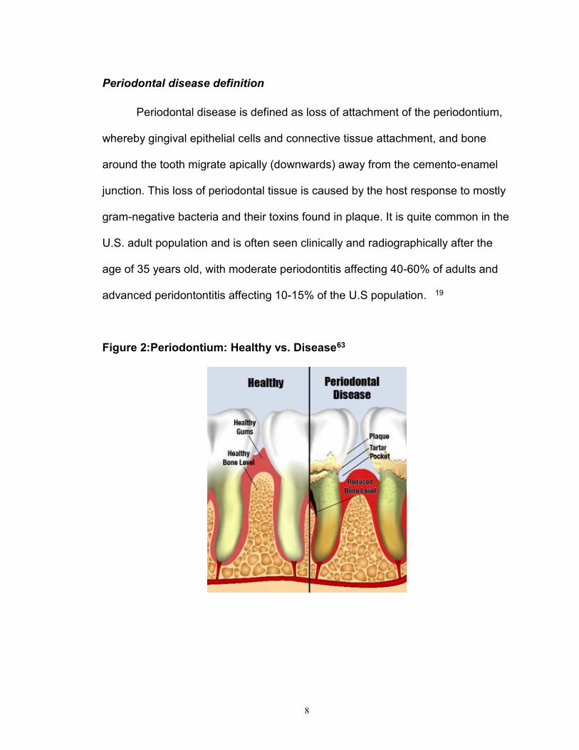
Figure 2-Periodontium: Healthy Vs Disease 8 Type 2 Diabetes and Clinical Periodontal Disease 9
Table 2-Effect of periodontal disease on glycemic control 10 Periodontal Disease and Systemic Inflammation 12 Systemic inflammatory Markers and Type 2 Diabetes 13 Insulin Resistance at the Cellular Level 14 Effect of Periodontal Treatment on Type 2 Diabetes 15
The Directionality of Periodontal Disease and Diabetes 16 Figure 3: Relationship of Inflammatory Cytokines with Induction of Insulin 18 Resistance
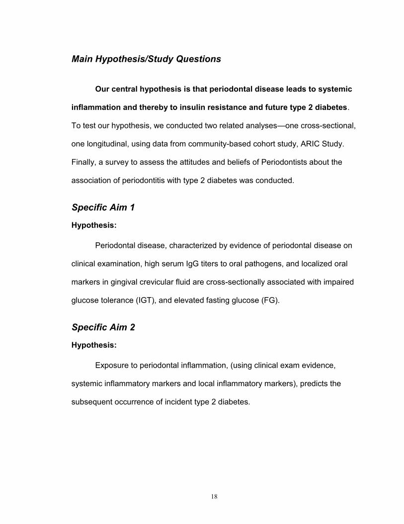
Main Hypothesis/Study Questions 19 Specific Aim 1 19
Hypothesis: 19 Specific Aim 2 19
Hypothesis: 19 Specific Aim 3 20
Hypothesis: 20
Chapter 2 21
The cross-sectional association of periodontal disease and pre-diabetes and undiagnosed diabetes 21 Abstract 21 Introduction 22
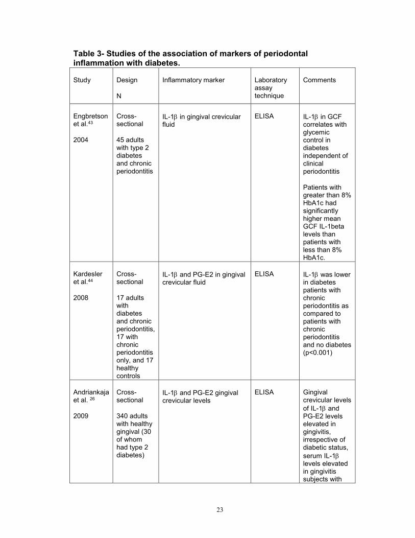
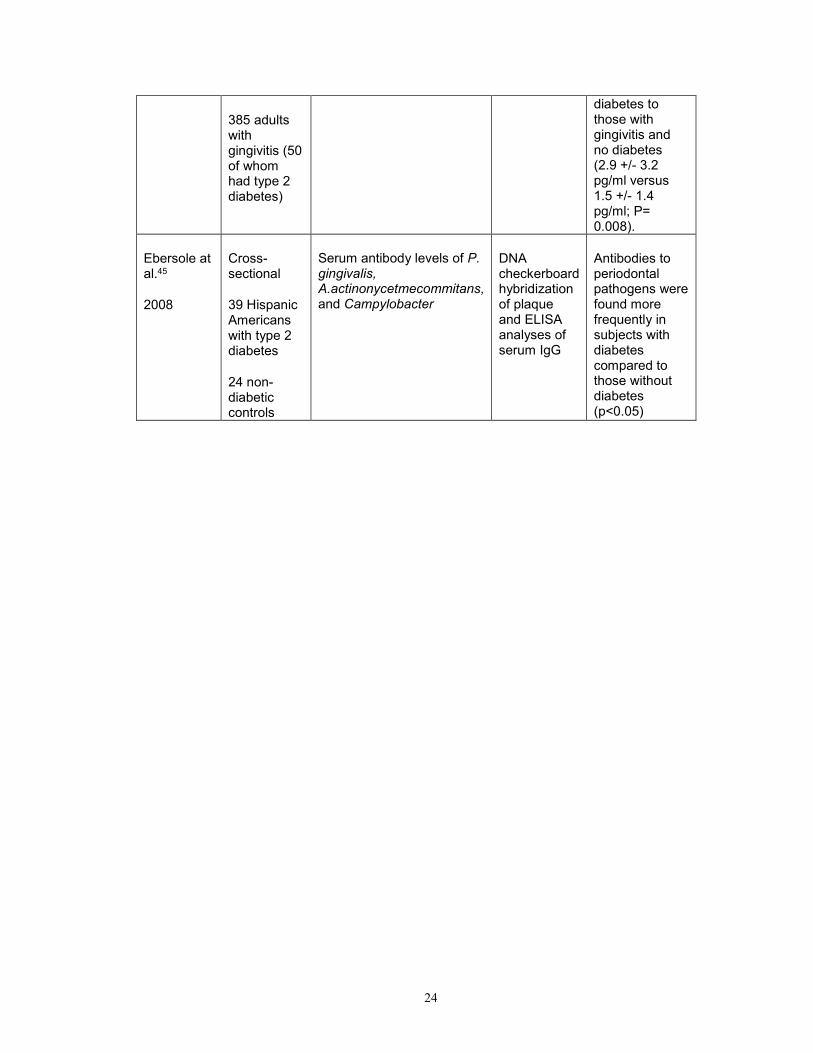
Table 3- Studies of periodontal markers and diabetes 24 Research Design and Methods 26
Description of Cohort 26 Figure 4-Participants available for cross-sectional analysis 27
Periodontal Disease 28 Diabetic Status Categorization 30 Other Variables 31 Data Analysis 31
Results 33 Table 4-Baseline characteristics by glycemic status 37 Table 5-Baseline characteristics by periodontal status 38 Table 6-Prevalence of glycemia by periodontal status 39 Table 7-Prevalence of glycemia by inflammatory marker 40 Table 8-Odds ratios for impaired glucose tolerance by periodontal status 41 Table 9-Odds ratios for impaired fasting glucose by periodontal status 42
vii
Table 10-Odds ratios for undiagnosed diabetes by periodontal status 43 Table 11-Odds ratios for impaired glucose tolerance by inflammatory marker 44 Table 12-Odds ratios for impaired fasting glucose by inflammatory marker 45 Table 13-Odds ratios for undiagnosed diabetes by inflammatory marker 46
Discussion 47 Conclusions 52
Chapter 3 53
The prospective longitudinal association of periodontal disease and the risk of type 2 diabetes 53 Abstract 53 Introduction 54 Methods 56
Study Population 56 Periodontal Disease 57 Prevalent Type 2 Diabetes 59 Incident Type 2 Diabetes 59 Other Variables 59 Data Analysis 60
Results 62 Table 14-Baseline characteristics by periodontal status 67 Table 15-Baseline characteristics by bleeding upon probing 68 Table 16-Relative hazard of diabetes by periodontal status 69 Table 17-Relative hazard of diabetes by bleeding upon probing 70 Table 18-Relative hazard of diabetes by inflammatory marker 71 Figure 5-KM plot of relative hazard of diabetes by periodontal status 72 Figure 6-KM plot of relative hazard of diabetes by P. gingivalis 73 Figure 7-KM plot of relative hazard of diabetes by A.a. 74
Figure 8-KM plot of relative hazard of diabetes by IL-1 75 Figure 9-KM plot of relative hazard of diabetes by PG-E2 76
Discussion 77 Conclusions 81
Chapter 4 83
Periodontists’ attitudes, beliefs and standard of care in treating dental patients at risk for diabetes: A survey in Washington DC area 83 Abstract 83 Introduction 84
Hypothesis: 86 Methods 86
Identification of Potential Study Population 86 Survey Content 86 Survey Process 87 Measurement and Data Analysis 88
Results 89 Study Participants 89 Risk Factors for Diabetes 89 Screening for Type 2 Diabetes in the Dental Setting 91
viii
Figure 10-Survey response to question #1 93 Table 19-Survey response to question #1 93 Figure 11-Survey response to question #2 94 Table 20-Survey response to question #2 94 Figure 12-Survey response to question #3 95 Table 21-Survey response to question #3 96 Figure 13-Survey response to question #4 97 Table 22-Survey response to question #4 97 Figure 14-Survey response to question #5a 98 Figure 15--Survey response to question #5b 99
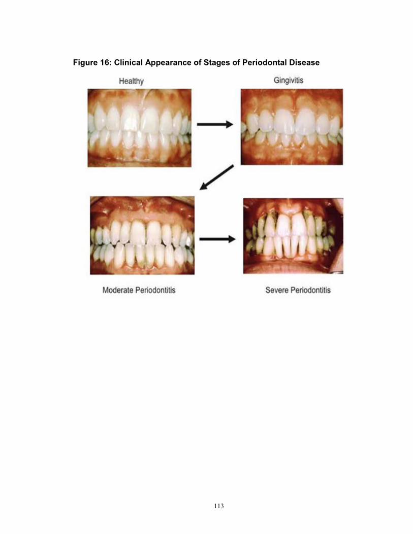
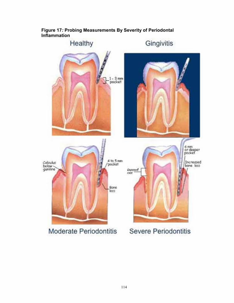
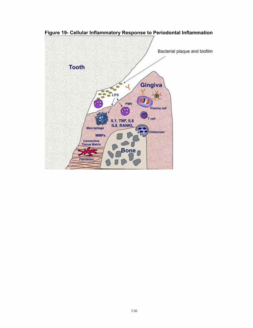
Appendix 113 Figure 16-Clinical appearance of periodontal disease 114 Figure 17-Probing measurements by severity of inflammation 115 Figure 18-Example checklist for periodontal patients 116 Figure 19-Cellular inflammatory response to periodontal inflammation 117 Figure 20- Diagnostic criteria of pre-diabetes and diabetes by glycemic test 118 Preamble to Telephone and Internet Survey from Chapter 4 119 Survey Questions to Local Periodontists 120
References 122
Curriculums Vitae 128
1
CHAPTER 1
Periodontitis and Diabetes- Review of a Two-way
Relationship
Background and Rationale
Type 2 diabetes is a known risk factor for diabetes in the dental literature,
where most studies were cross-sectional in design and included individuals with
normoglycemia and those with pre-diabetes in the same control groups. Despite
the discussion of a bi-directional relationship for the past twenty years, evidence
to support the effect of periodontal disease on the risk of incident diabetes is
lacking. This dissertation will explore the increased risk of insulin resistance and
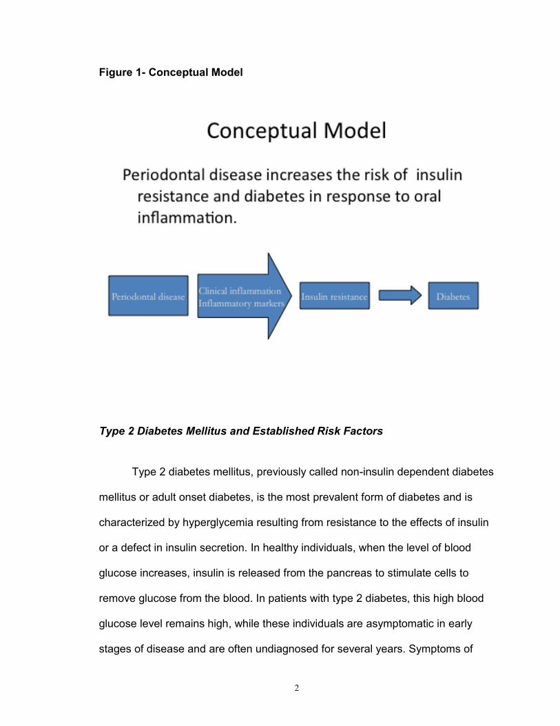
diabetes in response to oral inflammation. Figure 1 illustrates the conceptual
model, where exposure to periodontal inflammation, as measures by clinical
measures and systemic markers specific to this periodontal disease exposure,
increases the risk of insulin resistance and the subsequent onset of diabetes
2
Figure 1- Conceptual Model
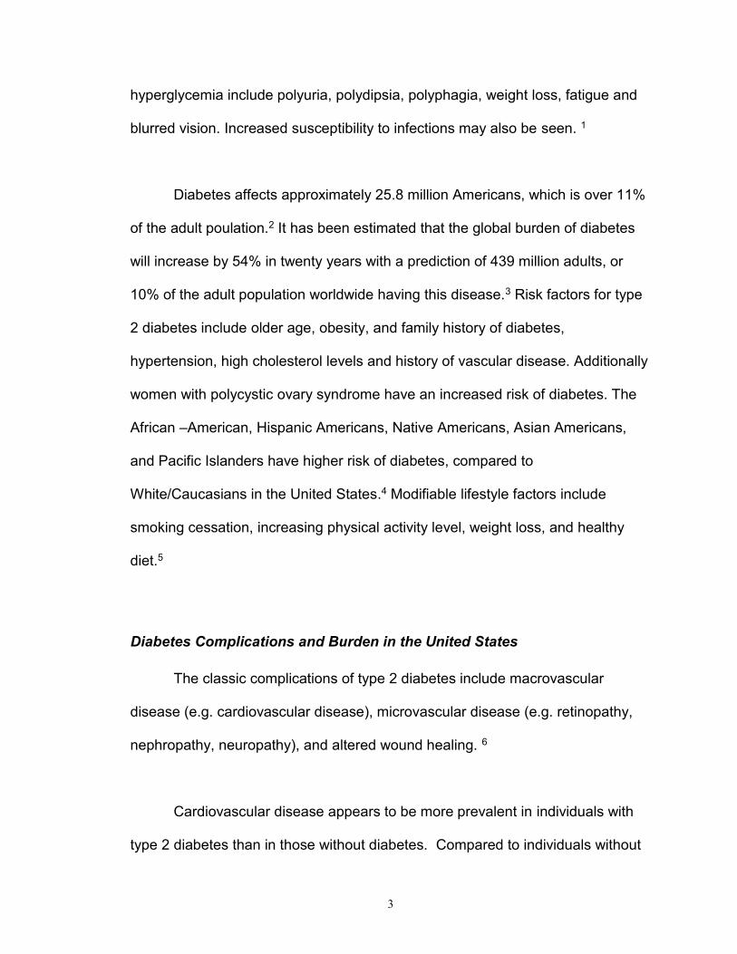
Type 2 Diabetes Mellitus and Established Risk Factors
Type 2 diabetes mellitus, previously called non-insulin dependent diabetes
mellitus or adult onset diabetes, is the most prevalent form of diabetes and is
characterized by hyperglycemia resulting from resistance to the effects of insulin
or a defect in insulin secretion. In healthy individuals, when the level of blood
glucose increases, insulin is released from the pancreas to stimulate cells to
remove glucose from the blood. In patients with type 2 diabetes, this high blood
glucose level remains high, while these individuals are asymptomatic in early
stages of disease and are often undiagnosed for several years. Symptoms of
3
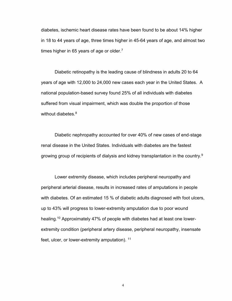
hyperglycemia include polyuria, polydipsia, polyphagia, weight loss, fatigue and
blurred vision. Increased susceptibility to infections may also be seen. 1
Diabetes affects approximately 25.8 million Americans, which is over 11%
of the adult poulation.2 It has been estimated that the global burden of diabetes
will increase by 54% in twenty years with a prediction of 439 million adults, or
10% of the adult population worldwide having this disease.3 Risk factors for type
2 diabetes include older age, obesity, and family history of diabetes,
hypertension, high cholesterol levels and history of vascular disease. Additionally
women with polycystic ovary syndrome have an increased risk of diabetes. The
African –American, Hispanic Americans, Native Americans, Asian Americans,
and Pacific Islanders have higher risk of diabetes, compared to
White/Caucasians in the United States.4 Modifiable lifestyle factors include
smoking cessation, increasing physical activity level, weight loss, and healthy
diet.5
Diabetes Complications and Burden in the United States
The classic complications of type 2 diabetes include macrovascular
cholesterol, fasting glucose and 2 hr glucose tolerance levels. (P<0.01) There
were no differences in age (P=0.34) and family history of diabetes (P=0.11).
The distribution of clinical category by glycemic status in 6, 138 ARIC
Dental Study participants is displayed in Table 6. High proportions of study
participants (between 29.0% in individuals with undiagnosed diabetes, and
42.9% in individuals with normoglycemia, P<0.0001) were displayed with
moderate periodontitis (Category IV periodontal status- one or more sites with
PD >4mm, bleeding upon probing >10% &<50%). Likewise, the distribution of
inflammatory markers in normoglycemia, IGT, IFG, and undiagnosed diabetes in
5,109 ARIC Dental Study participants without diagnosed diabetes is displayed in
Table 7, with all inflammatory markers showing no statistically significant
associations with glycemic status (all P-values were >0.05).
34
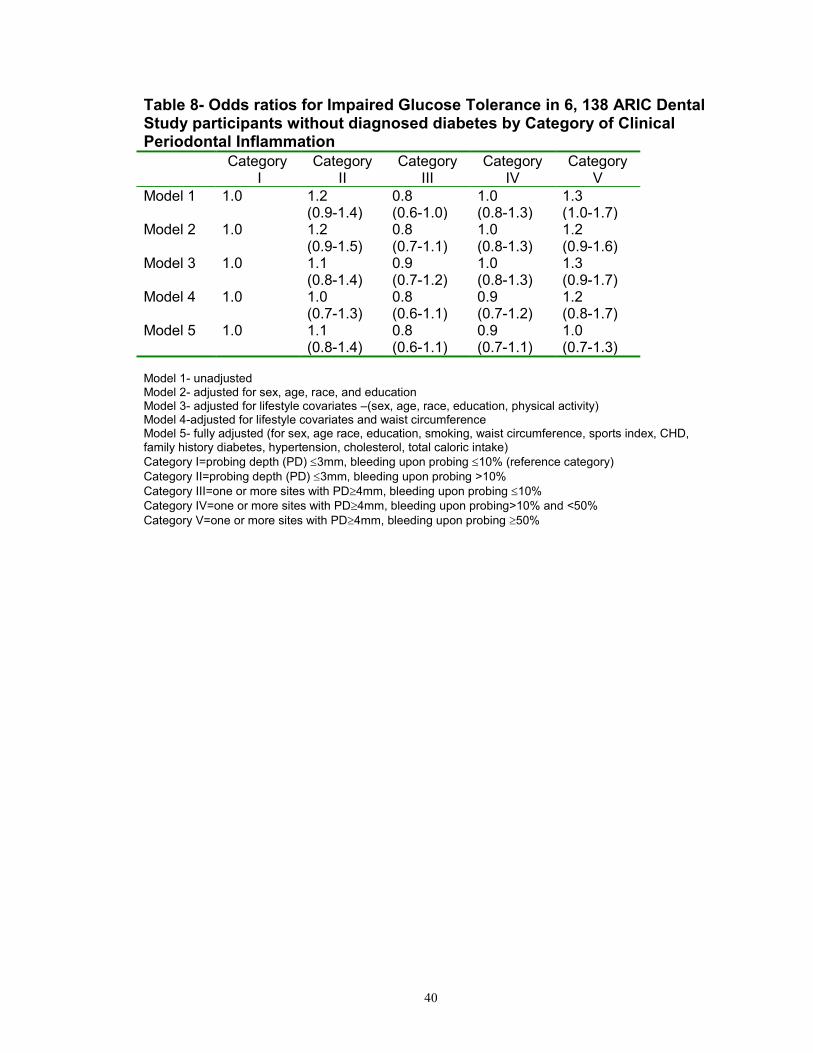
In the analysis of 2 hr GTT (Table 8), severe clinical periodontal
inflammation (Category V) was associated with elevated risk of impaired glucose
tolerance in an unadjusted model with an odds ratio of 1.3 (95% CI: 1.0-1.7).
However after adjustment for lifestyle and co-morbidity variables, this association
attenuated to null. (OR=1.0, 95% CI: 0.7-1.3).
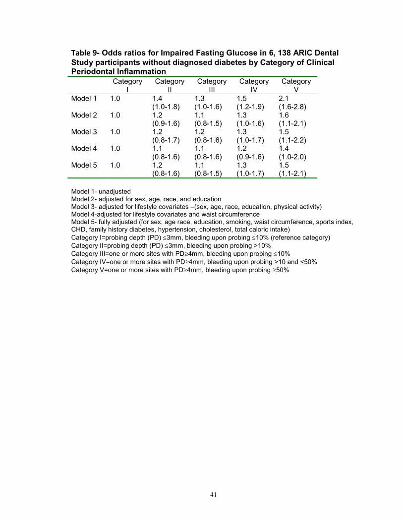
As Shown in Table 9, compared to individuals in Category I, participants
with more severe periodontal clinical inflammation had increased odds of
impaired fasting glucose. Compared to individuals in Category I, the odds ratio
for impaired fasting glucose in Category V was 2.1 (95% CI: 1.6-2.8) in an
unadjusted model. This relationship remained in the fully adjusted model with an
odds ratio of 1.5 (95% CI: 1.1--2.1) in the highest category of one or more sites
with a probing depth >4 mm and bleeding upon probing 50%.
Results in the undiagnosed diabetics mirrored the findings in the IFG
groups, showing that severe clinical periodontal inflammation was associated
with undiagnosed diabetes after adjusting for all covariates. (OR=1.5, 95% CI:
1.0-2.2). (Table 10)
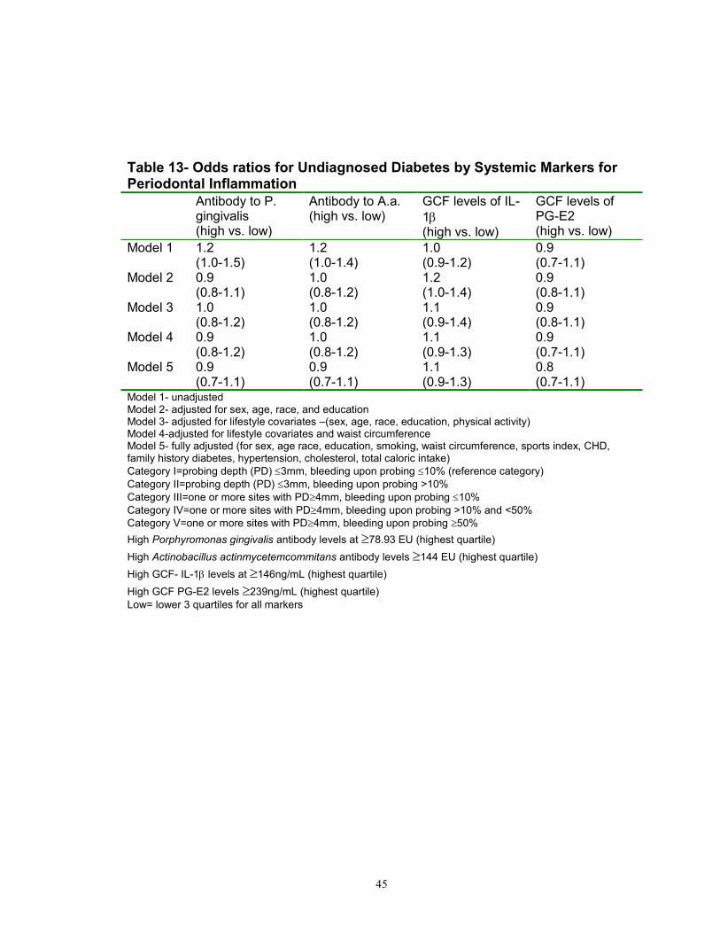
To further explore the relationship, between prediabetes and periodontal
inflammation, we performed four additional analyses using markers of systemic
inflammation (serum antibody levels to the periodontal pathogens
Porphyromonas gingivalis and Actinobacillus actinmycetemcommitans), and
35
markers of localized inflammation (gingival crevicular fluid levels of IL-1 (GCF-
IL-1) and gingival crevicular fluid levels of prostaglandin (PG-E2) (Tables 11-
13). However, no significant association was observed between pre-diabetes or
undiagnosed diabetes and any of those inflammation markers.
36
Table 4-Baseline characteristics of 6,138 middle-aged adults with periodontal exams according to glycemia status. ARIC Dental Study, 1996 – 1998
Normal Glucose
Impaired Glucose Tolerance
Impaired Fasting Glucose
Undiagnosed Diabetes
P value
N
2,154 1,572 1307 1105
Male sex (%) 61.8 62.0 58.5 57.6 <0.0001 Age (years) 61.55.6 63.35.6 61.35.6 62.85.4 <0.0001
African American (%)
11.0 12.0 16.3 23.9 <0.0001
Education <=12 Years (%)
56.2 46.0 56.0 50.6 <0.0001
Smoking Status (%)-Current
12.70.3 10.10.3 14.90.4 12.70.3 0.0082
Sports Index 2.60.8 2.50.8 2.60.8 2.50.8 <0.0001
Total Calorie Intake (Kcal/day)
1,578652 1,587603 1,637673 1,608690 0.1253
Body mass index (kg/m2)
26.84.3 28.94.8 28.95.2 29.45.9 <0.0001
Waist Circumference (cm)
95.912.6 102.513.6 102.613.2 103.714.2 <0.0001
Hypertension (%)
30.1 48.8 39.2 51.8 <0.0001
Previous Cardiovascular Disease (%)
4.9 6.0 5.5 6.7 0.3020
Family History of Diabetes (%)
11.5 15.3 12.1 14.8 0.013
High Density Lipoprotein (mmol/L)
1.40.5 1.30.4 1.20.4 1.30.3 <0.0001
Characteristics are statistically different if p<0.05 using ANOVA tests for continuous variables and
2 for categorical variables
Data are mean SD or percent. Normal glucose=FG<100mg/dL & 2hrGTT<140mg/dL & no diabetes Impaired Glucose Tolerance=2hr glucose of140-199mg/dL & no diabetes Impaired fasting glucose=FG of 100-125mg/dL & 2hr GTT<140mg/dL & no diabetes Undiagnosed diabetes=FG>125mg/dL, or 2hr GTT>199mg/dL & no diabetes diagnosis
Age (years) 62.25.6 62.35.6 62.45.5 62.35.6 62.75.4 0.3393
African American (%)
29.5 23.5 11.1 10.7 30.5 <0.0001
Education <=12 Years (%)
53.9 48.3 63.6 52.3 44.9 <0.0001
Smoking Status (%) Current
10.70.3 8.50.3 14.40.4 12.20.3 17.60.3 <0.0001
Sports Index 2.50.6 2.50.7 2.70.8 2.60.6 2.40.7 <0.0001
Total Calorie Intake (Kcal/day)
1548667 1556607 1564588 1627641 1739769 <0.0001
Body mass index (kg/m2)
28.33.6 28.54.5 27.55.6 28.44.4 29.05.6 <0.0001
Waist Circumference (cm)
101.912.6
104.513.6
102.611.2
103.812.2
10713.5 <0.0001
Hypertension (%)
46.6 43.4 32.7 39.9 49.3 <0.0001
Previous Cardiovascular Disease (%)
4.5 4.6 5.6 5.2 6.9 0.0096
Family History of Diabetes (%)
12.2
13.4
12.4
10.8
13.2
0.11
High Density Lipoprotein (mmol/L)
1.40.5 1.60.5 1.30.6 1.40.5 1.70.3 <0.0001
Mean Fasting Glucose (mg/dL)
100.013.2
102.413.6
100.014.6
101.614.1
108.113.2
<0.0001
Mean 2hr Glucose Tolerance Test (mg/dL)
135.017.1
141.118.4
127.419.2
135.920.0
143.4-
18.6
<0.0001
Characteristics are statistically different if p<0.05 (ANOVA tests for continuous variables and 2 for categorical variables), Category I=probing depth (PD) 3mm, bleeding upon probing 10% (reference
38
category),Category II=probing depth (PD) 3mm, bleeding upon probing >10%, Category III=one or more
sites with PD4mm, bleeding upon probing 10%,Category IV=one or more sites with PD4mm, bleeding
upon probing >10% and <50%,Category V=one or more sites with PD4mm, bleeding upon probing 50.
Data are mean SD or percent.
Table 6-Prevalence(%) with normal glycemia, IGT, IFG, and undiagnosed diabetes by clinical category in 6, 138 ARIC Dental Study participants
Category II=probing depth (PD) 3mm, bleeding upon probing >10%
Category III=one or more sites with PD4mm, bleeding upon probing 10%
Category IV=one or more sites with PD4mm, bleeding upon probing >10% and <50%
Category V=one or more sites with PD4mm, bleeding upon probing 50% Normal glucose=FG<100mg/dL & 2hrGTT<140mg/dL & no diabetes Impaired Glucose Tolerance=2hr glucose of140-199mg/dL & no diabetes Impaired fasting glucose=FG of 100-125mg/dL & 2hr GTT<140mg/dL & no diabetes Undiagnosed diabetes=FG>125mg/dL, or 2hr GTT>199mg/dL & no diabetes diagnosis
39
Table 7- Prevalence (%) of normoglycemia, IGT, IFG, and undiagnosed diabetes by inflammatory marker in 5,109 ARIC Dental Study participants without diagnosed diabetes
N
Antibody to P. gingivalis
Antibody to A.a.
GCF levels
of IL-1
GCF levels of PG-E2
High Low High Low High Low High Low Normal 4054 6.6 93.4 5.0 95.0 3.5 97.5 6.6 93.4 IGT 620 8.1 91.9 5.6 94.4 3.7 96.2 7.0 93.0 FG 314 7.8 92.2 4.8 95.2 3.5 97.5 5.7 94.3 Undiagnosed 121 5.7 94.3 4.9 95.1 3.7 96.2 5.4 94.6
P- value 0.45 0.34 0.09 0.59
All P-values used 2 tests for each inflammatory marker
Normal glucose=FG<100mg/dL & 2hrGTT<140mg/dL & no diabetes Impaired Glucose Tolerance=2hr glucose of140-199mg/dL & no diabetes Impaired fasting glucose=FG of 100-125mg/dL & 2hr GTT<140mg/dL & no diabetes Undiagnosed diabetes=FG>125mg/dL, or 2hr GTT>199mg/dL & no diabetes diagnosis
High Porphyromonas gingivalis antibody levels at 78.93 EU (highest quartile)
High Actinobacillus actinmycetemcommitans antibody levels 144 EU (highest quartile)
High GCF- IL-1 levels at 146ng/mL (highest quartile)
High GCF PG-E2 levels 239ng/mL (highest quartile) Low= lower 3 quartiles for all markers
40
Table 8- Odds ratios for Impaired Glucose Tolerance in 6, 138 ARIC Dental Study participants without diagnosed diabetes by Category of Clinical Periodontal Inflammation
Category I
Category II
Category III
Category IV
Category V
Model 1 1.0
1.2 (0.9-1.4)
0.8 (0.6-1.0)
1.0 (0.8-1.3)
1.3 (1.0-1.7)
Model 2 1.0 1.2 (0.9-1.5)
0.8 (0.7-1.1)
1.0 (0.8-1.3)
1.2 (0.9-1.6)
Model 3
1.0 1.1 (0.8-1.4)
0.9 (0.7-1.2)
1.0 (0.8-1.3)
1.3 (0.9-1.7)
Model 4
1.0 1.0 (0.7-1.3)
0.8 (0.6-1.1)
0.9 (0.7-1.2)
1.2 (0.8-1.7)
Model 5
1.0 1.1 (0.8-1.4)
0.8 (0.6-1.1)
0.9 (0.7-1.1)
1.0 (0.7-1.3)
Model 1- unadjusted Model 2- adjusted for sex, age, race, and education Model 3- adjusted for lifestyle covariates –(sex, age, race, education, physical activity) Model 4-adjusted for lifestyle covariates and waist circumference Model 5- fully adjusted (for sex, age race, education, smoking, waist circumference, sports index, CHD, family history diabetes, hypertension, cholesterol, total caloric intake)
Category II=probing depth (PD) 3mm, bleeding upon probing >10%
Category III=one or more sites with PD4mm, bleeding upon probing 10%
Category IV=one or more sites with PD4mm, bleeding upon probing>10% and <50%
Category V=one or more sites with PD4mm, bleeding upon probing 50%
41
Table 9- Odds ratios for Impaired Fasting Glucose in 6, 138 ARIC Dental
Study participants without diagnosed diabetes by Category of Clinical Periodontal Inflammation
Category I
Category II
Category III
Category IV
Category V
Model 1 1.0 1.4 (1.0-1.8)
1.3 (1.0-1.6)
1.5 (1.2-1.9)
2.1 (1.6-2.8)
Model 2 1.0 1.2 (0.9-1.6)
1.1 (0.8-1.5)
1.3 (1.0-1.6)
1.6 (1.1-2.1)
Model 3
1.0 1.2 (0.8-1.7)
1.2 (0.8-1.6)
1.3 (1.0-1.7)
1.5 (1.1-2.2)
Model 4
1.0 1.1 (0.8-1.6)
1.1 (0.8-1.6)
1.2 (0.9-1.6)
1.4 (1.0-2.0)
Model 5 1.0 1.2 (0.8-1.6)
1.1 (0.8-1.5)
1.3 (1.0-1.7)
1.5 (1.1-2.1)
Model 1- unadjusted Model 2- adjusted for sex, age, race, and education Model 3- adjusted for lifestyle covariates –(sex, age, race, education, physical activity) Model 4-adjusted for lifestyle covariates and waist circumference Model 5- fully adjusted (for sex, age race, education, smoking, waist circumference, sports index, CHD, family history diabetes, hypertension, cholesterol, total caloric intake)
Category II=probing depth (PD) 3mm, bleeding upon probing >10%
Category III=one or more sites with PD4mm, bleeding upon probing 10%
Category IV=one or more sites with PD4mm, bleeding upon probing >10 and <50%
Category V=one or more sites with PD4mm, bleeding upon probing 50%
42
Table- 10-Odds ratios for undiagnosed diabetes in 6, 138 ARIC Dental Study participants without diagnosed diabetes by Category of Clinical Periodontal Inflammation
Category I
Category II
Category III
Category IV
Category V
Model 1 1.0 1.3 (1.0-1.7)
0.7 (0.6-1.0)
1.1 (0.8-1.3)
2.0 (1.4-2.9)
Model 2 1.0
1.2 (1.0-1.8)
0.9 (0.7-1.3)
1.2 (0.9-1.6)
1.9 (1.3-2.7)
Model 3
1.0 1.2 (0.9-1.7)
0.9 (0.7-1.3)
1.3 (1.0-1.7)
1.9 (1.3-2.7)
Model 4
1.0 1.2 (0.9-1.7)
0.9 (0.7-1.3)
1.1 (0.9-1.5)
1.7 (1.2-2.4)
Model 5 1.0
1.2 (0.9-1.8)
0.9 (0.6-1.3)
1.1 (0.8-1.5)
1.5 (1.0-2.2)
Model 1- unadjusted Model 2- adjusted for sex, age, race, and education Model 3- adjusted for lifestyle covariates –(sex, age, race, education, physical activity) Model 4-adjusted for lifestyle covariates and waist circumference Model 5- fully adjusted (for sex, age race, education, smoking, waist circumference, sports index, CHD, family history diabetes, hypertension, cholesterol, total caloric intake)
Category II=probing depth (PD) 3mm, bleeding upon probing >10%
Category III=one or more sites with PD4mm, bleeding upon probing 10%
Category IV=one or more sites with PD4mm, bleeding upon probing >10% and <50%
Category V=one or more sites with PD4mm, bleeding upon probing 50%
43
Table 11- Odds ratios for impaired Glucose in 5,109 ARIC Dental Study participants without diagnosed diabetes by Inflammatory Markers for Periodontal Inflammation
Antibody to P. gingivalis (high vs. low)
Antibody to A.a. (high vs. low)
GCF levels of IL-
1 (high vs. low)
GCF levels of PG-E2 (high vs. low)
Model 1 1.2 (1.0-1.4)
1.2 (1.0-1.4)
1.1 (0.9-1.4)
1.1 (0.9-1.3)
Model 2 1.2 (1.0-1.4)
1.1 (0.9-1.3)
1.1 (0.9-1.4)
1.2 (1.0-1.4)
Model 3
1.1 (0.9-1.4)
1.1 (0.9-1.3)
1.1 (0.9-1.3)
1.2 (1.0-1.4)
Model 4
1.1 (0.9-1.4)
1.1 (0.9-1.3)
1.1 (0.9-1.3)
1.1 (1.0-1.4)
Model 5 1.2 (0.9-1.4)
1.1 (0.9-1.3)
1.1 (0.9-1.4)
1.1 (0.9-1.3)
Model 1- unadjusted Model 2- adjusted for sex, age, race, and education Model 3- adjusted for lifestyle covariates –(sex, age, race, education, physical activity) Model 4-adjusted for lifestyle covariates and waist circumference Model 5- fully adjusted (for sex, age race, education, smoking, waist circumference, sports index, CHD, family history diabetes, hypertension, cholesterol, total caloric intake)
Category II=probing depth (PD) 3mm, bleeding upon probing >10%
Category III=one or more sites with PD4mm, bleeding upon probing 10%
Category IV=one or more sites with PD4mm, bleeding upon probing >10% and <50%
Category V=one or more sites with PD4mm, bleeding upon probing 50%
High Porphyromonas gingivalis antibody levels at 78.93 EU (highest quartile)
High Actinobacillus actinmycetemcommitans antibody levels 144 EU (highest quartile)
High GCF- IL-1 levels at 146ng/mL (highest quartile)
High GCF PG-E2 levels 239ng/mL (highest quartile) Low= lower 3 quartiles for all markers
44
Table 12- Odds ratios for Impaired Fasting Glucose in 5,109 ARIC Dental Study participants without diagnosed diabetes by Systemic Markers for Periodontal Inflammation
Antibody to P. gingivalis (high vs. low)
Antibody to A.a. (high vs. low)
GCF levels of IL-
1 (high vs. low)
GCF levels of PG-E2 (high vs. low)
Model 1 1.1 (0.9-1.3)
1.0 (0.8-1.2)
1.0 (0.8-1.2)
0.9 (0.8-1.1)
Model 2 1.0 (0.8-1.2)
0.9 (0.8-1.1)
1.0 (0.8-1.2)
1.0 (0.8-1.2)
Model 3
1.0 (0.9-1.2)
0.9 (0.7-1.1)
0.9 (0.8-1.2)
1.0 (0.8-1.2)
Model 4
1.0 (0.8-1.2)
0.9 (0.7-1.1)
0.9 (0.7-1.1)
1.0 (0.8-1.2)
Model 5 1.0 (0.8-1.2)
0.9 (0.8-1.1)
1.0 (0.8-1.2)
0.9 (0.7-1.1)
Model 1- unadjusted Model 2- adjusted for sex, age, race, and education Model 3- adjusted for lifestyle covariates –(sex, age, race, education, physical activity) Model 4-adjusted for lifestyle covariates and waist circumference Model 5- fully adjusted (for sex, age race, education, smoking, waist circumference, sports index, CHD, family history diabetes, hypertension, cholesterol, total caloric intake)
Category II=probing depth (PD) 3mm, bleeding upon probing >10%
Category III=one or more sites with PD4mm, bleeding upon probing 10%
Category IV=one or more sites with PD4mm, bleeding upon probing >10% and <50%
Category V=one or more sites with PD4mm, bleeding upon probing 50%
High Porphyromonas gingivalis antibody levels at 78.93 EU (highest quartile)
High Actinobacillus actinmycetemcommitans antibody levels 144 EU (highest quartile)
High GCF- IL-1 levels at 146ng/mL (highest quartile)
High GCF PG-E2 levels 239ng/mL (highest quartile) Low= lower 3 quartiles for all markers
45
Table 13- Odds ratios for Undiagnosed Diabetes by Systemic Markers for Periodontal Inflammation
Antibody to P. gingivalis (high vs. low)
Antibody to A.a. (high vs. low)
GCF levels of IL-
1 (high vs. low)
GCF levels of PG-E2 (high vs. low)
Model 1 1.2 (1.0-1.5)
1.2 (1.0-1.4)
1.0 (0.9-1.2)
0.9 (0.7-1.1)
Model 2 0.9 (0.8-1.1)
1.0 (0.8-1.2)
1.2 (1.0-1.4)
0.9 (0.8-1.1)
Model 3 1.0 (0.8-1.2)
1.0 (0.8-1.2)
1.1 (0.9-1.4)
0.9 (0.8-1.1)
Model 4 0.9 (0.8-1.2)
1.0 (0.8-1.2)
1.1 (0.9-1.3)
0.9 (0.7-1.1)
Model 5 0.9 (0.7-1.1)
0.9 (0.7-1.1)
1.1 (0.9-1.3)
0.8 (0.7-1.1)
Model 1- unadjusted Model 2- adjusted for sex, age, race, and education Model 3- adjusted for lifestyle covariates –(sex, age, race, education, physical activity) Model 4-adjusted for lifestyle covariates and waist circumference Model 5- fully adjusted (for sex, age race, education, smoking, waist circumference, sports index, CHD, family history diabetes, hypertension, cholesterol, total caloric intake)
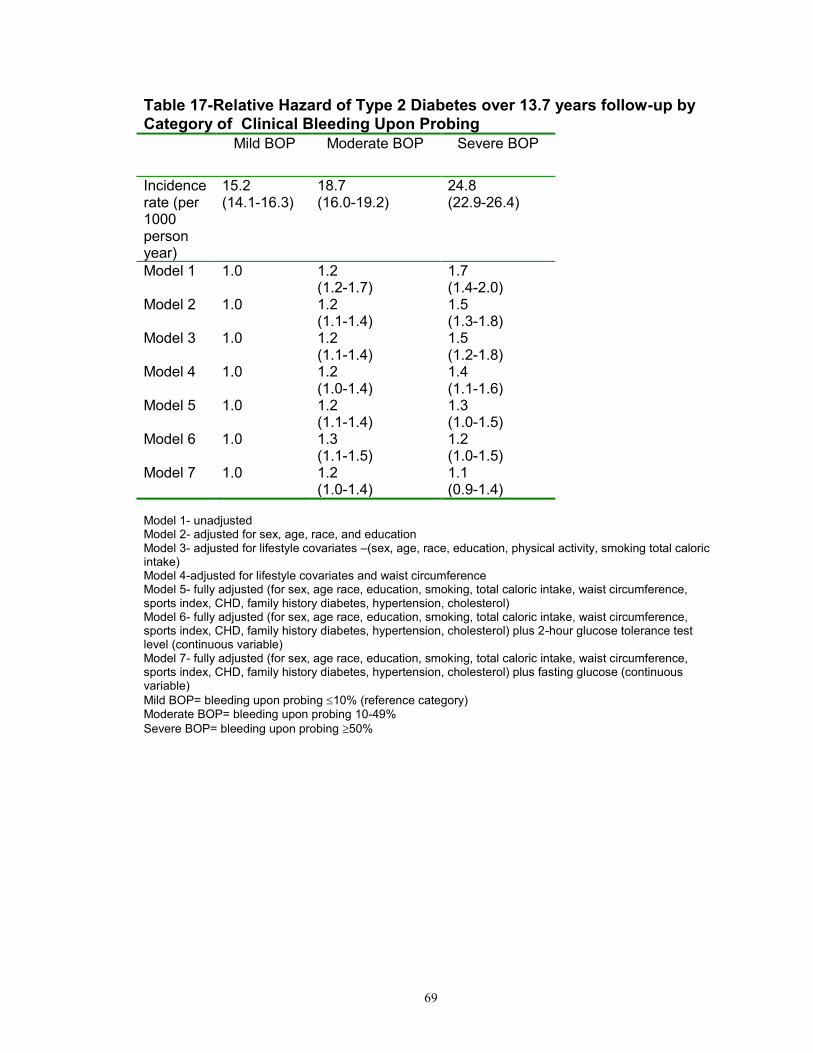
Category III did not display this increasing significant trend with a hazard ratio of
1.0 (95% CI: 0.8-1.20). As expected, additional adjustment including fasting
glucose (model 6) or 2-hr GTT (model 7) further attenuated the association,
because they were both in the causal pathway. A dose-response relationship
with clinical inflammation could be seen by using only bleeding upon probing as a
measure for clinical inflammation. (Table 17). Censoring of individuals who died
during follow-up (n= 211) also did not change the associations observed with
incident diabetes (data not shown).
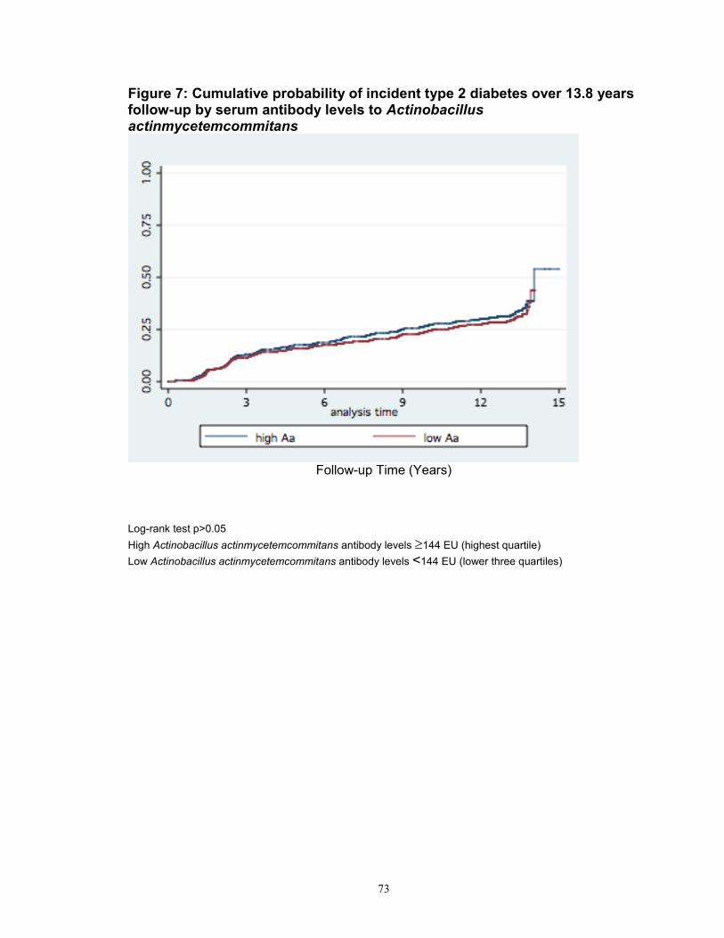
To investigate the relationship of incident diabetes to other inflammatory
measures of periodontal disease exposure, additional analyses including
participants with assays of periodontal inflammation were performed. First, to
determine if systemic markers specific to exposure to periodontal inflammation
might help explain the relationship of periodontal disease to diabetes risk,
antibodies to the periodontal pathogens Porphyromonas gingivalis and
Actinobacillus actinmycetemcommitans were included into multivariable models
adjusted for age, sex, race, smoking, waist circumference, cardiovascular
disease, family history of diabetes, total caloric intake, and cholesterol levels.
The hazard of incident diabetes appeared no different in adults with high levels of
antibodies to Porphyromonas gingivalis compared to low serum levels. The
hazard ratios for antibodies to Actinobacillus actinmycetemcommitans also
65
appeared to be in these ranges but did not reach statistical significance. (Table
18)
Data on localized markers for periodontal inflammation were available,
and additional adjusted multivariable analyses using gingival crevicular fluid were
performed. High levels of gingival crevicular fluid IL-1 were associated with no
change in hazard of incident diabetes (HR=1.0, CI:0.8-1.2). High levels of1 PG-
E2 were also associated with no increased hazard of incident diabetes (1.0: 95%
CI: 0.8-1.1). (Table 18)
66
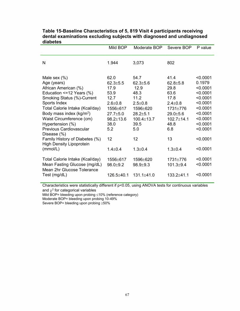
Table 14- Baseline Characteristics of 5, 819 Visit 4 participants receiving dental examinations excluding diagnosed and undiagnosed subjects with diabetes-ARIC Dental Study (1996-1998 to present).
Category I
Category II
Category III
Category IV
Category V
P value
N
860 861 1,084 2,326 688
Male sex (%) 73.4 64.2 53.0 50.8 40.3 <0.0001 Age (years) 62.25.5 62.35.8 62.45.6 62.35.6 62.75.8 0.198
African American (%)
28.4 23.0 9.5 10.1 29.5 <0.0001
Education <=12 Years (%)
53.9 48.3 63.6 52.3 44.9 <0.0001
Smoking Status (%)-Current
10.60.3 8.60.3 14.40.4 12.40.3 18.10.4 <0.0001
Sports Index 2.50.8 2.50.8 2.70.8 2.60.8 2.40.8 <0.0001
Family History of Diabetes (%) 12 12 13 <0.0001 High Density Lipoprotein (mmol/L)
1.40.4
1.30.4
1.30.4
<0.0001
Total Calorie Intake (Kcal/day) 1556617 1596620 1731776 <0.0001
Mean Fasting Glucose (mg/dL) 98.09.2 98.99.3 101.39.4 <0.0001
Mean 2hr Glucose Tolerance Test (mg/dL)
126.540.1
131.141.0
133.241.1
<0.0001
Characteristics were statistically different if p<0.05, using ANOVA tests for continuous variables
and 2 for categorical variables Mild BOP= bleeding upon probing 10% (reference category) Moderate BOP= bleeding upon probing 10-49%
Severe BOP= bleeding upon probing 50%
68
Table 16- Relative Hazard of Type 2 Diabetes over 13.8 years follow-up by Category of Clinical Periodontal Inflammation
Category 1 Category 2 Category 3 Category 4
Category 5
Incidence rate (per 1000 person years)
17.4 (17.2-19.0)
22.3 (20.9-23.7)
13.6 (12.5-14.1)
17.8 (16.2-18.1)
23.9 (22.2-25.2)
Model 1 1.0
1.3 (1.0-1.6)
0.8 (0.6-1.0)
1.1 (0.9-1.3)
1.4 (1.1-1.8)
Model 2 1.0
1.3 (1.1-1.6)
0.9 (0.7-1.1)
1.1 (0.9-1.4)
1.4 (1.1-1.7)
Model 3
1.0 1.3 (1.1-1.6)
0.9 (0.7-1.1)
1.1 (0.9-1.3)
1.3 (1.1-1.7)
Model 4
1.0 1.3 (1.0-1.6)
0.9 (0.7-1.1)
1.1 (0.9-1.3)
1.2 (1.0-1.6)
Model 5
1.0 1.4 (1.1-1.7)
1.0 (0.8-1.3)
1.2 (1.0-1.4)
1.3 (1.0-1.6)
Model 6
1.0 1.4 (1.1-1.8)
1.1 (0.9-1.5)
1.3 (1.0-1.6)
1.3 (1.0-1.7)
Model 7
1.0 1.3 (1.0-1.7)
1.0 (0.8-1.3)
1.2 (1.0-1.4)
1.1 (0.9-1.5)
Model 1- unadjusted Model 2- adjusted for sex, age, race, and education Model 3- adjusted for lifestyle covariates (sex, age, race, education, physical activity, smoking total caloric intake) Model 4-adjusted for lifestyle covariates and waist circumference Model 5- fully (for sex, age race, education, smoking, total caloric intake, waist circumference, sports index, CHD, family history diabetes, hypertension, cholesterol) Model 6- fully adjusted (for sex, age race, education, smoking, total caloric intake, waist circumference, sports index, CHD, family history diabetes, hypertension, cholesterol) plus 2-hour glucose tolerance test level (continuous variable) Model 7- fully adjusted (for sex, age race, education, smoking, total caloric intake, waist circumference, sports index, CHD, family history diabetes, hypertension, cholesterol) plus fasting glucose (continuous variable)
Category II=probing depth (PD) 3mm, bleeding upon probing >10%
Category III=one or more sites with PD4mm, bleeding upon probing 10%
Category IV=one or more sites with PD4mm, bleeding upon probing >10% and <50%
Category V=one or more sites with PD4mm, bleeding upon probing 50%
69
Table 17-Relative Hazard of Type 2 Diabetes over 13.7 years follow-up by Category of Clinical Bleeding Upon Probing
Mild BOP Moderate BOP Severe BOP
Incidence rate (per 1000 person year)
15.2 (14.1-16.3)
18.7 (16.0-19.2)
24.8 (22.9-26.4)
Model 1 1.0
1.2 (1.2-1.7)
1.7 (1.4-2.0)
Model 2 1.0 1.2 (1.1-1.4)
1.5 (1.3-1.8)
Model 3
1.0 1.2 (1.1-1.4)
1.5 (1.2-1.8)
Model 4
1.0 1.2 (1.0-1.4)
1.4 (1.1-1.6)
Model 5
1.0 1.2 (1.1-1.4)
1.3 (1.0-1.5)
Model 6
1.0 1.3 (1.1-1.5)
1.2 (1.0-1.5)
Model 7
1.0 1.2 (1.0-1.4)
1.1 (0.9-1.4)
Model 1- unadjusted Model 2- adjusted for sex, age, race, and education Model 3- adjusted for lifestyle covariates –(sex, age, race, education, physical activity, smoking total caloric intake) Model 4-adjusted for lifestyle covariates and waist circumference Model 5- fully adjusted (for sex, age race, education, smoking, total caloric intake, waist circumference, sports index, CHD, family history diabetes, hypertension, cholesterol) Model 6- fully adjusted (for sex, age race, education, smoking, total caloric intake, waist circumference, sports index, CHD, family history diabetes, hypertension, cholesterol) plus 2-hour glucose tolerance test level (continuous variable) Model 7- fully adjusted (for sex, age race, education, smoking, total caloric intake, waist circumference, sports index, CHD, family history diabetes, hypertension, cholesterol) plus fasting glucose (continuous variable)
Mild BOP= bleeding upon probing 10% (reference category) Moderate BOP= bleeding upon probing 10-49%
Severe BOP= bleeding upon probing 50%
70
Table 18- Relative Hazard of Type 2 Diabetes over 13.7 years follow-up by Systemic Markers for Periodontal Inflammation
Antibody to P. gingivalis (high vs. low)
Antibody to A.a. (high vs. low)
GCF levels of
IL-1 (high vs. low)
GCF levels of PG-E2 (high vs. low)
Incidence rate(per 1000person year)
0.058 (-0.02-0.42)
0.057 (-0.02-0.75)
0.058 (0.006-0.091)
0.060 (-0.0018-0.13)
Model 1 1.1 (1.0-1.3)
1.1 (1.0-1.3)
1.0 (0.9-1.1)
1.0 (0.9-1.1)
Model 2 1.1 (0.9-1.2)
1.0 (0.9-1.2)
1.0 (0.9-1.2)
1.0 (0.9-1.1)
Model 3
1.1 (0.9-1.2)
1.0 (0.9-1.2)
1.0 (0.9-1.2)
1.0 (0.9-1.1)
Model 4
1.1 (0.9-1.2)
1.1 (1.0-1.2)
1.0 (0.9-1.1)
1.0 (0.9-1.1)
Model 5 1.0 (0.9-1.2)
1.0 (0.9-1.2)
1.0 (0.9-1.2)
1.0 (0.8-1.1)
Model 6 1.0 (0.9-1.2)
1.0 (0.8-1.2)
1.0 (0.8-1.2)
1.0 (0.8-1.2)
Model 7 1.0 (0.8-1.2)
1.1 (0.9-1.3)
1.1 (1.9-1.3)
1.0 (0.8-1.2)
Model 1- unadjusted Model 2- adjusted for sex, age, race, and education Model 3- adjusted for lifestyle covariates –(sex, age, race, education, physical activity, smoking, total caloric intake) Model 4-adjusted for lifestyle covariates and waist circumference Model 5- fully adjusted (for sex, age race, education, smoking, total caloric intake, waist circumference, sports index, CHD, family history diabetes, hypertension, cholesterol) Model 6- fully adjusted (for sex, age race, education, smoking, total caloric intake, waist circumference, sports index, CHD, family history diabetes, hypertension, cholesterol) plus 2-hour glucose tolerance test level (continuous variable) Model 7- fully adjusted (for sex, age race, education, smoking, total caloric intake, waist circumference, sports index, CHD, family history diabetes, hypertension, cholesterol) plus fasting glucose (continuous variable)
High Porphyromonas gingivalis antibody levels at 78.93 EU (highest quartile)
High Actinobacillus actinmycetemcommitans antibody levels 144 EU (highest quartile)
High GCF- IL-1 levels at 146ng/mL (highest quartile)
High GCF PG-E2 levels 239ng/mL (highest quartile) Low= lower 3 quartiles for all markers
71
Figure 4: - Cumulative probability of incident type 2 diabetes over 13.8 years follow-up by category of clinical periodontal inflammation
Follow-up Time (Years)
Log-rank test p<0.001
72
Figure 6:Cumulative probability of incident type 2 diabetes over 13.8 years follow-up by serum antibody levels to Porphyromonas gingivalis
Follow-up Time (Years)
Log-rank test p>0.05
High Porphyromonas gingivalis antibody levels at 78.93 EU (highest quartile)
Low Porphyromonas gingivalis antibody levels at <78.93 EU (lower three quartiles)
73
Figure 7: Cumulative probability of incident type 2 diabetes over 13.8 years follow-up by serum antibody levels to Actinobacillus actinmycetemcommitans
Follow-up Time (Years)
Log-rank test p>0.05
High Actinobacillus actinmycetemcommitans antibody levels 144 EU (highest quartile)
Low Actinobacillus actinmycetemcommitans antibody levels <144 EU (lower three quartiles)
74
Figure 8:Cumulative probability of incident type 2 diabetes over 13.8 years follow-up by
GCF levels of IL-1
Follow-up Time (Years)
Log-rank test p>0.05
High GCF- IL-1 levels at 146ng/mL (highest quartile)
Low GCF- IL-1 levels at <146ng/mL (lower three quartiles)
75
Figure 9:Cumulative probability of incident type 2 diabetes over 13.8 years follow-up by GCF levels of PG-E2
Follow-up Time (Years)
Log-rank test p>0.05
High GCF PG-E2 levels 239ng/mL (highest quartile)
Low GCF PG-E2 levels <239ng/mL (lower three quartiles)
76
Discussion
In a longitudinal analysis of this cohort, clinical parameters of periodontal
inflammation at baseline increased the risk if incident diabetes over a 13.84 year
follow-up. As observed in the cross-sectional design (Chapter 2), serum markers
for inflammation were not associated strongly with incident diabetes. No
association with incident diabetes was seen with high baseline levels of IgG
antibody levels to Porphyromonas gingivalis, and Actinobacillus
actinmycetemcommitans, and this lack of association remained consistent when
analyzing gingival crevicular fluid IL- and PG-E2 levels.
Another study found no association of clinical periodontal disease with
incident diabetes in Japan. This study used fasting glucose levels similar to our
study with a similar sample size (n=5,848), but the study duration was only 7
years, which may not be sufficiently long enough to observe incident cases.57
Only one other study has found a positive association of baseline clinical
periodontal disease and risk of subsequent diabtetes.56 In the National Health
and Nutrition Examination Survey (NHANES) including 7,168 eligible
participants, after 17 years of follow-up, the odds ratios for incident diabetes
ranged from 1.5 (95% CI; 0.99-2.27) in advanced periodontal disease to 2.26
(95%CI: 1.56-3.27) in moderate periodontitis. That study used the periodontal
index to classify severity of periodontal inflammation, which looked at the visual
77
extent of gingival inflammation, presence or absence of pockets and tooth
mobility to assign an averaged score. Our study used a comprehensive
examination of probing measurements and bleeding upon probing, which are
both the standard of care in clinical practice for diagnosing periodontal disease.
The NHANES study also used death certificates, self-reports of diabetes
requirement of pharmacologic treatment, and a health care facility stay with a
discharge code of diabetes, which may have overestimated the number of new
cases. Those participants were followed up at least one time. Our study was
strengthened by yearly follow-up telephone calls, which was more likely to
identify true incident diabetes as they occurred.
Our results did not support the findings found in CVD outcome studies
where systemic markers for periodontal inflammation were associated with an
increased risk of cardiovascular disease. Both high antibody levels,
Porphyromonas gingivalis and Actinobacillus actinmycetemcommitans, have
been found to increase the risk of CVD by an overall odds ratio of 1.75 (95%CI:
1.32 to 2.34)54 No studies have assessed local inflammatory markers such as
gingival crevicular fluid IL- and PG-E2 with cardiovascular or diabetes risks.
Periodontal treatment for advanced peridontitis has not been shown to reduce
inflammatory mediators in diabetic subjects, though A1C levels were significantly
improved. 55
78
Our study had several strengths. First, ARIC is a large, community-based,
biracial population in which there was standardized ascertainment of follow-up for
approximately 14 years. Second, there were standardized measures of
exposures, outcomes, and confounding variables in a rigorously monitored
observational study, allowing us to explore the associated risk of incident
diabetes with prior periodontal disease exposures. This study is novel by
combining both clinical and systemic measures specific to periodontal
inflammation to assess diabetes as an outcome. This approach has been used to
assess cardiovascular disease as an outcome, but unlike these other
cardiovascular studies, an association of systemic inflammatory mediators with
increased risk of diabetes was not shown.50, 51 Our study suggests that the
association of periodontal inflammation with risk of diabetes is not the same as
the association with the risk of cardiovascular disease. In the cardiovascular
disease infection hypothesis, several studies have validated the use of serum
antibody level to the periodontal pathogens Porphyromonas gingivalis and
Actinobacillus actinmycetemcommitans as a surrogate of periodontal clinic exam
when assessing CVD risk. 51, 54 While these serum antibody levels do not indicate
active or current periodontal disease, they have been used to study the level of
prior exposure to periodontal inflammation and CVD risk.
Nonetheless, the limitations should be kept in mind when interpreting our
data. Firstly, this study also lacked longitudinal dental and medical exams. Teeth
and their surrounding tissues provide the niche for periodontal pathogens and
79
gingival crevicular fluid. One study found that the elevated serologies no longer
conferred increased cardiovascular risk in edentulous subjects. 51 Tooth loss data
was not available after the baseline visit and dietary data was scant in this
dataset. Tooth loss may also influence dietary choices, caloric intake,
cholesterol levels, body mass index and diabetes. While these were included in
the model as confounders, tooth loss and diet may be a distinct separate
pathway in the direction from periodontal disease to diabetes. The longitudinal
NHANES study found that participants with no teeth, had an odds ratio for
incident diabetes of 1.3 (95%CI: 1.0-1.7), and those with advanced tooth loss (1-
7 teeth remaining) had an odds ratio of 1.7 (P<0.05). 56 Blood glucose
assessment was also not available at follow-up to confirm incident diabetes in
our study to confirm the telephone questionnaire responses.
A subsidiary analysis of baseline characteristics of 4, 864 individuals
available at visit 4, but excluded from the periodontal examination, was
performed (results not shown). The individuals excluded from our primary
analysis had a higher proportion of African-Americans (39% vs. 24%), smokers
(19% vs. 13%), increased caloric intake (1773 kcal/day vs. 1587kcal/day) and
higher body mass-indices (31kg/m2 vs. 28.9kg/m2 ). These aforementioned
characteristics are known risk factors for diabetes. It is possible that exclusion of
these individuals from the analysis may have resulted in an underestimated risk
of diabetes. Additionally, 15 % (n=1,478) of the visit 4 participants were
edentulous. If we assume that tooth loss is a surrogate for severe periodontal
80
disease status, then it is possible a large proportion of individuals with prior
exposure to severe periodontal inflammation were not available for analysis, also
resulting in an underestimated risk of diabetes.
We used the serum levels for inflammatory mediators’ highest quartile as
the cut-point for high vs. low levels. Other studies used the highest tertile or
quartile for the high level category for studying the association of periodontal
disease with cardiovascular disease risk. 51 It is possible that the highest tertile or
quartile may represent a unique population with the possibility for residual
confounding.
Performing multiple regressions for the five clinical and four systemic
markers of inflammation increased the possibility of Type I error. The possibility
of a false positive merely due to chance may also be due to the large number of
models produced for dividing diabetes diagnosis into several categories.
Conclusions
The Atherosclerosis Risk in Communities (ARIC) Study is a community-
based prospective cohort providing a rich database with which to assess the
effect of periodontal disease exposure on incident diabetes. This study helps
answer the recent call by the Joint EFP/AAP consensus report for studies with
81
comprehensive clinical data, extent and severity of periodontal disease, level of
glycemic control, and consideration of local and systemic pathways affected both
periodontal disease and diabetes.53 This study served to contribute to the body of
evidence that is largely lacking in the directionality of periodontal disease and
subsequent incident diabetes.
This study supports the hypothesis that clinical periodontal inflammation
increases the risk of incident diabetes several years later. An increase in
bleeding upon probing appears to be both cross-sectionally associated with
impaired glucose tolerance and longitudinally associated with the onset of
incident diabetes. Since even minimal bleeding upon probing in this study was
associated with prevalent pre-diabetes and future incident diabetes, a strong
case is made to support patient education for prevention of periodontal disease
to and study the effect of prevention of even mild periodontal inflammation on
impaired glucose tolerance and diabetes.
The serum markers specific to periodontal disease used in cardiovascular
disease models do not appear to be helpful in assessing risk of incident diabetes.
Though the American Heart Association’s Scientific Statement on Diabetes
stated that “diabetes is a cardiovascular disease”, the mechanism of action may
be very different.58 It may not be enough to study just a few systemic and local
markers for periodontal disease to understand the mechanistic pathway of
periodontal diseases and increased risk of type 2 diabetes.
82
CHAPTER 4
Periodontists’ attitudes, beliefs and standard of
care in treating dental patients at risk for diabetes:
A survey in Washington DC area
Abstract
The two-way relationship of periodontal disease and diabetes has been
discussed in the literature for almost two decades, while the evidence to support
the risk of diabetes associated with periodontal disease exposure is sparse. The
association of periodontitis with type 2 diabetes is recognized by local
Periodontists (using a convenience sample survey of Washington DC area
Periodontists), and the attitudes and beliefs of these specialists influence the
standard of care in treating dental patients. When asked if it was appropriate to
probe further about of diabetes risk factors in patients with periodontal disease
and no diabetes diagnosis, most respondents (92.9%) agreed (agreed/ strongly
agreed, n=39). This survey suggests that practicing periodontists are aware that
an association between periodontal disease and onset of type 2 diabetes, and
83
they appear aware of the importance of HbA1c testing in assessing glycemic
control, whether this test is performed in the dental office or medical setting. This
appears to parallel the consensus report of the Joint European Federation/
American Academy Workshop (EFP/AAP) guidelines to dentists for patients
without a diabetes diagnosis, but obvious risk factors for type 2 diabetes.
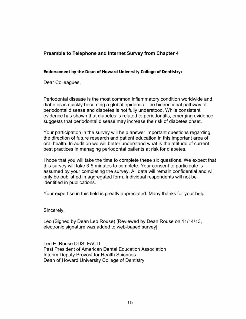
Introduction
Periodontal disease is the most common inflammatory condition
worldwide and diabetes is quickly becoming a global epidemic. The bidirectional
pathway of periodontal disease and diabetes is not fully understood. While
consistent evidence has shown that diabetes is related to periodontitis, emerging
evidence suggests that periodontal disease may increase the risk of diabetes
onset.
Risk factors for type 2 diabetes include older age, obesity, and family
history of diabetes, hypertension, high cholesterol levels and history of vascular
disease.5 Modifiable lifestyle risk factors include smoking, physical activity level,
weight loss, and healthy diet.5 While periodontal disease as a risk factor for
incident diabetes has been proposed, sufficient evidence to quantify this
association is lacking.59
The consensus report of the Joint European Federation/ American
Academy Workshop (EFP/AAP) on periodontitis and systemic disease recently
reviewed the role of periodontitis and the associated the risk of type 2 diabetes.
84
Their guidelines to dentists for patients without a diabetes diagnosis, but obvious
risk factors for type 2 diabetes, include that the patients:
“should be informed of their risk for having diabetes, assessed using a
chair-side HbA1C test, and/or referred to a physician for appropriate
testing and diagnostic care.” 53
This joint EFO/AAP suggests that evidence is emerging about the role of
periodontal inflammation and the risk of incident diabetes, but concluded, “there
is lack of clarity in the literature regarding the strength of this latter association”. 53
This joint consensus report concluded that because of the “relative immaturity of
the body of evidence for this purported relationship, the field is wide open and the
gaps in knowledge are large”.53 Therefore, we conducted this survey to better
understand the beliefs, perceptions, and current practices among local
Periodontists in treating periodontal patients who may be at risk for diabetes.
85
Hypothesis:
The association of periodontitis with diabetes with type 2 diabetes is
recognized by Peridontists , and the attitudes and beliefs of these specialists
influence the standard of care in treating dental patients.
Methods
Identification of Potential Study Population
We identified potential participants by examining the Periodontist listed by
the American Academy Periodontology (AAP) as active members of the AAP.
Additionally, only those listed within a 50-mile radius of Howard University were
contacted. These periodontists self-selected for inclusion by choosing to
participate in the survey. The institutional review board of Howard University
approved this study with a waiver for informed consent.
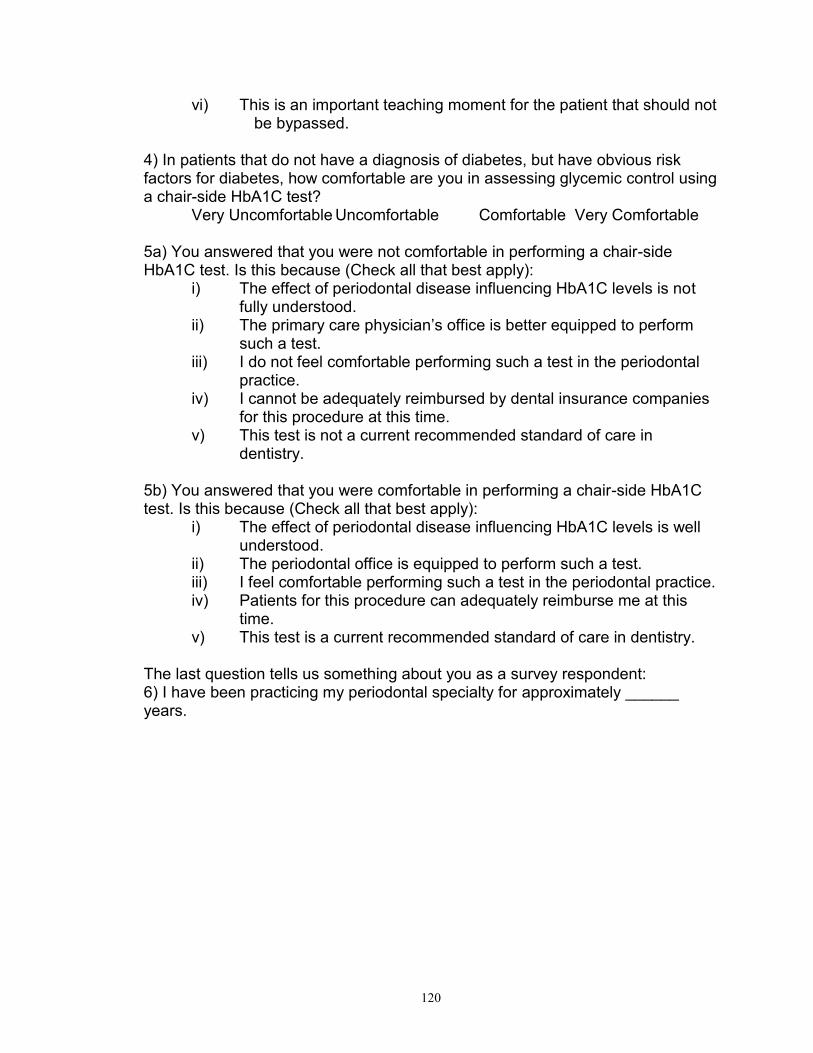
Survey Content
The survey consisted of 6 questions. Three questions were rated on a
four-point Likert-type scale and asked Periodontists about their practices in
86
treating patients who have not yet been diagnosed with type 2 diabetes. Two
more survey items were unique questions that were asked based on
respondent’s previous answers (using skip logic functions), and assessed the
beliefs of the providers for practice decisions (ranking answers, and multiple
answers). The final question was open-ended, which asked information about the
number of years the respondents have been practicing.
Survey Process
A confidential, self-administered survey instrument was developed with
consultation with experts in survey design and methodology. Specialists in the
field of dentistry assisted with the content and pre-tested the survey tool. The
survey was modified to reflect changes suggested from these reviewers, and
then it was transferred to an electronic format using a web-based survey service
(www.surveymonkey.com). The electronic and written versions of the survey
were then pilot tested by having reviewers complete the survey. Based on our
pilot testing, the survey took between 3-5 minutes to complete, regardless
whether the survey was done on paper or via the web-based format.
An e-mail invitation with an imbedded html link to the web-survey was sent
to all 100 participants who agreed to take the survey, with two subsequent
reminder e-mails sent at five days and ten days to non-responders, and a second
telephone call at 7 days to this group. The invitation included an endorsement
Those who disagreed (n=2) in the second question provided reasons for
not probing further about risk factors for diabetes in their patients. Neither
responder cited inadequate time during the dental visit as an important reason for
not discussing risk factors for diabetes, but did feel, in the order of most
important to least important: this is a discussion best addressed by the primary
care physician (average rating= 2.5/5), the patient would not expect the
Periodontist to do this (average rating= 3/5), there is not enough evidence about
the risk factors for diabetes (average rating= 4/5), there is not enough evidence
to suggest that periodontal disease increases the risk of diabetes(average
rating= 5/5) , and they were not comfortable discussing these risk factors
(average rating= 5/5).
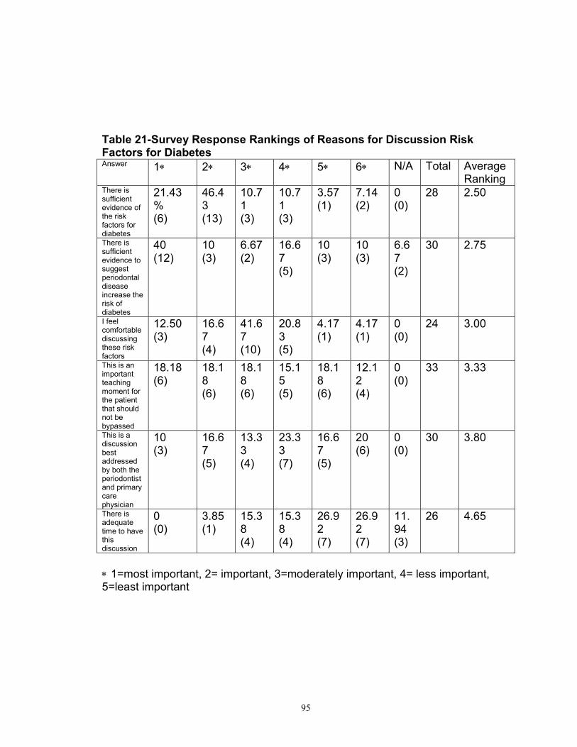
Those who agreed in the second question (n=37), skipped to a question
regarding the reasons for probing further about risk factors for diabetes in their
patients. (Figure 12) In this discussion of risk factors of diabetes with patients,
most felt; 1) This is an important teaching moment for the patient (89%, N=3), 2)
There is sufficient evidence to suggest that periodontal disease increases the risk
90
of diabetes (81%, N=30), 3) This is a discussion best addressed by both the
periodontist and the primary care physician (81%, N=30), 4) There is sufficient
evidence about the risk factors for diabetes (76%, N=28), 5) There is adequate
time during the appointment to have this discussion (70%, N=26), and 6) Feel
comfortable discussing these risk factors (65%, N=24). They felt, in order of most
important to least important: There is sufficient evidence about the risk factors for
diabetes (average rating 2.5/5), there is sufficient evidence to suggest the
periodontal disease increases the risk of diabetes (average rating= 2.75/5), they
were comfortable discussing these risk factors (average rating= 3/5), this is an
important teaching moment for the patient that should not be bypassed (average
rating=3.3/5), this is a discussion best addressed by both the periodontist and the
primary care physician (average rating=3.8/5), and there is adequate time during
the appointment to have this discussion (average rating= 4.7/5). (Table 21)
Screening for Type 2 Diabetes in the Dental Setting
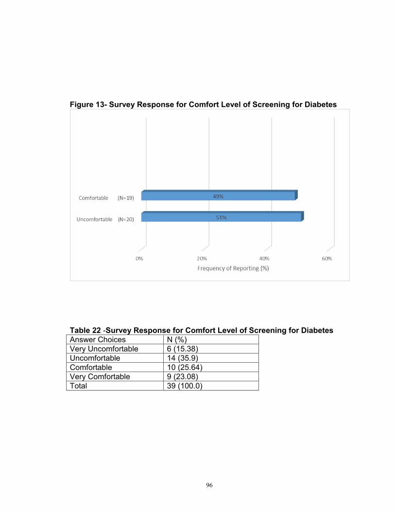
All survey respondents (n=39) were asked if they were comfortable
performing a chair-side HbA1c test for assessing glycemic control, and 54.95%
(n=20) felt uncomfortable, while 48. 7% (n= 19) were comfortable performing an
HbA1c test. (Figure 13). Those uncomfortable in performing an in-office HbA1c
test were asked the reasons for their discomfort, and 80% (n=16) felt that the
physicians office was better equipped to perform such a test, 30% (n-6) did not
feel comfortable performing this test in their office, 20% (n=4) did not feel they
could be adequately reimbursed, 20% (n=4) said it was not a current standard of
91
care in dentistry, and 5% (n=1) reported that the effect of periodontal disease
influencing HbA1c levels is not fully understood. (Figure 14)
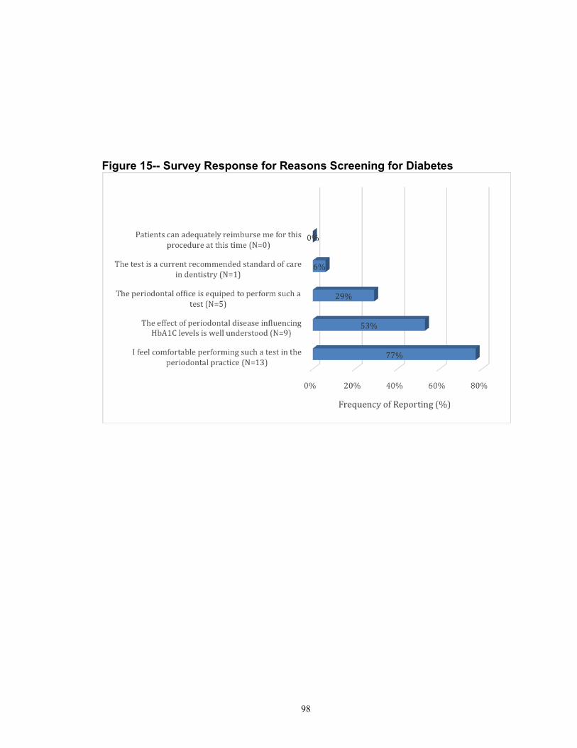
Those comfortable in performing an in-office HbA1c test were asked the
reasons for their comfort level and 76.5% (n=13) reported that they felt
comfortable performing the test in the periodontal practice, 53% (n=9) reported
that the effect of periodontal disease influencing HbA1c levels is well understood,
29.4% (n=5) felt the periodontal office is equipped to perform such a test, and
5.9% (n=1) said that it is a current recommended standard of care in dentistry.
This group did not report a concern for being reimbursed for this procedure (0%,
n=0). (Figure 15)
Of those uncomfortable in performing the HbA1c test in the dental office,
80% (N=16) felt that the physician’s office is better equipped to perform such as
test. (Figure 14) Over 50% of those comfortable in performing this test for
glycemic control in the dental office felt that the effect of periodontitis on HbA1c is
well understood. (Figure 15)
92
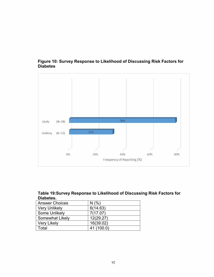
Figure 10: Survey Response to Likelihood of Discussing Risk Factors for Diabetes
Table 19:Survey Response to Likelihood of Discussing Risk Factors for Diabetes.
Answer Choices N (%)
Very Unlikely 6(14.63)
Some Unlikely 7(17.07)
Somewhat Likely 12(29.27)
Very Likely 16(39.02)
Total 41 (100.0)
93
Figure 11-Survey Response to Appropriateness of Discussing Risk Factors for Diabetes
Table 20-Survey Response to Appropriateness of Discussing Risk Factors of Diabetes
Answer Choices N (%)
Strongly Disagree 1 (2.38)
Disagree 2 (4.76)
Agree 30 (71.43)
Strongly Agree 9 (21.43)
Total 42 (100.0)
94
Figure 12-Survey Response to Reasons for Discussion Risk Factors for Diabetes
*ranking in order of importance not summarized in this chart
95
Table 21-Survey Response Rankings of Reasons for Discussion Risk Factors for Diabetes Answer 1 2 3 4 5 6 N/A Total Average
Ranking There is sufficient evidence of the risk factors for diabetes
21.43% (6)
46.43 (13)
10.71 (3)
10.71 (3)
3.57 (1)
7.14 (2)
0 (0)
28
2.50
There is sufficient evidence to suggest periodontal disease increase the risk of diabetes
40 (12)
10 (3)
6.67 (2)
16.67 (5)
10 (3)
10 (3)
6.67 (2)
30 2.75
I feel comfortable discussing these risk factors
12.50 (3)
16.67 (4)
41.67 (10)
20.83 (5)
4.17 (1)
4.17 (1)
0 (0)
24 3.00
This is an important teaching moment for the patient that should not be bypassed
18.18 (6)
18.18 (6)
18.18 (6)
15.15 (5)
18.18 (6)
12.12 (4)
0 (0)
33 3.33
This is a discussion best addressed by both the periodontist and primary care physician
10 (3)
16.67 (5)
13.33 (4)
23.33 (7)
16.67 (5)
20 (6)
0 (0)
30 3.80
There is adequate time to have this discussion
0 (0)
3.85 (1)
15.38 (4)
15.38 (4)
26.92 (7)
26.92 (7)
11.94 (3)
26 4.65
1=most important, 2= important, 3=moderately important, 4= less important, 5=least important
96
Figure 13- Survey Response for Comfort Level of Screening for Diabetes
Table 22 -Survey Response for Comfort Level of Screening for Diabetes
Answer Choices N (%)
Very Uncomfortable 6 (15.38)
Uncomfortable 14 (35.9)
Comfortable 10 (25.64)
Very Comfortable 9 (23.08)
Total 39 (100.0)
97
Figure 14- Survey Response for Reasons Not Screening for Diabetes
98
Figure 15-- Survey Response for Reasons Screening for Diabetes
99
Discussion
The position papers from the AAP have been discussing the two-way
relationship for almost two decades. While evidence of the effect periodontal
disease on glycemic control in type 2 diabetes populations has been well
documented, the Joint EFP/AAP consensus report concluded that evidence is
just emerging.53 This EFP/AAP group also gave recommendations for future
research that will strengthen what is known about this association. Approximately
76% of respondents to our survey felt that there is sufficient evidence to support
periodontal disease increase the risk of type 2 diabetes, though a recent review
concluded such studies are “sparse”.59
Over 50% of those comfortable in performing the test for glycemic control
in the dental office felt that the effect of periodontitis on HbA1c is well
understood. A review of literature identified a study (n=961) in Japan reporting
over ten years, each millimeter increase in periodontal probing depth
corresponded to an HbA1c of 0.13% (p=0.007).23 In contrast, a meta-analysis of
10 interventional studies of periodontal treatment found that successful
periodontal therapy did not result in statistically significant changes in glycemic
control in diabetic subjects, with 0.57% reduction in A1c measures (p=0.82).24
Only 456 subjects were included in all ten studies and larger studies with
randomized clinical trials are needed to determine the benefit of periodontal
therapy on glycemic control in patients with diabetes.
100
Respondents to this survey were all Members of the American Academy
of Periodontology (AAP) and displayed, on average over two decades of
experience in private practice. Members of this Academy have a subscription to
the Journal of Periodontology and are sent position papers, consensus reports,
and reviews from the AAP about topics in Periodontology. This is a professional
population who we expect to be up to date in periodontal literature. Most
responders were likely to discuss risk factors for diabetes (68%, n=28), and
probe further about these risk factors at initial appointments for patients with a
history of periodontitis. This would be expected since, medical history
questionnaires, and review of the medical history by the provider, are standards
of care for initial consultations. In these medical history forms, smoking, diet, and
cardiovascular disease are common items that are included. (See Appendix-
Figure 18- Sample checklist for dentists provided by the AAP) While these
are risk factors for type-2 diabetes, patients positive for these risk factors may
also warrant frequent oral cancer screenings, be on prescription medication, or
be contra-indicated for some procedures. Thus, the 93% (n=39) responders that
would probe further in patients with risk factors for diabetes is not surprising.
This study had several limitations. This study was a convenience sample
of the Washington, D.C. metropolitan area. It gives an initial look at what the
beliefs of local Peridontists have about the standard of care in the profession.
The web-based survey made it easy to send, receive and complete the questions
and no recipients requested a paper format of the survey, thus shortening the
101
time to receive completed surveys for analysis. Response rate may not have
been increased if paper surveys were mailed. Our response rate (39%) is in the
typical range from 20-47% for electronic surveys.60
The sampling the AAP members may not be representative of other
practicing periodontists, who do not have easy access to the Journal of
Periodontology, position papers, consensus reports, and reviews from the AAP
about topics in Periodontology. This group would be expected to be the most
informed group, with an information bias due to the availability of AAP
publications, as their knowledge may be better than other specialists in the
community. Additionally, general dentists often treat mild, to moderate forms of
periodontal disease, so limiting the survey to periodontists may miss the beliefs
of the standard of care in treating the many patients with early periodontal
disease in this geographic area. Our findings may not be generalizable to all
Periodontists and cannot be applied to all providers (general dentists) treating
patients with periodontal disease.
Respondents who answer web-based e-mails may be a biased towards
providers who are inter-net savvy. These individuals can access the most current
literature on the web and may possess an informational bias. Additionally, a local
Periodontist, with whom some of the survey recipients were acquainted, made
the initial telephone calls. Thus responders may also have responded with an
appeasement bias to please a fellow colleague.
102
Questions in the survey had closed-ended answers to which respondents
were asked to choose answers. These answers were presumed to be the most
likely answers based by experts in both dentistry and survey design. While such
a survey is places less burden of time on respondents and is simple to analyze, it
is possible however, that if the questions were open-ended, local Periodontists
may have provided quite different and varied responses.
Conclusion
The association of periodontitis with diabetes with type 2 diabetes is
accepted by local Peridontists (using a convenience sample survey of
Washington DC area Periodontists), and the attitudes and beliefs of these
specialists influence the standard of care in treating dental patients. When asked
if it was appropriate to probe further about of diabetes risk factors in patients with
periodontal disease and no diabetes diagnosis, most respondents (92.9%)
agreed (agreed/ strongly agreed, n=39).
The local Periodontists surveyed felt, in order of most important to least
important: there is sufficient evidence about the risk factors for diabetes, there is
sufficient evidence to suggest that periodontal disease increases the risk of
diabetes, they were comfortable discussing these risk factors, this is an important
teaching moment for the patient that should not be bypassed, this is a discussion
best addressed by both the periodontist and the primary care physician, and
103
there is adequate time during the appointment to have this discussion. This
survey suggests that practicing periodontists are aware that there is an
association between periodontal disease and the onset of type 2 diabetes.
Those surveyed also appear aware of the importance of HbA1c testing in
assessing glycemic control, whether this test is performed in the dental office or
medical setting. This appears to parallel the consensus report of the Joint
European Federation/ American Academy Workshop (EFP/AAP) guidelines to
dentists for patients without a diabetes diagnosis, but obvious risk factors for type
2 diabetes, where patients:
“ should be informed of their risk for having diabetes, assessed using a
chair-side HbA1C test, and/or referred to a physician for appropriate
testing and diagnostic care.” 53
Overall, the local Periodontists responding to the survey appear to be
implementing current and best practices recommended as the standard of care in
dentistry.
104
CHAPTER 5
Conclusions
Summary
Periodontal disease has been proposed as one source of inflammation
that might predispose adults to developing diabetes. Though the hypothesis of a
bidirectional pathway between periodontal disease and diabetes has been
proposed, few studies have addressed periodontal disease before the
occurrence of diabetes.19 Localized periodontal inflammation is now known to
have systemic effects on general health. 40,41 Compromised oral health may
increase the risk of a pre-diabetic status meditated through inflammation. Our
study, which used both clinical exams and markers for inflammation, looked at
the association of periodontal disease exposure and its’ association with pre-
diabetes and diabetes risks.
In our cross- sectional analysis, we found clinical periodontal measures for
inflammation were associated with the likelihood of impaired fasting glucose. This
association seemed to hold true for individuals with only slight bleeding upon
probing, with deeper probing measurements, or more severe gingival bleeding
upon probing. However, a dose response relationship was not observed with
increasing severity of periodontal inflammation. Compared with individuals with
normo-glycemic levels, adults with pre-diabetes using fasting glucose levels, had
105
an increased odds of periodontal clinical inflammation that remained even after
adjustment for lifestyle and co-morbidity covariates. Participants with more
severe periodontal clinical inflammation had an increased odds of impaired
fasting glucose. Compared to individuals in Category I, the odds ratio for
impaired fasting glucose in Category V was 2.1 (95% CI: 1.6-2.8) in an
unadjusted model. This relationship remained in the fully adjusted model with an
odds ratio of 1.5 (1.1--2.1) in this highest category (one or more sites with a
probing depth >4 mm and bleeding upon probing 50%).
To further explore the relationship, between prediabetes and periodontal
inflammation, we performed four additional analyses using markers of systemic
inflammation (serum antibody levels to the periodontal pathogens
Porphyromonas gingivalis and Actinobacillus actinmycetemcommitans), and
markers of localized inflammation (gingival crevicular fluid levels of IL-1 (GCF-
IL-1) and gingival crevicular fluid levels of prostaglandin (PG-E2) However, no
significant association was observed between pre-diabetes or undiagnosed
diabetes and any of those inflammation markers.
In a longitudinal design, adults with clinical periodontal measures for
inflammation were associated with incident diabetes. During 13.84 years of follow
up 1,967 individuals developed Type 2 diabetes of the total (n= 5,819)
participants initial visit 4. Compared with individuals with minimal bleeding and
probing measures, the hazard of incident diabetes appears to be 1.2 times higher
in adults with moderate to severe clinical periodontal inflammation (Category IV
106
and Category V both having 95%CI: 1.0-1.6, P<0.001). However Category III did
not display this increasing significant trend with a hazard ratio of 1.0 (95% CI:0.
8-1.20). Our results did not support the findings of CVD outcomes where
systemic markers for periodontal inflammation were associated with an increased
risk of cardiovascular disease. The hazard of incident diabetes appeared to be
the same in adults with high levels vs. low levels of antibodies to Porphyromonas
gingivalis and Actinobacillus actinmycetemcommitans. Similarly, no increase in
incident diabetes could be seen in high vs. low levels of gingival crevicular fluid
levels of IL-1 and PG-E2.
Using a convenience sample survey of Washington DC area
Periodontists, and the attitudes, beliefs, and the standard of care in treating
dental patients at risk for type 2 diabetes were assessed. Respondents to this
survey were all Members of the American Academy of Periodontology (AAP) and
displayed, on average over two decades of experience in private practice.
Approximately 76% of respondents to our survey felt that there is sufficient
evidence to suggest that periodontal disease increases the risk of type 2
diabetes, though a recent review concluded such studies are “sparse”.59 Almost
50% of those surveyed were comfortable in performing the HbA1c test for
glycemic control in the dental office and of those comfortable, 53% felt that the
effect of periodontitis on HbA1c is well understood.
107
Overall Discussion
Our studies supports that hypothesis that clinical periodontal inflammation
is associated with impaired glucose tolerance and this exposure may increase
the risk of incident diabetes several years later. An increase in bleeding upon
probing appears to be both cross-sectionally associated with impaired fasting
glucose and longitudinally associated with the onset of incident diabetes. A dose-
response relationship was not observed with increasing severity of periodontal
inflammation. These results are to be expected since probing depths give a
measure of prior periodontal attachment loss, while bleeding upon probing
assesses current inflammation. A periodontal exam that combines probing depth
and bleeding scores gives an accurate assessment of prior and current
periodontal status, and both are used together in the examination and diagnosis
of dental patients in the clinical setting.
An assessment of risk of impaired fasting glucose may be performed
easily in the dental office. This requires a dental exam commonly and currently
performed by dental providers. The use of immunoassays, which are costly, and
technically demanding, may not be necessary to define the relationship of
periodontal disease with pre-diabetes.
108
Presumably, the prevention of gingival inflammation may reduce the risk
of diabetes onset. The reduction of periodontal disease, and then diabetes, has
potential to slow the growing epidemic proportions of both diseases. Our studies
contribute to the sparse evidence for the association of periodontal association
with pre-diabetes and future incident diabetes. Additionally, we have provided
preliminary evidence to support the hypothesis that exposure to even mild
gingival inflammation may have systemic effects on glycemic control. This finding
is novel, since other studies found a dose-response relationship, with more
severe inflammation having more effect on glycemic control and type 2
diabetes.22, 23
The Atherosclerosis Risk in Communities (ARIC) Study, a community-
based prospective cohort provided a rich database with which to assess the
effect of periodontal disease exposure on incident diabetes. This study helps
answer the recent call by the Joint EFP/AAP consensus report for studies with
comprehensive clinical data, extent and severity of periodontal disease, level of
glycemic control, and consideration of local and systemic pathways affected by
both periodontal disease and diabetes.53 Our studies served to contribute to the
body of evidence that is largely lacking in the directionality of periodontal disease
and subsequent incident diabetes.
109
Implications
The strength of this overall dissertation is that it serves to contribute to the
lack of evidence to support the directionality of periodontal disease exposure and
the associated risk of pre-diabetes and diabetes. The recent recommendation for
future research in this area by the for more evidence in consensus report of the
Joint European Federation/ American Academy Workshop (EFP/AAP) includes a
“comprehensive assessment of clinical measures of periodontal inflammation
and biochemical markers of inflammation in blood and saliva”.53 Our studies
used a database with complete periodontal assessments, serum antibody levels
to periodontal pathogens, and gingival crevicular fluid levels of inflammatory
markers to asses periodontal disease exposure.
The use serum antibody levels to periodontal pathogens as a surrogate for
disease exposure is controversial. While validated in other studies51,54 , use of
antibody levels to Porphyromonas gingivalis and Actinobacillus
actinmycetemcommitans as measure of past severity of periodontal inflammation
is not accepted as a standard measurement of periodontal disease by the
Academy of Periodontology and not a current recommendation for assessment of
type 2 diabetes risk. However, these markers have been used in mechanistic
studies of cardiovascular disease risk, and helped suggest in our study that a
different biologic pathway may be involved when assessing type 2 diabetes risk.
110
This data will help define the biologic mechanisms and provide direction for
future interventions.
As the prevalence of diabetes and periodontitis increases, dentists will
likely see an increasing number of patients with diagnosed and undiagnosed
diabetes. The American Dental Association now recommends that dental offices
be equipped with glucometers. Patient education about the importance of
prevention and treatment of periodontal disease and diabetes is the role of both
physicians and dentists.61
Insurance coverage for dental care should be mandated for people with
diabetes. The health of the public will be served by public policies which focus on
the prevention and control of periodontitis and diabetes.62 Since the prognosis of
periodontal disease is best treated at its earliest stages, programs aimed at
patient education and health promotion may limit the burden of sequelae
associated with diabetes and periodontal disease.
Future Research
Since even minimal bleeding upon probing in this study was associated
with prevalent pre-diabetes and future incident diabetes, a strong case is made
to support patient education for prevention of periodontal disease to and study
111
the effect of prevention of even mild periodontal inflammation on impaired
glucose tolerance and diabetes.
Assessment younger populations with mild gingival inflammation,
followed longitudinally, may provide evidence of the earliest effects of periodontal
inflammation on the risk of impaired glucose levels and incident diabetes.
Studying gingivitis, the earliest form of periodontal inflammation, may yield the
most sensitive ascertainment of the effect of subtle changes of oral health on
diabetes.
The cost-effectiveness and outcomes of screening programs in the pre-
diabetic state should be reliably assessed in relation to oral health. This can
performed if medical providers routinely refer “at risk” individuals to dental
professionals.
Finally, the relationship between periodontal disease and diabetes is
complex and not likely to be understood by a single study regardless of its
design. Synthesis of existing and future studies will be helpful in elucidating these
relationships and provide the direction for public health policies aimed at
reducing their burden on the general public.
112
Appendix
113
Figure 16: Clinical Appearance of Stages of Periodontal Disease
114
Figure 17: Probing Measurements By Severity of Periodontal Inflammation
115
Figure 18: Example of a Checklist for Periodontal Patients in the Clinical Setting 63
116
Figure 19- Cellular Inflammatory Response to Periodontal Inflammation
117
Figure 20: Diagnostic Criteria of Pre-diabetes and Diabetes by Glycemic Test64
118
Preamble to Telephone and Internet Survey from Chapter 4
Endorsement by the Dean of Howard University College of Dentistry:
Dear Colleagues, Periodontal disease is the most common inflammatory condition worldwide and diabetes is quickly becoming a global epidemic. The bidirectional pathway of periodontal disease and diabetes is not fully understood. While consistent evidence has shown that diabetes is related to periodontitis, emerging evidence suggests that periodontal disease may increase the risk of diabetes onset. Your participation in the survey will help answer important questions regarding the direction of future research and patient education in this important area of oral health. In addition we will better understand what is the attitude of current best practices in managing periodontal patients at risk for diabetes. I hope that you will take the time to complete these six questions. We expect that this survey will take 3-5 minutes to complete. Your consent to participate is assumed by your completing the survey. All data will remain confidential and will only be published in aggregated form. Individual respondents will not be identified in publications.
Your expertise in this field is greatly appreciated. Many thanks for your help. Sincerely, Leo (Signed by Dean Leo Rouse) [Reviewed by Dean Rouse on 11/14/13, electronic signature was added to web-based survey] Leo E. Rouse DDS, FACD Past President of American Dental Education Association Interim Deputy Provost for Health Sciences Dean of Howard University College of Dentistry
119
Survey Questions from Chapter 4: 1) If your patient has a history of periodontal disease, but no reported medical history of diabetes, how likely are you to discuss the risk factors of diabetes (such family history of diabetes, smoking, diet, exercise, cardiovascular disease) during the initial consultation appointment:
Very Unlikely Somewhat Unlikely Somewhat likely Very Likely 2) Do you think it is appropriate to probe further about risk factors of diabetes (such family history of diabetes, smoking, diet, exercise, cardiovascular disease) in your patients without a current medical history of diabetes? Strongly Disagree Disagree Agree Strongly Agree 3a) You answered that you do not agree/strongly disagree that asking about risk factors of diabetes is appropriate in your non-diabetic patients. Is this because: (Number all that apply, if any, in order of importance, i.e. #1-most important reason)
i) There is not enough evidence to suggest periodontal disease increases the risk of diabetes.
ii) There is not enough evidence about the risk factors for diabetes. iii) I don’t feel comfortable discussing these risk factors. iv) There is insufficient time during the appointment to have this
discussion. v) This is a discussion best addressed by the patient’s primary care
physician. vi) I think the patient will not expect the Periodontist to do this.
3b) You answered that you agree/strongly agree that asking about risk factors of diabetes is appropriate in your non-diabetic patients. Is this because: (Number all that apply, if any, in order of importance, i.e. #1-most important reason)
i) There is sufficient evidence to suggest periodontal disease increases the risk of diabetes.
ii) There is sufficient evidence about the risk factors for diabetes. iii) I feel comfortable discussing these risk factors. iv) There is adequate time during the appointment to have this
discussion. v) This is a discussion best addressed by both the periodontist and
the primary care physician.
120
vi) This is an important teaching moment for the patient that should not be bypassed.
4) In patients that do not have a diagnosis of diabetes, but have obvious risk factors for diabetes, how comfortable are you in assessing glycemic control using a chair-side HbA1C test? Very Uncomfortable Uncomfortable Comfortable Very Comfortable 5a) You answered that you were not comfortable in performing a chair-side HbA1C test. Is this because (Check all that best apply):
i) The effect of periodontal disease influencing HbA1C levels is not fully understood.
ii) The primary care physician’s office is better equipped to perform such a test.
iii) I do not feel comfortable performing such a test in the periodontal practice.
iv) I cannot be adequately reimbursed by dental insurance companies for this procedure at this time.
v) This test is not a current recommended standard of care in dentistry.
5b) You answered that you were comfortable in performing a chair-side HbA1C test. Is this because (Check all that best apply):
i) The effect of periodontal disease influencing HbA1C levels is well understood.
ii) The periodontal office is equipped to perform such a test. iii) I feel comfortable performing such a test in the periodontal practice. iv) Patients for this procedure can adequately reimburse me at this
time. v) This test is a current recommended standard of care in dentistry.
The last question tells us something about you as a survey respondent: 6) I have been practicing my periodontal specialty for approximately ______ years.
121
References
1 American Diabetes Association. Diagnosis and Classification of Diabetes Mellitus.
Diabetes Care January 2013 Vol. 36 no Supplement 1 S67-S74.
2 Centers for Disease Control and Prevention (2011) National diabetes fact sheet: national
estimates and general information on diabetes and prediabetes in the United States, 20111
[WWW document]. Atlanta, GA: U.S. Department of Health and Human Services,
Centers for Disease Control and Prevention. URL
http:www.cdc.gov/dibates/pubs/pdf/ndfs_2011.
3 Chen L., Magliano, D.J. & Zimmet, P.Z. (2012) The worldwide epidemiology of type 2
A. Positions and Honors Positions and Employment: 1995-1998 Periodontal Residency, Mayo Clinic 1998-2006 Private practice in Periodontics, Washington, D.C. 1999-2005 Assistant Professor, Howard University College of Dentistry 2000-2005 Assistant Professor, University of Maryland College of Dentistry 2006- present Private practice in Periodontics, Washington, D.C. 2011-present Assistant Professor, Howard University College of Dentistry Other Experience and Professional Memberships 1995-99 Member of American Academy of Periodontology (AAP) 1997 Participant AAP Annual Meeting, New Orleans, LA 1998 Presenter, IADR “The effect of P-15 Growth Factor Around Endosseous Implants 1999-present Diplomate of American Board of Peridontology 2002 Participant AAP Annual Meeting, New Orleans, LA 2007 Ad hoc reviewer, Indian Journal of Dental Research 2007 Participant AAP Annual Meeting, Washington, DC 2006-2014 Graduate Training Program in Clinical Investigation, Johns
Hopkins University
128
Honors/Awards 1995 Dean’s Award Howard University College of Dentistry 1995 Who’s Who Recognition American Colleges 2006 Teacher of the Year in Clinic Modules- Howard University 2006-2011 T-32 NIH Training Grant at NYU and Johns Hopkins B. Peer-reviewed Publications (in chronological order)
Mustapha, IZ, Reeve, CM, Sheridan PJ. The effect of P-15 growth factor around endosseous implants. (Abstract) IADR, 1998. Meraw, S, Mustapha, IZ, Rogers, R. The effect of tobacco on oral mucosa. Dental Clinics of North America. 1998. Mustapha, IZ, Boucree SA, Mucocele of the lower lip-an uncommon presentation and review. JCDA.May 2004. Vol. 70, No.5 Mustapha, IZ, Debrey, S, Oladabu, M, Ugarte, R. Markers of systemic bacterial exposure in periodontal disease and cardiovascular disease risk-A systematic review and meta-analysis. J Periodontol. 2007; 78: 12. 2287-2302. Clark, Jeanne M., Gibbons, Christopher H, Mustapha, Indra Z. Chpt. 11: The Associations of Diabetes with Digestive, Oral and Liver Disease and Autonomic Neuropathy. Diabetes Public Health: From Data to Policy. Center for Disease Control. Oxford University Press. 2010.