Page 1/19 Optimization of Ligature/Bone Defect-Induced Periodontitis Model in Rats Jingyi Gao Southern Medical University Simin Cai Southern Medical University Dan Li Southern Medical University Zijie Wang Southern Medical University Minyi Ou Southern Medical University Xinlu Zhang Southern Medical University Zhihui Tian ( [email protected]) Southern Medical University Research Article Keywords: periodontitis, animal model, bone defect, ligature, rat Posted Date: September 20th, 2021 DOI: https://doi.org/10.21203/rs.3.rs-880589/v1 License: This work is licensed under a Creative Commons Attribution 4.0 International License. Read Full License
Transcript
Page 1/19
Optimization of Ligature/Bone Defect-InducedPeriodontitis Model in RatsJingyi Gao
BackgroundThe destruction of alveolar bone is a crucial manifestation of severe periodontitis, which stem cell-basedbioengineered therapies are expected to cure. Therefore, a cost-effective, reproducible, quanti�ability and easier-to-administrate animal model that mimics human periodontitis is of great importance for further endeavor.
MethodsIn this study, we created periodontitis rat models in silk ligation group, bone defect group and bone defect/silkligation group respectively. he clinical indexes of periodontitis were observed and recorded. The mandible wastaken for micro-computed tomographic, histological and histomorphometric analysis to assess the periodontalin�ammation and bone remodeling
ResultsObvious periodontal in�ammation but slight alveolar bone resorption were observed in the ligation group, whilesurgical trauma was not robust enough to continually worsen the constructed bone defect area in the bonedefect group. In the bone defect/ligature group, obvious and stable periodontal in�ammation could be the mostlasting with similar evolving pathological patterns of human periodontitis. It also exhibited enhanced clinicalsimilarity and con�rmed its superiority in quantitativeness.
ConclusionsThe present rat model is the �rst study to reproduce a pathological process similar to human periodontitis withreliable stability and repeatability, manifesting a priority to previous methods. Day 9 to Day 12 is the best timefor reproducing severe periodontitis syndromes with vertical bone resorption in this model.
BackgroundPeriodontitis is a chronic, multifactorial, infectious disease which can lead to the damage of periodontal tissuesincluding gingiva, alveolar bone, cementum, periodontal ligament and even serious tooth loss (1). Worldwideepidemiological data show that a fraction of around 10% of those over 40–50 years in all populations exhibitingsevere periodontitis (2). Periodontitis has become the prevalent cause of tooth loss in 90% of adults in the worldtoday (3). It is also associated with systemic diseases such as heart disease, diabetes, Alzheimer's disease andpregnancy complications (4). At present, the mainstream clinical treatment of periodontitis is to control thedevelopment of periodontitis through initial therapy and periodontal surgery, but the ravaged periodontal tissuecan seldom be restored (5). The regeneration of tissue loss caused by periodontitis is the ultimate goal ofperiodontal therapy (6). With the development of stem cells and bio-engineering, the research of periodontalregeneration therapy is making headway and it also points out the direction for the clinical treatment of
Page 3/19
periodontitis (7). While the more promising periodontal tissue engineering treatment is still in the research stage,it is necessary to evaluate the effect of periodontal tissue regeneration through in vivo animal experiments.
Animal experiments are the indispensable pathways to evaluate any new treatments. At present, animal modelsused for the study of periodontal tissue regeneration are often created by bone defect modeling method, whichestablishes the acute bone defect by surgically removing part of the alveolar bone, periodontal ligament andcementum (6). Surgical creation of bone defects allows for rapid, stable, and quanti�able access to periodontalbone tissue for study (8). However, due to the lack of in�ammatory microenvironment caused by accumulationof microbial plaque, the model obtained by acute bone defect is not satisfactory in etiology, development andprognosis of periodontitis (9, 10). The acute bone defects caused by surgery lack in�ammatory inductionprocess. Therefore, it is not widely used in periodontitis-related research (11). Instead, this method is moresuitable for mechanical traumatic etiology (12, 13).
In order to overcome the shortcomings of bone defect modeling, some studies have combined the bone defectmethod with periodontitis silk ligation and successfully established periodontitis models of large animals (suchas in miniature pigs and beagle dogs) (14, 15). Intraosseous defect is created on the alveolar bone and directlyexposed to the oral environment(16), then the ligated silk threads are placed around the cervical region of theteeth to ensure long-term plaque deposition and accelerate the natural process of in�ammation (17). Thismethod not only ensures a more standardized morphology of the surgically created defect but also allows forreliable reproducibility of the study according to any given scheme, combining the rapid creation of periodontalbone tissue defects with the in�ammatory microenvironment maintained by silk thread ligation method. It is amore appropriate model for the occurrence, development and prognosis of periodontal diseases in anin�ammatory environment.
At present, there has been no report on the establishment of regenerative periodontitis model in rats by bonedefect combined with silk ligature. In this regard, we proposed a novel surgical procedure to create bone defectsin the mandibular molar region of rats by removing alveolar bone and tying the teeth with silk ligature. Weobserved and recorded the clinical manifestations of periodontitis. Micro-computed tomographic, histologicaland histomorphometric were also performed to analyze the in�ammation and bone remodeling degrees. Ourprotocol have overcome the surgical operational obstacles of narrow oral region, small teeth of rats (18) andadministrated reproducible and standardized bone defects in a rapider and greater manner. Afterwards, silkligature was sutured around the cervical portion to simulate chronic periodontitis in rats. These optimizationsare expected to better mimic the pathological process of periodontitis in both acute and chronic in�ammation.We believe the present solution can facilitate the use of periodontitis models in periodontal regenerationresearch and shed light for the future study in the clinical effect of periodontal tissue regeneration therapy.
Methods
Establishment of animal modelForty female SPF Wistar Rats, aging 8–12 weeks and weighing 220-240g (19), were obtained from the AnimalScience Center of Southern Medical University (Guangzhou, China). Animals were acclimated for 1 week beforeperiodontitis induction and they were housed under conventional condition with free access to water and foodafter periodontitis induction. Animal experiments were approved by the Institutional Review Board of the
Page 4/19
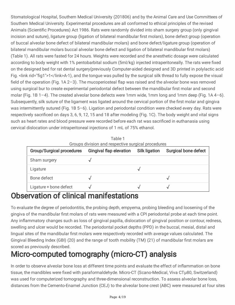
Stomatological Hospital, Southern Medical University (201806) and by the Animal Care and Use Committees ofSouthern Medical University. Experimental procedures are all conformed to ethical principles of the revisedAnimals (Scienti�c Procedures) Act 1986. Rats were randomly divided into sham surgery group (only gingivalincision and suture), ligature group (ligation of bilateral mandibular �rst molars), bone defect group (operationof buccal alveolar bone defect of bilateral mandibular molars) and bone defect/ligature group (operation ofbilateral mandibular molars buccal alveolar bone defect and ligation of bilateral mandibular �rst molars)(Table 1). All rats were fasted for 24 hours. Weights were recorded and the anesthetic dosage were calculatedaccording to body weight with 1% pentobarbital sodium (5ml/kg) injected intraperitoneally. The rats were �xedon the designed bed for rat dental surgery(previously Computer-aided designed and 3D printed in polylactic acidFig. <link rid="�g1">1</link>A-1), and the tongue was pulled by the surgical silk thread to fully expose the visual�eld of the operation (Fig. 1A 2–3). The mucoperiosteal �ap was raised and the alveolar bone was removedusing surgical bur to create experimental periodontal defect between the mandibular �rst molar and secondmolar (Fig. 1B 1–4). The created alveolar bone defects were 1mm wide, 1mm long and 1mm deep (Fig. 1A 4–6).Subsequently, silk suture of the ligament was ligated around the cervical portion of the �rst molar and gingivawas intermittently sutured (Fig. 1B 5–6). Ligation and periodontal condition were checked every day. Rats wererespectively sacri�ced on days 3, 6, 9, 12, 15 and 18 after modeling (Fig. 1C). The body weight and vital signssuch as heart rates and blood pressure were recorded before each rat was sacri�ced in euthanasia usingcervical dislocation under intraperitoneal injections of 1 mL of 75% ethanol.
Table 1Groups division and respective surgical procedures
Group/Surgical procedures Gingival �ap elevation Silk ligation Surgical bone defect
Sham surgery √
Ligature √
Bone defect √ √
Ligature + bone defect √ √ √
Observation of clinical manifestationsTo evaluate the degree of periodontitis, the probing depth, empyema, probing bleeding and loosening of thegingiva of the mandibular �rst molars of rats were measured with a CPI periodontal probe at each time point.Any in�ammatory changes such as loss of gingival papilla, dislocation of gingival position or contour, redness,swelling and ulcer would be recorded. The periodontal pocket depths (PPD) in the buccal, mesial, distal andlingual sites of the mandibular �rst molars were respectively recorded with average values calculated. TheGingival Bleeding Index (GBI) (20) and the range of tooth mobility (TM) (21) of mandibular �rst molars arescored as previously described.
Micro-computed tomography (micro-CT) analysisIn order to observe alveolar bone loss at different time points and evaluate the effect of in�ammation on bonetissue, the mandibles were �xed with paraformaldehyde. Micro-CT (Scano-Medical, Viva CTµ80, Switzerland)was used for computerized tomography and three-dimensional reconstruction. To assess alveolar bone loss,distances from the Cemento-Enamel Junction (CEJ) to the alveolar bone crest (ABC) were measured at four sites
Page 5/19
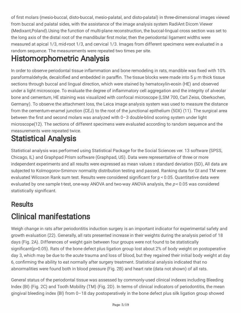
of �rst molars (mesio-buccal, disto-buccal, mesio-palatal, and disto-palatal) in three-dimensional images viewedfrom buccal and palatal sides, with the assistance of the image analysis system RadiAnt Dicom Viewer(Medixant,Poland).Using the function of multi-plane reconstruction, the buccal-lingual cross section was set tothe long axis of the distal root of the mandibular �rst molar, then the periodontal ligament widths weremeasured at apical 1/3, mid-root 1/3, and cervical 1/3. Images from different specimens were evaluated in arandom sequence. The measurements were repeated two times per site.
Histomorphometric AnalysisIn order to observe periodontal tissue in�ammation and bone remodeling in rats, mandible was �xed with 10%paraformaldehyde, decalci�ed and embedded in para�n. The tissue blocks were made into 5 µ m thick tissuesections through buccal and lingual direction, which were stained by hematoxylin-eosin (HE) and observedunder a light microscope. To evaluate the degree of in�ammatory cell aggregation and the integrity of alveolarbone and cementum, HE staining was visualized with confocal microscope (LSM 700, Carl Zeiss, Oberkochen,Germany). To observe the attachment loss, the Leica image analysis system was used to measure the distancefrom the cementum-enamel junction (CEJ) to the root of the junctional epithelium (50X) (11). The surgical areabetween the �rst and second molars was analyzed with 0–3 double-blind scoring system under lightmicroscope(12). The sections of different specimens were evaluated according to random sequence and themeasurements were repeated twice.
Statistical AnalysisStatistical analysis was performed using Statistical Package for the Social Sciences ver. 13 software (SPSS,Chicago, IL) and Graphpad Prism software (Graphpad, US). Data were representative of three or moreindependent experiments and all results were expressed as mean values ± standard deviation (SD), All data aresubjected to Kolmogorov-Smirnov normality distribution testing and passed. Ranking data for GI and TM wereevaluated Wilcoxon Rank sum test. Results were considered signi�cant for p < 0.05. Quantitative data wereevaluated by one sample t-test, one-way ANOVA and two-way ANOVA analysis, the p < 0.05 was consideredstatistically signi�cant.
Results
Clinical manifestationsWeigh change in rats after periodontitis induction surgery is an important indicator for experimental safety andgrowth evaluation (22). Generally, all rats presented increase in their weights during the analysis period of 18days (Fig. 2A). Differences of weight gain between four groups were not found to be statisticallysigni�cant(p>0.05). Rats of the bone defect plus ligation group lost about 2% of body weight on postoperativeday 3, which may be due to the acute trauma and loss of blood, but they regained their initial body weight at day6, con�rming the ability to eat normally after surgery treatment. Statistical analysis indicated that noabnormalities were found both in blood pressure (Fig. 2B) and heart rate (data not shown) of all rats.
General status of the periodontal tissue was assessed by commonly-used clinical indexes including BleedingIndex (BI) (Fig. 2C) and Tooth Mobility (TM) (Fig. 2D). In terms of clinical indicators of periodontitis, the meangingival bleeding index (BI) from 0–18 day postoperatively in the bone defect plus silk ligation group showed
Page 6/19
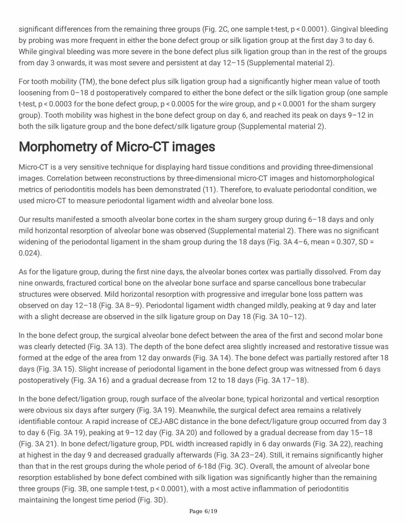
signi�cant differences from the remaining three groups (Fig. 2C, one sample t-test, p < 0.0001). Gingival bleedingby probing was more frequent in either the bone defect group or silk ligation group at the �rst day 3 to day 6.While gingival bleeding was more severe in the bone defect plus silk ligation group than in the rest of the groupsfrom day 3 onwards, it was most severe and persistent at day 12–15 (Supplemental material 2).
For tooth mobility (TM), the bone defect plus silk ligation group had a signi�cantly higher mean value of toothloosening from 0–18 d postoperatively compared to either the bone defect or the silk ligation group (one samplet-test, p < 0.0003 for the bone defect group, p < 0.0005 for the wire group, and p < 0.0001 for the sham surgerygroup). Tooth mobility was highest in the bone defect group on day 6, and reached its peak on days 9–12 inboth the silk ligature group and the bone defect/silk ligature group (Supplemental material 2).
Morphometry of Micro-CT imagesMicro-CT is a very sensitive technique for displaying hard tissue conditions and providing three-dimensionalimages. Correlation between reconstructions by three-dimensional micro-CT images and histomorphologicalmetrics of periodontitis models has been demonstrated (11). Therefore, to evaluate periodontal condition, weused micro-CT to measure periodontal ligament width and alveolar bone loss.
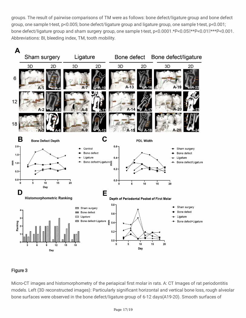
Our results manifested a smooth alveolar bone cortex in the sham surgery group during 6–18 days and onlymild horizontal resorption of alveolar bone was observed (Supplemental material 2). There was no signi�cantwidening of the periodontal ligament in the sham group during the 18 days (Fig. 3A 4–6, mean = 0.307, SD = 0.024).
As for the ligature group, during the �rst nine days, the alveolar bones cortex was partially dissolved. From daynine onwards, fractured cortical bone on the alveolar bone surface and sparse cancellous bone trabecularstructures were observed. Mild horizontal resorption with progressive and irregular bone loss pattern wasobserved on day 12–18 (Fig. 3A 8–9). Periodontal ligament width changed mildly, peaking at 9 day and laterwith a slight decrease are observed in the silk ligature group on Day 18 (Fig. 3A 10–12).
In the bone defect group, the surgical alveolar bone defect between the area of the �rst and second molar bonewas clearly detected (Fig. 3A 13). The depth of the bone defect area slightly increased and restorative tissue wasformed at the edge of the area from 12 day onwards (Fig. 3A 14). The bone defect was partially restored after 18days (Fig. 3A 15). Slight increase of periodontal ligament in the bone defect group was witnessed from 6 dayspostoperatively (Fig. 3A 16) and a gradual decrease from 12 to 18 days (Fig. 3A 17–18).
In the bone defect/ligation group, rough surface of the alveolar bone, typical horizontal and vertical resorptionwere obvious six days after surgery (Fig. 3A 19). Meanwhile, the surgical defect area remains a relativelyidenti�able contour. A rapid increase of CEJ-ABC distance in the bone defect/ligature group occurred from day 3to day 6 (Fig. 3A 19), peaking at 9–12 day (Fig. 3A 20) and followed by a gradual decrease from day 15–18(Fig. 3A 21). In bone defect/ligature group, PDL width increased rapidly in 6 day onwards (Fig. 3A 22), reachingat highest in the day 9 and decreased gradually afterwards (Fig. 3A 23–24). Still, it remains signi�cantly higherthan that in the rest groups during the whole period of 6-18d (Fig. 3C). Overall, the amount of alveolar boneresorption established by bone defect combined with silk ligation was signi�cantly higher than the remainingthree groups (Fig. 3B, one sample t-test, p < 0.0001), with a most active in�ammation of periodontitismaintaining the longest time period (Fig. 3D).
Page 7/19
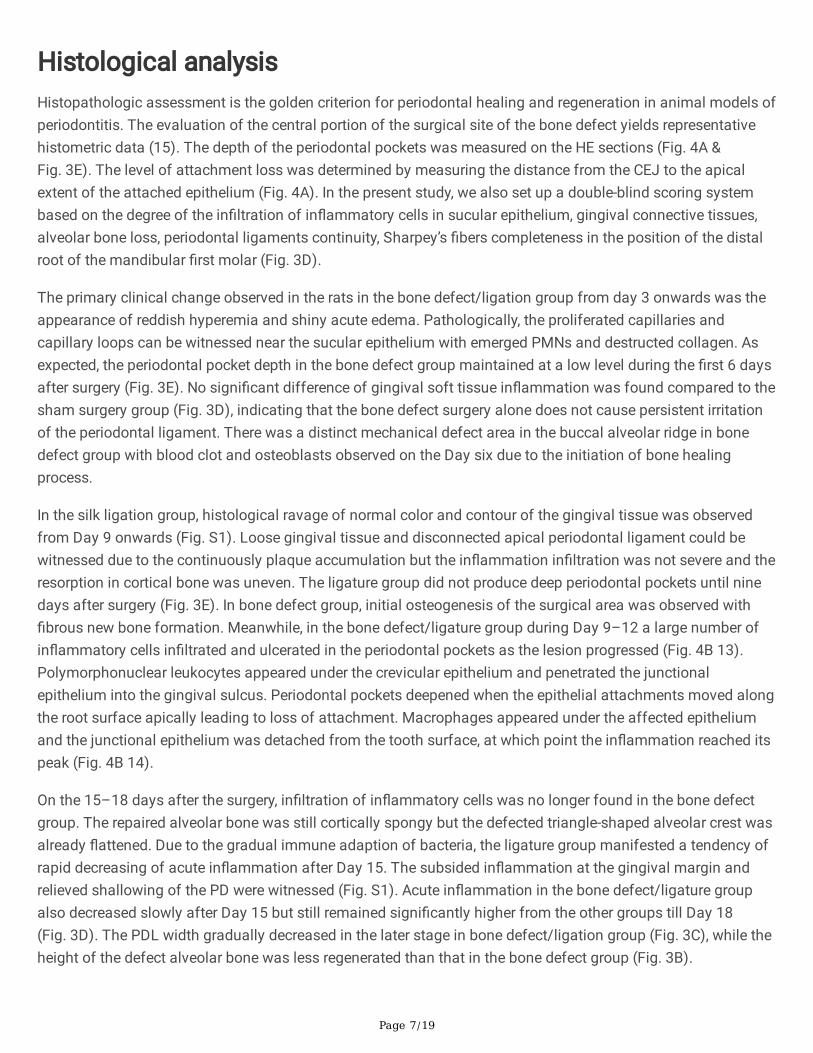
Histological analysisHistopathologic assessment is the golden criterion for periodontal healing and regeneration in animal models ofperiodontitis. The evaluation of the central portion of the surgical site of the bone defect yields representativehistometric data (15). The depth of the periodontal pockets was measured on the HE sections (Fig. 4A &Fig. 3E). The level of attachment loss was determined by measuring the distance from the CEJ to the apicalextent of the attached epithelium (Fig. 4A). In the present study, we also set up a double-blind scoring systembased on the degree of the in�ltration of in�ammatory cells in sucular epithelium, gingival connective tissues,alveolar bone loss, periodontal ligaments continuity, Sharpey’s �bers completeness in the position of the distalroot of the mandibular �rst molar (Fig. 3D).
The primary clinical change observed in the rats in the bone defect/ligation group from day 3 onwards was theappearance of reddish hyperemia and shiny acute edema. Pathologically, the proliferated capillaries andcapillary loops can be witnessed near the sucular epithelium with emerged PMNs and destructed collagen. Asexpected, the periodontal pocket depth in the bone defect group maintained at a low level during the �rst 6 daysafter surgery (Fig. 3E). No signi�cant difference of gingival soft tissue in�ammation was found compared to thesham surgery group (Fig. 3D), indicating that the bone defect surgery alone does not cause persistent irritationof the periodontal ligament. There was a distinct mechanical defect area in the buccal alveolar ridge in bonedefect group with blood clot and osteoblasts observed on the Day six due to the initiation of bone healingprocess.
In the silk ligation group, histological ravage of normal color and contour of the gingival tissue was observedfrom Day 9 onwards (Fig. S1). Loose gingival tissue and disconnected apical periodontal ligament could bewitnessed due to the continuously plaque accumulation but the in�ammation in�ltration was not severe and theresorption in cortical bone was uneven. The ligature group did not produce deep periodontal pockets until ninedays after surgery (Fig. 3E). In bone defect group, initial osteogenesis of the surgical area was observed with�brous new bone formation. Meanwhile, in the bone defect/ligature group during Day 9–12 a large number ofin�ammatory cells in�ltrated and ulcerated in the periodontal pockets as the lesion progressed (Fig. 4B 13).Polymorphonuclear leukocytes appeared under the crevicular epithelium and penetrated the junctionalepithelium into the gingival sulcus. Periodontal pockets deepened when the epithelial attachments moved alongthe root surface apically leading to loss of attachment. Macrophages appeared under the affected epitheliumand the junctional epithelium was detached from the tooth surface, at which point the in�ammation reached itspeak (Fig. 4B 14).
On the 15–18 days after the surgery, in�ltration of in�ammatory cells was no longer found in the bone defectgroup. The repaired alveolar bone was still cortically spongy but the defected triangle-shaped alveolar crest wasalready �attened. Due to the gradual immune adaption of bacteria, the ligature group manifested a tendency ofrapid decreasing of acute in�ammation after Day 15. The subsided in�ammation at the gingival margin andrelieved shallowing of the PD were witnessed (Fig. S1). Acute in�ammation in the bone defect/ligature groupalso decreased slowly after Day 15 but still remained signi�cantly higher from the other groups till Day 18(Fig. 3D). The PDL width gradually decreased in the later stage in bone defect/ligation group (Fig. 3C), while theheight of the defect alveolar bone was less regenerated than that in the bone defect group (Fig. 3B).
Page 8/19
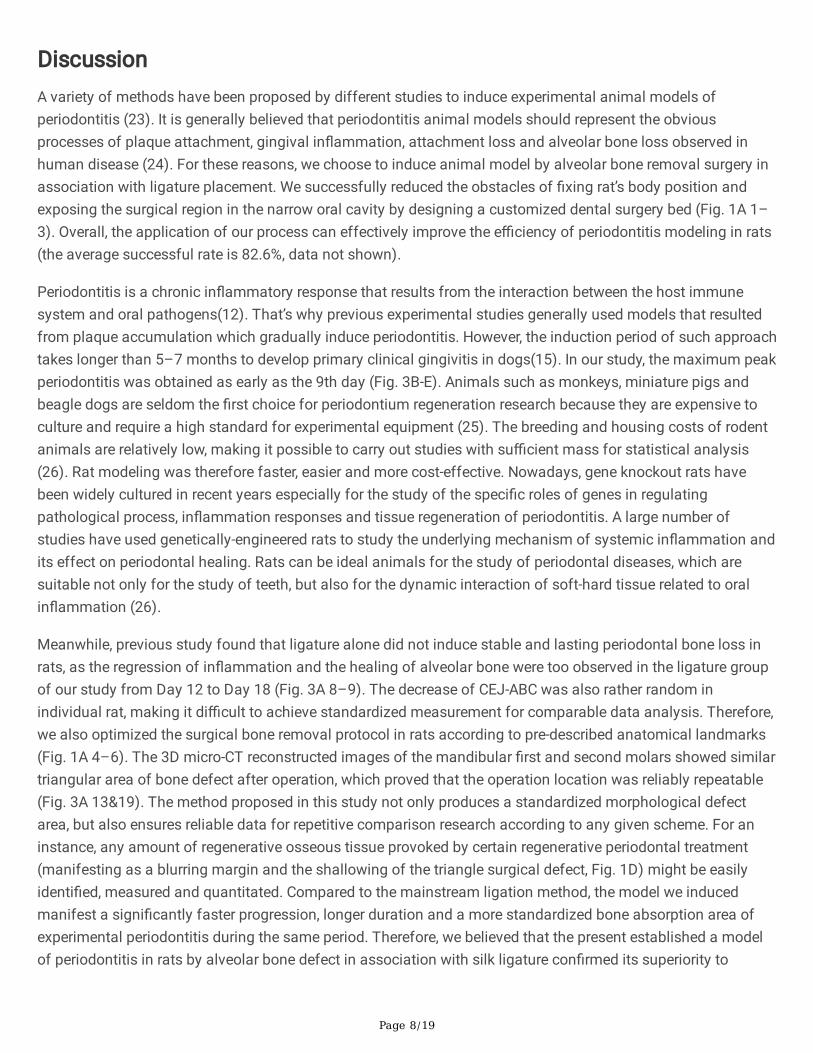
DiscussionA variety of methods have been proposed by different studies to induce experimental animal models ofperiodontitis (23). It is generally believed that periodontitis animal models should represent the obviousprocesses of plaque attachment, gingival in�ammation, attachment loss and alveolar bone loss observed inhuman disease (24). For these reasons, we choose to induce animal model by alveolar bone removal surgery inassociation with ligature placement. We successfully reduced the obstacles of �xing rat’s body position andexposing the surgical region in the narrow oral cavity by designing a customized dental surgery bed (Fig. 1A 1–3). Overall, the application of our process can effectively improve the e�ciency of periodontitis modeling in rats(the average successful rate is 82.6%, data not shown).
Periodontitis is a chronic in�ammatory response that results from the interaction between the host immunesystem and oral pathogens(12). That’s why previous experimental studies generally used models that resultedfrom plaque accumulation which gradually induce periodontitis. However, the induction period of such approachtakes longer than 5–7 months to develop primary clinical gingivitis in dogs(15). In our study, the maximum peakperiodontitis was obtained as early as the 9th day (Fig. 3B-E). Animals such as monkeys, miniature pigs andbeagle dogs are seldom the �rst choice for periodontium regeneration research because they are expensive toculture and require a high standard for experimental equipment (25). The breeding and housing costs of rodentanimals are relatively low, making it possible to carry out studies with su�cient mass for statistical analysis(26). Rat modeling was therefore faster, easier and more cost-effective. Nowadays, gene knockout rats havebeen widely cultured in recent years especially for the study of the speci�c roles of genes in regulatingpathological process, in�ammation responses and tissue regeneration of periodontitis. A large number ofstudies have used genetically-engineered rats to study the underlying mechanism of systemic in�ammation andits effect on periodontal healing. Rats can be ideal animals for the study of periodontal diseases, which aresuitable not only for the study of teeth, but also for the dynamic interaction of soft-hard tissue related to oralin�ammation (26).
Meanwhile, previous study found that ligature alone did not induce stable and lasting periodontal bone loss inrats, as the regression of in�ammation and the healing of alveolar bone were too observed in the ligature groupof our study from Day 12 to Day 18 (Fig. 3A 8–9). The decrease of CEJ-ABC was also rather random inindividual rat, making it di�cult to achieve standardized measurement for comparable data analysis. Therefore,we also optimized the surgical bone removal protocol in rats according to pre-described anatomical landmarks(Fig. 1A 4–6). The 3D micro-CT reconstructed images of the mandibular �rst and second molars showed similartriangular area of bone defect after operation, which proved that the operation location was reliably repeatable(Fig. 3A 13&19). The method proposed in this study not only produces a standardized morphological defectarea, but also ensures reliable data for repetitive comparison research according to any given scheme. For aninstance, any amount of regenerative osseous tissue provoked by certain regenerative periodontal treatment(manifesting as a blurring margin and the shallowing of the triangle surgical defect, Fig. 1D) might be easilyidenti�ed, measured and quantitated. Compared to the mainstream ligation method, the model we inducedmanifest a signi�cantly faster progression, longer duration and a more standardized bone absorption area ofexperimental periodontitis during the same period. Therefore, we believed that the present established a modelof periodontitis in rats by alveolar bone defect in association with silk ligature con�rmed its superiority to
Page 9/19
previous methods, proving its essentiality to be a suitable experimental model for regenerative periodontaltreatment evaluation.
By far, no satisfying model similar to the pathologic process of human periodontitis has been proposed (23). Inpresent study, we obtained various methods to evaluate the model outcomes of periodontitis at different timepoints, including the two-dimensional CT panel of labial-lingual section, the micro-CT reconstructed three-dimensional model and HE stained histopathological sections, all reporting obvious time-pattern changes andspeci�city. The present rat models we established, induced by acute alveolar bone defect and chronic silkligature, is the �rst to successfully mimic the pathological changes in periodontal tissue and stages divisions inhuman periodontitis (Table 2).
Page 10/19
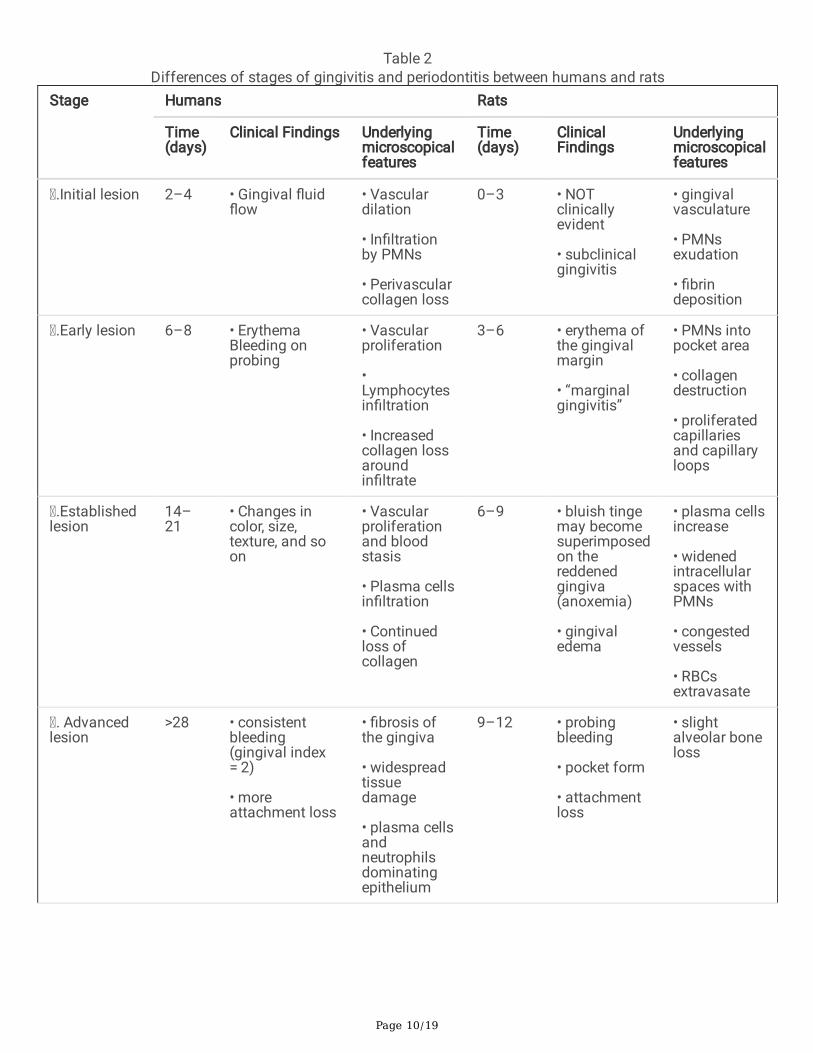
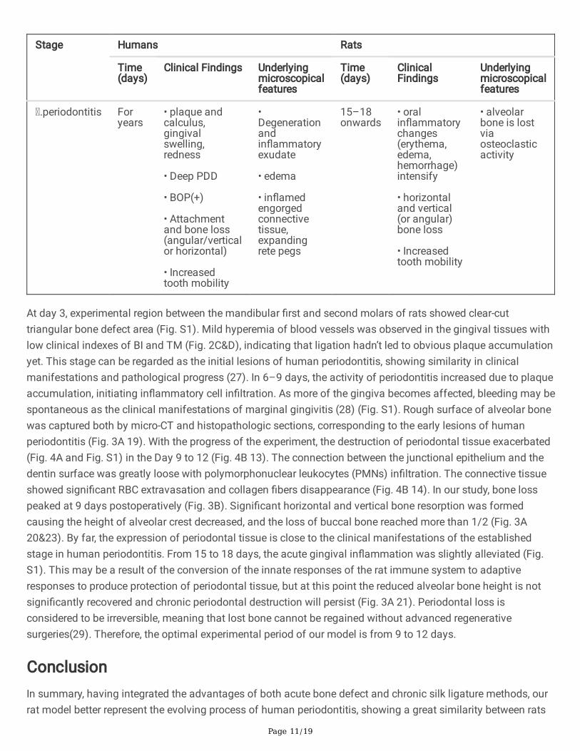
Table 2Differences of stages of gingivitis and periodontitis between humans and rats
At day 3, experimental region between the mandibular �rst and second molars of rats showed clear-cuttriangular bone defect area (Fig. S1). Mild hyperemia of blood vessels was observed in the gingival tissues withlow clinical indexes of BI and TM (Fig. 2C&D), indicating that ligation hadn’t led to obvious plaque accumulationyet. This stage can be regarded as the initial lesions of human periodontitis, showing similarity in clinicalmanifestations and pathological progress (27). In 6–9 days, the activity of periodontitis increased due to plaqueaccumulation, initiating in�ammatory cell in�ltration. As more of the gingiva becomes affected, bleeding may bespontaneous as the clinical manifestations of marginal gingivitis (28) (Fig. S1). Rough surface of alveolar bonewas captured both by micro-CT and histopathologic sections, corresponding to the early lesions of humanperiodontitis (Fig. 3A 19). With the progress of the experiment, the destruction of periodontal tissue exacerbated(Fig. 4A and Fig. S1) in the Day 9 to 12 (Fig. 4B 13). The connection between the junctional epithelium and thedentin surface was greatly loose with polymorphonuclear leukocytes (PMNs) in�ltration. The connective tissueshowed signi�cant RBC extravasation and collagen �bers disappearance (Fig. 4B 14). In our study, bone losspeaked at 9 days postoperatively (Fig. 3B). Signi�cant horizontal and vertical bone resorption was formedcausing the height of alveolar crest decreased, and the loss of buccal bone reached more than 1/2 (Fig. 3A20&23). By far, the expression of periodontal tissue is close to the clinical manifestations of the establishedstage in human periodontitis. From 15 to 18 days, the acute gingival in�ammation was slightly alleviated (Fig.S1). This may be a result of the conversion of the innate responses of the rat immune system to adaptiveresponses to produce protection of periodontal tissue, but at this point the reduced alveolar bone height is notsigni�cantly recovered and chronic periodontal destruction will persist (Fig. 3A 21). Periodontal loss isconsidered to be irreversible, meaning that lost bone cannot be regained without advanced regenerativesurgeries(29). Therefore, the optimal experimental period of our model is from 9 to 12 days.
ConclusionIn summary, having integrated the advantages of both acute bone defect and chronic silk ligature methods, ourrat model better represent the evolving process of human periodontitis, showing a great similarity between rats
Page 12/19
and humans in the divisions of clinical syndromes and pathological changes (27). It can be fully applied to thestudy during the period of Day 9–12 when reaches the most active peak. Present protocol proves to establish asuitable experimental model for the regenerative research of periodontitis, as the stability and reproducibility ofalveolar bone resorption triumphs over the rest of the methods as demonstrated above. The optimization of thismodel is anticipated to contribute to the application of periodontitis animal model in the future research,especially in the evaluation of clinical e�cacy as well as the underlying mechanism of periodontal regenerationtherapy.
DeclarationsEthics approval and consent to participate
Animal experiments were approved by the Institutional Review Board of the Stomatological Hospital, SouthernMedical University (201806) and by the Animal Care and Use Committees of Southern Medical University.Experimental procedures are all conformed to ethical principles of the revised Animals (Scienti�c Procedures)Act 1986. The study was carried out in compliance with the ARRIVE guidelines.
Consent for publication
Not applicable.
Availability of data and materials
The datasets used and/or analysed during the current study are available from the corresponding author onreasonable request.
Competing interest
All authors report no competing interest for this paper.
Authors' contributions
All authors have made substantial contributions to conception and design, establishment of animal model,analysis or interpretation of data in this study. Jinyi Gao carried out histomorphometric analysis, observation ofclinical index and drafted the manuscript. Simin Cai carried out micro-CT analysis and participated inobservation of clinical index, all data analysis and manuscript drafting. Minyi Ou participated in the studydesign and the data analysis, helped to perform the tissue sample preparation and revise the manuscript. Dan Liperformed the tissue sample preparation, participated in the data analysis and advised on the study design. ZijieWang advised on the data analysis and helped to revise the manuscript. Xinlu Zhang advised on the dataanalysis and reviewed the manuscript. Zhihui Tian conceived of the study, carried out the study design, helpedto draft and revise the manuscript. All authors read and approved the �nal manuscript.
Funding
This work was supported by the National Natural Science Foundation of China [818003710], Foundation ofPresident of Nanfang Hospital [grant.MO 2018B014] and GuangDong Basic and Applied Basic Research
Page 13/19
Foundation [2021A1515011656]. This work was also supported by grants from Guangdong Basic and AppliedBasic Research Fund Project [Grant No.2019A1515011503, to YGT].
Acknowledgments
We thank Dr. Xujia Lai, for her review and technical support for the statistical analysis in this paper.
References1. Bouchard P, Carra MC, Boillot A, Mora F, Range H. Risk factors in periodontology: a conceptual framework. J
Clin Periodontol. 2017;44(2):125-31.
2. Eke PI, Dye BA, Wei L, Slade GD, Thornton-Evans GO, Borgnakke WS, et al. Update on Prevalence ofPeriodontitis in Adults in the United States: NHANES 2009 to 2012. J Periodontol. 2015;86(5):611-22.
3. Nibali L, Farias BC, Vajgel A, Tu YK, Donos N. Tooth loss in aggressive periodontitis: a systematic review. JDent Res. 2013;92(10):868-75.
4. Hayashi K, Hasegawa Y, Takemoto Y, Cao C, Takeya H, Komohara Y, et al. Continuousintracerebroventricular injection of Porphyromonas gingivalis lipopolysaccharide induces systemic organdysfunction in a mouse model of Alzheimer's disease. Exp Gerontol. 2019;120:1-5.
5. Tro�n EA, Monsarrat P, Kemoun P. Cell therapy of periodontium: from animal to human? Front Physiol.2013;4:325.
�. Petersen A, Princ A, Korus G, Ellinghaus A, Leemhuis H, Herrera A, et al. A biomaterial with a channel-likepore architecture induces endochondral healing of bone defects. Nat Commun. 2018;9(1):4430.
7. Pellegrini G, Seol YJ, Gruber R, Giannobile WV. Pre-clinical models for oral and periodontal reconstructivetherapies. J Dent Res. 2009;88(12):1065-76.
�. Shujaa Addin A, Akizuki T, Matsuura T, Hoshi S, Ikawa T, Maruyama K, et al. Histological healing afternonsurgical periodontal treatment with enamel matrix derivatives in canine experimental periodontitis.Odontology. 2018;106(3):289-96.
9. Fawzy El-Sayed KM, Dorfer CE. (*) Animal Models for Periodontal Tissue Engineering: A Knowledge-Generating Process. Tissue Eng Part C Methods. 2017;23(12):900-25.
10. Finger Stadler A, Patel M, Pacholczyk R, Cutler CW, Arce RM. Long-term sustainable dendritic cell-speci�cdepletion murine model for periodontitis research. J Immunol Methods. 2017;449:7-14.
11. Li CH, Amar S. Morphometric, histomorphometric, and microcomputed tomographic analysis of periodontalin�ammatory lesions in a murine model. J Periodontol. 2007;78(6):1120-8.
12. Xie R, Kuijpers-Jagtman AM, Maltha JC. In�ammatory responses in two commonly used rat models forexperimental tooth movement: comparison with ligature-induced periodontitis. Arch Oral Biol.2011;56(2):159-67.
13. Batool F, Strub M, Petit C, Bugueno IM, Bornert F, Clauss F, et al. Periodontal Tissues, Maxillary Jaw Bone,and Tooth Regeneration Approaches: From Animal Models Analyses to Clinical Applications. Nanomaterials(Basel). 2018;8(5).
14. Liu Y, Zheng Y, Ding G, Fang D, Zhang C, Bartold PM, et al. Periodontal ligament stem cell-mediatedtreatment for periodontitis in miniature swine. Stem Cells. 2008;26(4):1065-73.
Page 14/19
15. Koo KT, Polimeni G, Albandar JM, Wikesjo UM. Periodontal repair in dogs: analysis of histometricassessments in the supraalveolar periodontal defect model. J Periodontol. 2004;75(12):1688-93.
1�. Wang S, Liu Y, Fang D, Shi S. The miniature pig: a useful large animal model for dental and orofacialresearch. Oral Dis. 2007;13(6):530-7.
17. Vargas-Sanchez PK, Moro MG, Santos FAD, Anbinder AL, Kreich E, Moraes RM, et al. Agreement, correlation,and kinetics of the alveolar bone-loss measurement methodologies in a ligature-induced periodontitisanimal model. J Appl Oral Sci. 2017;25(5):490-7.
1�. Kim SE, Lee ER, Lee Y, Jeong M, Park YW, Ahn JS, et al. A modi�ed method for inducing periodontitis indogs using a silk-wire twisted ligature. J Vet Sci. 2012;13(2):193-7.
19. Duan X, Gleason RC, Li F, Hosur KB, Duan X, Huang D, et al. Sex dimorphism in periodontitis in animalmodels. J Periodontal Res. 2016;51(2):196-202.
20. da Silva FRP, M ESCP, de Carvalho Franca LF, Alves EHP, Dos Santos Carvalho J, Di Lenardo D, et al.Sulfated polysaccharides from the marine algae Gracilaria caudata prevent tissue damage caused byligature-induced periodontitis. Int J Biol Macromol. 2019;132:1-8.
21. Xu Y, Wei W. A comparative study of systemic subantimicrobial and topical treatment of minocycline inexperimental periodontitis of rats. Arch Oral Biol. 2006;51(9):794-803.
22. Yu T, Zhao L, Huang X, Xie M, Wang X, Ma C, et al. Postoperative Weight Loss Masks Metabolic Impacts ofPeriodontitis in Obese Rodents. J Periodontol. 2017;88(6):e97-e108.
23. Donos N, Park JC, Vajgel A, de Carvalho Farias B, Dereka X. Description of the periodontal pocket inpreclinical models: limitations and considerations. Periodontol 2000. 2018;76(1):16-34.
24. Graves DT, Correa JD, Silva TA. The Oral Microbiota Is Modi�ed by Systemic Diseases. J Dent Res.2019;98(2):148-56.
25. Chiu HC, Chiang CY, Tu HP, Wikesjo UM, Susin C, Fu E. Effects of bone morphogenetic protein-6 onperiodontal wound healing/regeneration in supraalveolar periodontal defects in dogs. J Clin Periodontol.2013;40(6):624-30.
2�. Kantarci A, Hasturk H, Van Dyke TE. Animal models for periodontal regeneration and peri-implantresponses. Periodontol 2000. 2015;68(1):66-82.
27. Takata T, Donath K. The mechanism of pocket formation. A light microscopic study on undecalci�edhuman material. J Periodontol. 1988;59(4):215-21.
2�. Page RC SH. Periodontitis in man and animals. A comparative review. Basel: Karger. 1982.
29. Dahlen G, Fejerskov O, Manji F. Current concepts and an alternative perspective on periodontal disease.BMC Oral Health. 2020;20(1):235.
Figures
Page 15/19
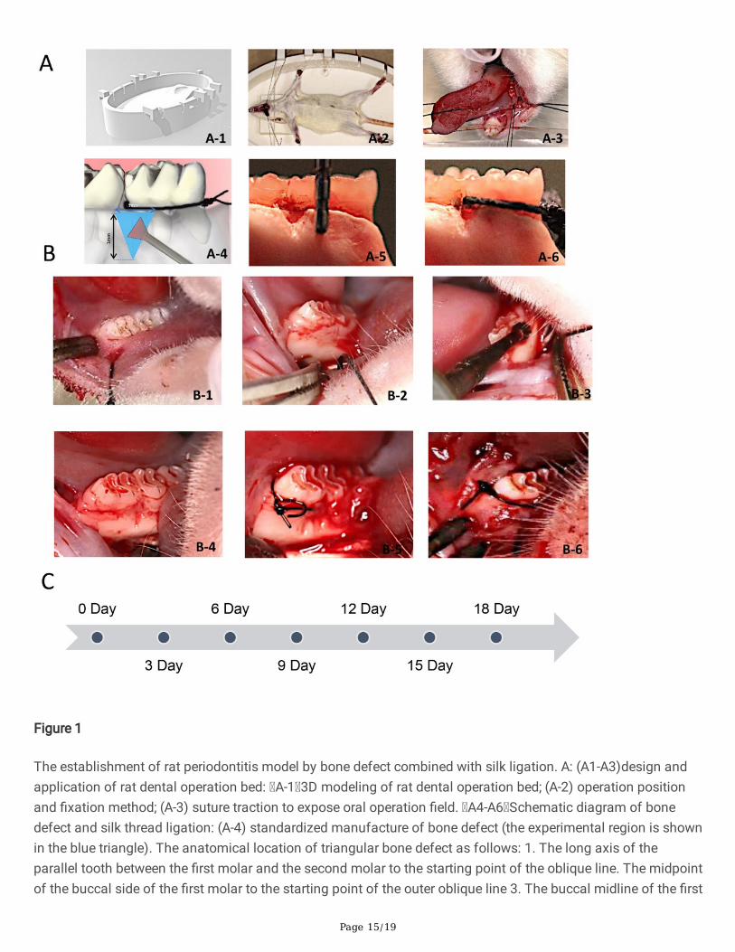
Figure 1
The establishment of rat periodontitis model by bone defect combined with silk ligation. A: (A1-A3)design andapplication of rat dental operation bed: A-1 3D modeling of rat dental operation bed; (A-2) operation positionand �xation method; (A-3) suture traction to expose oral operation �eld. A4-A6 Schematic diagram of bonedefect and silk thread ligation: (A-4) standardized manufacture of bone defect (the experimental region is shownin the blue triangle). The anatomical location of triangular bone defect as follows: 1. The long axis of theparallel tooth between the �rst molar and the second molar to the starting point of the oblique line. The midpointof the buccal side of the �rst molar to the starting point of the outer oblique line 3. The buccal midline of the �rst
Page 16/19
molars is between the �rst molars and the second molars; A-5 the measurement of bone defect area bygraduated periodontal probe (A-6) the ligated silk thread is closely attached to the cervical portion of themandibular �rst molars. B: operation steps: (B-1): Exposure of the operative �eld on the rat dental surgical bed.(B-2) Exposure of the mandible by gingival �ap on the buccal side of the mandibular �rst and second molars. (B-3) Removal of bone tissue from the operative area of the bone defect by the dental slow speed bur. (B-4) rinsingand hemostasis of the operative area. (B-5) Ligation of 5-0 sutures on the cervical part of the �rst molar in rats.(B-6) the overall appearance of oral cavity after primary suture of free gingival �ap. C: Timeline: the rats weresacri�ced and samples were taken on the 3rd, 6th, 9th, 12th, 15th and 18th day.
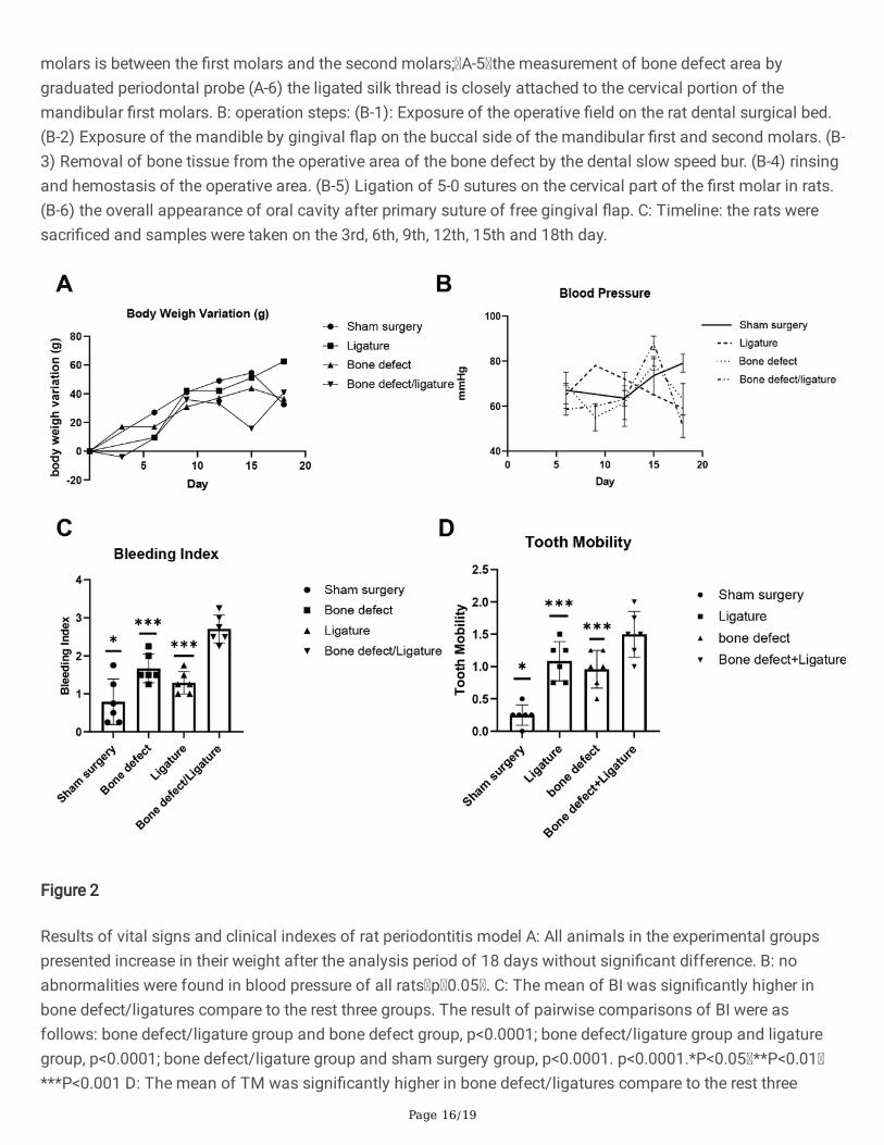
Figure 2
Results of vital signs and clinical indexes of rat periodontitis model A: All animals in the experimental groupspresented increase in their weight after the analysis period of 18 days without signi�cant difference. B: noabnormalities were found in blood pressure of all rats p 0.05 . C: The mean of BI was signi�cantly higher inbone defect/ligatures compare to the rest three groups. The result of pairwise comparisons of BI were asfollows: bone defect/ligature group and bone defect group, p<0.0001; bone defect/ligature group and ligaturegroup, p<0.0001; bone defect/ligature group and sham surgery group, p<0.0001. p<0.0001.*P<0.05 **P<0.01***P<0.001 D: The mean of TM was signi�cantly higher in bone defect/ligatures compare to the rest three
Page 17/19
groups. The result of pairwise comparisons of TM were as follows: bone defect/ligature group and bone defectgroup, one sample t-test, p<0.005; bone defect/ligature group and ligature group, one sample t-test, p<0.001;bone defect/ligature group and sham surgery group, one sample t-test, p<0.0001.*P<0.05 **P<0.01 ***P<0.001.Abbreviations: BI, bleeding index, TM, tooth mobility.
Figure 3
Micro-CT images and histomorphometry of the periapical �rst molar in rats. A: CT Images of rat periodontitismodels. Left (3D reconstructed images): Particularly signi�cant horizontal and vertical bone loss, rough alveolarbone surfaces were observed in the bone defect/ligature group of 6-12 days(A19-20). Smooth surfaces of
Page 18/19
alveolar bones were witnessed in the sham surgery group(A-1-3). The surgical defect areas were clear in thebone defect group(A13). Unevenly loss and rough surface of alveolar bones had been observed in the ligaturegroup (A8-9). Right (Two-dimensional micro-CT sections of periodontal ligament width): No signi�cant wideningof the periodontal ligament during the 18 days in sham surgery group and bone defect group (A4-6). In ligationgroup, the periodontium was slightly widened in the early period of 6 day(A10), and gradually returned to theoriginal level afterwards(A11-12). B: bone defect depth measurements of four groups. C: measurements of theperiodontal ligament width. D: histomorphometric measurements of PDD of �rst molar in HE staining sections.E: depth of PDD of �rst molar by Micro-CT panel sections.
Figure 4
Page 19/19
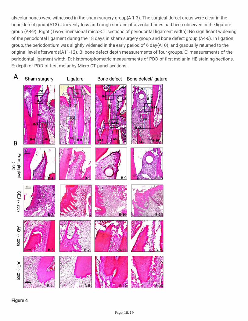
Periodontal tissue and alveolar bone of the distal mesial root of the �rst molar of rats under light microscopy.(A)Whole view of histopathologic of the distal root of the mandibular �rst molar by HE staining on Day 9.Measurement of the distance of CEJ-ABC (arrow bars) showing that the attachment loss of junction epitheliumin the bone defect/ligature group was longer than that in the other three groups, Scale bar = 100 μm. (B) Freegingiva and junctional epithelium of four groups. Spiky projections were thicker in the bone defect/ligaturegroup (B-14). Ulcer development in the surface of sulcular epithelium, signi�cant in�ltration of in�ammatorycells, hyperemia and edema of blood vessels, and gingival �bers loss could be observed in the connective tissuein the bone defect/ligature group (B-13-B14). Sucular epithelium was thinner, the epithelium pegs and dermalpapillae were shorter and blunter in the sham surgery group (B-2). The rete pegs or ridges and dermal papillaewere long and slender in the bone defect group with less in�ltration of in�ammatory cells underlying in theconnective tissue(B-10). Scale bar = 500 μm. Alveolar bone loss in four groups. Prominently horizontal andvertical bone resorption was present in the bone defect/ligature group (B-11), while irregular bone resorption wasobserved in the ligature group (B-7). Surgical region on the alveolar bone were clearly observed in the bonedefect group (B-11). The typical structure of Sharpey’s �ber and alveolar bone crest were observed in the shamsurgery group (B-3) but could no longer retain in the bone defect/ligature group (B-15). scale bar = 200 μm.In�ltration of in�ammatory cells in the apical periodontal ligament. The integrity of cementum and periodontalligament were lost in the bone defect/ligature group (B-16). Apical periodontal ligament was normal in the shamsurgery group without in�ammatory invasion (B-4) and partly reserved in the ligature group (B-8) and bonedefect group (B-12). scale bar = 200 μm.
Supplementary Files
This is a list of supplementary �les associated with this preprint. Click to download.