Facial Surgery W rinkle formation of the skin is a consequence of aging that is influenced by both intrinsic (biologic) and extrinsic (environmental) fac- tors. 1-4 The number and depth of wrinkles are linearly related to a person’s age. 5 Intrinsic aging is a largely genetically determined process that mimics the aging of all organs in the human body and results in cutaneous alterations. Those alterations include a flattening of the dermoepidermal junctions, atrophy of dermis and epi- dermis, and a reduction in the amount of collagen and reticuline, as well as the number of fibroblasts. 1,2 Skin that has been exposed to the sun, like the skin of the face and hands, develops permanent wrinkles, a process that is known as photoaging. 2,4,6 This process is the main contributor to extrinsic aging; as much as 80% of facial aging is caused by solar radiation. 1,4,6 The dam- aging effects of ultraviolet (UV) radiation lead to dermal elastosis, a reduction of collagen, and an initial thicken- ing of the epidermis. 1,2 Clinical signs of photoaging include dryness, hyperpigmentation, telangectesia, and deep wrinkles. 3 These wrinkles do not disappear when the skin is stretched; this is in contrast to temporary wrinkles that arise during aging even on sun-protected skin. 7 Other environmental factors that contribute to the extrinsic aging process are smoking, 1,3,8 diet, 1 and hor- mone replacement therapy (HRT). 9,10 We can distinguish between several types of perma- nent facial wrinkles, one of which is linear wrinkles. Linear wrinkles arise at the site of expression lines on the face, such as frown lines on the forehead, crow’s Volume 29 • Number 6 • November/December 2009 • 467 Aesthetic Surgery Journal Background: Women tend to develop more and deeper wrinkles in the perioral region than men. Although much is known about the complex mechanisms involved in skin aging, previous studies have described his- tologic differences between men and women with respect to skin aging only incidentally and have not inves- tigated the perioral region. Objective: The purpose of this study was to investigate gender-specific differences in the perioral skin. Methods: To determine wrinkle severity, skin surface replicas of the upper lip region in 10 male and 10 female fresh cadavers were analyzed by using the dermaTOP blue three-dimensional digitizing system (Breuckmann, Meersburg, Germany). In 30 fresh male and female cadavers, three full-thickness lip resections were investi- gated in a blinded fashion for specific histologic features. All results were statistically analyzed in a linear regres- sion model with SPSS software (version 15.0; SPSS, Chicago, IL). Results: The female replicas showed more and deeper wrinkles than the male replicas (P < .01). Histologic analysis revealed that the perioral skin of men displayed a significantly higher number of sebaceous glands (P = .000; 95% confidence interval [CI] 23.6–53.2), sweat glands (P = .002; 95% CI 2.1–8.1), and a higher ratio between vessel area and connective tissue area in the dermis (P = .009; 95% CI 0.003–0.021). The amount of hair follicles did not significantly differ between men and women, although the average number of sebaceous glands per hair follicle was greater in men (P = .002; 95% CI 0.33–1.28). Conclusions: Women exhibit more and deeper wrinkles in the perioral region and their skin contains a sig- nificantly smaller number of appendages than men, which could be a feasible explanation for why women are more susceptible to development of perioral wrinkles. (Aesthet Surg J; 29:467-472.) Dr. Paes and Dr. Kon are from the Department of Plastic, Reconstructive and Hand Surgery, University Medical Center, Utrecht, The Netherlands. Dr. Teepen is from the Department of Pathology, St. Elisabeth Hospital, Tilburg, The Netherlands. Dr. Koop is from the Department of Plastic, Reconstructive, and Hand Surgery, University Medical Center, Leeuwarden, The Netherlands. Dr. Kon is a member of the Dutch Society for Aesthetic Plastic Surgery. Perioral Wrinkles: Histologic Differences Between Men and Women Emma C. Paes, MD; Hans J. L. J. M. Teepen, MD, PhD; Willemijn A. Koop, MD; and Moshe Kon, MD, PhD I N T E R N A T I O N A L C O N T R I B U T I O N
Transcript
Facial Surgery
Wrinkle formation of the skin is a consequenceof aging that is influenced by both intrinsic(biologic) and extrinsic (environmental) fac-
tors.1-4 The number and depth of wrinkles are linearlyrelated to a person’s age.5 Intrinsic aging is a largelygenetically determined process that mimics the aging ofall organs in the human body and results in cutaneousalterations. Those alterations include a flattening of thedermoepidermal junctions, atrophy of dermis and epi-dermis, and a reduction in the amount of collagen andreticuline, as well as the number of fibroblasts.1,2
Skin that has been exposed to the sun, like the skin ofthe face and hands, develops permanent wrinkles, aprocess that is known as photoaging.2,4,6 This process isthe main contributor to extrinsic aging; as much as 80%of facial aging is caused by solar radiation.1,4,6 The dam-aging effects of ultraviolet (UV) radiation lead to dermalelastosis, a reduction of collagen, and an initial thicken-ing of the epidermis.1,2 Clinical signs of photoaginginclude dryness, hyperpigmentation, telangectesia, anddeep wrinkles.3 These wrinkles do not disappear whenthe skin is stretched; this is in contrast to temporarywrinkles that arise during aging even on sun-protectedskin.7 Other environmental factors that contribute to theextrinsic aging process are smoking,1,3,8 diet,1 and hor-mone replacement therapy (HRT).9,10
We can distinguish between several types of perma-nent facial wrinkles, one of which is linear wrinkles.Linear wrinkles arise at the site of expression lines onthe face, such as frown lines on the forehead, crow’s
Volume 29 • Number 6 • November/December 2009 • 467Aesthetic Surgery Journal
Background: Women tend to develop more and deeper wrinkles in the perioral region than men. Althoughmuch is known about the complex mechanisms involved in skin aging, previous studies have described his-tologic differences between men and women with respect to skin aging only incidentally and have not inves-tigated the perioral region.Objective: The purpose of this study was to investigate gender-specific differences in the perioral skin.Methods: To determine wrinkle severity, skin surface replicas of the upper lip region in 10 male and 10 femalefresh cadavers were analyzed by using the dermaTOP blue three-dimensional digitizing system (Breuckmann,Meersburg, Germany). In 30 fresh male and female cadavers, three full-thickness lip resections were investi-gated in a blinded fashion for specific histologic features. All results were statistically analyzed in a linear regres-sion model with SPSS software (version 15.0; SPSS, Chicago, IL).Results: The female replicas showed more and deeper wrinkles than the male replicas (P < .01). Histologicanalysis revealed that the perioral skin of men displayed a significantly higher number of sebaceous glands(P = .000; 95% confidence interval [CI] 23.6–53.2), sweat glands (P = .002; 95% CI 2.1–8.1), and a higher ratiobetween vessel area and connective tissue area in the dermis (P = .009; 95% CI 0.003–0.021). The amount ofhair follicles did not significantly differ between men and women, although the average number of sebaceousglands per hair follicle was greater in men (P = .002; 95% CI 0.33–1.28).Conclusions: Women exhibit more and deeper wrinkles in the perioral region and their skin contains a sig-nificantly smaller number of appendages than men, which could be a feasible explanation for why women aremore susceptible to development of perioral wrinkles. (Aesthet Surg J; 29:467-472.)
Dr. Paes and Dr. Kon are from the Department of Plastic,Reconstructive and Hand Surgery, University Medical Center, Utrecht,The Netherlands. Dr. Teepen is from the Department of Pathology, St.Elisabeth Hospital, Tilburg, The Netherlands. Dr. Koop is from theDepartment of Plastic, Reconstructive, and Hand Surgery, UniversityMedical Center, Leeuwarden, The Netherlands. Dr. Kon is a memberof the Dutch Society for Aesthetic Plastic Surgery.
Perioral Wrinkles:Histologic Differences
Between Men and WomenEmma C. Paes, MD; Hans J. L. J. M. Teepen, MD, PhD; Willemijn A. Koop, MD; and
Moshe Kon, MD, PhD
INTE
RNAT
IONAL CONTRIBUTION
468 • Volume 29 • Number 6 • November/December 2009 Aesthetic Surgery Journal
feet at the lateral canthus of the eye, the nasolabialcrease, and wrinkles at the skin of the perioral site.7
This last type seems to be observed more often inwomen.11,12 Most requests for the correction of perioralwrinkles are made by women. While this may beexplained in part by a greater concern among womenabout their appearance, it also appears that distinctivevertical wrinkles at the perioral region develop morefrequently in women than in men.
The growing demand for correction of these wrin-kles has led to an expansion of treatment choices.While dermabrasion and peeling13 were previously themost commonly used methods, laser treatment, botu-linum injections, and injectable or implantable wrin-kle fillers are the latest options.12,14 Nevertheless, thetreatment of wrinkles at the perioral region stillremains a difficult matter.
Although some papers have described the complexmechanism of aging of facial skin2,5,7,13-15 and the associ-ated histologic changes,3,5,11,15 little is known about spe-cific differences in facial skin aging between men andwomen, particularly with respect to the perioral region.Could there be a histologic explanation for why womenare more susceptible than men to development of wrin-
kles in that region? In this study, the perioral skin ofmen and women was investigated in an effort to resolvethis issue, in the hope that our results might contributeto the understanding of facial skin aging and ultimatelylead to the development of better strategies for the pre-vention and treatment of perioral wrinkles.
METHODSTo define wrinkle severity (amount and depth), a skinsurface replica was made of the perioral region in 10white male and 10 white female fresh cadavers (agerange 75–93 years). With the exceptions of date of birthand exterior appearance, nothing was known about theirmedical history. Subsequently, three full-thickness lipresections were taken for histologic analysis. To provideadditional histologic data, lip resections were also takenfrom another five white male and five white female freshcadavers with the same characteristics.
Skin Surface ReplicaThe perioral skin was replicated by imprinting it on asilicone elastomer. The elastomer was prepared by mix-ing a monomer (Xantopren L blue; Heraeus Kulzer,South Bend, IN) with a catalyst (Optosol-Xantopren;Heraeus Kulzer). This suspension was applied withlight pressure to the skin of the upper perioral region.The low viscosity and hydrophobic properties of thesuspension promoted its penetration into all the irregu-larities of the skin. After several minutes, the catalystconverted the silicone monomer into a harder polymer,producing a high-resolution, permanent negative replicaof the skin surface (Figure 1). The replicas were ana-lyzed at three set sites, using the dermaTOP bluethree-dimensional (3-D) digitizing system (Breuckmann,Meersburg, Germany). The replicas were oriented sothat the fine perioral wrinkles were horizontal and theregion of interest was centered in the field of view ofthe 3-D sensor (Figure 2). Three-dimensional topo-
Aesthetic Surgery Journal
Figure 1. Marked silicone skin surface replica of the perioral regionindicating the location of three set sites (G, C, and D) that were usedfor the measurements.
Figure 2. Measuring the skin surface replica. Orientation of thereplica during the measurement using a high-resolution camera. Inthis figure, site C is seen. Three-dimensional topography was thenextracted from each 12-mm � 20-mm site.
Volume 29 • Number 6 • November/December 2009 • 469Perioral Wrinkles: Histologic Differences Between Men and Women
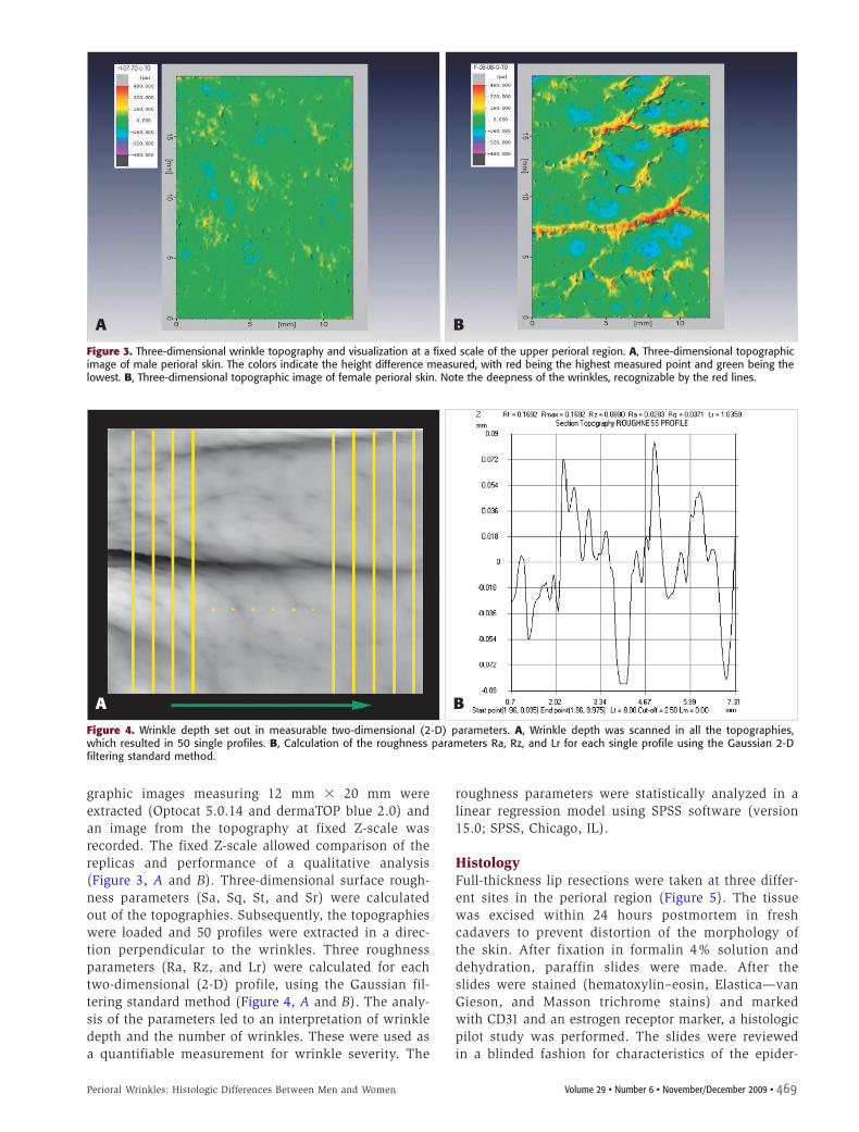
graphic images measuring 12 mm � 20 mm wereextracted (Optocat 5.0.14 and dermaTOP blue 2.0) andan image from the topography at fixed Z-scale wasrecorded. The fixed Z-scale allowed comparison of thereplicas and performance of a qualitative analysis(Figure 3, A and B). Three-dimensional surface rough-ness parameters (Sa, Sq, St, and Sr) were calculatedout of the topographies. Subsequently, the topographieswere loaded and 50 profiles were extracted in a direc-tion perpendicular to the wrinkles. Three roughnessparameters (Ra, Rz, and Lr) were calculated for eachtwo-dimensional (2-D) profile, using the Gaussian fil-tering standard method (Figure 4, A and B). The analy-sis of the parameters led to an interpretation of wrinkledepth and the number of wrinkles. These were used asa quantifiable measurement for wrinkle severity. The
roughness parameters were statistically analyzed in alinear regression model using SPSS software (version15.0; SPSS, Chicago, IL).
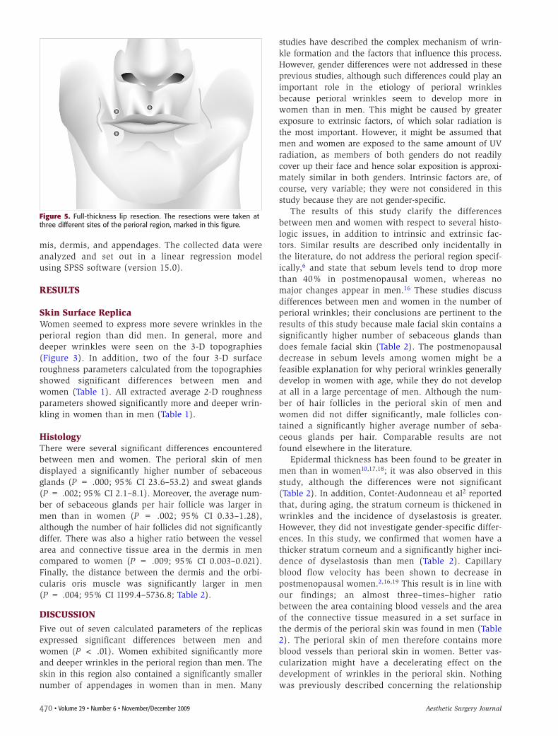
HistologyFull-thickness lip resections were taken at three differ-ent sites in the perioral region (Figure 5). The tissuewas excised within 24 hours postmortem in freshcadavers to prevent distortion of the morphology ofthe skin. After fixation in formalin 4% solution anddehydration, paraffin slides were made. After theslides were stained (hematoxylin–eosin, Elastica—vanGieson, and Masson trichrome stains) and markedwith CD31 and an estrogen receptor marker, a histologicpilot study was performed. The slides were reviewedin a blinded fashion for characteristics of the epider-
A B
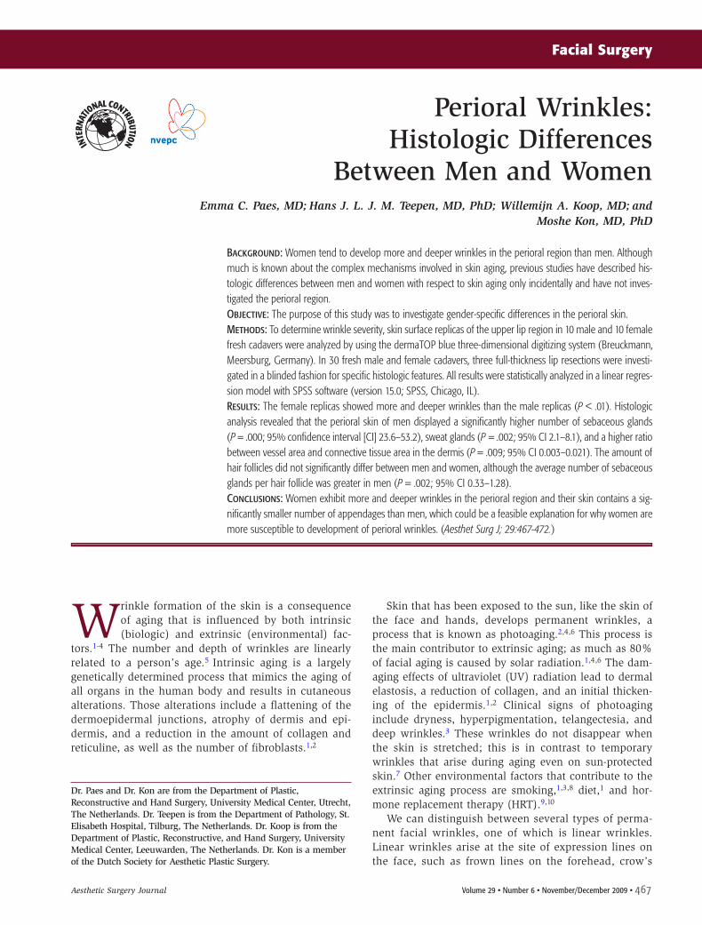
Figure 3. Three-dimensional wrinkle topography and visualization at a fixed scale of the upper perioral region. A, Three-dimensional topographicimage of male perioral skin. The colors indicate the height difference measured, with red being the highest measured point and green being thelowest. B, Three-dimensional topographic image of female perioral skin. Note the deepness of the wrinkles, recognizable by the red lines.
A B
Figure 4. Wrinkle depth set out in measurable two-dimensional (2-D) parameters. A, Wrinkle depth was scanned in all the topographies,which resulted in 50 single profiles. B, Calculation of the roughness parameters Ra, Rz, and Lr for each single profile using the Gaussian 2-Dfiltering standard method.
470 • Volume 29 • Number 6 • November/December 2009 Aesthetic Surgery Journal
mis, dermis, and appendages. The collected data wereanalyzed and set out in a linear regression modelusing SPSS software (version 15.0).
RESULTS
Skin Surface ReplicaWomen seemed to express more severe wrinkles in theperioral region than did men. In general, more anddeeper wrinkles were seen on the 3-D topog raphies(Figure 3). In addition, two of the four 3-D surfaceroughness parameters calculated from the topographiesshowed significant differences between men andwomen (Table 1). All extracted average 2-D roughnessparameters showed significantly more and deeper wrin-kling in women than in men (Table 1).
HistologyThere were several significant differences encounteredbetween men and women. The perioral skin of men displayed a significantly higher number of sebaceousglands (P = .000; 95% CI 23.6–53.2) and sweat glands(P = .002; 95% CI 2.1–8.1). Moreover, the average num-ber of sebaceous glands per hair follicle was larger inmen than in women (P = .002; 95% CI 0.33–1.28),although the number of hair follicles did not significantlydiffer. There was also a higher ratio between the vesselarea and connective tissue area in the dermis in mencompared to women (P = .009; 95% CI 0.003–0.021).Finally, the distance between the dermis and the orbi -cularis oris muscle was significantly larger in men(P = .004; 95% CI 1199.4–5736.8; Table 2).
DISCUSSIONFive out of seven calculated parameters of the replicasexpressed significant differences between men andwomen (P < .01). Women exhibited significantly moreand deeper wrinkles in the perioral region than men. Theskin in this region also contained a significantly smallernumber of appendages in women than in men. Many
studies have described the complex mechanism of wrin-kle formation and the factors that influence this process.However, gender differences were not addressed in theseprevious studies, although such differences could play animportant role in the etiology of perioral wrinklesbecause perioral wrinkles seem to develop more inwomen than in men. This might be caused by greaterexposure to extrinsic factors, of which solar radiation isthe most important. However, it might be assumed thatmen and women are exposed to the same amount of UVradiation, as members of both genders do not readilycover up their face and hence solar exposition is approxi-mately similar in both genders. Intrinsic factors are, ofcourse, very variable; they were not considered in thisstudy because they are not gender-specific.
The results of this study clarify the differencesbetween men and women with respect to several histo-logic issues, in addition to intrinsic and extrinsic fac-tors. Similar results are described only incidentally inthe literature, do not address the perioral region specif-ically,6 and state that sebum levels tend to drop morethan 40% in postmenopausal women, whereas nomajor changes appear in men.16 These studies discussdifferences between men and women in the number ofperioral wrinkles; their conclusions are pertinent to theresults of this study because male facial skin contains asignificantly higher number of sebaceous glands thandoes female facial skin (Table 2). The postmenopausaldecrease in sebum levels among women might be afeasible explanation for why perioral wrinkles generallydevelop in women with age, while they do not developat all in a large percentage of men. Although the num-ber of hair follicles in the perioral skin of men andwomen did not differ significantly, male follicles con-tained a significantly higher average number of seba-ceous glands per hair. Comparable results are notfound elsewhere in the literature.
Epidermal thickness has been found to be greater inmen than in women10,17,18; it was also observed in thisstudy, although the differences were not significant(Table 2). In addition, Contet-Audonneau et al2 reportedthat, during aging, the stratum corneum is thickened inwrinkles and the incidence of dyselastosis is greater.However, they did not investigate gender-specific differ-ences. In this study, we confirmed that women have athicker stratum corneum and a significantly higher inci-dence of dyselastosis than men (Table 2). Capillaryblood flow velocity has been shown to decrease inpostmenopausal women.2,16,19 This result is in line withour findings; an almost three–times–higher ratiobetween the area containing blood vessels and the areaof the connective tissue measured in a set surface inthe dermis of the perioral skin was found in men (Table2). The perioral skin of men therefore contains moreblood vessels than perioral skin in women. Better vas-cularization might have a decelerating effect on thedevelopment of wrinkles in the perioral skin. Nothingwas previously described concerning the relationship
Figure 5. Full-thickness lip resection. The resections were taken atthree different sites of the perioral region, marked in this figure.
Volume 29 • Number 6 • November/December 2009 • 471Perioral Wrinkles: Histologic Differences Between Men and Women
between gender and the presence of sweat glands inthe perioral skin. Our study shows that the perioralskin of men contains significantly more sweat glandsthan that of women (Table 2).
It has been reported that women who receive HRThave lower facial wrinkle scores than women who arenot treated with this therapy.9,10 In addition, mean levelsof epidermal skin moisture, elasticity, and skin thicknessare improved with HRT,10 although this has never beenshown with respect to the perioral region and the resultsof this study showed no difference between men andwomen regarding the presence of estrogen-positive seba-ceous glands in this area. This suggests that other fac-
tors may have a greater influence on the development ofperioral wrinkles.
As described, the perioral skin of men contains moreappendages than that of women, which could influence,in some way, the natural filling of the dermis. Becausethe dermis of the perioral skin in men includes consider-ably more sebaceous glands, sweat glands, and bloodvessels, one could imagine that the formation of wrin-kles in that region is more difficult in men.
Another interesting observation was that the orbicu-laris oris muscle, which surrounds the lips, is anchored1.5 times closer to the dermis in women than in men(Table 2). This difference has not been previously reported
Table 2. Average statistical results of the histologic analysis of 90 biopsies taken from the perioral region in 15 male and15 female fresh cadavers
Mean (SD)Variable Male Female Difference (95% CI) P*
Lr 1.03472 (.01265) 1.05258 (.01086) �0.018 (�0.029 – �0.007) .003
CI, confidence interval; LR, ratio of developed line to the profile length; Ra, arithmetic average of absolute values of roughness profile ordinates (Z), where Z isthe sum of the heights of the highest peaks and the lowest valley depth within a sampling length; Rz, arithmetic mean value of the single roughness depthsof consecutive sampling lengths; Sa, linear average surface roughness; SD, standard deviation; Sq, quadratic average surface roughness; Sr, ratio of developedarea to target area; St, maximum surface height difference (peak to peak value).*P < .05 was considered statistically significant.
472 • Volume 29 • Number 6 • November/December 2009 Aesthetic Surgery Journal
in the literature. It might play a role in wrinkle formationbecause fibrous connections between the muscle and thedermis can cause an inward traction, thereby creatingdeeper wrinkles.
It should be noted that during data collection, theassumption was made that the cadavers had a roughlyequal exposure to UV radiation during their lives. Wealso cannot exclude the possibility that other externalfactors that influence the aging process—such as smok-ing, hormones, and diet—may have been operative tovarying extents in the cadavers.
CONCLUSIONSThe treatment of perioral wrinkles remains a difficultmatter. To our knowledge, ours is the first study to inves-tigate skin surface and specific histologic differencesbetween the perioral skin of men and women and theirpossible relationship to wrinkle formation. We foundthat, in comparison to men, women exhibit more anddeeper wrinkles in the perioral region and that women’sperioral skin contains significantly fewer appendages.These findings provide a feasible explanation for whywomen are more susceptible to development of perioralwrinkles and contribute to our current understanding ofwrinkle formation. ◗
ACKNOWLEDGMENTS
The authors thank Mr. Simon Plomp and Mr. Willem van Wolferenfrom the anatomy department of the University Medical CentreUtrecht for their assistance in collecting the biopsies from thecadavers.
DISCLOSURES
The authors have no financial interest in and received no compen-sation from manufacturers of products mentioned in this article.
REFERENCES1. Baumann L. Skin ageing and its treatment. J Pathol
2007;211:241–251.2. Contet-Audonneau JL, Jeanmaire C, Pauly G. A histological study of
human wrinkle structures: comparison between sun-exposed areas ofthe face, with or without wrinkles, and sun-protected areas. Br JDermatol 1999;140:1038–1047.
3. Gilchrest BA. A review of skin ageing and its medical therapy. Br JDermatol 1996;135:867–875.
4. Jenkins G. Molecular mechanisms of skin ageing. Mech Ageing Dev2002;123:801–810.
5. Leveque JL, Goubanova E. Influence of age on the lips and perioralskin. Dermatology 2004;208:307–313.
6. Zouboulis CC, Boschnakow A. Chronological ageing and photoageingof the human sebaceous gland. Clin Exp Dermatol 2001;26:600–607.
7. Hatzis J. The wrinkle and its measurement—a skin surfaceProfilometric method. Micron 2004;35:201–219.
8. El-Domyati M, Attia S, Saleh F, et al. Intrinsic aging vs. photoaging: acomparative histopathological, immunohistochemical, and ultrastruc-tural study of skin. Exp Dermatol 2002;11:398–405.
9. Castelo-Branco C, Figueras F, Martínez de Osaba MJ, Vanrell JA. Facialwrinkling in postmenopausal women. Effects of smoking status andhormone replacement therapy. Maturitas 1998;29:75–86.
10. Sator PG, Schmidt JB, Sator MO, Huber JC, Honigsmann H. The influ-ence of hormone replacement therapy on skin ageing: a pilot study.Maturitas 2001;39:43–55.
11. Wojnarowska F. Clinical aspects of ageing skin. In: Fry L, editor. Skinproblems in the elderly, 2nd ed. Edinburgh: Churchill Livingstone;1985, pp 28-46.
12. Monhian N. Injectable implantable materials for facial wrinkles.Aesthetic Facial Surgery ed. 2008, pp 247-248.
13. Holmkvist KA, Rogers GS. Treatment of perioral rhytides: a comparisonof dermabrasion and superpulsed carbon dioxide laser. Arch Dermatol2000;136:725–731.
14. Semchyshyn N, Sengelmann RD. Botulinum toxin A treatment of perio-ral rhytides. Dermatol Surg 2003;29:490–495.
15. Piérard GE, Uhoda I, Piérard-Franchimont C. Update on the histologicalpresentation of facial wrinkles. Eur J Dermatol 2002;12:XIII–XXIV.
16. Sandby-Moller J, Poulsen T, Wulf HC. Epidermal thickness at differentbody sites: relationship to age, gender, pigmentation, blood content,skin type and smoking habits. Acta Derm Venereol 2003;83:410–413.
17. Castelo-Branco C, Duran M, Gonzalez-Merlo J. Skin collagen changesrelated to age and hormone replacement therapy. Maturitas1992;15:113–119.
18. Vaillant L, Callens A. Hormone replacement treatment and skin aging.Therapie 1996;51:67–70.
19. Raine-Fenning NJ, Brincat MP, Muscat-Baron Y. Skin aging andmenopause: implications for treatment. Am J Clin Dermatol2003;4:371–378.
Accepted for publication May 22, 2009.
Presented at the 20th Annual Meeting of the European Association of PlasticSurgeons, May 28–30, 2009, Barcelona, Spain.
Reprint requests: Moshe Kon, MD, PhD, University Medical Center Utrecht,Department of Plastic, Reconstructive and Hand Surgery, PO Box 85500,3508 GA Utrecht, the Netherlands. E-mail: [email protected].