19
Exceptional care, inspired by you Resource Manual Peripherally Inserted Central Catheters (PICCs) Percutaneous Central Catheters “Together…supporting quality care” Revised September 2016
Exceptional care, inspired by you
Resource Manual
Peripherally Inserted Central Catheters (PICCs) Percutaneous Central Catheters
“Together…supporting quality care” Revised September 2016

Page 2 of 19 Peripherally Inserted Central Catheters (PICCs) Percutaneous Central Catheters Quality, Patient Safety and Interprofessional Practice
TABLE OF CONTENTS
INTRODUCTION .................................................................................................. 3
PICCS .................................................................................................................. 4
LOCATION ........................................................................................................... 4 TYPES ................................................................................................................ 5 INDICATIONS ........................................................................................................ 5 CONTRAINDICATIONS ........................................................................................... 6 BENEFITS AND RISKS ............................................................................................ 6
PERCUTANEOUS CENTRAL LINES .................................................................. 7
LOCATION ........................................................................................................... 7 TYPES ................................................................................................................ 7 INDICATIONS ........................................................................................................ 8 CONTRAINDICATIONS ........................................................................................... 8 BENEFITS AND RISKS ............................................................................................ 8
NURSING USE AND CARE ................................................................................ 9
DAILY ASSESSMENTS ........................................................................................... 9 ACCESSING A CVAD ............................................................................................ 9 FLUSHING A CVAD ............................................................................................ 10 DRAWING BLOOD FROM A CVAD ......................................................................... 10 CHANGING THE DRESSING AND CAP OF A CVAD .................................................... 10
POTENTIAL CVAD COMPLICATIONS ............................................................. 11
QUICK REFERENCE ......................................................................................... 15
REFERENCES ................................................................................................... 16
CVAD QUIZ ....................................................................................................... 17

Page 3 of 19 Peripherally Inserted Central Catheters (PICCs) Percutaneous Central Catheters Quality, Patient Safety and Interprofessional Practice
INTRODUCTION
This learning package is a resource, designed to standardize education for all nurses
caring for patients with central lines. It is important that the reader know Quinte
Healthcare Corporation (QHC) policies on Central Venous Access Devices (CVAD’s)
and have successfully been certified on the care and maintenance of central lines
before managing central lines independently.
This package is intended to be a part of orientation to CVAD care and maintenance. If
at the completion of this program you feel that you are unable to perform these skills, it
is your responsibility to confer with your Clinical Educator, Nurse Manager, or Charge
Nurse/delegate.
Practice Guidelines
1. Nurses will attend in-service training as provided by QHC, which includes theory, anatomy & physiology, demonstration and practice of all CVAD applications currently used in QHC.
2. Nurses will complete a test and achieve a minimum of 80%.
3. Nurses will be supervised at least two times for each skill by a nurse competent in central line care and maintenance. Evaluating nurses will document on the appropriate “Skills Checklist” including date and signature. Comments should include whether or not the candidate is safe to practice independently.
4. Completed checklist should be photocopied, with one copy given to the staff member and original to Unit Manager.
Continuing Competence
It is strongly recommended that certified nurse’s review all skills related to the care and
maintenance of central lines on an ongoing basis to ensure continued competence. If at
any time the nurse feels additional review/retraining is required, it is the responsibility of
that nurse to seek additional education/resources from the manager, or clinical
educator/delegate to ensure continued competence related to CVAD care and
maintenance. Nurses are professionally responsible for ensuring that they have the
requisite knowledge, skill and judgment necessary to provide safe and effective infusion
therapy (CNO, 2002).

Page 4 of 19 Peripherally Inserted Central Catheters (PICCs) Percutaneous Central Catheters Quality, Patient Safety and Interprofessional Practice
PERIPHERALLY INSERTED CENTRAL CATHETER (PICC)
A Peripherally Inserted Central Catheter
(PICC) is a small gauge catheter that is
inserted peripherally but the tip sits in the
central venous circulation in the lower 1/3 of
the superior vena cava. It is suitable for long
term use and there are no restrictions for
age, or gender.
Location
PICCs are commonly placed at or above the antecubital space in the following veins:
Cephalic vein: The cephalic vein ascends along the outer
edge of the biceps muscle to the upper aspect of the arm.
It terminates in the axillary vein just below the clavicle.
Basilic vein: The basilic is larger than the cephalic vein. It
passes up the arm in a straight path along the inner side
of the biceps muscle. The basilic vein terminates in the
axillary vein
Medial-cubital vein: The medial cubital vein runs along the
anterior surface of the arm and is often visible in the
antecubital area. It creates interface between the cephalic
and basilic veins and often forms a ‘Y’ with one branch
going to the basilic vein (called the median cubital basilic)
and the branch going to the cephalic vein (called the
median cubital cephalic).
Page 5 of 19 Peripherally Inserted Central Catheters (PICCs) Percutaneous Central Catheters Quality, Patient Safety and Interprofessional Practice
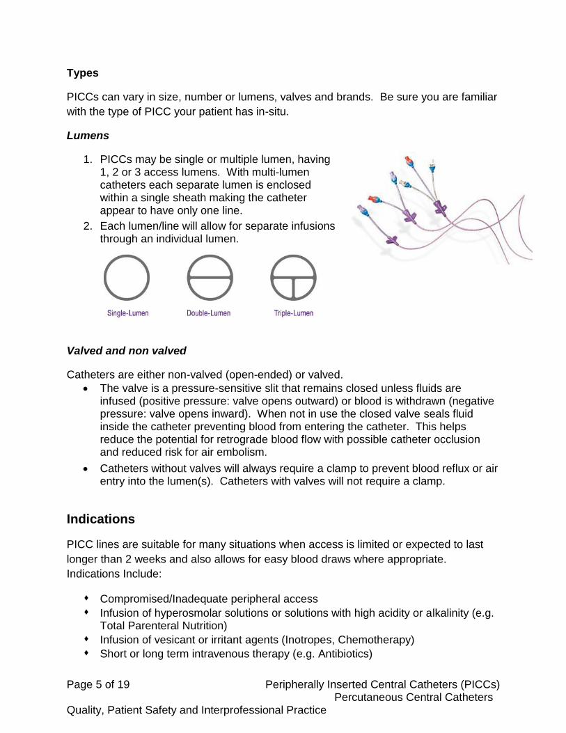
Types
PICCs can vary in size, number or lumens, valves and brands. Be sure you are familiar
with the type of PICC your patient has in-situ.
Lumens
1. PICCs may be single or multiple lumen, having 1, 2 or 3 access lumens. With multi-lumen catheters each separate lumen is enclosed within a single sheath making the catheter appear to have only one line.
2. Each lumen/line will allow for separate infusions through an individual lumen.
Valved and non valved
Catheters are either non-valved (open-ended) or valved.
The valve is a pressure-sensitive slit that remains closed unless fluids are infused (positive pressure: valve opens outward) or blood is withdrawn (negative pressure: valve opens inward). When not in use the closed valve seals fluid inside the catheter preventing blood from entering the catheter. This helps reduce the potential for retrograde blood flow with possible catheter occlusion and reduced risk for air embolism.
Catheters without valves will always require a clamp to prevent blood reflux or air entry into the lumen(s). Catheters with valves will not require a clamp.
Indications
PICC lines are suitable for many situations when access is limited or expected to last
longer than 2 weeks and also allows for easy blood draws where appropriate.
Indications Include:
Compromised/Inadequate peripheral access
Infusion of hyperosmolar solutions or solutions with high acidity or alkalinity (e.g. Total Parenteral Nutrition)
Infusion of vesicant or irritant agents (Inotropes, Chemotherapy)
Short or long term intravenous therapy (e.g. Antibiotics)

Page 6 of 19 Peripherally Inserted Central Catheters (PICCs) Percutaneous Central Catheters Quality, Patient Safety and Interprofessional Practice
Contraindications for PICC Insertion
Previous upper extremity venous thrombosis (DVT)
Trauma or vascular surgeries at or near the site of insertion
Presence of a device related infection, cellulitis, or bacteremia at or near the insertion site
Lymphedema.
Mastectomy surgery with axillary dissection +/- lymphedema on affected side (unless urgent condition requires it)
Allergy to materials
Irradiation of insertion site (Bard Access Systems, 2016)
Benefits and Risks
Advantages Disadvantages
Less traumatic to place (reduced intrathoracic or venipuncture complications)
No surgical requirements
Preservation of peripheral vascular system
Cost and time efficient
Reliable long-term access
Decreased peripheral bacterial colonies vs. jugular, thoracic or femoral area
Usually easily removed
Requires routine sterile dressing and injection cap changes
Care costs on a long-term basis
Some PICCs (small gauge) are not recommended for blood sampling
Patient self-care is difficult
External catheter breakage possible
Body image impact
Activity restrictions
Requires adequate peripheral access
Post-insertion phlebitis common

Page 7 of 19 Peripherally Inserted Central Catheters (PICCs) Percutaneous Central Catheters Quality, Patient Safety and Interprofessional Practice
PERCUTANEOUS CENTRAL LINES
Percutaneous central lines are inserted using a percutaneous approach into the jugular, subclavian, or femoral veins and advanced to the superior vena cava. This type of Central Venous Access Device (CVAD) is indicated for the acute care settings to administer short term therapies (usually less than 1 month). The percutaneous central line is identified by which vein it is inserted in (example: right internal jugular central line).
Location
Percutaneous central lines are commonly in the following veins:
External Jugular Vein: On the side of the neck the external jugular is easily recognized.
This vein connects to the subclavian vein along the center of the clavicle.
Internal Jugular Vein: The internal jugular vein initially descends behind and then to the
outer side of the internal and common carotid
arteries. It then joins the subclavian vein at the
base of the neck.
Subclavian Vein: The subclavian vein is a
continuation of the axillary vein and extends
from the outer edge of the first rib to the inner
end of the clavicle. Here it enters the inner
jugular vein to form the innominate vein.
Femoral Vein: The femoral vein is a
continuation of the popliteal vein upward
toward the external iliac vein. CVADs are
sometimes inserted here when other options
are limited or in emergency situations. It is a
site that is at high risk for infection and mal-
positioning or migration.
Types
This type of CVAD may have single or multiple lumens with the external catheter
segment separating and labeling the lumens so that each lumen can be accessed
individually and used as recommended.

Page 8 of 19 Peripherally Inserted Central Catheters (PICCs) Percutaneous Central Catheters Quality, Patient Safety and Interprofessional Practice
Lumens
1. Proximal (White port) - first opening on the catheter tip, closest to the hub
2. Medial (Blue port) - middle open on the catheter tip
3. Distal (Brown port) - opening at the very end of the catheter (ideal for CVP monitoring and blood draws)
Each lumen/line will allow for separate infusions through an individual lumen.
A single lumen percutaneous line is sometimes used in
critical care. It is also called a Cordis or Introducer. It is
typically used for temporary transvenous pacemakers,
or in traumas. They should only be used in critical care
areas.
The Cordis must have a minimum of 30 mL/hr. running at all times and cannot be clamped off.
Indications
Administration of multiple therapies or incompatible medications
Limited or poor peripheral venous access
Administration IV Medication Therapy (Including Vasoactive Medication Therapy)
IV Fluid Replacement (Including critically ill patients requiring fluid resuscitation)
Central Venous Pressure Monitoring
Therapy involving frequent blood sampling Contraindications for Insertion
Abnormal local anatomy to the area, for example injury, prior surgery, history of radiation
Infection at insertion site
Use caution when: o Presence of anticoagulation or bleeding disorder o Patient is excessively underweight or overweight
Risk and Benefits
Advantages Disadvantages
Can be inserted at the bedside
Economical
Easily removed
Exchanged over guidewire
High risk for air embolism and exsanguination (shorter catheter = greater risk)
Uncomfortable for the patient
Routine sterile dressing changes
Difficult to maintain occlusive dressing
Page 9 of 19 Peripherally Inserted Central Catheters (PICCs) Percutaneous Central Catheters Quality, Patient Safety and Interprofessional Practice
NURSING USE AND CARE PICCS AND PERCUTANEOUS LINES
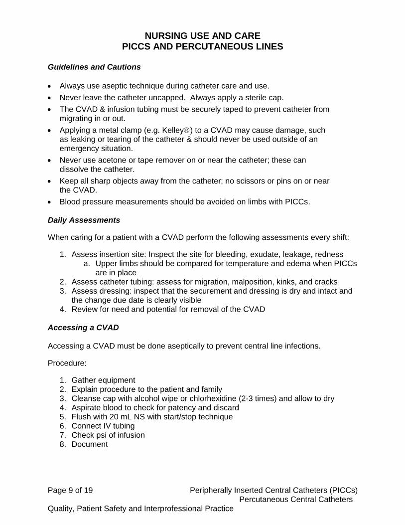
Guidelines and Cautions
Always use aseptic technique during catheter care and use.
Never leave the catheter uncapped. Always apply a sterile cap.
The CVAD & infusion tubing must be securely taped to prevent catheter from migrating in or out.
Applying a metal clamp (e.g. Kelley) to a CVAD may cause damage, such as leaking or tearing of the catheter & should never be used outside of an emergency situation.
Never use acetone or tape remover on or near the catheter; these can dissolve the catheter.
Keep all sharp objects away from the catheter; no scissors or pins on or near the CVAD.
Blood pressure measurements should be avoided on limbs with PICCs. Daily Assessments
When caring for a patient with a CVAD perform the following assessments every shift:
1. Assess insertion site: Inspect the site for bleeding, exudate, leakage, redness a. Upper limbs should be compared for temperature and edema when PICCs
are in place 2. Assess catheter tubing: assess for migration, malposition, kinks, and cracks 3. Assess dressing: inspect that the securement and dressing is dry and intact and
the change due date is clearly visible 4. Review for need and potential for removal of the CVAD
Accessing a CVAD Accessing a CVAD must be done aseptically to prevent central line infections.
Procedure:
1. Gather equipment 2. Explain procedure to the patient and family 3. Cleanse cap with alcohol wipe or chlorhexidine (2-3 times) and allow to dry 4. Aspirate blood to check for patency and discard 5. Flush with 20 mL NS with start/stop technique 6. Connect IV tubing 7. Check psi of infusion 8. Document

Page 10 of 19 Peripherally Inserted Central Catheters (PICCs) Percutaneous Central Catheters Quality, Patient Safety and Interprofessional Practice
Flushing a CVAD
Flushing checks for patency and helps to decrease the potential of a partial or complete occlusion.
**To avoid damaging the catheter NEVER use a syringe smaller than 10 mL** Flush all capped lumens once every 7 days for inactive CVADs and before and after all medications, infusions, and blood draws for CVADs in frequent use. Procedure:
1. Gather equipment 2. Explain procedure to the patient and family 3. Cleanse cap with alcohol wipe or chlorhexidine (2-3 times) and allow to dry 4. Aspirate blood to check for patency and discard 5. Flush with 20 mL NS using “start/stop” technique to create turbulent flow
ensuring the syringe size is 10 mL or larger 6. Document
Drawing blood from a CVAD
1. Gather equipment 2. Explain procedure to the patient and family 3. Stop infusion and flush with 20 mL NS 4. Wait a minimum of one minute 5. Cleanse cap with alcohol wipe or chlorhexidine (2-3 times) and let dry 6. Attach multi-sample vacutainer 7. Obtain a minimum 5 mL blood discard sample 8. Follow order of draw 9. Detach vacutainer and wipe any blood from connector with alcohol wipe 10. Flush all lumens with 20 mL NS 11. Restart any stopped infusions 12. Label blood samples at the bedside with 2 patient identifier 13. Send blood samples as per QHC policy 14. Document
Changing the Dressing and Cap of a CVAD The dressing is changed within 24 hours of line insertion and then every 7 days and PRN.
1. Gather equipment 2. Explain procedure to the patient and family 3. Remove old dressing (work towards center) 4. Assess vein, insertion site and any related anatomical areas 5. Clean site with a Chlorhexidine cleanse with a scrubbing circular motion 6. Allow to dry
**The most common cause of phlebitis and skin irritation is from applying the dressing before the area is completely dry. **

Page 11 of 19 Peripherally Inserted Central Catheters (PICCs) Percutaneous Central Catheters Quality, Patient Safety and Interprofessional Practice
7. Apply clear tegaderm dressing 8. Secure catheter ensuring that the catheter tubing is not kinked or bent at an
angle 90° or smaller 9. Label with date of dressing change 10. Document
Cap changes are required every 7 days and typically performed with the dressing change.
1. Gather equipment 2. Explain procedure 3. Prepare equipment (aseptic technique) 4. Cleanse cap and connections with chlorhexidine prior to removal 5. Clamp lumen 6. Remove old cap 7. Fill “dead space” with saline 8. Attach new cap 9. Unclamp lumen 10. Aspirate for blood return 11. Flush with 20 mL NS 12. Remove syringe 13. Clamp lumen if not in use (dependent on PICC type) 14. Document
POTENTIAL CVAD COMPLICATIONS
Complication Signs & Symptoms Prevention Intervention
Inflammation (Phlebitis)
Usually occurs within 7 days after insertion
Pain at site
Erythema
Edema
Secure dressing after prep solution is completely dry
Change dressing: - q 7 days and prn if semipermeable transparent dressing; - q 48 hrs. if gauze dressing
Evaluate patient for other sources of infection (seeding)
Reposition hub prn
Strict aseptic technique
Warm moist compresses
Assess site frequently
Alert physician of any temperature elevation, signs of infection.
Ensure dressing is secure
Assess patient before PICC insertion for preexisting infections
Occluded Catheter
Unable to flush
Unable to aspirate blood return
Slow/sluggish blood return or infusions.
Flush promptly after all intermittent infusions, blood draws
Maintain positive pressure in line when not in use
Assess cause
If blood clot: certified nurses can instill a thrombolytic with a physician order

Page 12 of 19 Peripherally Inserted Central Catheters (PICCs) Percutaneous Central Catheters Quality, Patient Safety and Interprofessional Practice
Assure all flush solutions & drugs are compatible.
If chemical occlusion: inform physician to consider PICC line removal
Septicemia, central line infection
Chills
Fever
Headache
Malaise
Glucose intolerance
Backache
Nausea & vomiting
Vascular collapse
Shock
Death
Use aseptic technique & maintain an adherent dressing
Use intact equipment and never use equipment/solutions with expired dates
Follow tubing change, solutions policy(s)
Monitor vital signs/ temp
Stop infusion & notify physician if Temp greater than 38°C
Administer therapeutic interventions as ordered
If discontinuing PICC line, send tip for culture
Broken Catheter
Leakage of fluid, blood from catheter or dressing
Keep sharps away from catheter
Check position of clamps before flushing
Do not use syringes smaller than 10mL
Clamp catheter with toothless clamp below break towards chest wall
Repair or replace
Embolism
Clot, catheter segment, or air becomes free-floating in blood & propelled into Pulmonary Artery
(PE/Catheter): sudden chest pain, tachycardia, dyspnea, productive cough and reddish-pink sputum. Mimic symptoms of MI
(Air): chest pain, pallor, cough, cyanosis, syncope, mill-wheel murmur, shock, coma and death
Do not forcibly flush against resistance to clear catheter occlusion
Prevent IV from running dry
Use only luer lock equipment, remove all air from tubing prior to starting infusion
Do not use scissors to remove tape from catheters
Do not remove cap without 2 appropriate clamps applied to catheter
PE/Catheter:
Administer O2 per orders
Notify physician immediately
Administer Meds as ordered
Air Emboli:
Close off source of air, if possible (i.e., empty glass bottle)
Place patient on Left side in Trendelenburg
Notify physician immediately
Administer ordered meds
Circulatory Overload
Presents as CHF.
jugular vein distention, peripheral edema, dyspnea, agitation, tachypnea, cough, crackles heard in lung bases,
I&Os
use pumps for infusions, especially if client history of CHF, renal failure
Do not attempt to catch up if IV fluids behind schedule and notify physician of noted signs/symptoms
Stop infusion
High fowler’s position,
Monitor Vital signs.
Administer O2
Notify physician immediately
Administer treatment as ordered

Page 13 of 19 Peripherally Inserted Central Catheters (PICCs) Percutaneous Central Catheters Quality, Patient Safety and Interprofessional Practice
Speed Shock Systemic toxic reaction when a substance foreign to the body is rapidly introduced i.e.: IV push medication or runaway IV Signs and symptoms: flushing, headache, syncope, shock, cardiac arrest
Administer drugs and fluids at rate prescribed Keep flow control devices out of reach if disoriented /confused /paediatric patients Monitor flow rate at least hourly, use infusion pump for delivery of all continuous infusions of medications
Slow IV; Maintain access Notify physician immediately Perform emergency care PRN to maintain ABCs Administer treatment ordered by physician
Catheter
Migration
CVAD tip
advances
proximally
into the heart
or distally
CVAD tip
withdraws out
of distal SVC
Proximally
Atrial or ventricular arrhythmias, short of breath, palpitations, cardiac tamponade if infusion into myocardium
Distally: Patient hears swishing / gurgling in same side ear when flushing (tip in jugular vein), infiltration / extravasation
Careful securement of CVAD at time of insertion
Continue to secure location with securement device each dressing change
Measure & document external catheter length
Do not use CVAD if migration is suspected until tip location confirmed by x-ray
Confirm tip location radiographically at time of insertion, and at any other time migration is suspected
Do not use until tip location is confirmed
Proximally migrated – tip may be withdrawn
Distally migrated – must remove & replace; cannot be advanced
Anaphylaxis Dyspnea, wheezing, choking, cyanosis, coughing, SOB, difficulty swallowing, tightness or chest pain, edema of hands / feet/ face / neck & eyelids, urticaria, hoarseness, generalized erythema, feeling of warmth, pruritus, nausea, vomiting, abdominal cramps, diarrhea, incontinence, rapidly falling BP, chills, diaphoresis, weakness, thready pulse, dizziness, flushing & pallor, drowsiness, agitation, anxiety, shaking, throbbing in the ears,
Awareness of cross-sensitivities (ex. PCN / cephalosporin’s) between meds
Know hospital anaphylaxis policy and frequently review protocols for administering emergency medications.
Know orders (Pre-Printed or Medical Directives) on all patients receiving infusions of IV medications to initiate anaphylaxis protocol PRN
Stop meds infusing, (Maintain IV access)
Call Code Blue if necessary
Maintain Airway
Administer emergency medications per Anaphylaxis Medical Directives or direct physician order
Administer Oxygen
Monitor vital signs continuously
Stay with patient
Document sequence of events and care provided
Note new allergy in patient health record

Page 14 of 19 Peripherally Inserted Central Catheters (PICCs) Percutaneous Central Catheters Quality, Patient Safety and Interprofessional Practice
paresthesia, coma, death
DVT redness, pain, swelling on the same side as PICC, edema distal to insertion site, difference in color & temperature of extremities between affected and unaffected sides, vein engorgement on affected side
Appropriate use of cannulated arm, securely anchor cannula to prevent movement in situ,
Be aware of patients with hypercoagulable states
Frequent site inspections, with thorough comparison of both arms during head to toe assessments,
Notify physician of Signs and symptoms Administer therapies as ordered
Page 15 of 19 Peripherally Inserted Central Catheters (PICCs) Percutaneous Central Catheters Quality, Patient Safety and Interprofessional Practice
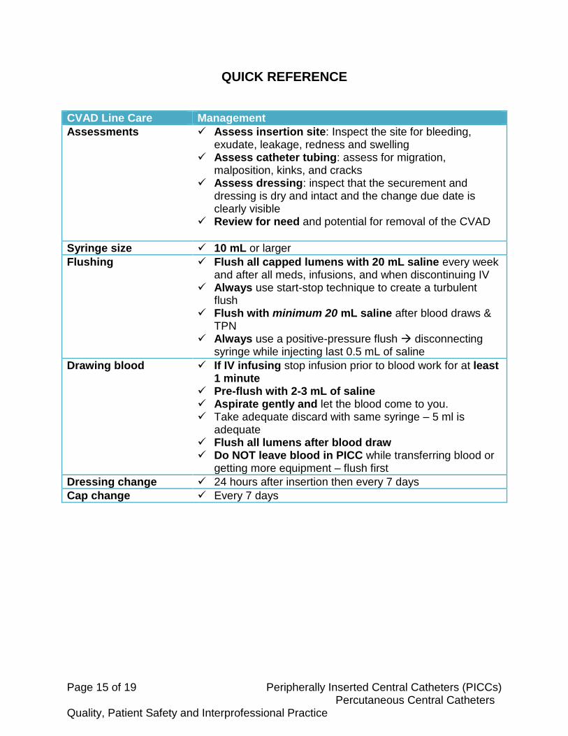
QUICK REFERENCE
CVAD Line Care Management
Assessments Assess insertion site: Inspect the site for bleeding, exudate, leakage, redness and swelling
Assess catheter tubing: assess for migration, malposition, kinks, and cracks
Assess dressing: inspect that the securement and dressing is dry and intact and the change due date is clearly visible
Review for need and potential for removal of the CVAD
Syringe size 10 mL or larger
Flushing Flush all capped lumens with 20 mL saline every week and after all meds, infusions, and when discontinuing IV
Always use start-stop technique to create a turbulent flush
Flush with minimum 20 mL saline after blood draws & TPN
Always use a positive-pressure flush disconnecting syringe while injecting last 0.5 mL of saline
Drawing blood If IV infusing stop infusion prior to blood work for at least 1 minute
Pre-flush with 2-3 mL of saline Aspirate gently and let the blood come to you. Take adequate discard with same syringe – 5 ml is
adequate Flush all lumens after blood draw Do NOT leave blood in PICC while transferring blood or
getting more equipment – flush first
Dressing change 24 hours after insertion then every 7 days
Cap change Every 7 days
Page 16 of 19 Peripherally Inserted Central Catheters (PICCs) Percutaneous Central Catheters Quality, Patient Safety and Interprofessional Practice
REFERENCES
College of Nurses of Ontario (CNO), (2002). Professional Standards.
The Canadian Intravenous Nurses Association, (1999). Intravenous therapy guidelines.
Kingston General Hospital, (2002). Central venous lines: removal.
Perry, A.G. & Potter, P.A. (2002). Clinical nursing skills and techniques (5th ed.). Toronto:
Mosby.
Quinte Health Care Corporation. (2007, September). Central venous access devices.
Guidelines for use and maintenance.
Registered Nurses Association of Ontario. (2005, April). Care and maintenance to reduce
vascular access complications. Toronto: RNAO.
Regional CVAD Committee, written by Vallee, K. (2006, February). Central venous access
devices. Nursing CVAD review: Care and Maintenance of CVADs. For the adult
client/patient population. (Rev.ed.).
Peripherally Inserted Central Catheter Learning Package. (2013). Retrieved 19
September 2016, from
http://www.nygh.on.ca/cernercbt/files%5CCONTENT_Day_3%5CPICC%202013
%5CPICC%20Learning%20Package,%20revised%20June%202013.1.pdf
Bard Access Systems, I. (2016). PowerPICC® Catheter | Nursing PICCs | Bard Access
Systems. Bardaccess.com. Retrieved 20 September 2016, from
http://www.bardaccess.com/products/nursing/powerpicc

Page 17 of 19 Peripherally Inserted Central Catheters (PICCs) Percutaneous Central Catheters Quality, Patient Safety and Interprofessional Practice
CVAD QUIZ
Read the following and circle the answer you feel best answers the question or best completes the sentence.
1. PICC lines are suitable for which of the following?
a. Long term vascular access for blood sampling
b. Chemotherapy
c. Long term antibiotic administration
d. TPN
e. All of the above
2. When blood sampling from an adult with a central line, a discard volume of ____
must be drawn pre-sample, with a _____ post-sample flush.
a. 5mL, 10mL
b. 5mL, 20mL
c. 10mL, 10mL
d. 20mL, 10mL
3. If the catheter appears infected, do all of the following EXCEPT:
a. Swab discharge and send for culture and sensitivity
b. Send catheter tip for culture and sensitivity
c. Assess patient for signs of systemic infection
d. Leave the site open to air to assist drainage

Page 18 of 19 Peripherally Inserted Central Catheters (PICCs) Percutaneous Central Catheters Quality, Patient Safety and Interprofessional Practice
4. The tip of the CVAD should ideally be placed in the:
a. lower one third of the superior vena cava
b. lower one third of the inferior vena cava
c. lower one third of the basilic vein
d. anywhere in the subclavian vein
5. When accessing a CVAD, always use at least a _______ - sized syringe.
a. 3 mL
b. 5 mL
c. 10 mL
d. 20 mL
6. Percutaneous Central lines are only placed in a femoral vein when:
a. The patient requests it
b. And emergency situation requires it
c. A PICC line is not available
d. A transvenous pacemaker is required
7. How much Normal Saline is used to flush a lumen after administering a
medication through a CVAD?
a. 5 mL
b. 10 mL
c. 20 mL
d. 60 mL
Page 19 of 19 Peripherally Inserted Central Catheters (PICCs) Percutaneous Central Catheters Quality, Patient Safety and Interprofessional Practice
8. A transparent film dressing on a CVAD insertion site is usually changed every:
a. 24 hours
b. 48 hours
c. 7 days
d. 10 days
9. Chest pain, shortness of breath and cyanosis are signs of:
a. a localized PICC-related infection
b. Pinch-Off Syndrome
c. a venous thrombosis
d. an embolism
10. A turbulent, pulsating flush method is used when flushing a PICC because it:
a. reduces the PPSI pressure on the catheter
b. reduces the risk of pneumothorax
c. eliminates the need for a heparin flush
d. helps to dislodge fibrin and medication residue from the internal lumen
![Focus on peripherally inserted central catheters in ... · as “medium term VADs”, while non-tunneled CICCs are regarded as “short term VADs”[8]. This review will not discuss](https://static.documents.pub/doc/80x56/5f4224d35277916ab3231c55/focus-on-peripherally-inserted-central-catheters-in-as-aoemedium-term-vadsa.jpg)


