ASCITES SECONDARY TO PERITONEAL TUBERCULOSIS, RULE OUT PERITONEAL CARCINOMATOSIS ____________________ A Case Study Presented to the College of Health Sciences Faculty Notre Dame University Cotabato City ____________________ In Partial Fulfillment of the Requirements for the Degree of BACHELOR OF SCIENCE IN NURSING By Alim, Suharto U. Ambolodto, Sandra Mae A. Cadungog, Evelyn Claire O. Gorospe, Irish Kate A. Rubi, Beverly Joy A.
Transcript
ASCITES SECONDARY TO PERITONEAL TUBERCULOSIS, RULE OUT PERITONEAL CARCINOMATOSIS
____________________
A Case Study Presented to theCollege of Health Sciences Faculty
Notre Dame UniversityCotabato City
____________________
In Partial Fulfillment of the Requirements for the Degree of
BACHELOR OF SCIENCE IN NURSING
By
Alim, Suharto U.Ambolodto, Sandra Mae A.Cadungog, Evelyn Claire O.
Gorospe, Irish Kate A.Rubi, Beverly Joy A.
Sero, Valerie P.Sumampao, Diamond M.
Suyom, Jessieden E.
December 13, 2012
Ascites Secondary to Peritoneal ii
ACKNOWLEDGEMENT
This case study would not have been provided, done and studied if not for the
support of the people who unselfishly contributed their time, knowledge, skills, and
effort. With grateful heart and minds, the group would like to extend their gratitude to the
following:
The Almighty Father, source of strength, wisdom, and knowledge for giving them
hope and enlightenment, which they need to accomplish these study.
Their beloved parents, for providing them financial assistance that made possible
the compilation of their study and for inspiring, and giving them enough strength, and
courage in pursuing their study.
Lyreyann A. Cordero, RN for assisting and guiding the group in their case study
and checking their case written output.
The Cotabato Regional and Medical Center and staff of medicine ward for the
trust and time, thus, giving us enough time to gather relevant data to our patient and the
staff of emergency department for supervising us upon duty hours and assisted us on the
delivery of quality nursing service.
To our client and her family, for their trust, willing participation, and allowing the
group to render appropriate nursing service and conduct an interview, assessment and
study on her disease process.
To Maureen Laurice T. Cases, RN, their adviser for critiquing and checking their
work, sharing her expertise, comments, and suggestions which added to the group’s
knowledge improved the study.
Ascites Secondary to Peritoneal iii
TABLE OF CONTENTS
Page
TITLE PAGE ...................................................................................................................... iACKNOWLEDGEMENT..............................................................................................
CHAPTER I INTRODUCTION Overview of the Case........................................................ Incidence........................................................................ Rationale for Choosing the Case..........................................
CHAPTER II OBJECTIVES General Objective.......................................................... Specific Objectives.................................................................
CHAPTER III PATIENT’S HISTORY.............................................................
CHAPTER IV PHYSICAL ASSESSMENT...............................................................General Physical Survey.................................................Focus Assessment.............................................................
CHAPTER V REVIEW OF ANATOMY & PHYSIOLOGY............................
CHAPTER VI PATHOPHYSIOLOGY ………………..………………………Narrative Discussion.........................................................Schematic Diagram............................................................
CHAPTER VII COURSE IN THE HOSPITAL …………………………………
CHAPTER VIII NURSING CARE PLAN ……………………………….……..
CHAPTER IX DRUG STUDY…………………….……………………………
CHAPTER X LABORATORY STUDY...........…………………………………
CHAPTER XI PROGNOSIS …………………………………………………..
CHAPTER XII DISCHARGE SUMMARY PLAN …………………………..….
CHAPTER XIII BIBLIOGRAPHY ……………………………………………...
i ii
1 1 2
3 3
4
7 7 11
13
181819
21
28
36
45
59
62
65
CHAPTER I
INTRODUCTION
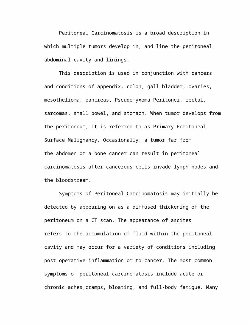
Peritoneal carcinomatosis (PC) is a type of secondary cancer that affects the lining
of the abdominal cavity, called the peritoneum. It occurs when cancer metastasizes from
another part of the body and implants into the lining. Peritoneal carcinomatosis most
commonly follows severe or untreated pancreas, ovarian, stomach, and colon cancer.
Symptoms can vary, but many people experience extreme fatigue and abdominal pain.
Quick, aggressive treatment in the form of medications and surgery is vital in preventing
fatal complications (Jeffress, 2012). Tumor growth on intestinal surfaces and associated
fluid accumulation eventually result in bowel obstruction and incapacitating levels of
ascites, which profoundly affect the quality of life for affected patients. Recently,
population-based studies have revealed that PC occurs relatively frequently among
patients with colorectal cancer (CRC). Risk factors for developing PC have been
Predisposing Factors Age (23 yrs. old) Gender (Female) Heredity
Precipitating Factors Environmental conditions Lifestyle Other health conditions
Damage to DNA in cell nucleus
Cell death
Cell Cycle Alteration
Imbalance between production and
absorption of fluid
Increased production and proliferation of enzymes and hormones
Tumor implants compress the bowel
by their volume
Carcinogenesis
New and rapid growth
Ascites (Abdominal distention: Girth-93cm)
Bowel obstruction
Compression and elevation of the diaphragm
DOB
Pain
Damaged to surrounding tissues and nerve
compression as tumor grows
Dissemination from the primary tumor
Invasion in the GIT
Paracentesis
Mechanical effects:
Palpable masses on the
abdomen
Ascites Secondary to Peritoneal 20
Systemic effects:
Cachexia(muscle wasting)
Body cannot synthesize amino
acids
Altered protein metabolism
Weight loss(From 50 kg to 42 kg)
Peritoneal Carcinomatosis
CHAPTER VII
COURSE IN THE HOSPITAL
DATE & TIME
SIDE NOTES ORDERS RATIONALE
December 5, 2012
11:10 am
Problem: Ascites secondary to Peritoneal TB, r/o Peritoneal carcinomatosis
Admit with consent under the service of green team.
Monitor vital signs every hour and record.
Small frequent feedings.
MIO every 4 hours and record.
IVF: D5LR 1L @ KVO (microset)
Laboratory:
CBC, BT
AFB peritoneal fluid
-Admission for referral of care.
-For close monitoring and to watch out for any unsualities.
-To prevent gastrointestinal reflux.
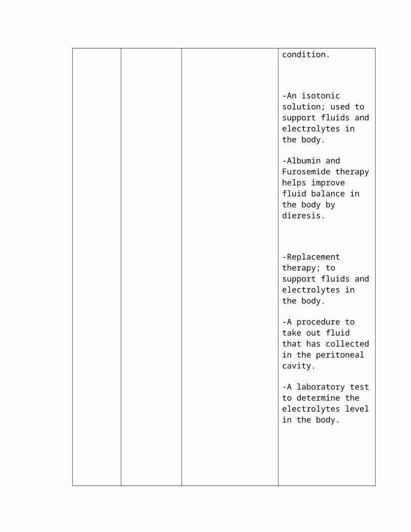
-Provides information about fluid status, circulating volume and replacement needs.
-Replacement therapy; to support fluids and electrolytes in the body.
-To use as baseline information in comparison to next repeated laboratory exams.
-A screening test to provide information about the cellular components of the patient’s blood; to determine presence of any abnormalities or disorders.
-Acid- fast bacilli, to identify pathogenic organisms present in the peritoneal fluid, as well as,
December 5, 20125:00 pm
(-) obstruction seen
Surgical notes;Thank you for the referral seen and examined
A/P carcinomatosis vs. PTB
Medications: Ceftriaxone 1mg
IVTT every 12 hours ANST
Ranitidine 50mg IVTT every 8 hours
Metoclopramide 10mg IVTT every 8 hours PRN for vomiting
Multivitamins + Amino acid 1 capsule once a day
For paracentesis, secure consent
Continue medication management
Refer
For: CEA
TSH
to identify the antimicrobial therapy that is best suited for the particular micobacteria identified.
-An antibiotic Cephalosphorin, for treatment of susceptible infection.
-An H2 receptor antagonist, used to decrease gastric secretion.
-An antiemetic, for management of nausea and vomiting associated with various GI disorders.
-To prevent low levels of vitamins, folic acid, and amino acids in the body.
-Secure consent, because the procedure to be done is an invasive procedure.
-Carcinoembryonic antigen, a test performed when cancer is suspected but not yet diagnosed and especially when doctor suspects that cancer has metastasized.
-Thyroid stimulating hormonetest, is a test that measures the amount of
Ascites Secondary to Peritoneal 22
Ascites Secondary to Peritoneal 23
Ascites Secondary to Peritoneal 27
CHAPTER VIII
NURSING CARE PLAN # 1
HRP NSG. Dx
AMB PATHO-PHYSIOLOGY
CLIENT OUTCOME
NURSING INTERVENTIONS
RATIONALE EVALUATION
EXCHANGING
Ineffective breathing pattern r/t decreased lung compliance secondary to ascites
Objective:-RR: 25 bpm-Nasal flaring noted-DOB noted-Uses accessory muscles-Abdominal distention noted due to ascites-Abdominal girth of 93 cm
The accumulation of fluid may cause breathing difficulties by compressing the diaphragm.A person with ascites has a swollen, rounded stomach. The skin on the abdomen is tight. The size of the abdomen is related to the amount of fluid present.Ascites may extend as far as the chest cavity. The presence of the fluid adds pressure to the lungs and may cause the individual to experience difficulty breathing.
Within the shift, patient will breathe with minimal difficulty as evidenced by not using accessory muscle and RR within normal range.
1. Monitor vital signs.
2. Place on semi-fowler’s position with arms supported with pillows.
3. Maintain calm attitude while dealing with client and to significant others.
4. Encourage adequate rest and sleep periods between activities.
5. Instructed to avoid overeating/ gas-forming foods.
-To watch out for abnormalities, assess condition.
-To relieve pressure on the diaphragm.
-To limit the level of anxiety.
-To limit fatigue and preserve energy.
-They can cause abdominal distention, thus, will aggravate difficulty of breathing.
Goal not met, patient’s respiratory rate was 27 bpm, evident use of her accessory muscles when breathing.
Ascites Secondary to Peritoneal 29
NURSING CARE PLAN # 2
HRP NSG. Dx
AMB PATHO-PHYSIOLOGY
CLIENT OUTCOME
NURSINGINTERVENTIONS
RATIONALE EVALUATION
EXCHANGING
Deficient fluid volume r/t active fluid volume loss (ascites: third spacing)
(Dec.7, 2012)
Subjective: “Kadalasan talaga gusto kong tubig.”
Objective:
-Abdominal distention (ascites)
- Muscle weakness
-Poor skin turgor
Ascites is the accumulation of fluid in the peritoneal cavity. Third spacing occurs when too much fluid moves from the intravascular to interstitial space causing a reduced blood volume in intravascular space.
Within the shift, the patient will able to maintain fluid volume at a functional level as evidenced by individually adequate urinary output with normal specific gravity, stable vital signs, moist mucous membranes, good skin turgor and prompt capillary refill.
1. Note possible condition that may create a fluid volume deficit such as fluid restriction, vomiting or use of diuretics.2. Monitor vital signs, noting low blood pressure—severe hypotension, rapid heartbeat, and thready peripheral pulses.3. Compare usual and current weight.
4. Measure abdominal girth.
5. Instruct the client to avoid foods very high in sodium content.
6. Monitor Intake and output accurately.
7. Instruct patient to avoid drinks containing caffeine e.g. beverages and coffee.8. Change position frequently.
-Help identify and prevent further fluid deprivation.
-Changes in vital signs are associated with fluid volume loss and/or hypovolemia.-To note for any significant fluid gain or loss.-To note for the extent of fluid retention in the abdomen.-To avoid excessive water retention and further fluid shifting (ascites).-To note for significant fluid loss and gain.
-To reduce effects of diuresis.
-To reduce pressure on fragile skin and tissues.
Goal partially met. The patient was able to maintain fluid volume at a functional level as evidenced by good vital sign, but skin turgor was still poor (3-4 sec).
Ascites Secondary to Peritoneal 30
NURSING CARE PLAN # 3
HRP
NSG. Dx
AMB PATHO-PHYSIOLOGY
CLIENT OUTCOME
NURSING INTERVENTONS
RATIONALE EVALUATION
FEELING
Acute pain r/t abdominal
fullness secondary to
ascites
(Dec.8,2012)
Subjective: “Masakitangtiyan ko ngayon” as verbalized-pain scale of 6/10
Objective:-pale and weak looking
-with limited movements noted
-facial grimace noted
-diaphoresis noted
Pain is a highly subjective state in which a variety of unpleasant sensations and a wide range of distressing factors may be experienced by the sufferer. Pain may be a symptom of injury or illness. Pain may also arise from emotional, psychological, cultural, or spiritual distress.
Within the shift, client will report
pain is relieved or controlled
and demonstrate
use of relaxation skills and
diversional activities.
1. Allow patient to verbalize pain.
2. Provide non-pharmacologic comfort measures such as repositioning, back rub and diversional activities such as listening to music and conversing about pleasant things.
3. Encourage use of stress management skills or complementary therapies such as guided imagery and therapeutic touch.
4. Observe or monitor signs and symptoms associated with pain, such as BP, HR, temp., color and moisture of skin, restlessness, and ability to focus.
5. Provide rest periods to facilitate comfort, sleep, and relaxation.
-Pain is subjective that can only be felt by the person affected.
-Promotes relaxation and helps refocus attention.
-Enables patient to participate actively in nondrug treatment of pain and enhances sense of control.
- Some people deny the experience of pain when it is present. Attention to associated signs may help the nurse in evaluating pain.
- Pain may result in fatigue, which may result in exaggerated pain and exhaustion.
Goal met, client appears calm and relaxed,
pain was decreased from
6/10 to 3/10; verbalized,
“Medyo hindi na masakit ngayon”.
Ascites Secondary to Peritoneal 31
NURSING CARE PLAN # 4
HRP NSG. Dx
AMB PATHO-PHYSIOLOGY
CLIENT OUTCOME
NURSING INTERVENTIONS
RATIONALE EVALUATION
EXCHANGING
Altered bowel elimination: Constipation r/t decreased motility of GI tract
(Dec. 8, 2012)
Subjective: -“Hindi parin ako nakakabawas simula ng naadmit ako” as verbalized.-Reports decreased frequency of bowel movement
Objective:-Abdominal distention noted due to ascites-Abdominal girth of 93 cm-Limited fluid intake of 1000mL-Inadequate fiber intake due to loss of appetite
Constipation is a condition characterized by infrequent or hard bowel movements, or having difficulty passing bowel movements. Also known as irregularity, Constipation can include pain when having a bowel movement, an inability to “go” after trying for more than ten minutes or having no bowel movement after more than three days.
Within the shift, patient will be able to establish or regain an elimination pattern as evidenced by bowel movement with at least normal consistency, thus, participate and understand the appropriate interventions or solutions in order to relieve self from constipation.
INDEPENDENT:1. Auscultate abdomen for
presence and location of bowel sounds and its characteristics.
2. Note color, odor, consistency, amount, and frequency of previous stool.
3. Identify factors (eg. Medications, bedrest, diet) that may cause or contribute to constipation.
4. Encourage on high fiber foods, and suggest warm stimulating fluids.
5. Encourage on light exercises as tolerated.
DEPENDENT:6. Administer laxative or stool
softeners as ordered.
-This reflects the bowel activity.
-This provides baseline comparison, promotes recognition of changes.
-Assessing causative factor is an essential first step in teaching and planning for improved bowel elimination.
-To improve consistency of stool and facilitate passage.
-Influences bowel elimination by improving muscle tone and stimulating peristalsis.
-May be necessary to gently stimulate peristalsis/ stool evacuation.
Goal not met, patient was still unable to regain her bowel movement.
Ascites Secondary to Peritoneal 32
NURSING CARE PLAN # 5
HRP NSG. Dx
AMB PATHO-PHYSIOLOGY
CLIENT OUTCOME
NURSING INTERVENTIONS
RATIONALE EVALUATION
EXCHANGING
Imbalanced nutrition less
than body requirements
related to feeling of
being full and mal-
absorption
(Dec.11, 2012)
Subjective: “Hanggang apat na kutsara lang kaya kong kainin kasi feeling ko wala ng mapaglagyan pagkain sa tyan ko” as verbalized.
Objective: -Weakness noted-Poor muscle tone-Decreased subcutaneous fat/ muscle mass
The client perceived that there is no space in her stomach that’s why she didn’t take lots of food. Her nutritional needs was very high due to poor eating habits. She seems to have poor nutritional status.
Within 8 hours of nursing interventions the client will be able to regain weight and verbalize understanding of causative factors when known and necessary inteventions
Assess weight, age, body build, strength, activity/ rest level
Auscultate bowel sounds. Note characteristics of stool.
Weigh weekly and document results.
Encourage to verbalize feelings and concerns
Discuss eating habits including food preferences, intolerance, aversions
Determine psychological factors
-Use as comparative baseline
-To identify if bowel movement is present for peristalsis
-To monitor effectiveness of dietary plan
-To know the real concern/ feeling of the client.
-To appeal to client likes/ desires.
-To assess body image and congruency with reality
Goal partially met, the client’s nutritional status enhances as evidenced by verbalization of “Medyo naging okay na ako ngayon, may lakas na ako” and having an energy during the conduct of assessment and during or within the activity period.
Ascites Secondary to Peritoneal 33
NURSING CARE PLAN # 6
HRP NSG. Dx
AMB PATHO-PHYSIOLOGY
CLIENT OUTCOME
NURSING INTERVENTIONS
RATIONALE EVALUATION
FEELING
Mild anxiety related to threat/ changes in health status secondary to peritoneal tuberculosis
(Dec.8, 2012)
Subjective:“Kinakabahan ako sa kalagayan ko ngayon”, as verbalized.
Objective:- -Awake with
blank stare- -Focus on self
- Pale and weak looking-Limited movements noted-Diaphoresis noted
Mild anxiety speaks for itself. Basically your body's natural warning system telling you to go on alert when there is no actual cause for alarm.Even though mild anxiety is slighter in terms of effects, it still can be a heavy baggage especially if it occurs more often than you think. On the case of our client she was anxious about her current condition, if there will be a good prognosis or not. Those suffering from mild anxiety will usually only suffer from the physical and mental symptoms.
Within the shift, client will be able to appear relaxed and report anxiety is reduced to a manageable level.
1. Explore client’s feelings.
2. Allow/ encourage client to speak openly about fears and concerns.
3. Establish a therapeutic relationship, conveying empathy and unconditional positive regard
4. Acknowledge anxiety or fear. Do not deny or reassure that everything will be alright
5. Monitor and record vital signs.
-To know what/ how does client really feels.
-To let him express what are those he think that makes him worry.
-To let patient feel that he’s not alone and to avoid the contagious effect or transmission of anxiety.
-Not to let client assure herself and blame anyone if something happen.
-To identify physical responses associated with both medical and emotional conditions.
Goal met, client was able to expressed feelings and concerns; appears relaxed and verbalized, “Mas okay sa ngayon kesa kanina”.
Ascites Secondary to Peritoneal 34
NURSING CARE PLAN # 7
HRP NSG. Dx
AMB PATHO-PHYSIOLOGY
CLIENT OUTCOME
NURSINGINTERVENTIONS
RATIONALE EVALUATION
FEELING
Anticipatory grieving related to perceived potential death
(Dec.11, 2012)
Subjective: “Malala na daw tong sakit ko”, as verbalized.
Objective:-Weakness noted- Alterations in sleep pattern
Grieving is an intellectual and emotional responses and behaviors by which the individual and family work through the process of modifying self concept based on the perception of potential loss. Since patient’s illness has a poor prognosis, and chance of survival is minimal, it is normal that the patient and family mourn.
Within the shift, the client will be able to identify and express feelings appropriately.
1. Establish rapport to the client.
2. Provide open, nonjudgmental environment. Use therapeutic communication skills.
3. Encourage verbalization of thoughts/concerns and accept expressions of sadness, anger, rejection. Acknowledge normality of these feelings.
4. Reinforce teaching regarding disease process and treatments and provide information as requested/appropriate about dying. Be honest; do not give false hope while providing emotional support.
5. Identify positive aspects of the situation.
-To establish trust and cooperation to the client.
- Promotes and encourages realistic dialogue about feelings and concerns.
- Patient may feel supported in expression of feelings by the understanding that deep and often conflicting emotions are normal and experienced by others in this difficult situation.
- Patient/SO benefit from factual information. Individuals may ask direct questions about death, and honest answers promote trust and provide reassurance that correct information will be given.
-Possibility of remission and slow progression of disease and/or new therapies can offer hope for the future.
Goal met, client and family were able to verbalize understanding of the dying process and feelings of being supported in grief work.
Ascites Secondary to Peritoneal 35
NURSING CARE PLAN # 8
HRP NSG. Dx
AMB PATHO-PHYSIOLOGY
CLIENT OUTCOME
NURSINGINTERVENTIONS
RATIONALE EVALUATION
MOVING
Self-care deficit r/t lack of motivation in performing good hygiene.
(Dec.7, 2012)
Subjective: “Hindi ko na magawang maligo at mag-ayos ng katawan ko dahil sa sakit ko”, as verbalized.
Objective:-discomfort noted
-dry skin
-slight unpleasant body odor noted
Self-care deficit is described as an impaired ability to perform complete feeding, bathing/ hygiene, dressing and grooming or toileting activities.Since the patient has weakness, it’s hard for her to move and do daily activities that’s why self-care is often depleted.
Within the shift, the client will be able to cooperate in the practice of good and proper hygiene.
1. Establish rapport to the client.
2. Encourage to verbalize feelings and concerns.
3. Assist on adaptation to accomplish activities of daily living.
4. Provide communication among those who are involved in caring for assisting the client.
5. Allow sufficient time for the client to accomplish task to fullest extent of ability.
-To establish trust and cooperation to the client.
-To discover barriers to participation.
-To encourage client and build on successes.
-Enhances coordination and continuity of care.
-To enhance client’s capabilities and promote independence.
Goal met, client and family were able to participate in promoting good hygiene to the patient by giving him a bed bath.
CHAPTER IX
DRUG STUDY # 1GEN.
NAMEBRAND NAME
DRUGCLASS
MODE OF ACTION
INDICATIONCONTRA-
INDICATIONACTUAL
DOSEUSUAL DOSE
SIDE EFFECTS
NURSING RESPONSIBILITIES
CEFTRIAXONE
ROCEPHIN
CEPHALOSPORIN
Inhibits bacterial wall synthesis, thus, promoting osmotic instability which eventually leads to bacterial cell death.
-Used to treat infection caused by staphyloco-ccus, streptococcus, E.coli, and other susceptible microorganism. Skin to skin structure infection and biliary tract infection.
Contra-indicated for patients who have known hypersensitive to cephalos-porins and any of its components.
Ceftriaxone 1gm q12 ANST ( )
Ceftriaxone 1-2 gms
once a day
Signs of allergy: skin rashes, fever.
Hematologic: leukopenia, reversible thrombo-penia
Digestive:nausea, vomiting, anorexia, diarrhea
1. Observe the 10R’s of administering drugs (RIGHT: client, medication, dosage, route, time, documentation, health education, to refuse, assessment, evaluation).
2. Assess patient’s previous sensitivity reaction to cephalosporins.
3. Monitor for signs of allergic reaction.
4. Monitor vital signs before and after giving the drug esp. HR,RR,BP. Report changes.
5. Explain that the patient may experience the following side effects: nausea, diarrhea.
6. Encourage patient to report for signs of abnormalities.
Ascites Secondary to Peritoneal 37
DRUG STUDY # 2
GEN.NAME
BRAND NAME
DRUGCLASS
MODE OF ACTION
INDICATIONCONTRA-
INDICATIONACTUAL
DOSEUSUAL DOSE
SIDE EFFECTS
NURSING RESPONSIBILITIES
RANITIDINE
ZANTAC
Histamine (H2)
receptor antagonist
Inhibits the action of histamine at H2 receptors of the parietal cells of the stomach, inhibiting basal gastric acid secretion that stimulates by food, insulin, histamine, cholinergic agonist, gastrin, and pentagastrin.
-Short-term treatment of active duodenal ulcer; treatment of gastro-esophageal reflux disease; short-term treatment of active, benign gastric ulcer; treatment of pathologic GI hypersecretory conditions (postoperative hypersecretion); heartburn.
-Contra-indicated with allergy to ranitidine.Use cautiously with impaired renal or hepatic function.
1. Observe the 10R’s of administering drugs (RIGHT: client, medication, dosage, route, time, documentation, health education, to refuse, assessment, evaluation).
2. Monitor vital signs and watch out for abnormalities such as tachycardia or bradycardia.
3. Monitor intake and output.
4. Explain to hat she may experience the following side effects: headache, malaise.
5. Check laboratory results for abnormalities and refer to the physician.
6. Check the insertion site for phlebitis.
7. Encourage to report immediately for any signs of abnormalities.
Ascites Secondary to Peritoneal 38
DRUG STUDY # 3
GEN.NAME
BRAND NAME
DRUGCLASS
MODE OF ACTION INDICATIONCONTRA-
INDICATIONACTUAL
DOSEUSUAL DOSE
SIDE EFFECTS
NURSING RESPONSIBILITIES
METOCLOPROMIDE
PLASIL
Anti-emetic
It binds to dopamine D2 receptors where it is a receptor antagonist, and is also a mixed 5-HT3 receptor antagonist/ 5-HT4 receptor agonist. The antiemetic action of metoclopramide is due to its antagonist activity at D2 receptors in the chemo- receptor trigger zone (CTZ) in the CNS—this action prevents nausea and vomiting triggered by most stimuli. At higher doses, 5-HT3 antagonist activity may also contribute to the antiemetic effect. The gastroprokinetic activity of metoclopramide is mediated by muscarinic activity, D2 receptor antagonist activity and 5-HT4 receptor agonist activity. The gastro-prokinetic effect itself may also contribute to the antiemetic effect.
-Disturbances of GI motility -For nausea andvomiting
-Contra-indicated withallergy to metoclopramide;GI hemorrhage; Mechanical obstruction or perforation; fluid overload, and renal impairment
1. Observe the 10R’s of administering drugs (RIGHT: client, medication, dosage, route, time, documentation, health education, to refuse, assessment, evaluation).
2. Check history: allergy to metoclopramide, GI hemorrhage, mechanical obstruction or perforation.
3. Monitor BP carefully during IV administration.
4. Monitor intake and output.
5. Tell patient that she may experience the said side effects: drowsiness, nausea, dizziness.
Ascites Secondary to Peritoneal 39
DRUG STUDY # 4
GEN.NAME
BRAND NAME
DRUGCLASS
MODE OF ACTION
INDICATIONCONTRA-
INDICATIONACTUAL
DOSEUSUAL DOSE
SIDE EFFECTS
NURSING RESPONSIBILITIES
MULTIVITAMINS
+
AMINO
ACIDS
NUTRI
WELL
Multi-vitamins
and supple-ments
Multivitamin is a combination
of many different
vitamins that are normally
found in foods and other natural
sources.Many act as
coenzymes or catalysts in numerous metabolic
processes. It also works by
providing extra vitamins, folic
acid, and amino acids to the
body when you need more than what you get in
your diet.
Treating or preventing low levels of vitamins, folic acid, and amino acids in the body.
-Contra-indicated if you are allergic to any ingredient in multivitamins with folic acid/amino acids and if you have high blood levels of arginine (argininemia).
Multi-vitamins +
Amino acids 1cap
OD
Multi-vitamins
1cap daily
Allergic reactions:Rash, hives, itching, difficulty breathing, tightness in the chest, swelling of the mouth, face, lips, or tongue
1. Observe the 10R’s of administering drugs (RIGHT: client, medication, dosage, route, time, documentation, health education, to refuse, assessment, evaluation).
2. Take multivitamins with folic acid/amino acids by mouth with or without food. If stomach upset occurs, take with food to reduce stomach irritation.
3. Take multivitamins with folic acid/amino acids with a full glass of water (8 oz/240 mL).
4. Explain that she may experience the following side effects: rash, difficulty breathing.
5. Encourage to report immediately for any signs of abnormalities.
Ascites Secondary to Peritoneal 40
DRUG STUDY # 5
GEN.NAME
BRAND NAME
DRUGCLASS
MODE OF ACTION
INDICATION CONTRA-INDICATION
ACTUAL DOSE
USUAL DOSE
SIDE EFFECTS
NURSING RESPONSIBILITIES
MULTIVITAMINS
+
MINERALS
SUPPLEMENTS
Multi-vitamins
and supple-ments
Multivitaminand minerals are used to provide vitamins and minerals that are not taken in through the diet. Multivitamin and minerals works by treating vitamin or mineral deficiencies caused by illness, pregnancy, poor nutrition, digestive disorders, certain medications, and many other conditions.
Dietary supplement for the treatment and prevention of vitamin and mineral deficiencies.
-Contra-indicated if you are allergic to any ingredient in multivitamins and minerals and any of its components.
Multi-vitamins + Minerals
(Supplements)1 vial OD x
12hours
Multi-vitamins + Minerals 1 vial once or twice a day
Less serious side effects:upset stomach,headache,unusual or unpleasant taste in your mouth
Allergic reaction: Hives, difficulty breathing, swelling of your face, lips, tongue, or throat.
1. Remember the 10R’s of administering drugs (RIGHT: client, medication, dosage, route, time, documentation, health education, to refuse, assessment, evaluation).
2. Monitor for manifestations of hypersensitivity appearance promptly.
3. Do not take this medication with milk, other dairy products, calcium supplements, or antacids that contain calcium. Calcium may make it harder for your body to absorb certain ingredients of the multivitamin.
4. Check for nutritional deficiencies.
5. Encourage to report immediately for any signs of abnormalities.
Metabolism of lactulose by bacteria results in reduced colonic pH which stimulates peristalsis & decreases stool transit time. In turn, decreased water reabsorption from the feces further facilitates the passage of soft, well-formed stools. Increased osmotic pressure of fecal material secondary to an increase in colonic organic acids results in accum. of fluid from surrounding tissues, helping to soften stool mass.
Treatment of constipation.
Prevention and treatment of portal- systemic encephalo-pathy
-Contra-indicated to patients with allergy to lactulose, low-galactose diet.
-Use cautiously with diabetes, pregnancy and lactation.
1. Observe the 10R’s of administering drugs (RIGHT: client, medication, dosage, route, time, documentation, health education, to refuse, assessment, evaluation).2. Instruct that this drug may be taken with fruit juice or milk to increase palatability.3. Do abdominal examination, check bowel sounds, and serum electrolyte levels.4. Do not administer if patient has already pass out stool especially if stool is liquid.5. Monitor intake and output.6. Tell patient that she may experience these side effects: flatulence, intestinal cramps, nausea)7. Report if unusualities occur.
Ascites Secondary to Peritoneal 42
DRUG STUDY # 7
GEN.NAME
BRAND NAME
DRUGCLASS
MODE OF ACTION
INDICATIONCONTRA-
INDICATIONACTUAL
DOSEUSUAL DOSE
SIDE EFFECTS
NURSING RESPONSIBILITIES
TRAMADOL
TRAMAL
Analgesic, opioid analgesic
Binds to –opiate receptors in the CNS causing inhibition of ascending pain pathways, altering the perception of and response to pain; also inhibits the reuptake ofnorepinephrine and serotonin, which also modifies the ascending pain pathway.
Moderate to severe acute or chronic pain and in painful diagnostic or therapeutic measures.
Hypersensi-tivity to tramadol, opioids, or any component of the formulation; opioid-dependent patients; acute intoxication with alcohol, hypnotics, centrally-acting analgesics, opioids, or psychotropic drugs.
1. Observe the 10R’s of administering drugs (RIGHT: client, medication, dosage, route, time, documentation, health education, to refuse, assessment, evaluation).
2. Assess type, location, and intensity of pain before and 2-3 hr (peak) after administration.
3. Assess BP & RR before and periodically during administration.
4. Assess bowel function routinely.
5. Encourage patient to cough and breathe deeply every 2 hr to prevent atelactasis and pneumonia.
6. Instruct client to report any adverse reaction to the physician or nurse.
Ascites Secondary to Peritoneal 43
DRUG STUDY # 8
GEN.NAME
BRAND NAME
DRUGCLASS
MODE OF ACTION
INDICATIONCONTRA-
INDICATIONACTUAL
DOSEUSUAL DOSE
SIDE EFFECTS
NURSING RESPONSIBILITIES
ALBUMIN
ALBUMINAR
Plasma expanders
Blood derivatives
Provides increase in intravascular oncotic pressure and causes mobilization of fluids from interstitial into intravascular space.
For plasma volume expansion and maintenance of cardiac output in the treatment of certain types of shock or impending shock; may be useful for burn, ARDS, peritonitis, and ascites.Unless the condition responsible for hypoproteinemia can be corrected, albumin can only provide symptomatic relief of supportive treatment.
-Contra-indicated with allergy to albumin and any of its components, with severe anemia and
Albumin 25% 50cc + furose-mide 20mg x 2 hours q12hours
1. Observe the 10R’s of administering drugs (RIGHT: client, medication, dosage, route, time, documentation, health education, to refuse, assessment, evaluation).
2. Monitor vital signs and watch out for abnormalities.
3. Monitor intake and output.
4. Explain to the parents that he may experience the following side effects: fever, chills, nausea.
5. Check laboratory results for abnormalities and refer to the physician.
6. Watch out for symptoms of overdose, such as: hypervolemia, CHF, pulmonary edema.
7. Encourage to report immediately for any signs of abnormalities.
-Treatment of fluid accumulation such as ascites, edema associated with CHF,hepatic cirrhosis, renal disease.
- Hypersen-sitivity to furosemide, sulfonylureas, or any other drugs.
- Contraindicated in patients with anuria, hyponatremia or hypovolemia.
Albumin 25% 50cc + furose-mide 20mg x 2 hours q12hours
Furosemide20-40mg
IV everyday of one to
two times a day
●Low blood pressure●Dehydration and electrolyte depletion●Orthostatic HPN●Pruritus●Vertigo●Dizziness●Fever ●Nausea●Vomiting●Constipation●Oral and gastric irritation ●Diarrhea●Increased blood sugar and uric acid levels may also occur.
1. Observe the 10R’s of administering drugs (RIGHT: client, medication, dosage, route, time, documentation, health education, to refuse, assessment, evaluation).
2. Check the BP first before administration.
3. Monitor Intake and Output of the patient.
4. Explain that she may experience these side effects: dizziness, nausea.
5. Instruct client to report any signs of side effects.
CHAPTER X
LABORATORY STUDY # 1
DETERMINATION ACTUAL VALUE NORMAL VALUE SIGNIFICANCE/INTERPRETATION NURSING RESPONSIBILTY
HEMATOLOGY(December 5, 2012)● WBC
● RBC
● HGB
HCT
PLT
MCV
MCH
MCHC
RDW
17.2 x 10⁹/ L
4.51 x 10¹²/L
118 g/L
0.38
957 x 109/L
84.0 fL
26 pg
340g/L
12.1 %
4.0-10.0 x 10⁹/ L
4.50-5.4 x 10¹²/L
115-155 g/L
0.36-0.47
100-300 x 109/L
86-100 fL
26-31 pg
310-375 g/L
11.6-13.7 %
Increased; indicative of impending infection or inflammation in the body due to disease process.
Normal; good oxygenation in the blood, may decrease because of disease process.
Normal; good circulation of oxygen in the blood.
Normal;there is good hydration status in the patient’s body; good oxygen supply.
Increased; or thrombocytosis, may result from iron deficiency anemia or inflammatory disorders.
Decreased; MCV measures the ratio of hematocrit to RBC count. May indicate iron deficiency anemia
Normal; MCH gives the hemoglobin to RBC ratio.
Normal: MCHC measures the ratio of hemoglobin weight to hematocrit.
Normal; RDW determines the measurement of RBCs.
Explain the procedure & purpose of performing the procedure, and that is to determine infection & its severity because of the disease. This test is very important as baseline data.
Tell patient as well as watcher that the test requires a blood sample and explain who will perform the venipuncture.
Give health teachings on patient’s diet and medication that may contribute to the result of the test.
Based on the result, instruct patient to eat nutritious foods especially rich in vitamins, minerals and iron, such as fish, vegetables, and fruits.
Advise to have adequate rest and sleep periods.
Stress out the importance of taking multivitamins as
Differential Count Neutrophils
Lymphocytes
Monocytes
Eosinophils
Basophils
80 %
10 %
9.0 %
1.0 %
0 %
40-70 %
19-42 %
3.0-9.0 %
2.0-8.0 %
0-5.0 %
Increased; may indicate infection, inflammatory processes during physical stress, or with tissue necrosis.
Decreased; may signal infection in the body and/or anemia.
Normal; may increase because of illness disease.
Decreased; signals infection because of illness.
Normal; aids in determining specific conditions.
prescribed by the physician.
Educate about the importance of medications and treatment regimen.
Note for any abnormalities on findings and refer the results to the physician.
Ascites Secondary to Peritoneal 47
LABORATORY STUDY # 2
DETERMINANTS ACTUAL VALUE
NORMAL VALUE
SIGNIFICANCE/ INTERPRETATION NURSINGINTERVENTIONS
Ascites Secondary to Peritoneal 46
CLINICAL CHEMISTRY(November 23, 2012) Creatinine
SGPT/ALT
SGOT/AST
ALP(Alkaline phosphatase)
Total Protein
Albumin
Globulin
73.4 umol/L
333.4 nKat/L
383.4 nKat/L
1300.3 nKat/L
62 g/L
33 g/L
53-97 mmol/L
0-517 nKat/L
0-517 nKat/L
700-1630 nKat/L
64-83 g/L
35-52 g/L
Normal; indicates that the kidneys are able to properly remove all creatinine. May increase if dehydrated or took certain medications.
Normal; indicates that liver and kidneys are functioning well. Low levels of ALT are normally found in the blood. But when the liver is damaged or diseased, it releases ALT into the bloodstream, which makes ALT levels go up. Most increases in ALT levels are caused by liver damage.
Normal; indicates no liver damage. High levels may indicate severe MI, severe infectious mononucleosis or alcoholic cirrhosis. Low levels indicate hemolytic anemia, metastatic hepatic tumors or fatty liver.
Normal;indicates no liver or bone disease. ALP test measures the amount of alkaline phosphatase released from the tissues into the blood and is a marker of the hepatobilary system function. Moderate increase indicates acute biliary obstruction. Low levels are linked to hypophosphatasia and protein or magnesium deficiency.
Decreased;may be indicative of certain diseases such as GI disease, protein deficiency, neoplastic disease, malnutrition or malabsorption.
Decreased; may indicate that not enough protein is being absorbed in the body, may also reflect diseases such as malnutrition or ascites.
Explain the procedure & purpose of performing the procedure, and that is to help diagnose the occurrence of disease and if there are complications, to test effectiveness of medications and find treatments for the disease.
Explain the procedure to the client that the medical technician will get sample of her blood for testing.
Give health teachings on patient’s diet and medication that may contribute to the result of the test.
Instruct patient to eat nutritious foods especially rich in vitamins, minerals and proteins, such as fish, vegetables, and fruits. Also, instruct to eat nutritious food that helps in cleansing the kidney.
Strictly monitor the intake and output.
Advise to have adequate rest and sleep periods.
Stress out the importance of
Ascites Secondary to Peritoneal 48
A/G Ratio
29 g/L
1.1
20-35 g/L
1.7-2.2
Normal;Globulin carries essential metals through the bloodstream and carries them to the various parts of the body and helps the body to fight infections. Globulin proteins include enzymes, antibodies and more than 500 other proteins. High levels indicate tuberculosis. Low levels indicate GI disease, malnutrition, or malabsorption.
Decreased;A low A/G ratio reflects overproduction of globulins, due to chronic infections, liver and kidney disease, fatty necrotic liver, rheumatoid arthritis, leukemia, increased amount of nonspecific protein, and autoimmunity disorders. On the other hand, a high A/G ratio suggests under production of immunoglobulin; this is seen in genetic deficiencies and in cases of nephrosis, liver dysfunction, acute hemolytic anemia, and hypogammaglobulinemia / agammaglobulinemia.
taking multivitamin and supplements as prescribed by the physician.
Note for any unusualities on findings and refer the results to the physician.
Abnormal; an increase in urinary albumin excretion is indicative of increased permeability of the filters of the kidney called, glomerulus which due caused by some kidney damage.
Normal; normally, glucose is not present in the urine because it is reabsorbed from the renal tubules.
Abnormal; cloudy urine may be caused by crystal deposits, white cells, epithelial cells or fat globules.
Normal; pH measures how acidic or alkaline the urine is. Sometimes urine pH is affected by certain treatments.
Normal; this checks the amount of substance in the urine. When you drink lots of fluid your specific gravity becomes low. When you are dehydrated your specific gravity becomes high.
Normal; there should be no yeast cells and bacteria or parasites in the urine, if present; it means that there is infection.
Normal; normally, there is no blood in the urine. One of the common causes of RBC in the urine is infection or inflammation of the urinary tract itself (cystitis).
Increased; Amorphous Urates indicates uric acid crystals in the urine. Higher than acceptable levels of uric acid crystals in urine
1. Instruct patient to void into a clean, dry container.
2. Sterile disposable container should be used always.
3. Cover all specimens tightly, label properly and send immediately to the laboratory.
4. Observe standard precaution when handling the specimen.
5. Avoid the specimen to be exposed to extreme temperature such as sunlight or heat.
6. The specimen should be preserved if not to send to laboratory to have accurate results.
7. Note for any unusualities on findings and refer the
Ascites Secondary to Peritoneal 50
Epithelial Cells
11 None to few
can be caused by gout, cardiovascular disease, diabetes, uric acid stone, urolithiasis, and metabolic syndrome.
Increased; may suggest inflammation within the bladder, but they may also originate from the skin and could be contaminated. Sometimes, it is normal not to have any epithelial cells present in a urine sample or to have occasional numbers of any of the three cell types. Large numbers of squamous cells may indicate contamination of the urine specimen, but large numbers of either the transitional or renal tubular cells may indicate a serious disease process.
Increased; may indicate tuberculosis and/or malignancy; could be low in malignant ascites
Increased; Elevated levels of LDH and changes in the ratio of the LDH isoenzymes usually indicate some type of tissue damage. LDH levels typically will rise as the cellular destruction begins, peak after some time period, and then begin to fall.
Decreased; may be indicative of a symptom of a disease, infection or an underlying condition. When there is inadequate protein intake, the body begins to breakdown muscle to obtain enough amino acids for the synthesis of serum albumin.
Increased; to distinguish exudates and transudates. Values above 1.1 g/dL are considered evidence of a transudate.
Normal; Globulins are proteins that include gamma globulins (antibodies) and a variety of enzymes and carrier/transport proteins. Low globulin levels signify a type of protein deficiency; high levels mean chronic infections.
Explain the procedure & purpose of performing the procedure, and that is to help distinguish between types of peritoneal fluid and help diagnose the cause of fluid accumulation (ascites).
Explain that in this procedure, a local anesthetic is applied to the area of operation and then a catheter is routed from the skin into the peritoneal cavity. As soon as this is done, the peritoneal fluid will start to flow out.
Monitor vital signs prior to the procedure.
Advise to empty the bladder first before the procedure becausethis is a lengthy test.
Note for any unusualities on findings and refer the results to the physician.
Ascites Secondary to Peritoneal 52
LABORATORY STUDY # 5
DETERMINANTS ACTUAL NORMAL SIGNIFICANCE/
VALUE VALUE INTERPRETATION NURSINGINTERVENTIONImmunology
CA 12-5
(November 28, 2012)
127 U/ml 0.35 U/ml Increased: indicates that the cancer antigen is increased in colon, upper gastrointestinal (GI), ovarian, and other gynaecologic cancers: pregnancy, peritonitis.
Explain the procedure and the purpose of performing such procedure, and that is to determine infection because of the disease, that this test is very important as baseline data.
Tell patient as well as watcher that the test requires a blood sample and explain who will perform the venipuncture.
Give health teachings on patient’s diet that may contribute to the result of the test.
Based on the result, instruct the patient to eat nutritious foods especially rich in iron, such as fish, vegetables, and fruits.
Advise to have adequate rest and sleep periods.
Stress out the importance of taking multivitamins as prescribed by the physician.
Ascites Secondary to Peritoneal 53
LABORATORY STUDY # 6
DETERMINANTS ACTUAL VALUE
NORMAL VALUE
SIGNIFICANCE/INTERPRETATION NURSINGINTERVENTION
Immunology(December 6, 2012) Free T4
TSH
CEA(Carcinoembryonic
Antigen)
0.95
4.08
531.12
0.58-1.64 ug/dl
0.34-5.60 µ U /ml
0-3 ng/ul
Normal; indicates that thyroid hormone feedback system is functioning well. This test was done to evaluate thyroid function. The free T4 test is a newer test that is not affected by protein levels. Since free T4 is the active form of thyroxine, the free T4 test is thought by many to be a more accurate reflection of thyroid hormone function.
Normal; indicates normal functioning of the thyroid. T4 will be ordered along with a TSH to give a more complete evaluation of the adequacy of the thyroid hormone feedback system. These tests are usually ordered when a person has symptoms of hyper or hypothyroidism.
Increased; can indicate possible cancerous activity. Increased CEA levels may also indicate some non-cancer-related conditions, such as some forms of inflammation, cirrhosis, and peptic ulcer. A CEA test is ordered when the patient’s symptoms suggest the possibility of cancer. CEA is an embryonic protein which could be secreted in adult as well, if there is any abnormality in protein producing organs, especially liver, but similar protein can also be secreted if there is a presence of cancer.
Explain the procedure and the purpose of performing such procedure, and that is to evaluate thyroid function, determine possibility of cancer, diagnosis of certain illness or to monitor the effectiveness of treatment.
Tell patient as well as watcher that the test requires a blood sample and explain who will perform the venipuncture.
Give health teachings on patient’s diet that may contribute to the result of the test.
Educate on the importance of strict compliance to medication and treatment regimen.
Advise to have adequate rest and sleep periods.
Advise to eat nutritious foods necessary to improve health and to hasten recovery.
Decreased; indicates an electrolyte disturbance in which the sodium concentration in the serum is lower than normal. Sodium is the dominant extraellular cation and cannot freelycross the cell membrane. Hyponatremia is most often a complication of other medical illnesses in which excess water accumulates in the body at a higher rate than can be excreted (for example in congestive heart failure, syndrome of inappropriate antidiuretic hormone, SIADH or polydipsia.
Normal; Potassium testing is frequently ordered, along with other electrolytes. The most common cause of hyperkalemia is kidney disease, but many drugs can decrease potassium excretion from the body and result in this condition. Hypokalemia can occur if someone has diarrhea and vomiting or if is sweating excessively. Potassium can be lost through the kidneys in urine; in rare cases, potassium may be low because someone is not getting enough in their diet.
Decreased; indicates an electrolyte imbalance. Hypocalcaemia either occurs as a result of too much calcium loss or insufficient calcium intake through food. Early symptoms of low serum calcium include frequent muscle cramps and joint pains. In addition to this, inability to perform tiresome activities, fatigue, brittle nails, and yellowness of teeth also occur as a result of abnormally low level of calcium in the blood stream.
Explain the procedure and the purpose of performing such procedure, and that is to determine electrolyte imbalance in the body due to disease process
Tell patient as well as watcher that the test requires a blood sample and explain who will perform the venipuncture.
Because of electrolyte imbalance, initial treatment consists of slow correction of the hyponatremia via fluid restriction.
To restore calcium to a normal level, advise patient to eat calcium-rich foods or calcium supplements on a regular basis or as prescribed.
Advise to eat nutritious foods necessary to improve health and to hasten recovery.
The uterus is anteverted with smooth contour and homogenous echopattern measuring 5.5 x 2.3 x 3.3 cm (The cervix
measures 2.6 x 19 x 1.3 cm with homogenous stoma and distinct endocervical canal).
The endometrium is hyperechoic measuring 0.2 cm thick with intact subendometrial halo.
The right ovary measures 3.0 x 1.7 x 1.8 cm.
The left ovary measures 2.5 x 1.8 x 1.8 cm.
There’s massive anechoic free fluid in the cul de sac.
The omentum is converted into a heterogenous mass measuring 18 x 10 cm.
Impression: Normal uterus
Thin endometrium
Normal ovaries
Consider GI pathology
Ascites Secondary to Peritoneal 58
CHEST AP (December 10, 2012):
There are no active lung infiltrates seen
Heart is not enlarged
Diaphragm is elevated
Bony thorax is unremarkable
Impression: Elevated Diaphragm
MISCELLANEOUS REPORT (November 25, 2012):
Specimen Submitted: Peritoneal Fluid
Final Report: No growth after 48 hours incubation
Gram Stain: No organism seen
AFB: None found
CHAPTER XI
PROGNOSIS
CRITERIAVERY GOOD
(5)GOOD
(4)FAIR
(3)POOR
(2)
VERYPOOR
(1)JUSTIFICATION
Severity/ Nature of disease
Ms. Bella’s disease is difficult to treat and is fatal. Onset of disease is rapidly progressive and for now, only supportive care can be rendered.
Financial Status
Though they are able to comply and provide financial support minimally, they are now referred to service consultant because of heavy expenses.
Family Support
The family of the patient’s partner supports her most of the time. Her parents seldom visit her and buy for her medicines.
Patient factor
The patient is cooperative and participative to treatment regimen, though weak and sometimes irritable.
Availability & accessibility of appropriate treatment
Most of the appropriate treatment and resources are available.
Ascites Secondary to Peritoneal 60
Respective Numerical Values:
Very Good= 5 Good = 4 Fair = 3 Poor = 2 Very Poor=1
Standard Rating:
Very Good = 4.20 – 5.00
Good = 3.41 – 4.20
Fair = 2.61 – 3.40
Poor = 1.81 – 2.60
Very Poor = 1.0 – 1.80
Formula:
Rate x Frequency No. of Criteria
Computaion:
Very Good: 5 x 0 = 0
Good: 4 x 1 = 4
Fair: 3 x 2 = 6
Poor: 2 x 1 = 2
Very Poor: 1 x 1 = 1 13 ÷ 5 criteria = 2.60 or POOR
General Prognosis:
Based on the criteria, Ms. Bella has poor prognosis with a result of 2.60. Specifically,
she has scores of zero (0) in very good; two (1) in good; two (2) in fair; one (1) in poor and
one (1) in very poor.
Ascites Secondary to Peritoneal 61
In general, the client has a poor prognosis due to the onset, severity and
progression of the disease and complications secondary to her health problems.
Peritoneal carcinomatosis represents a devastating form of cancer progression
with a very poor prognosis. It is the most common terminal feature of abdominal
cancers. peritoneal cancer can be hard to detect in the early stages. That's because its
symptoms are vague and hard to pinpoint. When clear symptoms do occur, the disease
has often progressed. Care at this time is focused on relieving symptoms and quality of
life issues post-treatment.
CHAPTER XII
DISCHARGE SUMMARY PLAN
I. MEDICATION
Instruct patient and watcher to administer the prescribed medications on a right
dose, frequency and time.
RATIONALE: To meet the therapeutic effect of the drug and prevent over
dosage of the medication.
Explain the purpose of the medication.
RATIONALE: This will provide information to both the client and the parent
as to why the patient needs to take the prescribed medication.
Explain the indication and possible side effects brought by each of the drug.
RATIONALE: This will give awareness on both the patient and the watcher
to prevent panic when side effects are experienced by the client.
Instruct the client and watcher that when adverse effect occurs and if there are
any unusualities consult the physician immediately.
RATIONALE: To prevent any complications and give appropriate
interventions
II. EXERCISE
Encourage client not to do strenuous activities and limit activities within own
capacity as possible.
RATIONALE: Activities that require great muscle strength should be
avoided to prevent injury and fatigue.
Ascites Secondary to Peritoneal 63
III. TREATMENT
Instruct to maintain the prescribed medication as regularly as ordered by the
physician.
RATIONALE: To have a pace of supportive care.
Let the patient and family know that they should maintain a conducive, peaceful,
and non-stressful environment.
RATIONALE: To promote relaxation and good palliative care.
Explain to the client and family the need for heightened quality of life until her
last days.
RATIONALE: To make the client and family aware that the care does not
end in the hospital and that their participation is a must in the continuation of care.
IV. HYGIENE
Encourage the client to observe proper hygiene like taking a bath everyday,
hand washing before and after performing activities especially when having
meals and brushing of teeth every after meal.
RATIONALE: Hygiene promotes comfort and cleanliness to the client
and it also increases the sense of wellness.
V. OUT PATIENT FOLLOW-UP
If possible, instruct the patient to follow physician’s order on when to consult
for checkup.
RATIONALE: To enable the physician to evaluate patient’s condition.
Ascites Secondary to Peritoneal 64
Advise the family to supervise the patient properly.
RATIONALE: To take note for any unusualities and can be referred
immediately.
VI. DIET
Inform the family that the patient must receive adequate & proper nutrition
(especially high fiber diet). Eat fruits and green leafy vegetables.
RATIONALE: To modify patient’s diet and prevent further complication.
VII. SEXUAL ACTIVITY
Instruct patient that sexual intercourse is not recommended.
RATIONALE: Care is focused on supportive and emotional care.
CHAPTER XIII
BIBLIOGRAPHY
Austin, F., Mavanur, A., Sathaiah, M., Steel, J., Lenzner, D., Ramalingam, L., Holtzman, M., Ahrendt, S., Pingpank, J., Zeh, H., Bartlett, D., & Choudry, H. (2012). Peritoneal Carcinomatosis. Retrieved December 11, 2012 from, http://pmppals.org/peritoneal-carcinomatosis.html
Brunner, L. S. &Suddarth’s D.S. (2008). Medical-Surgical Nursing 11th& 12th edition, Volume 1 & 2.
Doenges, M., Moorhouse, M., &Murr A. (2002). Nursing Care Plans: Guidelines for Individualizing patient care 6th edition.
Gould, B. (2007). Pathophysiology for the Health Professionals 3rd edition.
Gulanick, M., Klopp, A., Galanes, S., Gradishar, D., &Puzas, M. (1994). Nursing Care Plan 3rd edition.
Jeffress, D. (2012). What Is Peritoneal Carcinomatosis? Retrieved December 10, 2012 from,http://www.wisegeek.com/what-is-peritoneal-carcinomatosis.htm
Johnson, RJ.(1993). Radiology in the management of ovarian cancer. Retrieved December 11, 2012 from, http://radiology.rsna.org/content/221/1/173.full
Karch, A. (2007). Lippincott’s Nursing Drug Guide.
Kusamura, S., Baratti, D., Zaffaroni, N., Villa, R., Laterza, B., Balestra, MR., & Deraco, M. (2010). Pathophysiology and biology of peritoneal carcinomatosis. Retrieved December 12, 2012 from,http://www.ncbi.nlm.nih.gov/pmc/articles/PMC2999153/
McCann, J. (2004). Handbook of Diseases 3rd edition.
Mizumoto, A., Canbay, E., Hirano, M., Takao, N., Matsuda, T., Ichinose, M., & Yonemura, Y. (2012). Gastroenterology Research and Practice Volume 2012Retrieved December 10, 2012 from, http://www.hindawi.com/journals/grp/2012/836425/
Peritoneal Health Guide (2010). Peritoneal Carcinomatosis Survival Rate. Retrieved December 11, 2012 from, http://peritoneal-health.info/peritoneal-carcinomatosis-survival-rate/
Sugarbaker, PH., Esquivel, J., & Sticca, R., (2007). Cytoreductive surgery and hyperthermic intraperitoneal chemotherapy in the management of peritoneal surface malignancies of colonic origin:a consensus statement. Retrieved December 11, 2012 from, http://www.ncbi.nlm.nih.gov/pubmed/17072675