Permeability of Red Cell Membranes to Small Hydrophilic and Lipophilic Solutes R. I. SHA'AFI, C. M. GARY-BOBO, and A. K. SOLOMON From the Biophysical Laboratory, Harvard Medical School, Boston, Massachusetts 02115, and the Department of Physiology, American University of Beirut, Beirut, Lebanon. Dr. Sha'afi's present address is the Department of Physiology, American University of Beirut, Beirut, Lebanon. Dr. Gary-Bobo's present address is Laboratoire de Physiologie Cellulaire, College de France, Paris, France. Please sendall reprint requests to the Biophysical Laboratory, Harvard Medical School, Boston, Massachusetts 02115. ABSTRACT The permeability coefficients of a series of amides, ureas, and diols have been measured on red cells of man and dog using the minimum volume method of Sha'afi et al. When the molecules are grouped according to their ether-water partition coefficients, kther, the behavior of the hydrophilic molecules, with kether less than water, is different from that of the lipophilic molecules, characterized by kther greater than water. The rate of permeation of the hydrophilic molecules through an aqueous pathway is determined by the molar volume, a parameter in which the geometrical measure of molecular volume is modified by hydrogen-bonding ability. This indicates the importance of chemical interactions within the aqueous path. The permeation of the lipophilic molecules is determined in the first instance by kether, taken as a measure of the ease with which the molecule can escape from its aqueous environment. Within the membrane, lipophilic permeability is modified both by steric factors and by the formation of hydrogen bonds with membrane components. These data allow one to infer that lipid-soluble molecules travel through an organized structure within the lipid membrane and come into contact with polar moieties. Numerous classical studies have been carried out on the permeability of red cells to homologous series of nonelectrolytes, particularly by Jacobs and H6- ber and Orskov (see Danielli [1]). These investigators computed permeabil- ity coefficients indirectly from the time required for red cells to hemolyze when placed in a solution containing the permeant nonelectrolyte. The red cells swell because the entrance of a permeant solute moving down its con- centration gradient causes an imbalance of water activity. Water then moves down its own activity gradient and the process is terminated when 238 THE JOURNAL OF GENERAL PHYSIOLOGY VOLUME 58, 971 pages 238-258
Transcript
Permeability of Red Cell
Membranes to Small Hydrophilic
and Lipophilic Solutes
R. I. SHA'AFI, C. M. GARY-BOBO, and A. K. SOLOMON
From the Biophysical Laboratory, Harvard Medical School, Boston, Massachusetts 02115,and the Department of Physiology, American University of Beirut, Beirut, Lebanon. Dr.Sha'afi's present address is the Department of Physiology, American University of Beirut,Beirut, Lebanon. Dr. Gary-Bobo's present address is Laboratoire de Physiologie Cellulaire,College de France, Paris, France. Please sendall reprint requests to the Biophysical Laboratory,Harvard Medical School, Boston, Massachusetts 02115.
ABSTRACT The permeability coefficients of a series of amides, ureas, anddiols have been measured on red cells of man and dog using the minimumvolume method of Sha'afi et al. When the molecules are grouped according totheir ether-water partition coefficients, kther, the behavior of the hydrophilicmolecules, with kether less than water, is different from that of the lipophilicmolecules, characterized by kther greater than water. The rate of permeationof the hydrophilic molecules through an aqueous pathway is determined by themolar volume, a parameter in which the geometrical measure of molecularvolume is modified by hydrogen-bonding ability. This indicates the importanceof chemical interactions within the aqueous path. The permeation of thelipophilic molecules is determined in the first instance by kether, taken as ameasure of the ease with which the molecule can escape from its aqueousenvironment. Within the membrane, lipophilic permeability is modified bothby steric factors and by the formation of hydrogen bonds with membranecomponents. These data allow one to infer that lipid-soluble molecules travelthrough an organized structure within the lipid membrane and come intocontact with polar moieties.
Numerous classical studies have been carried out on the permeability of redcells to homologous series of nonelectrolytes, particularly by Jacobs and H6-ber and Orskov (see Danielli [1]). These investigators computed permeabil-ity coefficients indirectly from the time required for red cells to hemolyzewhen placed in a solution containing the permeant nonelectrolyte. The redcells swell because the entrance of a permeant solute moving down its con-centration gradient causes an imbalance of water activity. Water thenmoves down its own activity gradient and the process is terminated when
238 THE JOURNAL OF GENERAL PHYSIOLOGY VOLUME 58, 971 pages 238-258
cd39
Pencil
R. I. SHA'AFI ET AL. Permeability of Red Cell Membranes to Solutes 239
the cells hemolyze. The kinetics clearly depend on other parameters inaddition to solute permeability.
More recently Sha'afi et al. (2) have developed a method by which thepermeability coefficient may be measured at a time when solvent move-ment has virtually ceased. When a red cell is placed in a solution containingan isosmolal concentration of nonpermeant solute together with a suitableconcentration of the permeant solute, the cells initially shrink as watermoves out in response to the applied osmotic pressure gradient. The cellvolume then reaches a minimum value when the net volume flow of waterout equals the net volume flow of solute in. Subsequently the direction of netwater flux is reversed so that the cells begin to swell and finally return totheir initial volume when all the activity gradients have been dissipated.The permeability coefficient may be determined from the minimum cellvolume at the precise time when the net volume flow across the membraneis passing through zero.
We have used our method to measure the permeability coefficients of aseries of graded amides, of urea and two substituted urea derivatives, andof four diols in which the properties of isomers were of particular interest.The results of the earlier classical studies (1) had already shown that per-meability is governed by at least three factors: molecular size, lipid solubility,and the chemical nature of the solute. The results of our measurements haveenabled us to assess the importance of each of these three factors separatelyand to infer some of the important membrane properties that determinepermeability.
EXPERIMENTAL METHOD
The permeability coefficient, , was determined for membranes of human and dogred cells by the minimum method of which a detailed account has been given bySha'afi et al. (2). The method depends on measurements of the time course of red cellvolume change in the rapid reaction stop-flow apparatus described by Sha'afi et al. (3).Red cells are rapidly mixed with a buffer solution containing a hypoosmolar concen-tration of salts together with the solute whose permeability is being measured. Underthese conditions the cells first shrink to a minimum volume and then swell again totheir final volume when both the concentration gradient of the permeant solute andthe activity gradient of the water have fallen to zero. is determined from the ratioof the minimum volume to the original volume together with the first derivative ofthe rate of volume change at the minimum volume, (d2V/dt2)min, according to equa-tions previously given (2). The data obtained by this procedure are taken when netvolume flow is zero or close to zero, a necessary condition for accurate measurementsof as discussed extensively by Sha'afi et al. (2). In a few instances the reflectioncoefficient, a, was also measured by the method of Goldstein and Solomon (4).
Measurements were made on several homologous series of compounds: (a) all thestraight chain amides from formamide to valeramide, (b) the branch chain amides,
24 o THE JOURNAL OF GENERAL PHYSIOLOGY · VOLUME 58 · 1971
isobutyramide and isovaleramide, (c) urea and two methyl substituted ureas, and(d) four short chain diols including three isomers of butanediol. All solutes werereagent grade chemicals, or similar, obtained from Eastman Kodak Co., Rochester,N.Y., Fisher Scientific Co., Boston, Mass., or Aldrich Chemical Co., Cedar Knolls,N.J.
As has been discussed (reference 2; see also the following paper by Savitz andSolomon) the values of o determined by the minimum method, in which the con-centration of the permeant solute is 0.3-0.8 M, agree with w determinations in tracerexperiments, in which the solute concentration is 0.5-5 m. This suggests that theeffect of solute concentration on w is relatively unimportant. However, the twomethods of measurement are quite different, so it is still possible for a concentrationeffect to be masked by this difference. Tracer experiments in which the solute con-centration was varied over wide ranges would be required to evaluate the concentra-tion dependence of w.
RESULTS AND DISCUSSION
Table I gives the values of all the diffusional permeability coefficients, ,obtained. In Fig. 1, the permeability coefficients of the straight chain amidesfor both man and dog are plotted as a function of the number of unscreenedCH 2 groups in the solute. The presence of a minimum in the graph indicatesthat at least two parameters must be involved in amide diffusion through thered cell membrane. One of them decreases in relative importance with thenumber of CH2 groups, while the other increases with the same parameter.An examination of the selected physical properties of the diffusing solutesgiven in Table II shows that in the amide homologous series, the cylindricalradius increases rapidly from formamide to acetamide, and much moreslowly from propionamide to valeramide. On the other hand, the ether:water partition coefficient, kether, taken as an index of lipid solubility, in-creases rapidly with the number of CH 2 groups.
A second observation about Fig. 1 is that amide permeability in dog red
cell membrane is faster than in man, the difference being greater in the caseof lipophilic amides. This observation points to significant differences in theproperties of the two species, which corroborate previous observations onred cell water permeability by Vieira, Sha'afi and Solomon (5). The datain Fig. 1 appear to be consistent with Collander and Birlund's hypothesis(6) that biological membranes act not only as a selective solvent but alsoas a molecular sieve, and to support the principle of a porous model for thered blood cell membrane.
Parameters Governing Red Cell Membrane Permeability
There are three important variables which need to be considered separatelyin understanding the permeation process for the solutes that we have studied.The first is a parameter describing lipid solubility, the second a parameter
R. I. SA'AFI ET AL. Permeability of Red Cell Membranes to Solutes 241
dependent on molecular size, and the third a parameter which is concernedwith the chemical nature of the solute. Our studies indicate that three suchparameters are sufficient to account for the major permeability propertiesof all the small nonelectrolytes we have studied. The model is perforce em-pirical and its specific properties depend upon the exact nature of each ofthe parameters that have been selected.
The lipid solubility parameter which we have chosen is the ether:water
TABLE I
PERMEABILITY COEFFICIENTS, ,IN HUMAN AND DOG RED CELLS
Man' Dog
Solute Symbol C X 1015 o X 101
mol dyne- l
sect-
mol dyne-' sec-
Watert W 136 232
Formamide F 1841 (2) 2345 (4)Acetamide A 5.040.5 (4) 942 (2)Propionamide P 4.040.5 (3) 74-1 (5)Butyramide B 144-1 (3) 2144 (4)Isobutyramide IB 54-1 (3) 8.540.5 (2)Valeramide V 27-2 (2) 634-8 (4)Isovaleramide IV 7.2-0.5 (2) 2543 (2)
Urea U 154-1 (6)Methyl urea MU 2.04-0.3 (2)1,3-Dimethyl urea DMU 1.14-0.2 (2)
* The number of experiments is indicated in parentheses. Errors are standard errors of the mean.$ Data for water taken from Vieira, Sha'afi, and Solomon (5).
partition coefficient as determined by Collander and B/irlund (6) and Col-lander (7, 8). The olive oil: water partition coefficient might have been used,but we have found empirically that the use of kether gives a better fit to ourdata. The ratio between these two partition coefficients lies between rela-tively narrow limits as discussed in detail by Collander and Birlund (6).The partition coefficients of nonelectrolytes between water and a variety oforganic solvents have been studied by Hansch, Quinlan, and Lawrence (9)who found that aqueous solubility was the primary determinant of parti-tion between water and a wide variety of organic solvents including alcohols,ketones, esters, and ethers. Their experiments showed that virtually any
242 THE JOURNAL OF GENERAL PHYSIOLOGY · VOLUME 58 · 971
monofunctional organic liquid would serve equally well to represent thelipid phase in partition experiments with water. The values of kether givenin Table II have been generally determined at relatively high solute concen-trations of the order of 0.05-5 M in the water phase. In many cases (for ex-ample: urea, 5 M; formamide, 5 M; acetamide, 2 M) the concentrations werevery much higher than the concentrations at which our measurements were
6I K DOG
V
a'a,0
C
w
w
C-)
I-
-J
a
wa-
40-
20-
mu
0 I 2FOR ACET PROP
NO. OF UNSCREENED
3 4BUTY VALE
CH2 GROUPS
FIGURE 1. Permeability coefficients of a homologous series of straight chain amides in redcell membranes of man and dog.
made, which varied from 0.3 to 0.8 M. No values are known to us for ketherfor isobutyramide and valeramide. The values enclosed in square bracketsin Table II have been taken arbitrarily as equal to butyramide and iso-valeramide, respectively, a process which introduces an error in those in-stances of the order of 30%. Although the comparisons with kethe, haveturned out to be very illuminating, it must be kept in mind that more appro-priate coefficients than kether may later become available, as for example,partition coefficients determined in the appropriate concentration rangebetween plasma and a purified phospholipid, or a membrane lipid extract.
R. I. SHA'AFI ET AL. Permeability of Red Cell Membranes to Solutes 243
One of us (C. M. G-B.) has made preliminary measurements of partitioncoefficients of amides between red cell water and bulk water. The ratios arevery close to unity for formamide, acetamide, and propionamide. The par-tition coefficient is greater than unity for butyramide and isobutyramide;furthermore, it is not a function of concentration between 10-1 and 10- 3 M.
The cylindrical radius of the permeating molecule is the measure of molec-ular size which Soll (10) has shown to be of most importance in the steric
TABLE 1
PHYSICAL CHEMICAL PROPERTIES OF SOLUTES*
Solute Molar volume Cylindrical radius Density ketber
* The values for kethr have been taken from Collander (7, 8) except for water which was deter-mined by one of us (C. M. G-B) using THO. The molar volume is obtained from the molecularweight and the density of the pure crystal as given in the usual handbooks. The cylindricalradius is the minimum cylindrial radius which will contain the nonhydrated molecule as mea-sured with molecular models.
interactions which govern the values of the reflection coefficient, a, for humanred cells. Gary-Bobo, DiPolo, and Solomon (11) have also found the cylin-drical radius to be the parameter of choice in studies of the diffusion of smallnonelectrolytes through nonporous cellulose acetate membranes. In thepresent study, we have examined a number of other geometrical parametersand have found that the cylindrical radius gives the best fit in those cases inwhich purely geometrical factors are dominant.
The molar volume is another parameter which includes geometrical fac-
244 THE JOURNAL OF GENERAL PHYSIOLOGY · VOLUME 58 1971
tors, being equal to the molecular weight divided by the density of the purecompound. The molecular weight may be construed as a measure of molec-ular size based on a spherical model. Division by the density modifies thestrictly geometrical interpretation by introduction of the hydrogen-bondingability because, as Pimentel and McClellan (12) have pointed out, hydrogenbonds generally increase the density and lower the molar volume. For ex-ample, the 1.32 density of urea may be taken as an index of the hydrogen-bonding ability of this solute. The correlation of hydrogen-bonding abilitywith density is illustrated particularly effectively by the butanediol series inwhich the ability to hydrogen bond with other molecules decreases as thehydroxyl groups move closer together and become able to form intramolec-ular hydrogen bonds. As Table II shows the density of the isomers increaseswith increasing separation of the hydroxyl groups. Other convincing ex-amples are given by Pimentel and McClellan (12).
We interpret the molar volume as a mixed parameter, a geometrical con-struct modified by chemical properties, primarily hydrogen-bonding ability.Though the compound nature of this parameter makes it difficult to assignexact weights to the relative contributions of geometry and chemical reac-tivity, it has the important advantage of being a combined measure that canbe specified exactly by quantitative measurements, and one that is widelyavailable in physical chemical tables.
Some discussion of the effect of hydrogen bonding on diffusion coefficientsis pertinent to an understanding of membrane permeability. Horowitz andFenichel (13) have shown that hydrogen bonding hinders nonelectrolytediffusion in formamide-swollen dextran gels. These authors found that thegreater the number and the strength of solute-solvent hydrogen bonds, theslower the diffusion in a hydrogen-bonding solute such as formamide. Onthe other hand when similar studies were made with water-swollen dextrangels, the reverse phenomenon was observed, and an increase in hydrogenbonding led to an increase in diffusion. Thus, for the same molecular weight,ureas diffused faster than amides and amides faster than alcohols, formingthree quite distinct series when plotted against inverse molecular weight.Horowitz and Fenichel assumed that, in water, a second effect was super-imposed on the original hydrogen-bonding effect, through a solute-inducedmodification of the water structure which favored solute diffusion. This proc-ess is peculiar to water. A similar explanation has been given by Gary-Boboand Weber (14) to account for their observation that amides, as a class,diffuse faster than alcohols in bulk water. Horowitz and Fenichel also pointout that "the use of the molar volume will bring the series together, so thatdifferences between compounds of the same molar volume can be seen inmost cases only by careful examination of the data."
R. I. SHA'AFI ET AL. Permeability of Red Cell Membranes to Solutes 245
Relation of c to Partition Coefficient and Cylindrical Radius
In order to have an overview of two of the factors affecting permeation,In (/kether) has been plotted as a function of the cylindrical radius in Fig.2. Katchalsky and Curran (15) define as follows:
W = (J/Ar),,=o = K,/Ax (f/ + f) (1)
in which J represents either solute flow, J., or volume flow, J,. Ar is theosmotic pressure difference, K, is the partition coefficient of the solute be-
-23
-25 \REF
-27 MU
a at
-29
V 2-BD
1.5 2.0 2.5 3.0CYLINDRICAL RADIUS, r (A)
FIGutRE 2. Relation among permeability coefficient, partition coefficient, and cylindricalradius for a series of amides, diols, and ureas in human red cells. Table I gives the code for thesolutes.
tween membrane and external solution, and Ax is the path length throughthe membrane. Solute-water and solute-membrane frictions are denoted byf,, and fm. Assuming kth,, to serve as a qualitative indicator of K,, theratio o/kether should be inversely proportional to the sum of the frictionalcoefficients, Ax being assumed constant. By plotting In (/kther) as a func-tion of the cylindrical radius, as has been done in Fig. 2, it is possible toevaluate the importance of the purely steric factors.
Fig. 2 shows that steric hindrance has a consistent effect on the entireseries including both hydrophilic and lipophilic molecules. It is clear thatthe cylindrical radius is an important parameter to be considered in under-standing the permeation process. Fig. 2 also shows that chemical factors are
246 THE JOURNAL OF GENERAL PHYSIOLOGY · VOLUME 58 1971
of great importance since the urea family falls on an entirely different curvethan the amides. The cylindrical radius has little effect on the permeabilityof the diols, whose behavior is entirely different from either of the other twoseries. Water also occupies a unique place apparently unrelated to any ofthe other solutes. The lack of uniformity among the solutes in Fig. 2 indicatesquite clearly that no unitary hypothesis will serve to account for the be-havior of all the solutes that have been studied. We may conclude rather thatchemical properties play a role beyond that reflected in the partition coeffi-cient, and that geometrical factors are important in most but not all in-stances.
We have therefore formed a composite model by which the data shown inFig. 2 may be interpreted in a coherent fashion. Drawing on inferences fromthe data in Fig. 1, and previous observations reviewed by Solomon (16),we have assumed that the membrane has one pathway for hydrophilic mole-cules, and a second pathway for lipophilic molecules. Neither pathway isexclusive and for molecules such as propionamide which lies at the minimumin Fig. 1, both pathways are open. Steric factors are important to permeationby either route but are not sufficient in themselves to account for all the ob-servations. Chemical interactions between the solute and the membranealso play a significant role as do those between solute and solvent.
Permeation through Aqueous Pathways
We have defined hydrophilic solutes to include all those studied for whichkethe, is <0.003, the partition coefficient for water. The hydrophilic classincludes all three ureas, the two shortest chain amides, and, of course, water.The data in Fig. 2 show that a simple geometric approach will not be satis-factory even for these six molecules. Since they are all hydrophilic there isno reason to expect kether to be an important criterion.
The relevant distribution parameter for these solutes would be their par-tition coefficient between the water in the membrane and the water outside.These coefficients are unknown, and implicitly taken equal to unity. How-ever, the partition ratio for urea (17) between red cell water (including themembrane) and external solution water is greater than unity, having a valueof 1.06 at 0.3 M, probably as a result of the "salting-in" effect, which is par-ticularly important for polar molecules such as urea.
In view of Horowitz and Fenichel's finding (13) that the molar volumeis a parameter that brings together the diffusion coefficients in water of aseries of ureas, amides, and alcohols, we have plotted In w as a function ofthe molar volume for the hydrophilic solutes we have studied. This treat-ment: provides a very good empirical fit for all six molecules, as illustratedin Fig. 3. The implication is that hydrogen bonding with both membrane
R. I. SHA'AFI ET AL. Permeability of Red Cell Membranes to Solutes 247
and solvent cannot be neglected in understanding the mechanism of trans-port of hydrophilic molecules through aqueous channels. The available datafor the dog red cell membrane include only two hydrophilic solutes andwater. Though the data are few, the results are consistent with those in man.
As DiPolo, Sha'afi, and Solomon (18) have pointed out, the effect of hy-drogen bond formation is apparently much more important with respectto w than to a in porous cellulose acetate membranes. Thus it is not surprisingthat allowance for hydrogen bonding must be made in considering the be-havior of w in red cells whereas hydrogen bonding has hitherto appearedto be unimportant as a determinant of a for the human red blood cell.
WJIO-3
F UT- o
M \
20 40 60
20 40 60 80MOLAR VOLUME (cm3 mor
)
FiGURE 3. Permeability coefficient for hydrophilic solutes in human red cells as a functionof molar volume.
Permeation by Lipophilic Amides
Five of the amides that have been studied may be classified as lipophilic:propionamide and the two isomers each of butyramide and valeramide.The ether:water partition coefficient for the least lipid-soluble of theseamides is three times greater than that of water, and the coefficient for themost soluble is almost two orders of magnitude greater than for water. Thecylindrical radii of all these molecules lie between 2.6 and 3.1 A so that per-meation through the aqueous pathway may not be excluded, but a reason-able picture emerges if we consider all five as a lipophilic class whose routeof entrance is primarily by dissolution in the membrane. In Fig. 4, isplotted as a function of keth,, and it is seen that for the straight chain amides,w increases directly with the partition coefficient, in entire agreement with
248 THE JOURNAL OF GENERAL PHYSIOLOGY VOLUME 58 197
Overton's rule. Comparison of the results for dog and man indicates that thespecies differences are quite important, as was suggested by the data inFig. 1. Since the fractional difference in w increases as kether, increases, it islikely that important species differences lie in the lipid moiety of the mem-brane.
The most striking feature of Fig. 4 is the difference between the pairs ofisomers. The quantitative values of the difference might be altered if the
V60
/DOG
E40
a
20
:m. MA2 O/
PARTITION COEFFICIENT, keher
FIGURE 4. Relation between permeability coefficient and partition coefficient for lipophilicamides in red cell membranes of man and dog.
partition coefficient of the other member of each pair were known but,notwithstanding this possibility, it is clear that branched chain compoundsbehave consistently differently from straight chain ones. Introduction of thebranch in the chain increases both the cylindrical radius and the molarvolume as can be seen in Table II.
When W/kether is plotted as a function of molar volume, the differences,both between species and between isomers, remain as impressive as in Fig.4. This is not surprising since the molar volume is a composite of the sphericalvolume and the hydrogen-bonding ability. The isomers have similar hydro-
R. I. SHA'AFI ET AL. Permeability of Red Cell Membranes to Solutes 249
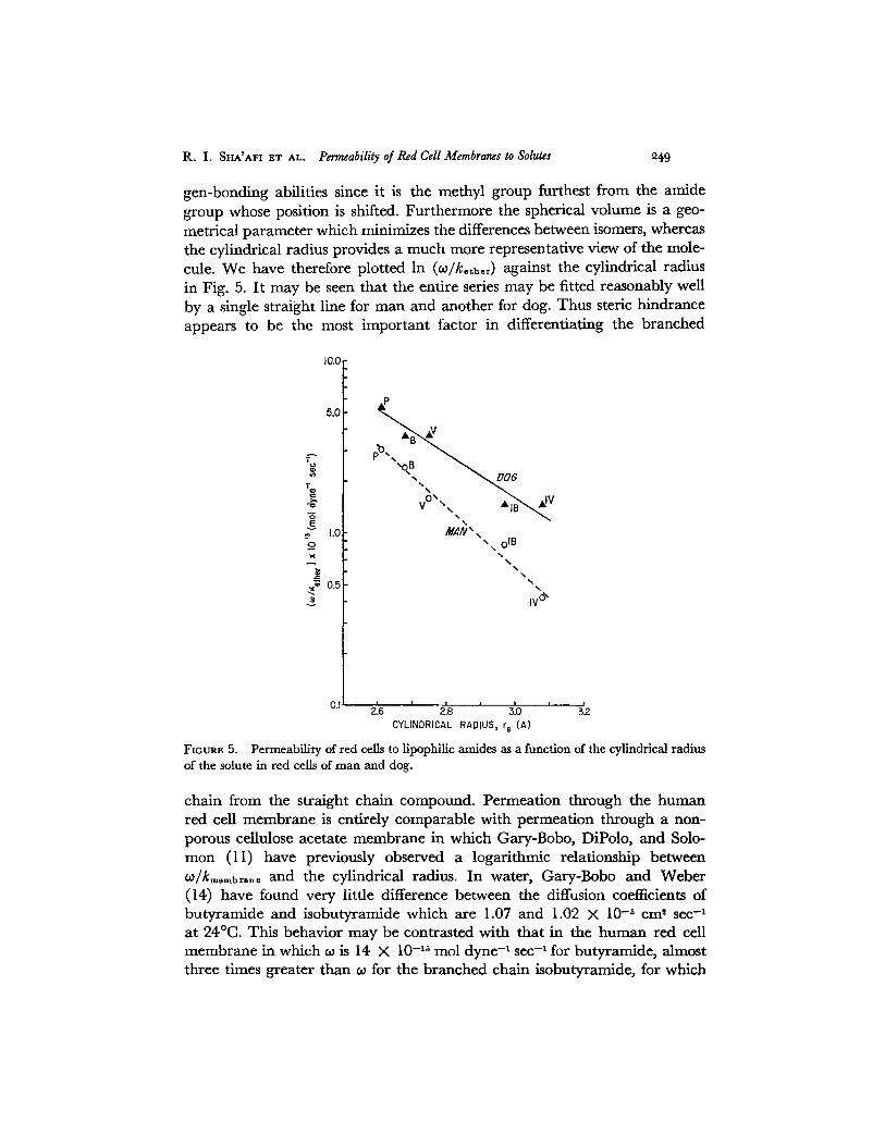
gen-bonding abilities since it is the methyl group furthest from the amidegroup whose position is shifted. Furthermore the spherical volume is a geo-metrical parameter which minimizes the differences between isomers, whereasthe cylindrical radius provides a much more representative view of the mole-cule. We have therefore plotted In (wl/kther) against the cylindrical radiusin Fig. 5. It may be seen that the entire series may be fitted reasonably wellby a single straight line for man and another for dog. Thus steric hindranceappears to be the most important factor in differentiating the branched
10.0r
7
G}
E.
o
0.5
A
V "
MAN
DOG
Nx IB\
N
I I
2.6 2.8 3.0CYLINDRICAL RADIUS, r (A)
32
FIGURE 5. Permeability of red cells to lipophilic amides as a function of the cylindrical radiusof the solute in red cells of man and dog.
chain from the straight chain compound. Permeation through the humanred cell membrane is entirely comparable with permeation through a non-porous cellulose acetate membrane in which Gary-Bobo, DiPolo, and Solo-mon (11) have previously observed a logarithmic relationship betweenco/kmembrane and the cylindrical radius. In water, Gary-Bobo and Weber(14) have found very little difference between the diffusion coefficients ofbutyramide and isobutyramide which are 1.07 and 1.02 X 10- 5 cm2 sec-'at 240C. This behavior may be contrasted with that in the human red cellmembrane in which is 14 X 10- ' 5 mol dyne-' sec- ' for butyramide, almostthree times greater than for the branched chain isobutyramide, for which
25 THE JOURNAL OF GENERAL PHYSIOLOGY · VOLUME 58 · 971
w is 5 X 10- 1 mol dyne-' sec- 1. It appears therefore that the lipids in thered cell membrane are very much less fluid than water and must be heldtogether in an organized structure.
The observation that the lines for the two species in Fig. 5 are not farfrom parallel is consistent with the view that the degree of organization inthe membranes, as reflected in the steric hindrance, is about the same. Thehigher permeability coefficients in the dog probably reflect differences inthe detailed lipid composition of the membrane leading to increased solu-bility of the amide series in the dog red cell membrane. Striking speciesdifferences in lipid content have been reported (19) which support thisview. For example, phosphatidylcholine makes up to 30-35% of the totalphospholipid in human red cell membranes as compared to 47% in the dog;in the case of sphingomyelin the figures are: 25-32% man; 11% dog.
Permeation by Diols
Two variables have been changed systematically in our study of the diolgroup. 1,3-butanediol differs from 1,3-propanediol by the addition of amethyl group. The positions of the two hydroxyl groups in the three butane-diols have been permuted to give further insight into the effect of hydrogenbonding on membrane permeation. It is instructive first to compare the be-havior of 1,3-propanediol with propionamide. These molecules have verysimilar physical properties, with molecular weights of 73.09:76.10, cylin-drical radii of 2.61:2.56 A, and kethe, of 0.013:0.012. The major differencearises from the difference in the hydrogen-bonding ability of the solute as aresult of replacing a single amide group with two hydroxyls.
Hydrogen-bonding ability is somewhat greater for amides than for alco-hols as illustrated by differences in NH, the number of possible hydrogenbonds that may be formed for the solute. Franks and Ives (20) give this num-ber as 2 for the alcohol group, whereas the most likely value for the amidegroup is 3 (12, 21). Gary-Bobo, DiPolo, and Solomon (11) have shown that aseries of amides experiences greater friction than an analogous series of alco-hols when diffusing across a nonporous cellulose acetate membrane and thatthe ratio of the frictions is about 3:2. Assuming simple additivity of hydrogen-bonding ability as a first approximation, it is apparent that the hydrogen-bonding ability of the diols should be greater than that of amides. The den-sity of 1,3-propanediol is also slightly greater than that of propionamide,though differences in density of solutes of different reactivity are less signifi-cant than differences between solutes with similar reactive groups. Sincethe hydrogen-bonding ability of 1,3-propanediol is greater than that ofpropionamide, the sharp decrease in co from 4.0 X 10 - 15 mol dyne-' sec- 1
for propionamide to 1.2 X 10 - 15 mol dyne-' sec- ' for 1,3-propanediol may
R. I. SHA'AFI ET AL. Permeability of Red Cell Membranes to Solutes 251
be attributed to an increase in solute-membrane friction due to the increasedhydrogen-bonding ability of the diol.
The effect of the addition of a methyl group as one goes from 1,3-propane-diol to 1,3-butanediol is to increase both kther and the cylindrical radius.As kether increases, o/kether decreases from 1.0 X 10-'3 mol dyne-1 sec- 1
for 1,3-propanediol to 0.48 X 10 - '1 mol dyne-' sec-1 for 1,3-butanediol.This effect is in the same direction, and of about the same magnitude, asthe steric hindrance effect on amide isomers as can be seen in Fig. 2. Thus,when hydrogen bonding is kept constant by keeping the separation betweenthe two hydroxyls the same, increases in cylindrical radius produce con-sistent effects in diols and amides alike.
However, the situation is very different when the separation of the twohydroxyls in the butanediol isomers is altered systematically. Under theseconditions, the permeability of the three butanediols, expressed as In w/keth,,in Fig. 2, increases with cylindrical radius, rather than decreasing as is thecase for the amide and urea series. More importantly the hydroxyl groupsare brought closer together as the hydroxyls are moved from the 1 ,4-positionto the 1,3- and finally reach the 2,3-isomer which is characterized by thelargest cylindrical radius. As the hydroxyl groups are brought into increas-ingly close apposition intramolecular hydrogen bonding increases at the ex-pense of the ability to form hydrogen bonds with external acceptors. Sincethe strength of the solute-membrane hydrogen bonds has decreased, thesolute diffuses through the membrane more readily and In w/keth,, increasesdespite the concomitant increase in cylindrical radius. This indicates clearlythat hydrogen bonding is a more important determinant of membrane per-meability than is cylindrical radius.
Davson and Danielli have reviewed (1) a number of earlier observationsindicating that apposition of hydroxyl groups increases solute permeability.Using the hemolysis method Jacobs (1) found that 1,2-propanediol per-meated the red cells of ox and rabbit more rapidly than 1,3-propanediol.Diamond and Wright (22) have also pointed out that the importance ofintramolecular hydrogen bonds increases as the hydroxyl groups movemore closely together and have shown that this process is reflected in a re-sultant decrease of solute reflection coefficients in the gallbladder.
Comparison of w with Permeability Coefficients Determined by the HemolysisMethod Hber and Orskov (23) measured the time of hemolysis of human redcells for a number of solutes including four whose permeability coefficients aregiven in Table I. Jacobs (24) has given an equation by which permeabilitycoefficients may be derived from hemolysis time. Use of this equation givesa value of w of 5 X 10-16 mol dyne-' sec- ' for acetamide, which is one orderof magnitude less than our value in Table I. According to Jacobs' equation,
252 THE JOURNAL OF GENERAL PHYSIOLOGY ·VOLUME 58 ·1971
w is inversely proportional to the hemolysis time, th, and is related to it by aseries of factors which are constant for any given species. In Table III wehave taken the permeability of acetamide as a standard and computed rela-tive permeability ratios both from the data in Table I and the inverse hemo-lysis times taken from Hober and Orskov. The agreement in the relativedata in these two series of quite different kinds of experiments is surprisinglygood. However, since the permeability coefficients computed from hemolysistimes differ by an order of magnitude from those obtained using rapid reac-tion techniques, it is not possible to combine data obtained by both methodsinto a single ratio, as was done, for example, by Stein (25) in a discussionof the equivalent pore radius in human red cells.
TABLE III
COMPARISON OF WITH HEMOLYSIS DATA
Relative inverse RelativeSolute Hemolysis time hemolysis time co
Acetamide 0.9 1.0 1.0
Propionamide 1.2 0.7 0.8Urea 0.3 3.0 3.0
Methyl urea 2.3 0.4 0.4
Membrane Properties Inferred from Permeability Measurements
The classical studies of Collander and Birlund (6) and Collander (26) haveprovided strong evidence that small hydrophilic molecules cross the mem-brane through a pathway different from that followed by lipophilic mole-cules. For Chara, Collander and Birlund give a figure of approximately 4 Aas the radius of the equivalent pore. These authors' evidence for the existenceof an aqueous path has been generally accepted, although Danielli (1)does not share this view. Recently, Lieb and Stein (27) have suggested thatcell membranes should be treated as homogeneous membranes in which thepermeability coefficient may be computed from an equation in which theonly variables are molecular weight and the oil-water partition coefficient.In the case of bovine red cells they have fitted permeability data obtainedfrom hemolysis measurements to the equation
P = PoBnM-" (2)
in which P is the permeability coefficient and Po, n, and p are adjustable con-stants, the oil-water partition coefficient, and M the molecular weight,relative to methanol. Lieb and Stein obtained values of 1.4 for n and 6.0 forp. The least squares fit to our data on human red cells in Table I gives the
R. I. SHA'AFI ET AL. Permeability of Red Cell Membranes to Solutes 253
following values: PO, 0.4; n, 1.0; p, 6.0, when M is used to represent molecu-lar weight. The correlation is very poor, as shown not only by the correla-tion coefficient of 0.64 but also by the very great scatter when the valuespredicted according to equation 2 are compared with the experimentallydetermined ones. There appears to be no simple physical exaplanation as towhy the molecular weight should enter the equation to the inverse sixthpower. In view of the absence of a convincing physical basis for the equationand the poor correlation of the equation with our data, we conclude thatthe Lieb and Stein treatment does not provide convincing evidence that thehuman red cell membrane behaves as a homogeneous structure.
On the contrary, the present results are consistent with the characteriza-tion of the red cell membrane in terms of equivalent pores. The primarysupport for this conclusion is given by the clear differences in the factorsgoverning the diffusion of hydrophilic and lipophilic molecules.
The equivalent pore radius was initially based on analysis of water diffusionand filtration data and of nonelectrolyte reflection coefficients in which theonly parameter considered was. steric hindrance. The present study of non-electrolyte diffusion clearly focuses attention on the importance of the physi-cal chemical factors governing membrane permeability. The study of thetemperature dependence of water diffusion and filtration, both in red bloodcells (5) and artificial membrane models (28), has already shown that chem-ical factors play an important role in addition to geometrical factors. Hence,the measurement of an equivalent pore radius must be affected by the chem-ical nature of the membrane fabric. This fact, together with the recent evi-dence of the duplex nature of the red blood cell membrane (29), makes itimpossible to interpret hydrophilic diffusion solely in terms of a quantitativeestimate of a geometrical equivalent pore radius.
Lipophilic solutes permeate by dissolution in the membrane. The parti-tion coefficient, which may be interpreted primarily as a measure of theease with which the lipophilic solute can escape from its aqueous environ-ment, is of overwhelming importance. Once the solute has entered the mem-brane, steric hindrance plays a role, as illustrated by the comparison betweenthe two pairs of lipophilic amide isomers. It is clear from these data thatthere is an organized structure within the membrane of a higher order thanthat characteristic of the liquid state and comparable to the relatively rigidmatrix of the cellulose acetate membrane. There are significant differencesin lipophilic amide diffusion in dog and man. This species specificity indi-cates that in biological membranes the lipid route cannot be consideredsimply as consisting of undifferentiated hydrocarbon layers. The study of thediols shows the importance of hydrogen bonding. From this we infer thatlipid-soluble solutes traverse a path that comes into contact with polar moie-ties within the membrane, such as are provided by phosphatides or proteins.
254 THE JOURNAL OF GENERAL PHYSIOLOGY ·VOLUME 58 · 1971
An entirely different situation prevails in the case of hydrophilic solutes.For molecules with kether smaller than that for water, the partition coefficientdoes not play any part. This is not surprising, for if these molecules cross anaqueous path, the only relevant ratio would be the partition ratio betweenthe water in the pathway and the water outside. Although this ratio is notknown, there is reason to think that it plays a significant part, inasmuch asthe solubility of nonelectrolytes in water in close contact with macromolec-ular surfaces may be very different from their solubility in the bulk phase.The predominant diffusion factor is steric hindrance. The extreme sensitiv-ity to molecular size and shape reflects not only the geometry of the aqueouspore but also molecular solubility in the water contained in the pore. Thesegeometrical constraints are further modulated by chemical factors as indi-cated by the role of hydrogen bonding.
Further evidence for the reality of aqueous paths and the effect of hydro-gen bonding is afforded by the results of Holz and Finkelstein (30) on non-electrolyte diffusion through aqueous pores induced by nystatin in thinlipid membranes. These authors have measured diffusion coefficients throughthe membrane of: H 2 0, urea, thiourea, ethylene glycol, and glycerol. Wehave plotted the natural log of their diffusion coefficients against molarvolume and have found a reasonable linear fit, much closer than the fitobserved when the same data are plotted against the cylindrical radius.
An increasing amount of evidence thus appears to support the porous modelof biological membranes. The concept of the equivalent pore, which ini-tially was purely geometrical, has been expanded by detailed considerationof further physical chemical interactions, which make it a functional entitybetter able to perform its multiple and complex physiological task.
APPENDIX
Frictional Interpretation of Phenomenological Coefficients
DiPolo, Sha'afi, and Solomon (18) have pointed out that the partition coefficientand hydrogen bonding exercise opposing actions on and a as measured in a porouscellulose acetate membrane. Phenomenologically w and a are entirely independentparameters. However, according to the frictional interpretation of phenomenologicalcoefficients (see reference 15) co and can be related to one another by the equation
(1 - a - Vs/L/,)/t = J.f (/Ax (3)
in which PV is partial molar volume of the solute and L,, the hydraulic conductivity.Kedem and Katchalsky (see reference 15) have pointed out that in a very loosemembrane f, approaches the value for free solution J, which is related to the dif-fusion coefficient according to D = RT/Jo. However the relationship between f,and D both in the red cell and in porous cellophane membranes is complex and verydifficult to understand. This means that the friction between solute and water in
R. I. SHA'AFI ET AL. Permeability of Red Cell Membranes to Solutes 255
aqueous pathways is very sensitive to the specific chemical and physical properties ofmembrane and solute, as has been discussed extensively in the main body of thispaper.
In the present study, values of r have been determined for four of the solutes whosei's are given in Table I. Table IV contains a comparison of the values computed forf,,, QP/x from these data with the diffusion coefficients in water for these four solutes
FIGUR 6. Relation between solute-water friction in porous membranes and solute diffusioncoefficients in water. G stands for glycerol and EG for ethylene glycol.
at 25°C. Since L, and are known functions of osmolality, and the parameters werenot measured at identical osmolalities, the comparison is qualitative. On the basis ofthe reasonable assumption that Ax and (D remain essentially constant during all themeasurements of a, , and L,, f,, should be inversely proportional to the diffusioncoefficient, except as modified by molecular size. Ginzburg and Katchalsky (31) havemeasuredy, for a series of solutes that penetrate porous artificial membranes throughaqueous channels and have shown that steric factors play an important and consistent
256 THE JOURNAL OF GENERAL PHYSIOLOGY ·VOLUME 58 · I971
role in this frictional term. This is not the case for the four molecules in Table IV.The acetamide-propionamide pair are particularly puzzling since propionamide islarger than acetamide and yet the frictional term is smaller by a factor of two. Thisis not to be attributed to that moiety of propionamide that dissolves in the membranefabric since the introduction of the term, wV,/L, should take account of that con-tribution.
The three solutes, acetamide, methyl urea, and urea, are all in the hydrophilicclass and might therefore be expected to behave consistently. Even this expectationis not satisfied since urea, whose cylindrical radius is approximately equal to that ofacetamide, is characterized by a very much smaller value off8 ,,A x/,', correspondingto the very large difference in in Table I.
We have also examined the relationship of a to co using the data from measurementson a porous cellulose acetate membrane given by DiPolo, Sha'afi, and Solomon(18). Their data were obtained on a thin porous membrane in which Ax and (, couldeach be measured.f,Ax/~4 is plotted in Fig. 6 as a function of D, for some membersof the amide series and for glycerol and ethylene glycol for which kether is 0.00066 and0.0053, respectively. The frictional term for the amide series decreases with decreasein molecular size, as expected on steric grounds. The frictional term for ethyleneglycol is greater than that for glycerol, a larger molecule. The two series fall on dif-ferent parts of the graph. This diversity of behavior in an artificial membrane onlyunderlines the fact that the diffusion coefficient in water does not bear a predictiverelationship tof,,, the solute-water friction in the membrane. In other words, evenin an artificial membrane, it is not possible to obtain reliable estimates of w frommeasurements of a.
The situation is even more complicated in red cells because evidence has beenpresented by Sha'afi et al. (2) and by Rich et al. (29) indicating that the human redcell membrane acts as a series membrane so that equation 3 would not necessarily beexpected to apply to this membrane. The point to be stressed is that the frictionaltreatment does not have any predictive value in the case of the human red cell mem-brane. The clear implication of this observation is that grave reservations must bemaintained about the results of studies in which a- is used as a measure of membranepermeability as has been done, for example, by Diamond and Wright (22) and Wrightand Prather (32).
This project has been supported in part by the Atomic Energy Commission.Dr. Gillian Rich collaborated in the early phases of this study and made many valuable contribu-tions.
We should like to express our thanks to Miss Sandra Czekanski for devoted technical assistance.
Received for publication 28 December 1970.
REFERENCES
1. DANIELLI, J. F. 1952. Permeability to non-electrolytes. The Permeability of NaturalMembranes. H. Davson and J. F. Danielli, editors. University Press, Cambridge,England. 80.
2. SHA'AFI, R. I., G. T. RICH, D. C. MIKULECKY, and A. K. SOLOMON. 1970. Determinationof urea permeability in red cells by minimum method. A test of the phenomenologicalequations. J. Gen. Physiol. 55:427.
R. I. SHA'AFI ET AL. Permeability of Red Cell Membranes to Solutes 257
3. SHA'AFI, R. I., G. T. RICH, V. W. SIDEL, W. BOSSERT, and A. K. SOLOMON. With an ap-pendix by A. Pandiscio. 1967. The effect of the unstirred layer on the human red cellwater permeability. J. Gen. Physiol. 50:1377.
4. GOLDSTEIN, D. A., and A. K. SOLOMON. 1960. Determination of equivalent pore radius forhuman red cell by osmotic pressure measurement. J. Gen. Physiol. 44:1.
5. VIEIRA, F. L., R. I. SHA'AFI, and A. K. SOLOMON. 1970. The state of water in human anddog red cell membranes. J. Gen. Physiol. 55:451.
6. COLLANDER, R., and H. BARLUND. 1933. PermeabilitAts Studien an Chara Ceratophylla.Acta Bot. Fenn. 11:1.
7. COLLANDER, R. 1949. Die Verteilung organischer Verbindungen zwischen Ather undWasser. Acta Chem. Scand. 3:717.
8. COLLANDER, R. 1954. The permeability of Nitella cells to non-electrolytes. Physiol. Plant.7:420.
9. HANSCH, C., J. E. QUINLAN, and G. L. LAWRENCE. 1968. The linear free-energy relation-ship between partition coefficients and the aqueous solubility of organic liquid. J. Org.Chem. 33:347.
10. SOLL, A. H. 1967. A new approach to molecular configuration applied to aqueous poretransport. J. Gen. Physiol. 50:2565.
11. GARY-BOBO, C. M., R. DIPOLO, and A. K. SOLOMON. 1969. Role of hydrogen-bonding innonelectrolyte diffusion through dense artificial membranes. J. Gen. Physiol. 54:369.
12. PMENTEL, G. C., and A. L. MCCLELLAN. 1960. The Hydrogen Bond. W. H. Freemanand Company, San Francisco.
13. HoRowITZ, S. B., and I. R. FENICHEL. 1964. Solute diffusional specificity in hydrogen-bonding systems. J. Phys. Chem. 68:3378.
14. GARY-BOBo, C. M., and H. W. WEBER. 1969. Diffusion of alcohols and amides in waterfrom 4 to 37 ° . J. Phys. Chem. 73:1155.
15. KATCHALSKY, A., and P. F. CURRAN. 1965. Nonequilibrium Thermodynamics in Bio-physics. Harvard University Press, Cambridge, Mass. 113.
16. SOLOMON, A. K. 1968. Characterization of biological membranes by equivalent pores. J.Gen. Physiol. 51:335.
17. GARY-BOBO, C. M., and A. B. LINDENBERG. 1961. Interpretation physico-chimique de ladistribution globulo-plasmatique variable de l'uree eh fonction de la concentration. J.Physiol. (Paris). 53:347.
18. DIPOLO, R., R. I. SHA'AFI, and A. K. SOLOMON. 1970. Transport parameters in a porouscellulose acetate membrane. J. Gen. Physiol. 55:63.
19. ROUSER, G., G. J. NELSON, S. FLEISCHER, and F. SIMON. 1968. Lipid composition of animalcell membranes, organelles and organs. In Biological Membranes Physical Fact andFunction. D. Chapman, editor. Academic Press, Inc., London. 24.
20. FRANKS, F., and D. J. G. IvEs. 1966. The structural properties of alcohol-water mixtures.Quart. Rev. 20:1.
21. BATES, W. W., and M. E. HOBBS. 1951. The dipole moments of some acid amides and thestructure of the amide group. J. Amer. Chem. Soc. 73:2151.
22. DIAMOND, J. M., and E. M. WRIGHT. 1968. Molecular forces governing non-electrolytepermeation through cell membranes. Proc. Roy. Soc. Ser. B. Biol. Sci. 172:273.
23. HBER, R., and S. L. ORSKOV. 1933. Untersuchungen fiber die Permeiergeschwindigkeitvon Anelektrolyten bei den roten Blutk6rperchen verschiedener Tierarten. Pfluegers Arch.Gesamte Physiol. Menschen Tiere. 231:599.
24. JACOBS, M. H. 1952. The measurement of cell permeability with particular reference to theerythrocyte. Mod. Trends Physiol. Biochem. 149.
25. STEIN, W. D. 1967. The Movement of Molecules across Cell Membranes. AcademicPress, Inc., New York. Fig. 3-16, p. 113.
26. COLLANDER, R. 1949. The permeability of plant protoplasts to small molecules. Physiol.Plant. 2:300.
27. LIEB, W. R., and W. D. STEIN. 1969. Biological membranes behave as non-porous poly-meric sheets with respect to the diffusion of non-electrolytes. Nature (London). 224:240.
258 THE JOURNAL OF GENERAL PHYSIOLOGY VOLUME 58 · 1971
28. GARY-BOBO, C. M., and A. K. SOLOMON. 1971. Effect of geometrical and chemical con-straints on water flux across artificial membranes. J. Gen. Physiol. 57:610.
29. RICH, G. T., R. I. SHA'AFI, A. ROMUALDEZ, and A. K. SOLOMON. 1968. Effect of osmolalityon the hydraulic permeability coefficient of red cells. J. Gen. Physiol. 52:941.
30. HOLZ, R., and A. FINKELSTEIN. 1970. The water and nonelectrolyte permeability inducedin thin lipid membranes by the polyene antibiotics nystatin and amphotericin B. J.Gen. Physiol. 56:125.
31. GINZBURG, B. Z., and A. KATCHALSKY. 1963. The frictional coefficients of the flows ofnon-electrolytes through artificial membranes. J. Gen. Physiol. 47:403.
32. WRIGHT, E. M., and J. W. PRATHER. 1970. The permeability of the frog choroid plexusto nonelectrolytes. J. Membrane Biol. 2:127.