Page 1
REVISED, EMBO-2011-77375
Supplementary Information
Nedd4-1 Binds and Ubiquitylates Activated FGFR1 to Control its Endocytosis
and Function
Avinash Persaud1*
, Philipp Alberts1*
, Madeline Hayes1, Sebastian Guettler
2, Ian Clarke
1, Frank
Sicheri2, Peter Dirks
1, Brian Ciruna
1 and Daniela Rotin
1#
1Programs in Cell Biology and Developmental & Stem Cell Biology, The Hospital for Sick
Children, 2Samuel Lunenfeld Research Institute, Mt. Sinai Hospital, Toronto, and
1,2Departments
of Biochemistry and Molecular Genetics, University of Toronto, Toronto, Canada.
#Corresponding author:
Dr. Daniela Rotin
Program in Cell Biology
The Hospital for Sick Children
MaRS-TMDT, 11-305, 101 College St.
Toronto, Ont. M5G 1L7
Te: 416-813-5098
FAX:416-813-8456
Email:[email protected]
Page 2
2
Supplementary Figure Legends:
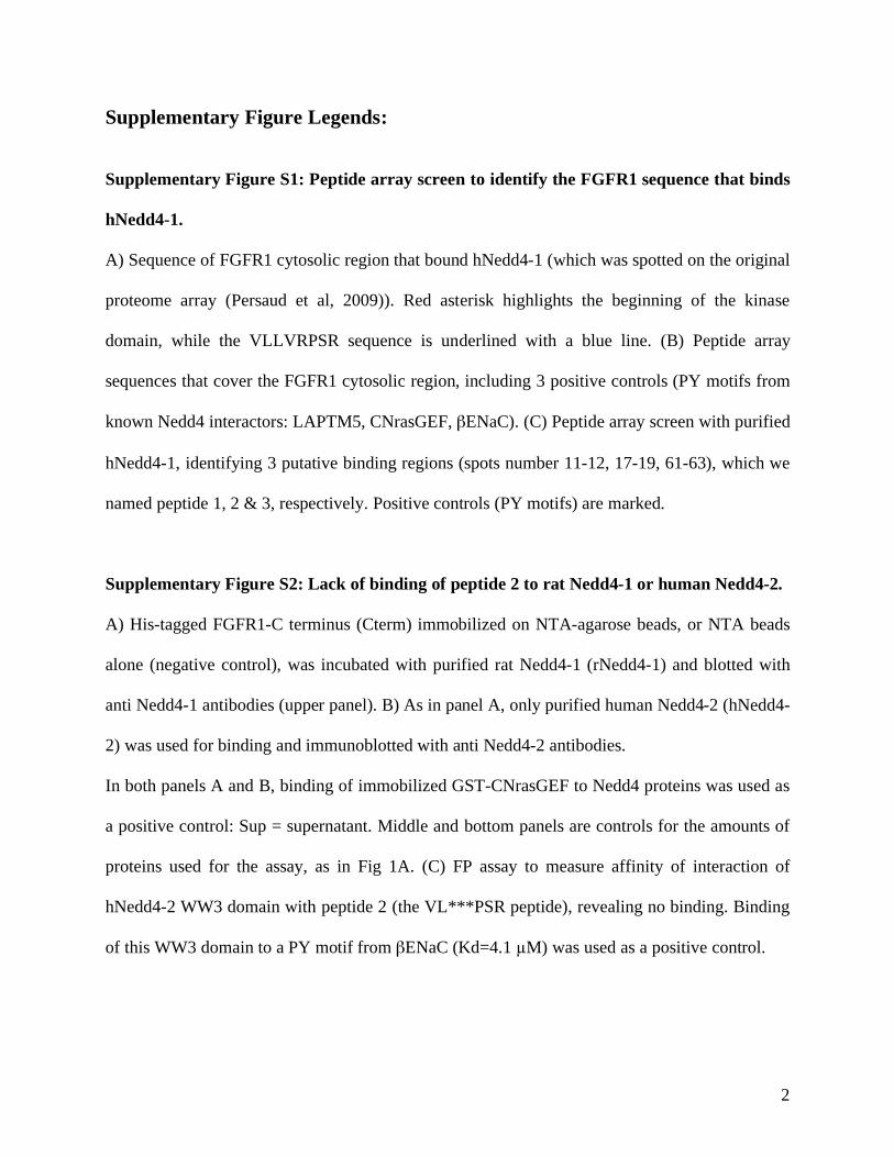
Supplementary Figure S1: Peptide array screen to identify the FGFR1 sequence that binds
hNedd4-1.
A) Sequence of FGFR1 cytosolic region that bound hNedd4-1 (which was spotted on the original
proteome array (Persaud et al, 2009)). Red asterisk highlights the beginning of the kinase
domain, while the VLLVRPSR sequence is underlined with a blue line. (B) Peptide array
sequences that cover the FGFR1 cytosolic region, including 3 positive controls (PY motifs from
known Nedd4 interactors: LAPTM5, CNrasGEF, ENaC). (C) Peptide array screen with purified
hNedd4-1, identifying 3 putative binding regions (spots number 11-12, 17-19, 61-63), which we
named peptide 1, 2 & 3, respectively. Positive controls (PY motifs) are marked.
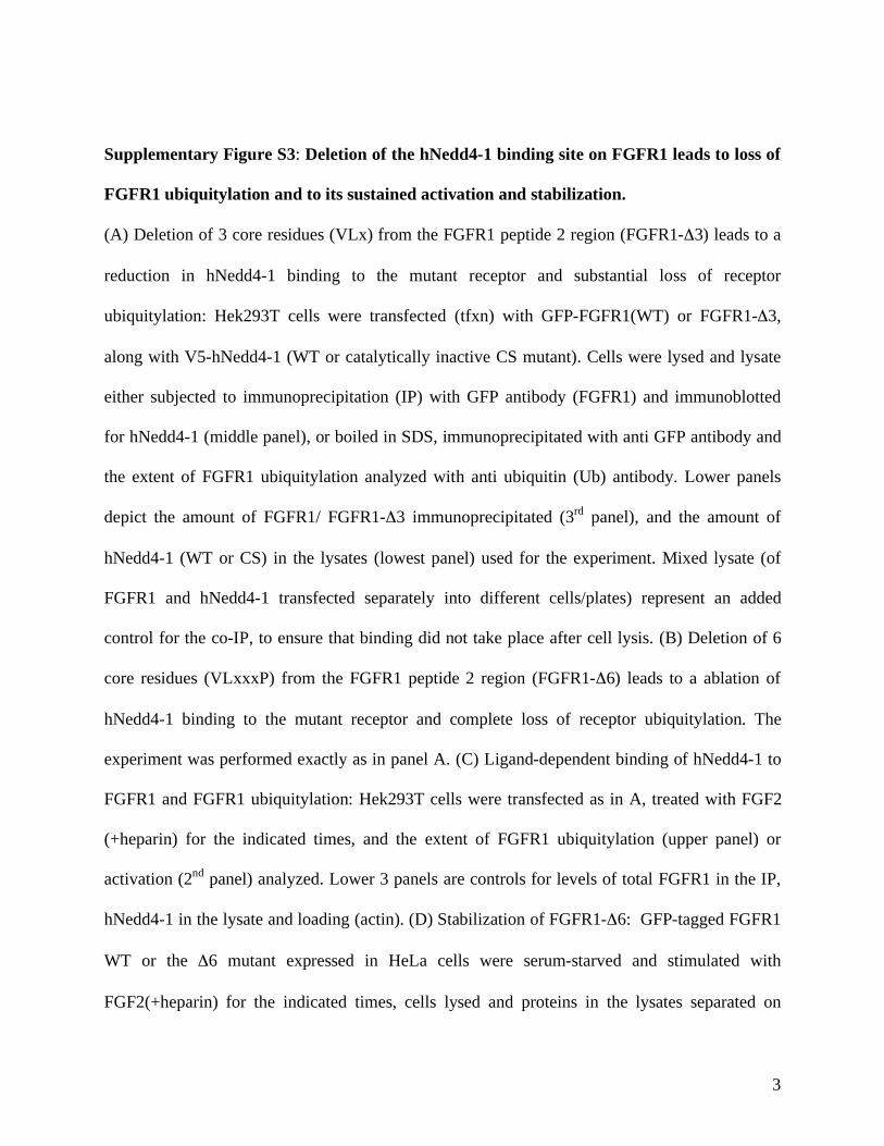
Supplementary Figure S2: Lack of binding of peptide 2 to rat Nedd4-1 or human Nedd4-2.
A) His-tagged FGFR1-C terminus (Cterm) immobilized on NTA-agarose beads, or NTA beads
alone (negative control), was incubated with purified rat Nedd4-1 (rNedd4-1) and blotted with
anti Nedd4-1 antibodies (upper panel). B) As in panel A, only purified human Nedd4-2 (hNedd4-
2) was used for binding and immunoblotted with anti Nedd4-2 antibodies.
In both panels A and B, binding of immobilized GST-CNrasGEF to Nedd4 proteins was used as
a positive control: Sup = supernatant. Middle and bottom panels are controls for the amounts of
proteins used for the assay, as in Fig 1A. (C) FP assay to measure affinity of interaction of
hNedd4-2 WW3 domain with peptide 2 (the VL***PSR peptide), revealing no binding. Binding
of this WW3 domain to a PY motif from ENaC (Kd=4.1 M) was used as a positive control.
Page 3
3
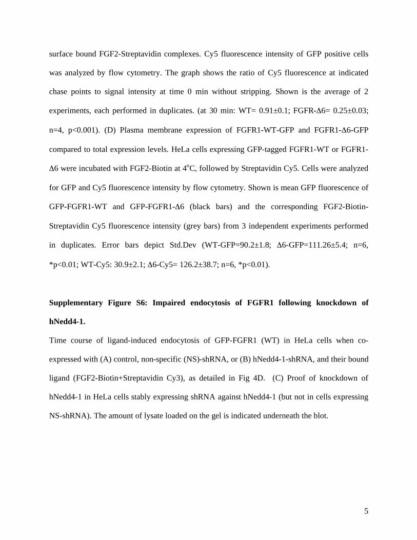
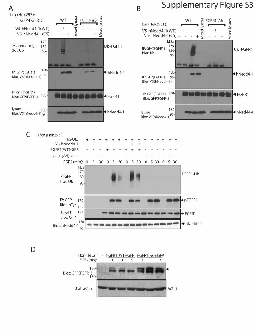
Supplementary Figure S3: Deletion of the hNedd4-1 binding site on FGFR1 leads to loss of
FGFR1 ubiquitylation and to its sustained activation and stabilization.
(A) Deletion of 3 core residues (VLx) from the FGFR1 peptide 2 region (FGFR1- 3) leads to a
reduction in hNedd4-1 binding to the mutant receptor and substantial loss of receptor
ubiquitylation: Hek293T cells were transfected (tfxn) with GFP-FGFR1(WT) or FGFR1- 3,
along with V5-hNedd4-1 (WT or catalytically inactive CS mutant). Cells were lysed and lysate
either subjected to immunoprecipitation (IP) with GFP antibody (FGFR1) and immunoblotted
for hNedd4-1 (middle panel), or boiled in SDS, immunoprecipitated with anti GFP antibody and
the extent of FGFR1 ubiquitylation analyzed with anti ubiquitin (Ub) antibody. Lower panels
depict the amount of FGFR1/ FGFR1- 3 immunoprecipitated (3rd
panel), and the amount of
hNedd4-1 (WT or CS) in the lysates (lowest panel) used for the experiment. Mixed lysate (of
FGFR1 and hNedd4-1 transfected separately into different cells/plates) represent an added
control for the co-IP, to ensure that binding did not take place after cell lysis. (B) Deletion of 6
core residues (VLxxxP) from the FGFR1 peptide 2 region (FGFR1- 6) leads to a ablation of
hNedd4-1 binding to the mutant receptor and complete loss of receptor ubiquitylation. The
experiment was performed exactly as in panel A. (C) Ligand-dependent binding of hNedd4-1 to
FGFR1 and FGFR1 ubiquitylation: Hek293T cells were transfected as in A, treated with FGF2
(+heparin) for the indicated times, and the extent of FGFR1 ubiquitylation (upper panel) or
activation (2nd
panel) analyzed. Lower 3 panels are controls for levels of total FGFR1 in the IP,
hNedd4-1 in the lysate and loading (actin). (D) Stabilization of FGFR1- 6: GFP-tagged FGFR1
WT or the 6 mutant expressed in HeLa cells were serum-starved and stimulated with
FGF2(+heparin) for the indicated times, cells lysed and proteins in the lysates separated on
Page 4
4
8%SDS-PAGE and immunoblotted for GFP (FGFR1) or actin. Note the disappearance of the
upper band (arrow) over time in the WT FGFR1, but its stabilization in the FGFR1- 6 mutant.
This upper band represents the cell-surface pool of FGFR1, based on our previous work.
Supplementary Figure S4: Cbl knockdown does not affect stability of active FGFR1, and
normal binding of FGFR1- 6 to FRS2 and PLC .
(A) Knockdown of both cCbl and CblB with siRNA in HeLa cells has no effect on stability of
activated (Tyr-phosphorylated) FGFR1: Cells were transfected with GFP-tagged FGFR1 and
incubated with siRNAs for cCbl and CblB. FGFR1 was then subjected to IP with anti GFP
antibodies (FGFR1) and blotted with anti pTyr or GFP antibodies. Lower 3 panels are controls
for loading (actin), and for demonstration of knockdown of cCbl and CblB. (B) Activated
FGFR1- 6 can bind its substrates FRS2 and PLC : HeLa cells were transfected with GFP-
tagged FGFR1-WT, 6 or the kinase inactive (KI) mutants. Cells were serum-starved and then
stimulated (or not) with FGF2(+heparin) for 5 min, and co-IP (and Tyr phosphorylation) of their
targets FRS2 and PLC analyzed. Note similar co-IP of these substrates between WT and the
6 mutant, but not with the kinase inactive mutant.
Supplementary Figure S5: Impaired endocytosis of FGFR1- 6.
Time course of ligand-induced endocytosis of (A) GFP-FGFR1-WT or (B) GFP-FGFR1- 6 and
their bound ligand (FGF2-Biotin+Streptavidin Cy3), as detailed in Fig 4B. (C) Intracellular
accumulation of FGF2 in HeLa cells expressing GFP-FGFR1-WT or GFP-FGFR1- 6. Cells
were pulsed with FGF2-Biotin on ice, followed by Streptavidin Cy5 incubation. Cells were then
incubated at 37oC for the indicated timepoints followed by acid stripping to remove remaining
Page 5
5
surface bound FGF2-Streptavidin complexes. Cy5 fluorescence intensity of GFP positive cells
was analyzed by flow cytometry. The graph shows the ratio of Cy5 fluorescence at indicated
chase points to signal intensity at time 0 min without stripping. Shown is the average of 2
experiments, each performed in duplicates. (at 30 min: WT= 0.91±0.1; FGFR- 6= 0.25±0.03;
n=4, p<0.001). (D) Plasma membrane expression of FGFR1-WT-GFP and FGFR1- 6-GFP
compared to total expression levels. HeLa cells expressing GFP-tagged FGFR1-WT or FGFR1-
6 were incubated with FGF2-Biotin at 4oC, followed by Streptavidin Cy5. Cells were analyzed
for GFP and Cy5 fluorescence intensity by flow cytometry. Shown is mean GFP fluorescence of
GFP-FGFR1-WT and GFP-FGFR1- 6 (black bars) and the corresponding FGF2-Biotin-
Streptavidin Cy5 fluorescence intensity (grey bars) from 3 independent experiments performed
in duplicates. Error bars depict Std.Dev (WT-GFP=90.2±1.8; 6-GFP=111.26±5.4; n=6,
*p<0.01; WT-Cy5: 30.9±2.1; 6-Cy5= 126.2±38.7; n=6, *p<0.01).
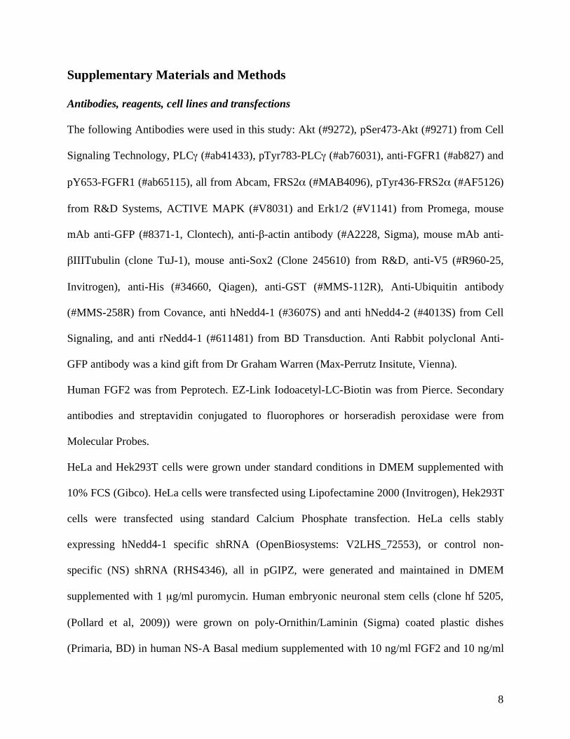
Supplementary Figure S6: Impaired endocytosis of FGFR1 following knockdown of
hNedd4-1.
Time course of ligand-induced endocytosis of GFP-FGFR1 (WT) in HeLa cells when co-
expressed with (A) control, non-specific (NS)-shRNA, or (B) hNedd4-1-shRNA, and their bound
ligand (FGF2-Biotin+Streptavidin Cy3), as detailed in Fig 4D. (C) Proof of knockdown of
hNedd4-1 in HeLa cells stably expressing shRNA against hNedd4-1 (but not in cells expressing
NS-shRNA). The amount of lysate loaded on the gel is indicated underneath the blot.
Page 6
6
Supplementary Figure S7: Transient knockdown of hNedd4-1 in neural embryonic stem
cells, lack of binding of FGFR3 to hNedd4-1, and maternal deposition of zNedd4-1 in
zebrafish embryos.
(A) Knockdown of hNedd4-1 in human embryonic neural stem cells with shRNA is transient and
normal expression of hNedd4-1 is restored by day 7 post knockdown. NS: non-specific shRNA
control. (B) FGFR3 does not bind hNedd4-1. HeLa cells were transfected with V5-hNedd4-1
plus FGFR1-GFP or FGFR3-Flag, serum-starved and treated (or not) with FGF2(+heparin).
Cells were then lysed, hNedd4-1 immunoprecipitated and co-IP of FGFR3 or FGFR1 analyzed,
as indicated. Note that while both FGFR1 and FGFR3 are activated (as evident from their
activation of Akt (pAkt)), only FGFR1, but not FGFR3, can bind hNedd4-1. (C) Maternally
deposited zNedd4a (zNedd4-1) transcript in early stage embryos. Zebrafish zNedd4a transcript
was detected in 1-cell stage, 8-cell stage, and 1000-cell stage embryos (prior to onset of zygotic
transcription), following reverse transcriptase PCR from total RNA samples. The transcript size
is 794 base pairs. DNA standard, 100bp ladder.
Page 7
7
Supplementary Table:
Table SI: Human proteins that contain VL***PSR motif
Name Known or predicted features/function
DHX33_HUMAN Putative ATP-dependent RNA helicase
DHX33
FGFR1_HUMAN Fibroblast Growth Factor Receptor 1
FRMD1_HUMAN FERM domain-containing protein 1
LRC8A_HUMAN Leucine-rich repeat-containing protein 8A
LRIT3_HUMAN Leucine-rich repeat, immunoglobulin-like
domain and transmembrane domain-containing
protein 3
MAST2_HUMAN Microtubule-associated serine/threonine-
protein kinase 2
PHLB1_HUMAN Pleckstrin homology-like domain family B
member 1
TM209_HUMAN Transmembrane protein 209
WDR18_HUMAN WD repeat-containing protein 18
ZC12D_HUMAN Probable ribonuclease ZC3H12D
Page 8
8
Supplementary Materials and Methods
Antibodies, reagents, cell lines and transfections
The following Antibodies were used in this study: Akt (#9272), pSer473-Akt (#9271) from Cell
Signaling Technology, PLC (#ab41433), pTyr783-PLC (#ab76031), anti-FGFR1 (#ab827) and
pY653-FGFR1 (#ab65115), all from Abcam, FRS2 (#MAB4096), pTyr436-FRS2 (#AF5126)
from R&D Systems, ACTIVE MAPK (#V8031) and Erk1/2 (#V1141) from Promega, mouse
mAb anti-GFP (#8371-1, Clontech), anti- -actin antibody (#A2228, Sigma), mouse mAb anti-
IIITubulin (clone TuJ-1), mouse anti-Sox2 (Clone 245610) from R&D, anti-V5 (#R960-25,
Invitrogen), anti-His (#34660, Qiagen), anti-GST (#MMS-112R), Anti-Ubiquitin antibody
(#MMS-258R) from Covance, anti hNedd4-1 (#3607S) and anti hNedd4-2 (#4013S) from Cell
Signaling, and anti rNedd4-1 (#611481) from BD Transduction. Anti Rabbit polyclonal Anti-
GFP antibody was a kind gift from Dr Graham Warren (Max-Perrutz Insitute, Vienna).
Human FGF2 was from Peprotech. EZ-Link Iodoacetyl-LC-Biotin was from Pierce. Secondary
antibodies and streptavidin conjugated to fluorophores or horseradish peroxidase were from
Molecular Probes.
HeLa and Hek293T cells were grown under standard conditions in DMEM supplemented with
10% FCS (Gibco). HeLa cells were transfected using Lipofectamine 2000 (Invitrogen), Hek293T
cells were transfected using standard Calcium Phosphate transfection. HeLa cells stably
expressing hNedd4-1 specific shRNA (OpenBiosystems: V2LHS_72553), or control non-
specific (NS) shRNA (RHS4346), all in pGIPZ, were generated and maintained in DMEM
supplemented with 1 μg/ml puromycin. Human embryonic neuronal stem cells (clone hf 5205,
(Pollard et al, 2009)) were grown on poly-Ornithin/Laminin (Sigma) coated plastic dishes
(Primaria, BD) in human NS-A Basal medium supplemented with 10 ng/ml FGF2 and 10 ng/ml
Page 9
9
EGF and 2 μg/ml Heparin (all from Stem Cell Technologies) according to published procedures
(Sun et al, 2008). Transfection of neuronal stem cells was achieved using the Amaxa
nucleofector device and corresponding mouse neuronal stem cells kit (#VPG-1004).
cDNA constructs
Human FGFR1 cDNA (AC#BC015035) was sub-cloned into pcDNA6.2emGFP using the
Gateway system (Invitrogen). The deletion mutants hFGFR1- 3-GFP (439-441), hFGFR1- 6-
GFP (439-444) and hFGFR1-KI-GFP (Y653F/Y654F, kinase inactive (Mohammadi et al, 1996))
were created using a Site Directed Mutagenesis kit (Qiagen) with WT hFGFR1-GFP in
pcDNA6.2emGFP as template. Zebrafish FGFR1was amplified from Zebrafish cDNA library
using primers designed against the 5’ and 3’ ends of zFGFR1 (NM_152962). zFGFR1 cDNA
was cloned into pDONR221 (Invitrogen) and then sub-cloned into the expression vector
pCSDEST2-GFP (Invitrogen) using the Gateway system. zFGFR1- 6 (424-429) was created
using Site Directed Mutagenesis kit. Zebrafish Nedd4a WW3 (a.a.429-467) was amplified from
the pDONR221-zFGFR1 template and zNedd4L WW3 was created by mutating residues T500S
and K501R in hNedd4-2 WW3 domain (a.a.476-514). Both zNedd4a and zNedd4L WW3
domains were subcloned into the bacterial expression vector pETMHtb.
Purification of epitope tagged and fusion proteins for in-vitro experiments
hFGFR1 C terminus residues 399-616 (in pQE30), hNedd4-1 WW1, WW2, WW3, WW4
domains (in pQE30), hNedd4-1 Hect domain (in pGEX6p-1) and hNedd4-2 WW3 domain (in
pQE30) were expressed in E.coli strain BL21 (RIL) as described (Persaud et al, 2009). Proteins
were purified from 100 ml of growth medium using either glutathione–sepharose resin (for GST
Page 10
10
tagged proteins) or Ni2+
NTA resin (for 6xHis tagged proteins) and eluted using either 80 mM
reduced glutathione (Amersham Biosciences) or 80 mM Imidazole (Sigma), respectively. The C
terminus of CNrasGEF (PY motif containing protein and a known substrate for Nedd4 (Pham &
Rotin, 2001)), hNedd4-1, hNedd4-2 and rNedd4-1 were also purified as described (Persaud et al,
2009).
In-vitro binding experiments
All binding reactions were conducted at 4°C in PBS supplemented with 1 mM DTT for 2h.
Purified 6xHis-hFGFR1 C terminus (residues 399-616) (0.5 g) and GST-CNrasGEF C terminus
(0.5 g) immobilized on Ni2+
NTA and glutathione–sepharose resin, respectively, was incubated
with purified hNedd4-1, hNedd4-2 or rNedd4-1 (0.5 g). Biotinylated peptides 1, 2, 3 and
ENaC PY motif peptide (10 g) immobilized on streptavidin agarose were incubated with 10
g purified hNedd4-1, and to detect binding of peptide 2 to hNedd4-1 domains, biotinylated
peptide 2 (10 g) immobilized on streptavidin agarose was incubated with 10 g of purified
hNedd4-1 6xHis WW1, WW2, WW3, WW4 or GST-Hect domains. For the reciprocal
experiment, the hNedd4-1 domains were precipitated using either glutathione–sepharose or Ni2+
NTA beads and binding to biotinylated peptide 2 was determined. In addition, biotinylated
zebrafish peptide 2 (10 g) immobilized on streptavidin agarose was incubated with 10 g
purified zNedd4a or zNedd4L 6xHis-GST-WW3 domains to detect binding to these domains;
binding of hFGFR1 peptide 2 (10 g) to hNedd4-1 6xHis-GST-WW3 domains was used as a
positive control. All samples were washed (x2) with HNTG (50 mM HEPES (pH 7.5), 150 mM
NaCl, 0.1% Triton X-100, 10% glycerol). Binding was detected by immunoblotting with anti-
Page 11
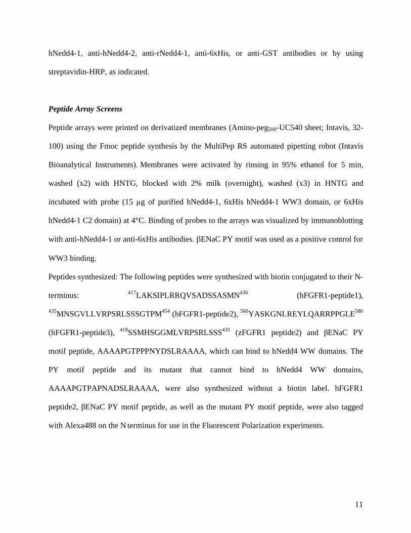
11
hNedd4-1, anti-hNedd4-2, anti-rNedd4-1, anti-6xHis, or anti-GST antibodies or by using
streptavidin-HRP, as indicated.
Peptide Array Screens
Peptide arrays were printed on derivatized membranes (Amino-peg500-UC540 sheet; Intavis, 32-
100) using the Fmoc peptide synthesis by the MultiPep RS automated pipetting robot (Intavis
Bioanalytical Instruments). Membranes were activated by rinsing in 95% ethanol for 5 min,
washed (x2) with HNTG, blocked with 2% milk (overnight), washed (x3) in HNTG and
incubated with probe (15 g of purified hNedd4-1, 6xHis hNedd4-1 WW3 domain, or 6xHis
hNedd4-1 C2 domain) at 4°C. Binding of probes to the arrays was visualized by immunoblotting
with anti-hNedd4-1 or anti-6xHis antibodies. ENaC PY motif was used as a positive control for
WW3 binding.
Peptides synthesized: The following peptides were synthesized with biotin conjugated to their N-
terminus: 417
LAKSIPLRRQVSADSSASMN436
(hFGFR1-peptide1),
435MNSGVLLVRPSRLSSSGTPM
454 (hFGFR1-peptide2),
560YASKGNLREYLQARRPPGLE
580
(hFGFR1-peptide3), 418
SSMHSGGMLVRPSRLSSS435
(zFGFR1 peptide2) and ENaC PY
motif peptide, AAAAPGTPPPNYDSLRAAAA, which can bind to hNedd4 WW domains. The
PY motif peptide and its mutant that cannot bind to hNedd4 WW domains,
AAAAPGTPAPNADSLRAAAA, were also synthesized without a biotin label. hFGFR1
peptide2, ENaC PY motif peptide, as well as the mutant PY motif peptide, were also tagged
with Alexa488 on the N terminus for use in the Fluorescent Polarization experiments.
Page 12
12
Supplementary References
Mohammadi M, Schlessinger J, Hubbard SR (1996) Structure of the FGF receptor tyrosine
kinase domain reveals a novel autoinhibitory mechanism. Cell 86: 577-587
Persaud A, Alberts P, Amsen EM, Xiong X, Wasmuth J, Saadon Z, Fladd C, Parkinson J, Rotin
D (2009) Comparison of substrate specificity of the ubiquitin ligases Nedd4 and Nedd4-2 using
proteome arrays. Mol Syst Biol 5: 333
Pham N, Rotin D (2001) Nedd4 regulates ubiquitination and stability of the guanine-nucleotide
exchange factor CNrasGEF. J Biol Chem 276: 46995-47003
Pollard SM, Yoshikawa K, Clarke ID, Danovi D, Stricker S, Russell R, Bayani J, Head R, Lee
M, Bernstein M, Squire JA, Smith A, Dirks P (2009) Glioma stem cell lines expanded in
adherent culture have tumor-specific phenotypes and are suitable for chemical and genetic
screens. Cell Stem Cell 4: 568-580
Sun Y, Pollard S, Conti L, Toselli M, Biella G, Parkin G, Willatt L, Falk A, Cattaneo E, Smith A
(2008) Long-term tripotent differentiation capacity of human neural stem (NS) cells in adherent
culture. Mol Cell Neurosci 38: 245-258
Page 13
Supplementary Figure S1
A B
C
*
Page 14
A BHis-FGFR1-Cterm
GST-CNrasGEF(Cterm)
NTA Beads
hNedd4-2
hNedd4-2
GST-CNrasGEF(Cterm)
Blot: GSTHis-FGFR1-Cterm
Blot: Nedd4-2
Blot: Nedd4-2
Blot: His
130-
95-
His-FGFR1-Cterm
GST-CNrasGEF(Cterm)
NTA Beads
rNedd4-1
rNedd4-1
GST-CNrasGEF(Cterm)
Blot: GSTHis-FGFR1-Cterm
Blot: His
Blot: Nedd4-1
Blot: Nedd4-1
130-
95-
Sup
Sup
C
Supplementary Figure S2
ΔfP
(mP)
μProtein ( M)
PY peptide+hNedd4-2-WW3 (Kd=4.1 μM)
VL***PSR peptide + hNedd4-2-WW3
Page 15
GFP-FGFR1: WT [ [
V5-hNeed4-1(WT)V5-hNedd4-1(CS) M
ixed
lysa
tes
Mix
ed ly
sate
s
Mix
ed ly
sate
s
- - + - - + -- - - + - - +
Ub-FGFR1
FGFR1
hNedd4-1
lysateBlot: V5(hNedd4-1) hNedd4-1
IP: GFP(FGFR1)Blot: Ub
IP: GFP(FGFR1)Blot: V5(hNedd4-1)
IP: GFP(FGFR1)Blot: GFP(FGFR1)
95-130-170-
Δ3
Supplementary Figure S3
FGFR1-
IP: GFPBlot: Ub
IP: GFPBlot: pTyr
Blot: hNedd4-1
IP: GFPBlot: GFP
FGFR1-Ub
pFGFR1
FGFR1
hNedd4-1130-
130-
170-
170-
95-
95-
130-
170-
kDa
V5-hNedd4-1: - - - - - - + + + - - - + + + FGFR1(WT)-GFP: - - - + + + + + + - - - - - -
FGFR1(Δ6)-GFP: - - - - - - - - - + + + + + + FGF2 (min): 0 5 30 0 5 30 0 5 30 0 5 30 0 5 30
His-Ub: + + + + + + + + + + + + + + + Tfxn (Hek293):
A BTfxn (Hek293):
C
WT Δ6[ [
Mix
ed ly
sate
s
Mix
ed ly
sate
s
- - + - - + -V5-hNedd4-1(WT)- - - + - - +V5-hNedd4-1(CS)
hNedd4-1
FGFR1
hNedd4-1lysateBlot: V5(hNedd4-1)
IP: GFP(FGFR1)Blot: Ub
IP: GFP(FGFR1)Blot: V5(hNedd4-1)
IP: GFP(FGFR1)Blot: GFP(FGFR1)
95-130-170-
130-95-
170-
130-95-
kDa
FGFR1-Tfxn (Hek293T):
Ub-FGFR1
130-95-
170-
130-95-
D
actin
170-
FGFR1(Δ6)-GFPFGFR1(WT)-GFPTfxn(HeLa): -
Blot: GFP(FGFR1)
Blot: actin
FGF2(hrs): 0 1 3 0 1 3____________ ___________
130-
Page 16
pFGFR1
FGFR1-Δ6
FGF2(min): 0 5 0 5 0 5
pFRS2α
pPLCγ
Supplementary Figure S4
170- FGFR1
IgG
IgG
FGFR1-WT
FGFR1-KI
_____ _____ _____Tfxn: GFP-tagged:
170-
130-
95-
130-
95-
72-
55-
72-55-
IP:GFP(FGFR1)Blot:
pTyr
GFP(FGFR1)
pPLCγ
pFRS2α
cCBL
CblB
actin
pFGFR1
FGFR1
Control RNAi RNAi cCbl/CblB0 5 30 60 120 0 5 30 60 120FGF2(min)______________ ______________
Tfxn: FGFR1-GFP___________________________
IP: GFP(FGFR1)Blot: pTyr
IP: GFP(FGFR1)Blot: GFP(FGFR1)
Blot: actin
Blot: cCbl
Blot: CblB
____
____
____
___
Lysa
tes
43-
170-
170-
95-
130-
A
B
95-
Page 17
FGF2-Biotin-Cy3A
Supplementary Figure S5
B.
-0.1
0.2
0.5
0.8
1.1
0 10 20 30Time(min)+acid stripping
Δ6-FGFR1-GFP
WT-FGFR1-GFP
Rela
tive
inte
nsity
(a.u
.)
0
20
40
60
80
100
120
140
160
180
FGFR1-WTTotal Cell Surface
FGFR1-WT FGFR-Δ6FGFR-Δ6
mea
n flu
oesc
ence
inte
nsity
(a.u
.)
C0min
37C, 20min
37C, 40min
WT-FGFR1-GFPGFP
FGFR1-Δ6-GFPB 0min
37C, 20min
37C, 40min
Merge
D
**
Page 18
FGF2-Biotin-Cy3A
Supplementary Figure S6
0min
37C, 20min
37C, 40min
FGFR1-GFP+NS-shRNAGFP
FGFR1-GFP+hNedd4-1 shRNA
0min
37C, 20min
37C, 40min
Merge
B
Stable tfxn: NS-shRNA hNedd4-1-shRNA
40 20 10 40 20 10
Nedd4-1
actin
___________ __________
43-
95-
130-kDa
lysate (μg)
C
Page 19
Supplementary Figure S7
IP: V5 (hNedd4-1)
Blot: GFP Flag
Blot: V5 (hNedd4-1)
IgG
IgG
____
____
____
__
hNedd4-1
____
____
____
____
____
____
Lysa
tes
FGFR1FGFR3
actin
FGFR1-GFP +V5-hNedd4-1
Tfxn:
FGF2(min): 0 5 0 5
Blot: actin
Blot: pAkt
Blot: Akt
Blot: GFP Flag
170-
IP: V5 (hNedd4-1)
130-
72-
55-
130-
95-
72-
55-
170-
130-
43-
72-
55-
55-
FGFR3-Flag +V5-hNedd4-1
_______ _______
pAkt
Akt
FGFR1FGFR3
95-
B
A
130-
95-
NS-shRNA
NS-shRNA
hNedd4-1-shRNA
hNedd4-1-shRNA
hNedd4-1-shRNA
Days post Tfxn: 5 5 12 12 19 19NS-shRNA
Blot:hNedd4-1
Actin
hNedd4-1
actin
C
zNedd4-1
- 1 8 1000Cell-stage embryo:
500-
1000-bp