Persistent HIV-1 infection in Persistent HIV-1 infection in duodenal mucosa and memory CD8+ duodenal mucosa and memory CD8+ T cell differentiation T cell differentiation Liliana Belmonte Liliana Belmonte 1 1 PhD PhD ; Alberto Zalar ; Alberto Zalar 2 MD; Patricia Baré MD; Patricia Baré 1 PhD; Noel Badano PhD; Noel Badano 1 1 PhD st; Valentina Araya PhD st; Valentina Araya 2 MD; Eduardo MD; Eduardo Piskorz Piskorz 2 MD; Maria Inés Figueroa MD; Maria Inés Figueroa 3 MD; Cecilia Parodi MD; Cecilia Parodi 1 PhD, Beatriz Ruibal-Ares PhD, Beatriz Ruibal-Ares 1 MD, Pedro Cahn MD, Pedro Cahn 3 MD, PhD; Maria MD, PhD; Maria Marta de E de Bracco Marta de E de Bracco 1 PhD. PhD. Laboratory of Immunology and Virology 1 , Academia Nacional de Medicina, Buenos Aires, Argentina. Gastroenterology 2 and Infectious Diseases 3 Units, Hospital Juan A Fernandez, Buenos Aires, Argentina.
Transcript
Persistent HIV-1 infection in Persistent HIV-1 infection in duodenal mucosa and memory duodenal mucosa and memory
CD8+ T cell differentiationCD8+ T cell differentiation
Liliana BelmonteLiliana Belmonte11 PhD PhD; Alberto Zalar; Alberto Zalar22 MD; Patricia Baré MD; Patricia Baré11 PhD; Noel PhD; Noel
BadanoBadano1 1 PhD st; Valentina ArayaPhD st; Valentina Araya22 MD; Eduardo Piskorz MD; Eduardo Piskorz22 MD; Maria MD; Maria
Pedro CahnPedro Cahn33 MD, PhD; Maria Marta de E de Bracco MD, PhD; Maria Marta de E de Bracco11 PhD. PhD.
Laboratory of Immunology and Virology 1, Academia Nacional de Medicina,
Buenos Aires, Argentina.
Gastroenterology2 and Infectious Diseases3 Units, Hospital Juan A Fernandez,
Buenos Aires, Argentina.
IntroductionIntroduction
Despite the clinical improvement associated with
HAART, current antiviral drugs are not able to
eradicate HIV due to the persistence of virus in
tissue reservoirs. Viral reservoirs therefore
represent a potentially life-long site of replication-
competent
forms of HIV that cannot be suppressed by current
antiretroviral treatment
The objectives of the study were:The objectives of the study were:
¤ To verify if HIV-1 persists in the To verify if HIV-1 persists in the intestine of HIV+ patients in spite intestine of HIV+ patients in spite of successful antiretroviral of successful antiretroviral treatment (HAART)treatment (HAART)
¤ To identify the cells that harbour To identify the cells that harbour HIV-1 in the duodenal mucosa HIV-1 in the duodenal mucosa
Material and MethodsMaterial and Methods
A.-A.- Distal duodenal biopsies were obtained from 13 patients with chronic HIV-1 Distal duodenal biopsies were obtained from 13 patients with chronic HIV-1 infection under HAART:infection under HAART: 9 males, 4 females, 9 males, 4 females, 7 with pVL < 50 copies/ml, Median CD4 cell count 354 cell/mm7 with pVL < 50 copies/ml, Median CD4 cell count 354 cell/mm33(31-563) (31-563) 6 with pVL>100.000 copies/ml, Median CD4 cell count 58 cell/mm6 with pVL>100.000 copies/ml, Median CD4 cell count 58 cell/mm33(7-198) (7-198)
Distal duodenal biopsies from 10 HIV-1 seronegative individuals were used as Distal duodenal biopsies from 10 HIV-1 seronegative individuals were used as control (C).control (C).
B.-B.- A biopsy was labeled as A biopsy was labeled as “HIV +”“HIV +” if HIV-DNA was detected by standard if HIV-DNA was detected by standard PCR PCR (using primers SK145 and SKCC1B to define a sequence of 155 nucleotides within (using primers SK145 and SKCC1B to define a sequence of 155 nucleotides within the highly conserved region of the HIV-1 the highly conserved region of the HIV-1 gag gag gene)gene)
C.-C.- The phenotype of leukocytes present in the tissue was assessed by flow The phenotype of leukocytes present in the tissue was assessed by flow cytometry using anti CD4, CD64, CD27, CD28, CD45RO. HIV-1 detection was cytometry using anti CD4, CD64, CD27, CD28, CD45RO. HIV-1 detection was performed by intracellular staining with KC57 antibody (anti-p24). performed by intracellular staining with KC57 antibody (anti-p24).
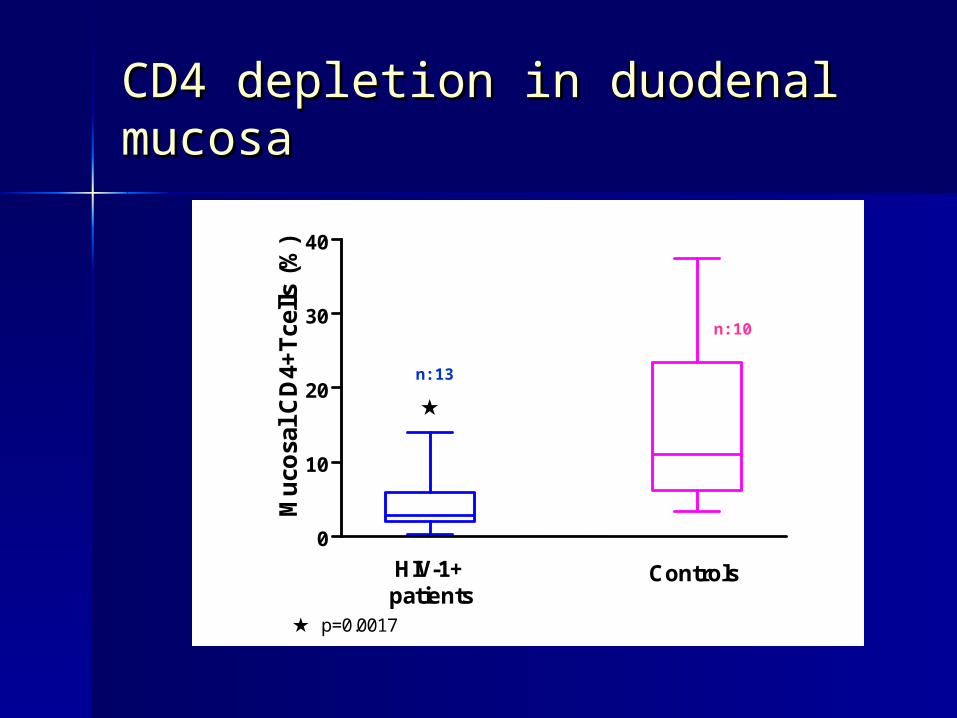
CD4 depletion in duodenal CD4 depletion in duodenal mucosamucosa
0
10
20
30
40
HIV-1+patients
Controls
p=0.0017
Mu
cosa
l C
D4+
Tce
lls
(%)
n:13
n:10
CD8+ T cells in duodenal CD8+ T cells in duodenal mucosamucosa
0
25
50
75
100
HIV-1+patients
Controls
Mu
cosa
l C
D8+
Tce
lls
(%)
n:13 n:10
Characteristics of duodenal Characteristics of duodenal CD8+ T cell populationCD8+ T cell population
HIV-1 + patients
HIV+ biopsies (n=6)
HIV- biopsies (n=7)
Controls
(n=10)
Early
Differentiated cells
(CD28+,CD27+)
18 ± 8 10 ± 4 13 ± 6
Fully
Differentiated cells
(CD28-, CD27-)
32 ± 8
61 ± 8
24 ± 5
Mean±SEM
p=0.001p=0.027
% % %
% % %
p=NS p=NS
Patients
ND pVL
(n=7)
Patients
pVL > 100.000 copies/ml (n=6)
HIV + HIV + Biopsies Biopsies 44 44
HIV-HIV- BiopsiesBiopsies 33 22
Lack of correlation of pVL with HIV Lack of correlation of pVL with HIV persistence in duodenal mucosapersistence in duodenal mucosa
p=0.72, chi-square test
p24 positivity in duodenal p24 positivity in duodenal CD64+ cellsCD64+ cells
Control BiopsyHIV- biopsy(by PCR)
HIV+ biopsy(by PCR)
Control isotype
p24 expression
KC57 FITC KC57 FITC KC57 FITC
Median FI KC57: 193Median FI isotype: 52
Median FI KC57: 56Median FI isotype: 47
Median FI KC57: 45Median FI isotype: 35
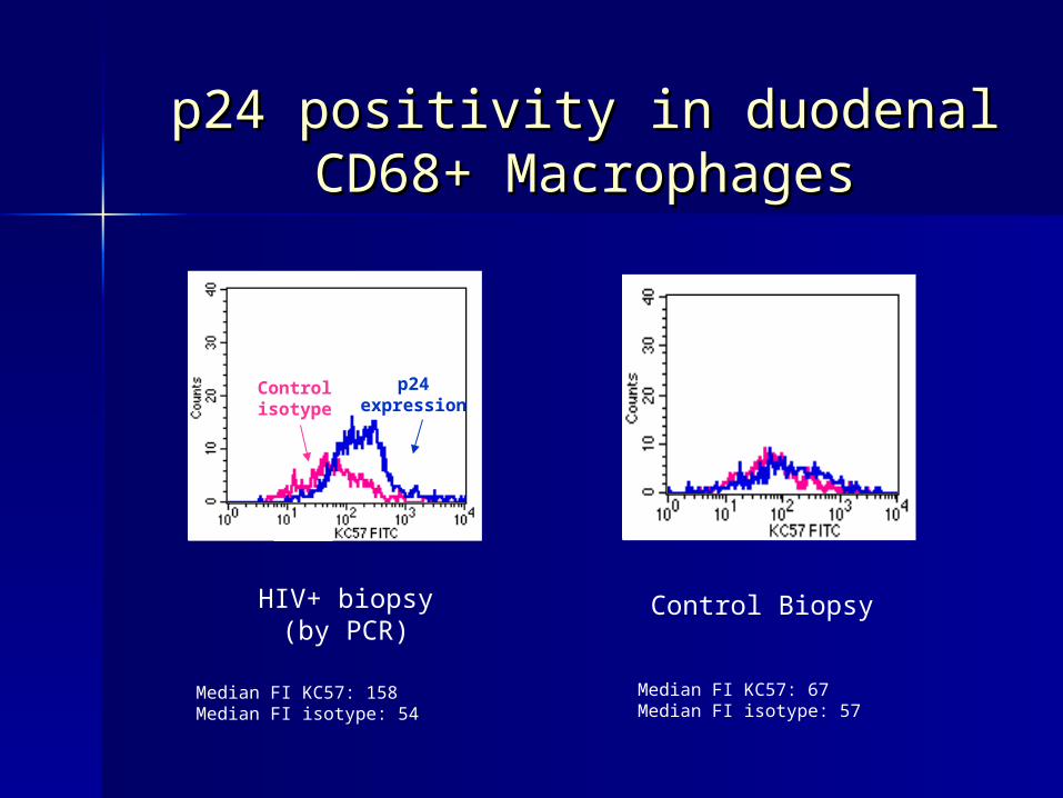
p24 positivity in duodenal p24 positivity in duodenal CD68+ MacrophagesCD68+ Macrophages
HIV+ biopsy(by PCR)
Control Biopsy
Control isotype
p24 expression
Median FI KC57: 158Median FI isotype: 54
Median FI KC57: 67Median FI isotype: 57
ConclusionsConclusions
As reported in other segments of the GI tract, we found that
CD4 T cells were depleted in duodenal mucosa of HIV patients
when compared to controls, while no differences were
observed in
the proportion of CD8 T cells in both groups.
However, the persistence of HIV in the duodenal tissue affects
the differentiation pattern of CD8 T cells. Thus, the proportion
of
fully differentiated CD8+ T cells was higher in the absence of
HIV
in the duodenum of HIV patients.
ConclusionsConclusions
HIV-DNA could be detected in the duodenal mucosa both in
patients who failed treatment and in those with undetectable
pVL.
Concerning the nature of cells that harbour HIV infection, we
have shown that macrophages may host persistent HIV
infection
and could be considered as a reservoir of HIV even after