Page 1
Persistent Hypermetabolism after Severe Burn Injury:
Effects of Hepatic Stress and Regeneration
by
Li Diao
A thesis submitted in conformity with the requirements
for the degree of Doctor of Philosophy
Institute of Medical Science
University of Toronto
© Copyright by Li Diao, 2019
Page 2
ii
Persistent Hypermetabolism after Severe Burn Injury: Effects of Hepatic Stress and Regeneration
Li Diao
Doctor of Philosophy
Institute of Medical Science, University of Toronto
2019
Abstract
Burn injury represents one of the most severe forms of trauma. Persistent
hypermetabolism and inflammatory response are common in major burned patients,
contributing to morbidity and mortality. The underlying mechanisms are largely unknown and
therefore novel and effective treatments are lacking. Liver is the fundamental mediator of post-
burn immunologic and metabolic derangement and significant hepatomegaly is universally
present and associated with the persistent hypermetabolism and inflammatory response in
severely burned patients. We sought to understand if such a hepatomegaly is the consequence
of 1) increased hepatic fat infiltration due to intensified lipolysis in white adipose tissue (WAT)
and inter-organ crosstalk between liver and WAT, or 2) aberrant liver regeneration induced by
stress response and liver damage which carries on hypermetabolic and pro-inflammatory
signaling, contributing to persistent hypermetabolism and inflammatory response after major
burn injury.
Page 3
iii
Rodent models of burn plus LPS administration, high fat diet (HFD) plus burn, and 30%
TBSA burn of Sox9-cre/ERT2:ROSA26-EYFP mice were used for the studies.
In the rat model of burn plus LPS, we demonstrated 1) increased ER stress,
inflammasome activation, apoptosis and lipolysis in WAT, contributing to liver steatosis
(Chapter 2); 2) hepatic ER stress and inflammasome activation, contributing to liver damage
and organ dysfunction (Chapter3). In the mouse model of HFD plus burn, we showed that
hepatic fat infiltration and metaflammation augment the liver damage and metabolic
dysfunction post-burn (Chapter 4). We lineage-traced the facultative liver progenitor cells after
burn injury and demonstrated that liver regeneration by this group of cells peaked around 2
weeks post-burn. Significant activation of multiple inflammatory and metabolic signaling
pathways was indicated by transcriptomic analysis and verified by further analysis in the liver
stem cells and their progeny post-burn as compared with both sham and self-renewal mature
hepatocytes. Concomitant down-regulation of LXR signaling in the liver stem cells post-burn
implicated the therapeutic potential of LXR agonist in ameliorating pro-inflammatory response
and restoring lipid homeostasis after major burn injury (Chapter 5).
In conclusion, severe burn injury leads to hepatic stress response, liver damage and
steatosis, stimulating liver regeneration from facultative stem cells which contributes to
persistent hypermetabolism and pro-inflammatory response.
Page 4
iv
Acknowledgement
This will be my 2nd PhD degree (also most likely the last one) in my life and I have the opportunity
to compare the learning experience between the young and the senior. I can tell the increased difficulty
of a senior student in learning new things. However, coming back to school after 2 decades as a busy
surgeon taking care of critically injured burn patients, it has always been very delightful to work on
better understanding of the pathology of severe trauma patients and to appreciate the great potential
of the advancement of modern biomedical science in improving the quality of the medical care and thus
the quality of life of severely injured patients.
Upon the completion of the current PhD study, I would like to express my sincere thanks to my
supervisor, Dr. Marc G Jeschke, for his mentorship, patience, and encouragement. It would be
impossible for me to have this marvelous learning experience if without his many years of kindly support.
Of equal importance is his role model of academic excellence, which, to me, is the perfect mixture of
rigorous German and open-minded American. I will always remember: “…Mike, your research needs to
be hypothesis driven!” I would also like to thank the members of my program advisory committee: Dr.
Avery Nathens, Dr. Sandro Rizoli, and Dr. Ori Rotstein, for their support and suggestions that have been
extremely helpful and added much value to my research. As what the Chinese sage Confucius said: “how
happy I am, when meeting friends from far away”, I came a long way from the other side of the earth
and also from a different cultural background to meet them and cherish the elegance and glamour of
surgery and surgical research. I extend my special gratitude to Dr Saeid Amini-Nik for his generous help
and guidance in detail in scientific research, from how to choose the proper mouse strain for animal
experiments to the recommendation of science symposiums like Gordon and Keystone. As the
participant of the Collaborative Program of Resuscitation Science, I would also express my thanks to Dr
Page 5
v
Laurie Morrison who, together with Dr Rizoli and Dr Rotstein, hosted the program and provided
enjoyable experience of group study for graduate students.
In the past 6 years, I spent most of the time in the Jeschke lab and I treasure all the happy
memories with previous and current members of the lab. I have been learning a lot from everybody. As
a Buddhist, I also benefited from the few hostile peers on how to keep calm, be humble, and be flexible.
I want to express my gratitude to all the members of the IMS office for their continuous care and
support in my PhD study. Special thanks to Dr Mingyao Liu for his encouragement, inspiration and
support all the way during my PhD study.
I feel so lucky to have many good friends around me and to be constantly blessed by them for the
success of the scientific research in the past few years. Some friends are physically around as the family
of Jed, Fiona and Anqi; Ma Bing and Michael; the family of Angus, Sophia and Shania; the family of Andy,
Jenny and Lingling. Some are distant and I still clearly feel their warmth of care and support: Guocheng,
Chen Hui, Helen, and their families.
I want to express my heartiest thanks to my parents, my parents-in-law, my brother and his family,
my brothers-in-law and their families for their keen expectation and good wishes for my academic
success. My gratitude goes to my parents and my parents-in-law for their altruistic support and I feel so
blessed to see that they are happy and healthy and I wish them all the best for happy longevity. I
appreciate my brother’s support in every aspect as we immigrate to Canada and I think a great deal of
the industrious family of Rock, Jenny and Changchang and wish them good luck for future success.
I dedicate my thesis to my wife Alina and my son Joseph. It is their love and support that
accompany me in our new life in Canada. I hope that this thesis may signify the future success of Alina
together with our joint endeavor and motivate Joseph for his academic excellence.
Page 6
vi
Table of Contents
Abstract………………………………………………………………………………………………………………………………………………………….…. ii
Acknowledgements………………………………………………………………………………………………………………………………………….. iv
Table of Contents……………………………………………………………………………………………………………………………………………… vi
List of Tables…………………………………………………………………………………………………………………………………………………….. xi
List of Figures……………………………………………………………………………………………………………………………………………………. xii
List of Abbreviations…………………………………………………………………………………………………………………………………………. xv
List of Publications (PhD study period)……………………………………………………………………………………………………….….. xviii
Prologue………………………………………………………………………………………………………………………………………………………….... 1
Chapter 1 Introduction ……………………………………………………………………………………………………………………………….... 3
1.1 Persistent pro-inflammatory response and hypermetabolism in major burned
patients: liver as the mediator and the functional hub…………………………………………………………..….. 3
1.2 Cellular stress response: the cellular basis of post-burn pathology……………………….………………..… 11
1.2.1 Historical perspectives…………………………………………………………………………………………………….. 12
1.2.2 Heat shock responses (HSR)..………………………………………………………………………………………..…. 15
1.2.3 ER stress and UPR………………………………………………………………………………………………………...…. 17
1.2.4 Mitochondrial stress response and mitochondrial UPR (UPRmt
)………………………………..…….. 20
1.2.5 Integrated stress response (ISR) determines cell function and cell fate and its
Implication in the pathophysiology of critical illness…………………………………………………..….. 24
1.2.5.1 ISR in hypoxia and ischemia and reperfusion injury…………………………………………….. 24
1.2.5.2 ISR upon infection and inflammation………………………………………………………………….. 26
1.2.5.3 ISR in cell death and tissue and organ damage……………………………………………………. 30
1.2.6 Summary……………………………………………………………………………………………………………………..…. 32
1.3 Hepatic immunometabolic disorder, liver damage and regeneration after
severe trauma injury………………………………………………………………………………………………………………. 33
1.3.1 Immunometabolism and hepatic inflammasome activation under stress conditions…….. 33
1.3.2 Liver regeneration under profound stress condition and severe liver damage………………. 38
1.4 Research problem, rationale and working hypotheses……………………………………….………………..… 42
Page 7
vii
1.4.1 Research problem and rationale………………………………………………………………………………….... 42
1.4.2 Hypotheses and specific aims of the study…………………………………………………..……………..…. 44
Chapter 2 Increased lipolysis in WAT and its contribution to hepatic fat infiltration ………….......................... 47
2.1 Introduction………………………………………………………………………………………………………………………….... 47
2.2 Materials and Methods……………………………………………………………………………………………………………. 49
2.2.1 Animal model…………………………………………………………………………………………………………..…… 49
2.2.2 Cell culture ………………………………………………………………………………………………………………..… 50
2.2.3 Plasma and tissue collection …………………………………………………………………………………..…… 50
2.2.4 Gene expression analysis …………………………………………………………………………………………..… 51
2.2.5 Western blotting ……………………………………………………………………………………………………..….. 51
2.2.6 Immunofluorescent multi-channel staining of WAT……………………………………………………… 52
2.2.7 H&E, Oil Red O (ORO), IHC and TUNEL staining of tissue sections…………………………….…… 53
2.2.8 Determination of FFA, glycerol and triglyceride levels in blood………………………………….…. 53
2.2.9 Statistical analysis……………………………………………………………………………………………………..….. 53
2.3 Results…………………………………………………………………………………………………………………………………..… 54
2.3.1 Burn and LPS induce significant catabolism and hepatic fat infiltration ………………………... 54
2.3.2 Increased WAT lipolysis in the 2-hit rat model of burn plus LPS ……………………………..….…. 56
2.3.3 Increased lipolysis in WAT after burn plus LPS is associated with
reduced AMPK signaling ………………………………………………………………………………………………. 58
2.3.4 Burn plus LPS increases adipocyte apoptosis ………………………………………………………….……. 61
2.3.5 Burn plus LPS synergistically induce apoptosis in WAT ………………………………………………... 64
2.3.6 Increased macrophage infiltration and inflammasome activation correlate
with apoptosis in WAT……………………………………………………………………………………………..…… 67
2.4 Discussion………………………………………………………………………………………………………………………….……. 70
Chapter 3 Hepatic ER stress, inflammasome activation, liver dysfunction and damage …………….…................ 78
3.1 Introduction……………………………………………………………………………………………………………………….……. 78
3.2 Materials and Methods……………………………………………………………………………………………………………. 81
3.2.1 Animal model…………………………………………………………………………………………………………….….. 81
Page 8
viii
3.2.2 Plasma and tissue collection ………………………………………………………………………………………… 82
3.2.3 Real-time quantitative RT-PCR …………………………………………………………………………………….. 82
3.2.4 Western blotting ……………………………………………………………………………………………………….... 84
3.2.5 Blood glucose level, plasma assay and IHC analysis for liver damage assessment ……….. 84
3.2.6 Statistical analysis ……………………………………………………………………………………………..…….….. 85
3.3 Results………………………………………………………………………………………………………………………………….… 86
3.3.1 The two-hit of burn injury with LPS injection induces liver damage ……………………..……... 86
3.3.2 Burn plus LPS injection augment hepatic NLRP3 inflammasome activation ……………..….. 88
3.3.3 Burn and LPS injection induce hepatic ER stress……………………………………………………….….. 89
3.3.4 Burn induces hypermetabolism …………………………………………………………………….………….…. 90
3.3.5 Unlike burn which down-regulates SIRT1, LPS inhibits PKA C/AMPK………………………….. 92
3.4 Discussion………………………………………………………………………………………………………………………….…… 95
Chapter 4 Hepatic fat infiltration and liver damage …………………..………………………………………………………….…. 102
4.1 Introduction…………………………………………………………………………………………………………………….…… 102
4.2 Materials and Methods……………………………………………………………………………………………………..…. 103
4.2.1 Animal model……………………………………………………………………………………………………………… 103
4.2.2 Plasma and tissue collection……………………………………………………………………………………….. 104
4.2.3 Western blotting…………………………………………………………………………………………………………. 104
4.2.4 In-gel mitochondrial ETC activity assays………………………………………………………………………. 105
4.2.5 Immunofluorescent multi-channel staining of liver……………………………………………………… 105
4.2.6 H&E staining and TEM of tissue sections……………………………………………………………………… 105
4.2.7 Determination of FFA, glycerol and triglyceride levels in blood……………………………………. 105
4.2.8 Statistical analysis………………………………………………………………………………………………………… 105
4.3 Results…………………………………………………………………………………………………………………………………… 107
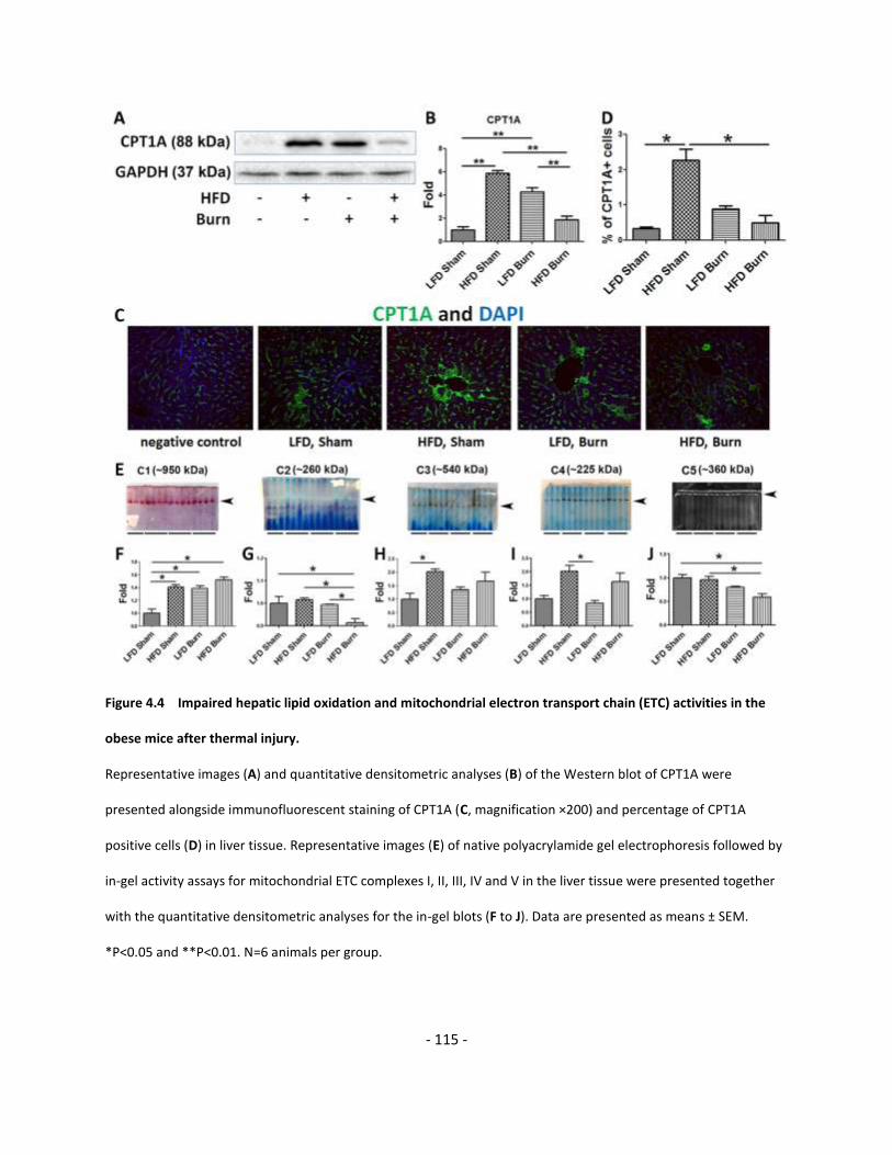
4.3.1 HFD and burn lead to hepatic fat infiltration and increased lipolysis……………………………. 107
4.3.2 De novo lipogenesis is not activated in HFD mice after thermal injury……………………….… 111
4.3.3 Decreased hepatic lipid -oxidation and attenuated mitochondrial ETC
Page 9
ix
function associate with hepatic fat infiltration…………………………………………………………….. 113
4.3.4 Perturbed inter-organelle Ca2+ homeostasis correlates with decreased
ER-mitochondrial contact……………………………………………………………………………………………. 116
4.3.5 Augmented hepatic ER stress, inflammasome activation and aggravated
cell damage in HFD mice after thermal injury……………………………………………………………… 120
4.4 Discussion…………………………………………………………………………………………………………………………….. 125
Chapter 5 Stress induces periportal ductal progenitor cells proliferation, contributing to
prolonged pro-inflammatory response and hypermetabolism …….………………………………….......... 129
5.1 Introduction………………………………………………………………………………………………………………………….. 129
5.2 Materials and Methods…………………………………………………………………………………………………………. 133
5.2.1 Animal model………………………………………………………………………………………………………………… 133
5.2.2 Liver tissue collection and digestion………………………………………………………………………………. 134
5.2.3 Reagents and antibodies……………………………………………………………………………………………….. 135
5.2.4 Western blotting……………………………………………………………………………………………………………. 135
5.2.5 Cell staining and flow cytometry……………………………………………………………………………………. 135
5.2.6 Immunofluorescent multi-channel staining of liver………………………………………………………… 136
5.2.7 Microarray transcriptomic analysis………………………………………………………………………………… 136
5.2.8 Statistical analysis………………………………………………………………………………………………………….. 137
5.3 Results…………………………………………………………………………………………………………….…………………….. 138
5.3.1 The proliferation of PDPCs increases, contributing to hepatomegaly after major
burn injury…………………………………………………………………………………………………………………….. 138
5.3.2 The hepatic stress response correlates with the increased proliferation of
PDPCs after major burn injury……………………………………………………………………………………….. 143
5.3.3 Increased proliferation of PDPC-derived hepatocytes contributes to persistent
pro-inflammation and hypermetabolism after major burn injury………………………………….. 146
5.4 Discussion……………………………………………………………………………………………………………………………… 155
Chapter 6 Thesis summary and future directions………………………………………………………………………………………. 162
6.1 General discussion…………………………………………………………………………………………………………………. 162
6.1.1 Rodent animal models for translational research…………………………………………………………… 163
Page 10
x
6.1.2 Immunometabolic disorder after trauma: what we can learn from metaflammation……. 166
6.1.3 Inter-organ crosstalk between adipose tissue and liver: lipolysis and hepatic
fat infiltration………………………………………………………………………………………………………………... 168
6.1.4 “Birth and death, concomitant processes”…………………………………………………………………….. 170
6.2 Conclusions……………………………………………………………………………………………………………………………. 174
6.3 Limitations of the current study and future directions………………………………….……………………….. 176
References…………………………………………………………………………………………………………………………………….................. 180
Page 11
xi
List of Tables
Table 3.1 Primers sequences for qRT-PCR………………………………………………………………………………………………………….. 83
Table 5.1 Primers for genotyping……………………………………………………………………………………………………………………… 133
Table 5.2 Microarray samples………………………………………………………………………………………………………………………….. 146
Table 5.3 Comparison of the changes in canonical signaling pathways in EYFP+
cells in mice of Sham versus PBD7 group……………………………………………………………………………………… 147
Table 5.4 Comparison of the changes in canonical signaling pathways in EYFP+
versus EYFP- cells in mice of PBD7 group……………………………………………………………………………………… 148
Table 6.1 Difference between the immunometabolic disorders after severe
trauma and metaflammation……………………………………………………………………………………………………….. 167
Page 12
xii
List of Figures
Figure 1.1 Specific aims of the study…………………………………………………..……………………………………………………………… 46
Figure 2.1 Burn and LPS induced catabolism and increased liver fat content……………………………………………………… 55
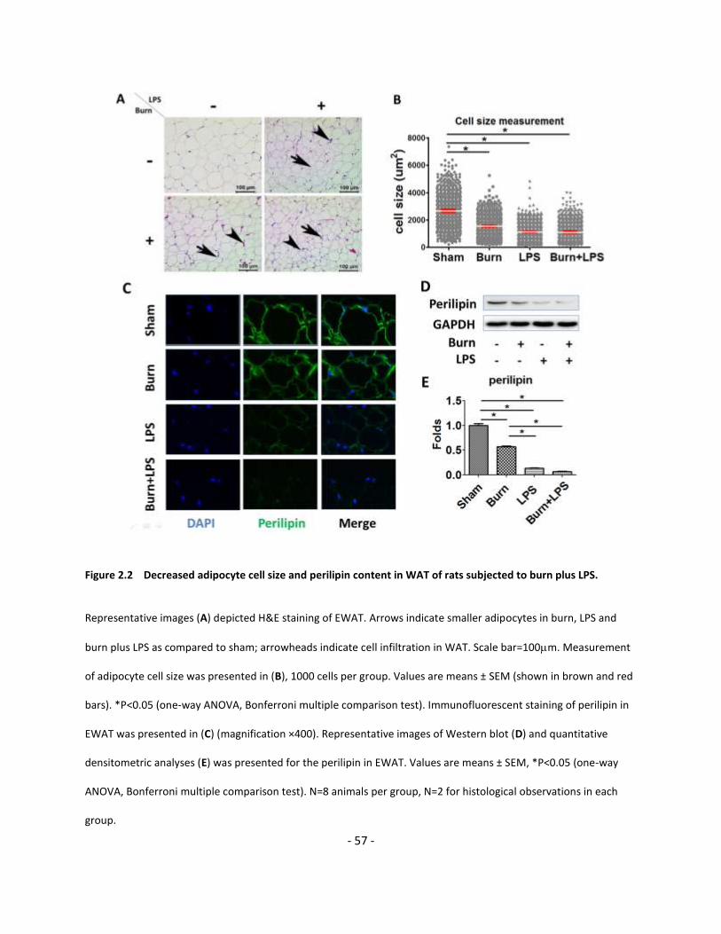
Figure 2.2 Decreased adipocyte cell size and perilipin content in WAT of rats subjected to
burn plus LPS………………………………………………..………………………………………………………………………………….. 57
Figure 2.3 Burn plus LPS do not directly activate HSL and MAPK lipolysis pathway…………………………………………….. 59
Figure 2.4 Burn and LPS increased lipolysis by inhibiting AMPK signaling in WAT………………………………………….…... 60
Figure 2.5 Burn plus LPS promoted apoptosis in WAT: TUNEL staining ……………………………………………………….…….. 62
Figure 2.6 Burn plus LPS promoted apoptosis in WAT: evidence of pro-apoptotic signaling………………………………. 63
Figure 2.7 Burn plus LPS increased ER stress which correlated with apoptosis in WAT………………………………….…... 65
Figure 2.8 Activation of pro-apoptotic signaling correlates with ER stress in adipose
tissue in burn plus LPS in rats……………………………………………………………………………………………………….…… 66
Figure 2.9 Burn and LPS stimulated macrophage infiltration and inflammasome activation
which correlated with apoptosis in WAT……………………………………………………………………………………….…. 68
Figure 2.10 Activation of pro-apoptotic signaling correlates with macrophage Infiltration
in adipose tissue in burn plus LPS in rats……………………...………………………………………………………………..... 69
Figure 2.11 No significant changes are detected in serum level of free fatty acid (A),
glycerol (B) or triglyceride (C) among different treatment groups……………………………………………..…..... 73
Figure 2.12 Increased WAT lipolysis and its contribution to immunological and metabolic
impairment in the 2-hit model of burn plus LPS………………………..…………………………..…………………..….... 77
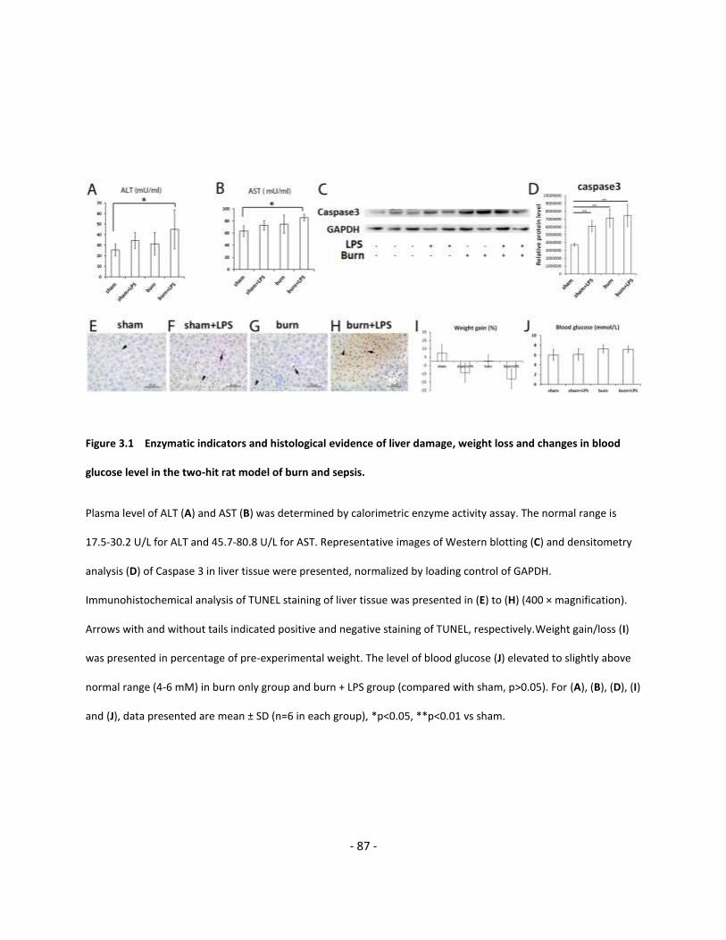
Figure 3.1 Enzymatic indicators and histological evidence of liver damage, weight loss and
changes in blood glucose level in the two-hit rat model of burn and sepsis…………..………………………… 87
Figure 3.2 Two-hit models of burn plus LPS injection augments inflammasome formation in rat liver………………. 88
Figure 3.3 Burn and LPS induced hepatic ER stress……………………………………………………………………………………….…… 89
Figure 3.4 Burn and LPS induced changes in gene expression of metabolic modulators in liver…………………….….. 91
Figure 3.5 Burn and LPS induced changes in PGC 1 in liver…………………………………………………………………………..…. 93
Figure 3.6 LPS reduced PGC-1 by inhibiting its upstream regulators…………………………………………………………..…… 94
Figure 3.7 Hepatic ER stress and NLRP3 inflammasome activation exacerbate hepatic
metabolic dysfunction and liver damage in the 2-hit rat model of burn plus LPS……………………………. 101
Page 13
xiii
Figure 4.1 16 weeks of HFD establishes obese mice with insulin resistance…………………………………………………….. 108
Figure 4.2 Augmented hepatic fat infiltration, increased lipolysis, and circulating FFA in
obese mice after thermal injury ……………………………………………………..………………………………………..….... 110
Figure 4.3 Repression of de novo lipogenesis in HFD mice after thermal injury…………………………………………..……. 112
Figure 4.4 Impaired hepatic lipid oxidation and mitochondrial electron transport
chain (ETC) activities in the obese mice after thermal injury………………………………………………………..….. 115
Figure 4.5 Mitochondrial metabolic dysfunction is correlated with the perturbed inter-organelle Ca2+
homeostasis and mitochondrial dynamics in the liver of obese mice after thermal injury……………….. 118
Figure 4.6 The decrease of hepatic ER-mitochondrial contact and mitochondrial
structural changes after burn injury ………………………………………………………………………………………….…….. 119
Figure 4.7 Augmented hepatic ER stress in HFD burned mice…………………………….…………………………………………...... 121
Figure 4.8 TUNEL staining of liver tissue demonstrated increased liver cell apoptosis in HFD burned mice….……. 122
Figure 4.9 NLRP3 inflammasome activation, enhanced pro-apoptotic signaling and
DNA damage in HFD burned mice……………………………………………………………………………………….….……….. 124
Figure 4.10 Hepatic fat infiltration is attributable to the vicious cycle of ER stress,
mitochondrial dysregulation and cell damage in HFD burned mice………………………………………….…….. 128
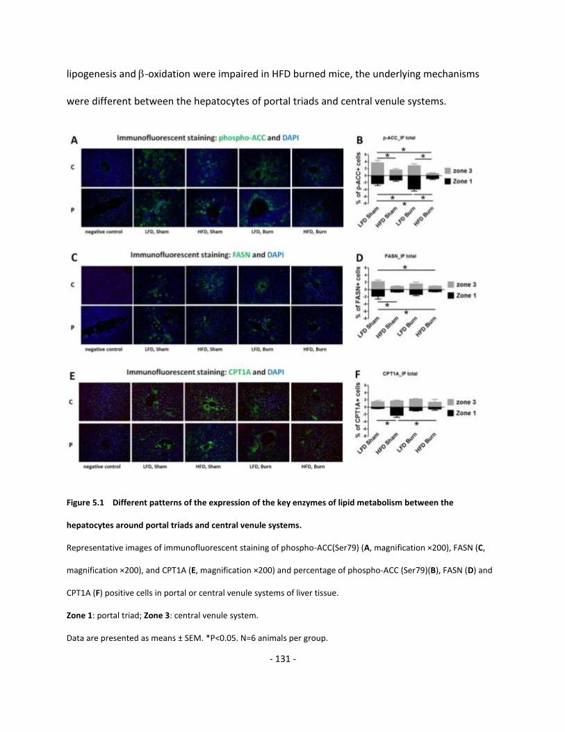
Figure 5.1 Different patterns of the expression of the key enzymes of lipid metabolism
between the hepatocytes around portal triads and central venule systems………………….…………………. 131
Figure 5.2 Increased proliferation of the PDPCs contributes to hepatomegaly after thermal injury…………….....… 140
Figure 5.3 Increased proliferation of the PDPCs is around portal venule after thermal injury……………………………. 141
Figure 5s.1 Optimization of the tamoxifen treatment protocol: dosage and route
of administration……………………………………………………………………………………………………………………...……. 142
Figure 5s.2 The changes in body weight in mice before and after burn injury…………………………………………...………. 142
Figure 5.4 Hepatic cellular stress response after thermal injury…………………………………………..…………………………..… 144
Figure 5.5 Hepatic cellular stress response correlates with PDPCs proliferation after thermal injury…………………. 145
Figure 5.6 Up-regulation of the acute phase response (A), p38 MAPK (B), and IL-6 (C)
signaling pathways in PDPCs after thermal injury………………………………………………………………….............. 149
Figure 5.7 On PBD7, acute phase response (A), p38 MAPK (B), and IL-6 (C) signaling pathways
are more activated in PDPCs as compared with that in mature hepatocytes……………………………….….... 150
Figure 5.8 LXR/RXR signaling pathway is significantly down-regulated in PDPCs…………………………………..……..…….. 151
Page 14
xiv
Figure 5.9 Up-regulated hepatic acute phase response and p38 MAPK signaling followed
the decrease of LXR expression and correlated with increased lipid oxidation
and cell damage in the liver after thermal injury………………………………………………………………………....... 154
Figure 5.10 Aberrant liver regeneration contributes to persistent pro-inflammatory response
and hypermetabolism after major burn injury…………………………………………………………………………….… 161
Figure 6.1 Hepatic stress response, liver damage and regeneration contribute to persistent
pro-inflammatory response and hypermetabolism after major burn injury……………………………..…….. 175
Page 15
xv
List of Abbreviations
ACC acetyl CoA carboxylase
ALT alanine aminotransferase
AMPK AMP-activated protein kinase
αMSH -melanocyte stimulating hormone
Arg 1 arginase 1
ASK1 apoptosis signaling kinase 1
AST aspartate aminotransferase
ATF activating transcription factor
ATGL desnutrin/adipose triglyceride lipase
BiP Binding immunoglobulin protein
C/EBPs CCAAT/enhancer-binding-proteins
CGI-58 comparative gene identification-58
CHOP CCAAT/Enhancer-Binding Protein Homologous Protein
cLDs cytoplasmic lipid droplets
CLP cecal ligation and puncture
CRTC2 CREB-regulated transcription coactivator 2
DAMP damage-associated molecular pattern
eIF2 eukaryotic translation initiation factor 2
ERAD ER-associated degradation
ESLD end-stage liver disease
FAO fatty acid oxidation
FFA free fatty acids
G-CSF granulocyte colony-stimulating factor
GH growth hormone
HG high glucose
HMGB1 high mobility group box protein 1
HOP HSP-organizing protein
HPA hypothalamic–pituitary–adrenal
HSE heat shock element
HSF1 heat shock factor 1
HSL hormonal-sensitive lipase
HSP heat shock protein
HSR heat shock response
IF immunofluorescent
IGF-1 insulin-like growth factor‐1
IHC immunohistochemical
IKK IB kinase
IL-6 interleukin-6
IMS intermembrane space
Page 16
xvi
iNOS inducible nitric-oxide synthase
IP3R inositol 1,4,5-triphosphate receptor
IPGTT intraperitoneal glucose tolerance test
IR insulin resistance
IRE1 inositol-requiring kinase 1
IRF interferon-regulatory factor
ISR integrated stress response
JNK c-Jun N-terminal kinase
LAL lysosomal acid lipase
LDL low-density lipoproteins
LFD low fat diet
LPS lipopolysaccharides
LXR liver X receptor
MAG 2-monoacylglycerol
MAPK mitogen-activated protein kinase
MCP-1 monocyte chemoattractant protein-1
M-CSF macrophage colony- stimulating factor
MCSR mitochondria to cytosol stress response
MFN2 mitofusin 2
MGL monoacylglycerol lipase
MLKL mixed lineage kinase like
mPOS mitochondrial precursor over-accumulation stress
MPT mitochondrial permeability transition
mTOR mechanistic target of rapamycin
MTS mitochondrial targeting sequences
NAFLD nonalcoholic fatty liver disease
NASH nonalcoholic steatohepatitis
NF-B nuclear factor-B
NLR NOD-like receptor
NLRP3 NOD-like receptor, pyrin domain containing 3
OPA1 Dynamin-like 120 kDa protein, mitochondrial
ORO oil red O
OXPHOS Oxidative phosphorylation
PAMP pathogen-associated molecular pattern
PARP poly ADP ribose polymerase
PBA phenylbutyrate
PBD post-burn day
PBS phosphate-buffered saline
PCNA proliferating cell nuclear antigen
PDI Protein disulfide isomerase
PDPC periportal ductal progenitor cells
Page 17
xvii
PERK pancreatic ER eIF2a kinase
PGC-1 Peroxisome proliferator-activated receptor gamma coactivator 1-
PHIR Persistent hypermetabolism and inflammatory responses
PINK1 PTEN-induced putative kinase 1
PKA C Protein kinase A catalyst unit
PMA phorbol myristate acetate
PPAR Peroxisome proliferator-activated receptor
PRR pattern recognition receptor
REE resting energy expenditure
RIDD regulated IRE1-dependent decay of mRNA
RIN RNA integrity number
RIP regulated intramembrane proteolysis
RIPK receptor interacting protein kinase
RLR RNA cytosolic helicases of the RIG-1-like receptors family
Rnase endoribonuclease
ROS reactive oxygen species
RXR retinoid X receptor
SCD1 Stearoyl-CoA desaturase
SIK1 salt inducible kinase 1
Sox9 Sry-related HMG box 9
STAT signal transducer and activator of transcription
TAG triacylglycerol
TAK1 Transforming growth factor beta-activated kinase 1
TBK1 TANK-binding kinase 1
TBSA total body surface area
TFE3 transcription factor E3
TFEB transcription factor EB
TG triglycerides
TLR Toll-like receptors
TNF tumor necrosis factor
TRAF2 tumor necrosis factor α receptor-associated factor 2
TRAP1 TNF Receptor-Associate Protein 1
TUNEL Terminal deoxynucleotidyl transferase dUTP nick end labeling
UCP uncoupling protein
UPR unfolded protein response
VLDL Very low density lipoprotein
VDAC Voltage-dependent anion channel
WAT white adipose tissue
XBP1 X-box-binding protein 1
Page 18
xviii
List of Publications (PhD study period)
1. Diao L, Yousuf Y, Amini-Nik S, Jeschke MG: Increased proliferation of hepatic periportal ductal progenitor cells
contributes to persistent hypermetabolism after trauma. Cell Death and Differentiation [submitted]
2. Diao L, Auger C, Konoeda H, Sadri A, Amini-Nik S, Jeschke MG: Hepatic steatosis associated with decreased -
oxidation and mitochondrial function contributes to cell damage in obese mice after thermal injury. Cell Death and
Disease 2018, 9(5): 530 (11 pages). doi: 10.1038/s41419-018-0531-z.
3. Amini-Nik S, Sadri A, Diao L, Belo C, and Jeschke MG: Accumulation of Myeloid Lineage Cells Is Mapping Out
Liver Fibrosis Post Injury: A Targetable Lesion Using Ketanserin. Experimental & Molecular Medicine 2018, 50(7): 81.
4. Xiu F, Diao L*, Qi P, Catapano M, Jeschke MG: Palmitate Differentially Regulates the Polarization of
Differentiating and Differentiated Macrophages. Immunology 2016, 147(1): 82-96. (*co-first author)
5. Diao L, Patsouris D, Sadri A, Dai X, Amini-Nik S, Jeschke MG: Alternative mechanism for white adipose tissue
lipolysis after thermal injury. Molecular Medicine 2015, 21:959-968.
6. Bogdanovic E, Kraus N, Patsouris D, Diao L, Wang V, Abdullahi A, Jeschke MG: Endoplasmic reticulum stress in
adipose tissue augments lipolysis. Journal of cellular and molecular medicine 2015, 19:82-91.
7. Xiu F, Catapano M, Diao L, Stanojcic M, Jeschke MG: Prolonged ER Stressed- Hepatocytes drives an Alternative
Macrophage Polarization. Shock 2015, 44(1):44-51.
8. Diao L, Marshall AH, Dai X, Bogdanovic E, Abdullahi A, Amini-Nik S, Jeschke MG: Burn plus lipopolysaccharide
augments endoplasmic reticulum stress and NLRP3 inflammasome activation and reduces PGC-1 in liver. Shock
2014, 41:138-44.
9. Xiu F, Stanojcic M, Diao L, Jeschke MG: Stress hyperglycemia, insulin treatment, and innate immune cells.
International journal of endocrinology 2014, 2014:486403.
10. Drennan IR, Allan KS, Diao L: Re: Use of rapid sequence intubation predicts improved survival among patients
intubated after out-of-hospital cardiac arrest. Resuscitation 2014, 85:e113.
Page 19
- 1 -
Prologue
In May 2012, right before entering into the PhD program, I read 3 published papers on
the clinical study of the major burned patients, written by my PhD supervisor Dr Jeschke, in
which he wrote:
“…it clearly demonstrated that burn induced metabolic and inflammatory changes
persisted for 3 years after the injury” in a study in which “Nine-hundred seventy-seven severely
burned children were included...” (Jeschke et al., 2011a). Such prolonged hypermetabolism and
inflammation is linked with multiple post-burn comorbidities including increased risk for
infection and sepsis and increases the mortality of the patients.
“… the change in serum triglycerides and free fatty acids, both of which are significantly
increased through almost the entire acute hospital stay...A therapeutic approach to decrease
lipolysis and fatty infiltration and reverse the acute phase response may thus improve
morbidity and mortality” (Jeschke et al., 2008a).
Concomitant to the catabolism seen in multiple organs and systems including muscle,
bone, and peripheral adipose tissue, there is a “massive hepatomegaly and hepatic fatty
infiltration” observed in both “…burn victim at autopsy” as well as “…in 242 surviving burn
patients” (Jeschke, 2009), depicting pivotal role of the liver in the post-burn pathophysiology in
which hypermetabolism and inflammation are featured.
In the past 6 years, I took on the journey of biomedical research, investigating the
nature of stress response and lipolysis in white adipose tissue (WAT), and seeking the
Page 20
- 2 -
relationship between such lipolysis and liver fat infiltration and the impact of the latter on liver
dysfunction and damage.
In addition to the aforementioned stress induced cell damage and organ dysfunction,
considering the huge potential of liver regeneration after injury and the central role of liver as
the hub integrating the whole body regulation of metabolism and immunology , I also
hypothesized and sought to prove that profound stress response after burn injury stimulates
liver regeneration which is different to physiological renewal of hepatic parenchyma, and
contributes to the prolonged hypermetabolism and hyper-inflammatory state.
Page 21
- 3 -
Chapter 1 Introduction
1.1 Persistent pro-inflammatory response and hypermetabolism in major
burned patients: liver as the mediator and the functional hub
Our previous clinical studies clearly demonstrate the presence of prolonged pro-
inflammatory and hypermetabolic responses that lead to hyper-dynamic circulation, increased
body temperature, glycolysis, proteolysis, lipolysis and futile substrate cycling in patients with
major burn over 30% total body surface area (TBSA) (Jeschke et al., 2008a; Jeschke et al.,
2011a). Such a prolonged post-burn metabolic and inflammatory changes are accompanied
with sustained increase in catecholamines and stress hormones, persistent elevation of resting
energy expenditure (REE), increased expression of inflammatory cytokines such as TNF, IL-6, IL-
8, granulocyte colony-stimulating factor (G-CSF), monocyte chemoattractant protein-1 (MCP-1),
and persistent elevated levels of blood glucose and insulin depicting significant insulin
resistance. The consequence of this persisting post-burn metabolic and inflammatory response
is detrimental, contributing to impaired wound healing, increased incidence of wound and
systemic infection, prolonged post-burn rehabilitation and even increased mortality. Indeed, in
our recent prospective cohort study, we have shown that significantly higher serum levels of IL-
6, IL-8, G-CSF, MCP-1, C-reactive protein, glucose, insulin, blood urea nitrogen, creatinine, and
bilirubin predicted higher likelihood of mortality and non-survivors exhibited a vastly increased
hypermetabolic response that was associated with increases in organ dysfunction and sepsis
Page 22
- 4 -
(Jeschke et al., 2014a). The research to elucidate the underlying mechanisms of aggravating
pro-inflammatory response and hypermetabolism is thus warranted for effective treatment.
Considering that the liver is the functional hub integrating metabolic response,
immunologic response, inflammatory response and acute phase response, we have long been
emphasizing the importance of the liver in mediating the metabolic and inflammatory disorders
post-burn (Jeschke, 2009; Jeschke et al., 2004).
Aberrant hepatic metabolic responses after major burn injury are manifested by the
derangement of glucose metabolism, increased proteolytic signaling, and dysregulated lipid
metabolism.
Hepatic glucose metabolism is regulated through diverse mechanisms. Hepatic glucose
production is regulated by 1) the provision of substrates, such as glucose or glycerol; 2)
allosteric control by metabolites, such as acetyl-CoA, glucose and glucose-6-phosphate; 3) the
balance of hormones, including insulin, glucagon, catecholamine and corticosteroids; and 4)
cellular redox state, which can be modified by treatment with metformin(Petersen et al., 2017).
Stress mediators, such as catecholamine, glucocorticoid, glucagon, and dopamine, stimulate
lipolysis in adipose tissue and proteolysis in skeletal muscle thus increase the substrates of
gluconeogenesis; catecholamine and glucagon can also mediate enhancement of hepatic
glycogenolysis, all contributing to hyperglycemia post-burn. Pro-inflammatory cytokines,
including TNF, IL-6 and MCP-1, directly act on the insulin signal transduction pathway through
modification of the signaling properties of insulin receptor substrates, contributing to post-burn
hyperglycemia via hepatic insulin resistance (Mecott et al., 2010).
Page 23
- 5 -
Increased proteolysis and muscle wasting are common pathology of major burned
patients. Although this is out of the scope of the current PhD research project, we noticed that
in recent years, in the area of the research of nonalcoholic fatty liver disease (NAFLD) and
nonalcoholic steatohepatitis (NASH), the concept of sarcopenia is called into attention which is
defined as a progressive and generalized loss of skeletal muscle mass, strength, and function
commonly seen in up to 60% of patients with end-stage liver disease (ESLD), depicting the
importance of hepatic pathophysiology in the changes in the protein catabolism under critical
illness (Bhanji et al., 2017). Interestingly, since mechanisms relating sarcopenia to NASH include
IR, increased inflammation, myokines secreted by skeletal muscle, myostatin, adiponectin,
vitamin D deficiency, and physical inactivity (Merli and Dasarathy, 2015), it is reasonable to
speculate the interaction between sarcopenia and post-traumatic IR and pro-inflammatory
state which are at least partly originated from liver pathology. Specifically, IR increases lipolysis
in adipose tissue with the consequent release of free fatty acids (FFAs) to the liver and high
levels of FFA inhibit the growth hormone (GH)/insulin-like growth factor‐1 (IGF‐1) axis,
contributing to muscle loss and decrease in muscle regeneration (Kalyani et al., 2014);
enhanced fatty acid oxidation (FAO) in the liver leads to generation of oxygen free radicals,
which causes lipid peroxidation and induces synthesis of pro-inflammatory cytokine such as
tumor necrosis factor‐ (TNF) which not only leads to direct liver injury, but also stimulate
protein catabolism, which results in loss of muscle mass and sarcopenia (Phillips and
Leeuwenburgh, 2005).
Page 24
- 6 -
Dysregulated hepatic lipid metabolism features hepatomegaly and hepatic fat
infiltration which could be attributed to excessive delivery of fatty acids to the liver as a
consequence of -adrenergic mediated stimulation of lipolysis and a diminished effectiveness
of insulin in suppressing lipolysis. It has also been suggested that decreased VLDL-triglyceride
secretion is seen in burn patients and is unresponsive to increased hepatic triglyceride synthesis
(Morio et al., 2002). Hepatomegaly and hepatic steatosis and dysfunction in severely burned
rats are associated with increased mortality and that liver integrity and function are crucial for
survival post-burn (Mittendorfer et al., 1998). In IL-6 knockout mice which developed
cholestasis, steatosis, and hepatocellular injury upon cecal ligation and puncture (CLP), there is
an aggravated hepatic dysfunction and increased mortality in sepsis (Deutschman et al., 2006).
All these observations indicate that hepatomegaly and hepatic fat infiltration are detrimental to
the outcome of major burn injury and infection.
In a recent review of the metabolic stress response to burn trauma (Porter et al., 2016),
it has been corroborated that persistent pathophysiological stress response of adrenergic and
inflammatory stress, hypermetabolism, metabolic dysfunction, and reduced lean body mass can
be presented for up to and beyond 3 years after burn injury of more than 20% TBSA. It has also
been clearly demonstrated that the activation of uncoupling protein 1 (UCP1) expression in
functional brown and subcutaneous white adipose tissues upon persistent adrenergic stress
post-burn contributes to increased energy expenditure and hypermetabolic response (Patsouris
et al., 2015; Sidossis et al., 2015). Accordingly, it has been suggested that browning of white
adipose tissue is causative to post-burn hypermetabolism and to inhibit or alleviate such
Page 25
- 7 -
browning is proposed to be therapeutic to decrease hypermetabolism and improve clinical
outcome (Abdullahi and Jeschke, 2017).
However, from the point of view of evolution, browning of the adipose tissue is one of
the most important adaptive mechanisms of thermogenesis and it has been strongly implicated
as protective and beneficial to the living organisms under different stress conditions. If such
beneficial thermogenesis in mammals generally hold true, we might speculate another
regulatory mechanism when taking into consideration the central insulin resistance of the liver:
persistent and profound adrenergic stress signaling post-burn stimulates lipolysis in the adipose
tissues and contributes to the increased hepatic lipid influx; hepatic fat infiltration contributes
to increased hepatic glucose production and output; browning of the adipose tissue may thus
be an adaptive and protective mechanism to neutralize the detrimental effect of such lipolysis
and consequent hepatic lipotoxicity. Hence, it is important to further clarify the impact of
browning of white adipose tissue in the post-burn pathology.
Regardless of the dispute and controversy in the significance of browning of the white
adipose tissue in the pathophysiology after major burn injury, it is clear that attenuating
lipolysis may decrease the hepatic lipid preload thus is beneficial to the restoration and
maintenance of hepatic homeostasis.
There are three mechanistically cooperate principal pathways of intracellular lipolysis,
which are neutral lipolysis of cytoplasmic lipid droplets (cLDs), acid lipolysis in lysosomes, and
lipophagy (Zechner et al., 2017). The most common neutral lipolysis in adipose and non-adipose
tissues initiates from triacylglycerol hydrolysis by adipose triglyceride lipase (ATGL) to form
Page 26
- 8 -
diacylglycerol and FFAs (Zimmermann et al., 2004). Hormone-sensitive lipase (HSL) and
monoacylglycerol lipase (MGL) complete the process by consecutively hydrolyzing
diacylglycerols into monoacylglycerols and FFAs and hydrolyzing monoacylglycerols into
glycerol and FFAs (Vaughan et al., 1964). Endocrine regulation of neutral lipolysis is complex
and involves numerous hormones, growth factors and adipokines that are linked to diverse
signal transduction pathways. Catecholamines, glucagon, thyroid-stimulating hormone and
melanocortins, natriuretic peptides, and pituitary growth hormone (somatotropin) are able to
activate neutral lipolysis via the cAMP–PKA pathway in which a number of cLDs-associated
proteins, including perilipin 1, HSL and comparative gene identification-58 (CGI-58), are
phosphorylated whereas insulin and insulin-like growth factors as well as non-hormone
inhibitors like lactate, adenosine, -hydroxybutyrate and nicotinic acid (niacin), mTOR complex
1 (mTORC1), mTORC2 and AMPK are the inhibitors of lipolysis. Perilipin 1 phosphorylation at
multiple residues leads to the release of CGI-58, which is then able to activate ATGL.
Simultaneously, phosphorylated HSL translocate from the cytosol to cLDs. HSL regulation by
enzyme phosphorylation is complex. Five distinct serine residues (Ser563, 565, 600, 659, 660)
are phosphorylated by either activating kinases (PKA, PKG and extracellular-signal-regulated
kinases (ERKs)) or inhibitory kinases (AMP-activated protein kinase (AMPK)),
calcium/calmodulin-dependent protein kinase type II and glycogen synthase kinase 4), which
respectively trigger or prevent HSL translocation and activation (Watt and Steinberg, 2008).
However, the role of AMPK in the regulation of lipolysis is less well defined since AMPK is
activated during fasting and exercise, when cellular AMP concentrations increase, but whether
or not this induction contributes to the upregulation of lipolysis is still controversial (Ceddia,
Page 27
- 9 -
2013; Gaidhu et al., 2009; Kim et al., 2016b). The second principal pathway of intracellular
lipolysis is acid lipolysis in lysosomes where triacylglycerol degradation is carried out by
lysosomal acid lipase (LAL) owing to its optimal activity at the lysosomal pH of 4.5-5. This
pathway was assumed to be mainly responsible for the degradation of exogenous plasma
lipoprotein-associated lipids, including triacylglycerol. LAL is highly glycosylated and exists in
various tissue-specific isoforms and it can be secreted from cells via the classical endoplasmic
reticulum (ER)–Golgi secretory pathway and can subsequently re-enter cells and lysosomes by
endocytosis. Since lysosomes are unable to store any degradation products, the catabolic
machinery, including LAL, and lysosomal export mechanisms are constitutively active. Therefore,
the regulation of acid lipolysis, and specifically LAL occurs predominantly at the gene
transcription stage and FOXO1, transcription factor EB (TFEB), transcription factor E3 (TFE3),
PPAR and its co-activator, PGC1 are among the many that promote the LAL transcription
(Emanuel et al., 2014; Settembre et al., 2013). The third lipolysis pathway is lipophagy which
relies on the same general mechanisms as macroautophagy involving more than 30 ATG-
encoding genes. This is strongly induced by the major metabolic kinases mTORC1 and AMPK
during lengthy fasting and the activity of these kinases depends on growth factor signaling, the
cellular energy status (ATP: AMP ratio) and nutrient availability. Nutrient-mediated
transcriptional regulation of hepatic autophagy also occurs through the nuclear receptors
PPAR and the liver X receptors (Lamb et al., 2013; Lee et al., 2014).
Page 28
- 10 -
Considering the above complex nature of the lipolysis and its importance in the
development of the hepatic metabolic derangement, it warrants further investigation how the
lipolysis is involved in the pathology of major burn injury.
It has become clear that hepatic immunologic and pro-inflammatory responses under
stress conditions after severe trauma injury are closely related and synergistically regulated
with the metabolic response, and such an interaction has been conceptualized as
immunometabolic disorder which will be discussed in detail in the third section of this chapter.
Another important aspect of hepatic involvement of post-burn pathology is the
activation of the acute phase response which is believed to represent a re-direction of the liver
to fulfill immune functions, metabolic responses, coagulation, and wound healing processes
(Jeschke et al., 2008a). It has been demonstrated that pro-inflammatory cytokines mediate the
acute phase response and the signal transcription cascade includes various pro- and anti-
inflammatory signal transcription factors such as c-jun/c-fos, nuclear factor-kappa B (NF-κB),
CCAAT/enhancer-binding-proteins (C/EBPs), tyrosine phosphorylation and activation of
intracellular tyrosine kinases (JAKs), latent cytoplasmic transcription factors, signal transducer
and activator of transcription 1 (STAT1), STAT3, and STAT5, or mitogen-activated protein (Klein
et al., 2003). The surge of acute phase response is concomitant with the down-regulation of the
synthesis of the constitutive hepatic proteins. Accordingly, although the acute phase response
could be beneficial to protect the body from further damage if all elements of the acute phase
response coalesce in a balanced fashion, a prolonged increase in pro-inflammatory cytokines
Page 29
- 11 -
and acute phase proteins has been shown to be indicative of a hyper-catabolic state, associated
with an increased risk of sepsis, multi-organ failure, morbidity and mortality.
In summary, the correlation between the liver dysfunction/damage and inflammatory
and metabolic disorders in major burn patients has been well-established, further mechanistic
studies are warranted to investigate the interaction among hepatic stress response, liver
dysfunction, and liver damage and to elucidate how such an interaction contributes to
prolonged inflammatory and metabolic derangement.
1.2 Cellular stress response: the cellular basis of post-burn pathology
Burn injury represents one of the most severe forms of trauma in which pervasive
perturbation of homeostasis occurs in almost all the organs and systems and lasts for a
prolonged period of time. Such a perturbation of homeostasis leads to stress responses at the
cellular level (Jeschke et al., 2012). While the cellular stress response is highly conserved
throughout the evolution, the hierarchical difference among the species is still significant. In
single cell organisms, the stress response solely aims at restoring homeostasis and thus pro-
survival as what we refer to as “to be or not to be, that is the only question”; in organisms of
multi-cellular and higher level when the benefit and risk of single cell death or survival should
be taken into consideration for the general interest of the whole body, evolutionary pressure is
Page 30
- 12 -
in favor of flexible thus multifaceted mechanisms of stress responses to ensure not merely the
protection of the individual cell but also that under certain circumstances when the cell damage
is inevitable, the pro-survival signaling can be quickly turned into pro-apoptotic one to facilitate
apoptosis thus effectively contain the detrimental effect of the insult within the damaged cells
for the best interest of the homeostasis and survival of unaffected cells and, ultimately, the
living body as a whole.
How such flexible and multifaceted stress responses are initiated and regulated is of
persevering interest of biological research for more than half of a century and a rich body of
literature has been accumulated. It is necessary to summarize the development of our
understanding toward the nature of the stress responses, especially in mammals and human, so
as to elaborate how the cellular stress responses are triggered, regulated, and linked with other
cell physiology at the subcellular level, and to speculate the pathological implications of such
stress responses in the injuries and illnesses.
1.2.1 Historical perspectives
In 1962, Ferruccio Ritossa published his seminal paper of the heat shock response (HSR)
in the larval of the Drosophila upon the raising of the incubating temperature (Ritossa, 1962).
This is the milestone of the initiation of our understanding of cellular stress response which
stands only one year after the discovery of mRNA (Brenner et al., 1961). 12 years later, Tissieres
Page 31
- 13 -
et al. reported that the induction of such heat shock response coincided with the synthesis of
the new proteins which were later named heat shock proteins (Tissieres et al., 1974).
In the following 10 more years, multiple heat shock (stress) proteins were identified and
isolated, classified into different groups according to the molecular weight. Their genes were
cloned, and they were gradually distinguished among each other for their functions in different
cellular physiological or pathological processes (Lindquist, 1986). It came out that except for the
increased temperature, different environmental changes can induce the increased gene
expression of this large group of proteins and thus they are preferably termed stress proteins
and, in most cases, they work as molecular chaperones which dynamically interact with the
unfolded or mis-folded target proteins at their exposed hydrophobic patches, specific peptide
sequences, or structural elements of the nonnative proteins thus facilitate the optimization of
the efficient and correct folding of these substrate proteins to facilitate proper folding or
stabilize the structure of the target proteins (Richter et al., 2010; Welch, 1992).
However, neither the mechanisms by which the cells recognize the adverse changes in
the environment and increase the expression of certain stress proteins, nor the exact location
of such stress proteins take effects was clearly defined until the publication of Kozutsumi’s work
in 1988 (Kozutsumi et al., 1988). In this paper, not only was it clearly demonstrated that the
increase in the unfolded or mis-folded proteins is the inducer of the expression of the stress
proteins, but also that two stress proteins studied, namely glucose regulated protein 78 and 94
(GRP78 and GRP94), are located in the endoplasmic reticulum (ER) while most of the canonical
heat shock proteins are cytosolic. This is the commencement of the research in ER stress which
Page 32
- 14 -
brings the studies of stress response to the subcellular level. Indeed, considering that the ER is
where the gene translation occurs and newly synthesized, nascent peptide chains fold to form
stereo structures for functional proteins, it is not surprising that, despite being called ER stress
proteins, these molecular chaperones play pivotal roles in maintaining physiological function of
the cells (Bukau et al., 2006). Nevertheless, more attention has been called to study the
unfolded protein response (UPR) and ER stress which proves to be involved in a wide spectrum
of illness (Jeschke et al., 2012; Ozcan et al., 2004).
In parallel with the initiation of the studies of stress response in the ER, scientists also
paid attention to the stress response in the mitochondria (Deshaies et al., 1988). However,
since 1) the stress response of the mitochondria involves stoichiometry of mitochondrial- and
nuclear-encoded proteins; 2) the mitochondria are double-membraned structures with dynamic
and fluctuating transmembrane potential; 3) there are constant biochemical reactions of
oxidative phosphorylation (OXPHOS) and reactive oxygen species (ROS) production; 4)
mitochondria are highly dynamic organelles (under constant fission and fusion) and subject to
cellular quality control mechanisms for degradation upon damage, the mitochondrial stress
response and unfolded protein response are more complicated and it took much longer time to
form a blueprint for it than that for the ER stress (Haynes and Ron, 2010).
Indeed, until very recently, with the better understanding of such a complicated
mitochondrial stress responses, there has been an increased appreciation of the integration of
the stress responses in the cytoplasm, ER, mitochondria, and nucleus which results in a cell-
autonomous reprogramming in different pathological conditions (D'Amico et al., 2017; Ruan et
Page 33
- 15 -
al., 2017; Schito and Rey, 2018; Sorrentino et al., 2017). Accordingly, in the foreseeable future,
we are looking forward to more systematic elucidation of the cellular stress response which
may shed lights on novel therapeutic interventions for more effective restoration and better
maintenance of homeostasis when facing harmful insults of different origin.
1.2.2 Heat shock response (HSR)
The cytosolic HSR is the first line of the adaptive mechanisms toward the stressful
conditions. However, this part is not within the scope of my current PhD research program. To
maintain the inclusiveness of the literature review, I briefly summarize here the basic concept
of the HSR. It is implicated that the structural changes of biomolecules in the cytosol happen in
advance of the genetic regulation. Specifically, deleterious environmental changes exemplified
as heat shock bring about intracellular changes including 1) reorganization of the cytoskeleton
from stress fiber formation of actin filaments, aggregation of vimentin or other filament-
forming proteins, to the collapse of intermediary, actin and tubulin networks (Toivola et al.,
2010); 2) loss of correct localization of intracellular organelles such as fragmentation of Golgi
system and ER, as well as decrease of the number of mitochondria and lysosomes (Welch and
Suhan, 1985); 3) formation of nucleoli (Boulon et al., 2010) and stress granules (Buchan and
Parker, 2009) containing incorrectly processed ribosomal RNAs, aggregating ribosomal proteins,
non-translating mRNAs, translation initiation components, and other proteins affecting mRNA
function; 4) changes in membrane morphology and the ratio of protein to lipids which result in
Page 34
- 16 -
higher fluidity of the membranes and increased membrane permeability and consequently,
drop in cytosolic pH and changes in ion homeostasis (Vigh et al., 2007).
Upon such perturbation of the homeostasis, HSR is triggered and mediated
predominantly by the heat-shock factor (HSF) family of transcription factors to maintain proper
protein-folding in the cytosol. Mechanistically, constitutive HSP70 and HSP90 bind to the trans-
activating domain of HSF1, thus repressing its transcriptional activity under normal conditions.
Following either heat shock or any other condition that perturbs protein folding within the
cytosol, HSP70 and HSP90 preferentially interact with the accumulating unfolded proteins, thus
releasing HSF1 and allowing it to translocate from the cytosol to the nucleus and bind as a
homotrimer to heat shock elements (HSEs), the promoter consensus sequences that regulate
the expression of heat shock genes, and transcriptionally activate the genes including HSP27,
HSP70, HSP90 and proteasome subunits (Velichko et al., 2013). Among these, HSP27
disaggregates nuclear proteins, provides significant resistance from heat shock and oxidative
stress, and plays a role in the repair and restoration of the cytoskeleton structures (Singh et al.,
2017); HSP70 and HSP90 facilitate nascent and mis-folded protein folding, protein trafficking
and subcellular sorting (Young et al., 2004); and ubiquitin tags the damaged or other targeted
proteins for their degradation in the proteasome (Varshavsky, 2017).
Page 35
- 17 -
1.2.3 ER stress and UPR
The emergence of the ER in the process of the evolution is concomitant with the
evolutionary jump from prokaryotes to eukaryotes. The membranous structures of eukaryotes,
including nuclear membrane, ER, Golgi complex, and mitochondria, compartmentalize the cell
to multiple interacting yet separate units. This not only makes it possible that, within the cell of
a much larger size, parallel and efficient biochemical reactions can be accurately modulated to
optimize the cell function, but also renders the cell much higher complexity when facing the
perturbation of the homeostasis. Since ER is where numerous secretory and structural proteins
are synthesized, folded to form functional structure, and further modified for trafficking and
quality control, molecular chaperones, such as GRP78/BiP, GRP94, and GRP170, play important
roles in all these processes and thus keep a high expression level under physiological conditions
(Schroder and Kaufman, 2005).
Furthermore, it has been clearly demonstrated that BiP, the ER resident HSP70
homologue, is binding with multiple ER transmembrane signaling molecules under physiological
conditions and thus keeping these molecules in the inactivated states. Upon stress conditions,
the ER protein synthesis increases and unfolded and mis-folded proteins accumulate. BiP
preferentially bind to these unfolded and mis-folded proteins and release the binding with
those transmembrane ER signaling molecules, including inositol-requiring kinase 1
(IRE1), pancreatic ER eIF2 kinase (PERK), and activating transcription factor 6 (ATF6), thus
activating the three branches of ER UPR (UPRER) (Cao and Kaufman, 2012).
Page 36
- 18 -
Mammalian IRE1 has two homologues: IRE1 and IRE1. IRE1 is expressed
ubiquitously and IRE1 is strictly expressed in the intestinal epithelial cells. The IRE1 has two
cytosolic domains of a serine/threonine kinase domain and an endoribonuclease (RNase)
domain, corresponding to two mechanisms of activation upon dimerization when released from
BiP binding under stress conditions (Kimata et al., 2004). Firstly, phosphorylation of IRE1 in
the cytosolic domain stimulates its interaction with tumor necrosis factor receptor-associated
factor 2 (TRAF2), an adaptor protein in the TNF signaling pathway, which recruits IB kinase
(IKK) to phosphorylate and degrade IB thus activates nuclear factor- B (NF-B) and its
downstream inflammatory pathways (Tam et al., 2012). The IRE1–TRAF2 complex also recruits
apoptosis signaling kinase 1 (ASK1), which activates c-Jun N-terminal kinase (JNK) to stimulate
pro-inflammatory response signaling by the AP1 transcription factor phosphorylation (Ron and
Walter, 2007). IRE1–JNK was also suggested to activate pro-apoptotic pathways and induce
insulin resistance by phosphorylating insulin receptor substrate 1 and 2 in response to ER stress
(Liang et al., 2015). Secondly, IRE1 dimerization activates luminal domain of RNase which
initiates the splicing of X-box-binding protein 1 (XBP1) and degradation of a subset of mRNA to
reduce protein synthesis to alleviate ER stress (regulated IRE1-dependent decay of mRNA,
RIDD). Spliced XBP-1 is a potent transcription activator, inducing the expression of a wide range
of genes that orchestrate ER protein folding, secretion, quality control and ER-associated
degradation (ERAD), and activates phospholipid biosynthesis and ER expansion upon ER stress.
It is thus implicated in a wide spectrum of biological processes, including differentiation,
metabolism, inflammation, tumorigenesis and neurodegeneration (He et al., 2010).
Page 37
- 19 -
PERK is activated upon releasing from BiP under stress conditions by oligomerization
and trans-autophophorylation. Activated PERK phosphorylates Ser51 of α subunit of eukaryotic
translation initiation factor 2 (eIF2), which, on the one hand, attenuates translation initiation
to reduce the ER protein-folding load, and on the other hand, stimulates translation of specific
mRNA including ATF4. ATF4, in turn, induces transcription of genes encoding ER chaperones,
such as BiP and GRP94, UPR-associated transcription factors, such as XBP1, ATF6, and
CCAAT/Enhancer-Binding Protein Homologous Protein (CHOP). Among these, CHOP is an
important mediator of ER stress-induced apoptosis and oxidative stress and regulator of ER
mitochondrial communications (Brewer, 2014).
When there is an accumulation of unfolded or mis-folded proteins in the ER, ATF6 is
released from BiP for trafficking to the Golgi apparatus where it is cleaved to yield a cytosolic
fragment known as ATF6 p50, which migrates to the nucleus to activate gene expression. This
process is termed regulated intramembrane proteolysis (RIP). In the nucleus, homodimeric
ATF6 bind to ER stress response element motifs in promoter regions to transactivate ER
chaperone genes including BiP. ATF6 can also form heterodimers with XBP-1 to induce the
expression of the ERAD components under stress conditions. ATF6 was also proposed to induce
ER quality control genes by recruiting the CREB-regulated transcription coactivator 2 (CRTC2) to
ER stress-inducible promoters. Hence, ATF6 is essential for optimal protein folding, secretion,
and degradation in response to ER stress (Cao and Kaufman, 2012).
As is described above, the three branches of UPR are cross activated by each other.
Their activation all contributes to increased expression of ER molecular chaperones, forming a
Page 38
- 20 -
feedback loop to restore the homeostasis. From the point of view of cybernetics, this mode of
transactivation and feedback signaling ensure fine-tuned regulation of the related gene
expression to optimize the outcome of the cellular adaptation to environmental changes
(Brewer, 2014).
1.2.4 Mitochondrial stress response and mitochondrial UPR (UPRmt)
In eukaryotes, mitochondria are the powerhouse of the cells, generating energy via
OXPHOS. This energy production process is also concomitant with the generation of the ROS
which, under physiological condition and at a low level, play important roles in cell signaling
and homeostasis (Yun and Finkel, 2014). This system is so complicated and delicately regulated
that constant perturbation occurs due to mismatch of the substrates or changes in the
intracellular signaling for different cell behaviors including proliferation, differentiation, stress
response to internal or external stimuli, etc. Hence, higher ROS production than normal level
and damage of structural and functional molecules in the mitochondria are pervasive and
persistent and the molecular chaperones are by no means dispensable and play pivotal roles in
the restoration and maintenance of the correct structure of the functional biomolecules in the
mitochondria. Moreover, precise maintenance of the mitochondrial proteome is challenged by
the partitioning of the protein encoding genes between the mitochondrial and nuclear
genomes. Not only that the gene expression in the mitochondria and the nucleus should be
concisely coordinated, but also that the nuclear transcribed and cytosolic ribosome translated
mitochondrial biomolecules should be efficiently trafficking to the mitochondria to fulfill their
Page 39
- 21 -
proper function. This depends on sophisticated mechanisms of mitochondrial protein sorting
via mitochondrial targeting sequences (MTS) and the coordinating endeavor of molecular
chaperones in the nucleus, cytosol, and mitochondria (D'Amico et al., 2017). Furthermore,
optional mechanisms are mandatory when the structural damage of the target proteins is too
severe to be repaired. Mitochondrial quality control assisted by mitochondrial molecular
chaperones is in charge of such clearance of the damaged proteins (Baker and Haynes, 2011).
It is still not fully understood the trigger and regulation of the mammalian mitochondrial
UPR. Owing to the research in C. elegans, in which ATFS-1 is found to be the pivotal regulator of
the UPRmt, it has been suggested that ATF5 works in a similar way to control the UPRmt in
mammals. In the absence of the mitochondrial stress, ATF5 localizes to mitochondria by MTS
and is supposed to be degraded subsequently while under stress conditions, when such a
mechanism of ATF5 clearance is impaired, ATF5 localizes to the nucleus to initiate gene
transcription to restore the mitochondrial homeostasis (Qureshi et al., 2017). Besides, it has
been demonstrated that CHOP is transcriptionally induced during the UPRmt via c-Jun activation
to play a role in the mitochondrial quality control mechanism (Horibe and Hoogenraad, 2007).
Perturbation of the mitochondrial matrix and intermembrane space (IMS) protein folding
environment activates the deacetylase SirT3 to promote mitochondrial recovery through the
activation of anti-oxidant machinery and the stimulation of mitophagy which is presumably
mediated by the FOXOA3 (Papa and Germain, 2014). Akt activates phosphorylation of the
estrogen receptor is also reported to be protective upon accumulation of unfolded or mis-
folded proteins in the IMS (Papa and Germain, 2011). However, further studies are needed to
Page 40
- 22 -
establish a better working model to connect all the dots of the above pieces of information
together.
The mitochondrial molecular chaperones involved in the UPRmt include mortalin
(mtHSP70), HSP10/60, HSP40, TRAP1, and GRP170. As the endosymbionts of prokaryotes origin,
HSP10/60 protein folding machinery is conserved to take charge of the proper folding of the
proteins in the mitochondrial matrix. Similar with the BiP in the ER, mortalin is the major
mitochondrial chaperone elaborating translocation of proteins in and out of the mitochondrial.
Also, it works in conjunction with the HSP40 and together with the HSP10/60 protein folding
complex, guarantees the timely and precise protein trafficking and communication among
nucleus, cytoplasm and mitochondria to ensure the proper function of the organelle (Kaul et al.,
2007). TRAP1 is the mitochondrial homologue of HSP90 involved in the maintenance of
mitochondrial integrity and protecting cells against oxidative stress and apoptosis. It may also
localize at the interface of the ER and mitochondria contact and interact with the proteasome
regulatory particle thus involves co-translational quality control of the target proteins (Amoroso
et al., 2012; Montesano Gesualdi et al., 2007). Mitochondrial GRP170 is upregulated by CHOP
and it is potent chaperone to stabilize and prevent aggregation of damaged proteins due to
severe cellular stress (Arrington and Schnellmann, 2008).
It is interesting to notice that, as the important protective and pro-survival transcription
factors in UPRmt, CHOP and ATF5 are also activated as pro-apoptotic in the ER stress response.
Further research is warranted to answer below questions: 1) Are there different mechanisms of
protein structure modification that renders the different function of these transcription factors
Page 41
- 23 -
in the ER or in the mitochondria? 2) Does this implicate the relationship between the ER UPR
and mitochondrial UPR? And how?
It is also intriguing that mitochondrial damage can induce UPRmt in distal tissues by cell
non-autonomous signaling through “mitokines” (Durieux et al., 2011). It has been reported that
neurotransmitter serotonin and secretory neuropeptide FLP-2 can relay stress signals and
stimulate neuronal stress responses in the distal tissues (Berendzen et al., 2016; Shao et al.,
2016). This sheds light on a promising novel area of research to elucidate the mechanisms of
inter-organ crosstalk in different pathophysiological conditions.
From the point of view of evolution, mitochondria are endosymbionts of prokaryotes’
origin inside the eukaryotic cells. A single eukaryote may contain several thousand
mitochondria. The biological interaction between nucleus and mitochondria can thus be taken
as communications between thousands of small functional individuals (the mitochondria with a
small genome, the function of energy production, and the ability to initiate the persecution of
mitophagy) and a single large command center (the nucleus which perceives and integrates the
signals from the rest part of the cells and responds by giving orders to synthesize functional and
structure molecules accordingly). There exists some uncertainty of the behavior of each single
mitochondrion considering the diverging nuclear-mitochondrial communication and the
individualized mitochondrial import efficiency, such as seen in PTEN-induced putative kinase 1
(PINK1)-Parkin mediated mitophagy, among mitochondria even within the same cell.
Nevertheless, generally applied mechanisms such as the ATF5 induced UPRmt activation are
Page 42
- 24 -
capable of posing overall impact on whole mitochondria inside the cell. All these contribute to
the complexity of the responses and the outcomes of the mitochondria under stress conditions.
1.2.5 Integrated stress response (ISR) determines cell function and cell fate and its
implication in the pathophysiology of critical illness
With better understanding of the cytosolic heat shock responses, UPRER and UPRmt,
there has been an increasing appreciation of the cooperation among these different stress
response pathways which is termed ISR. This includes cooperation between heat shock
response and ER stress/UPRER (Duennwald, 2015; Liu and Chang, 2008), anterograde and
retrograde communication between mitochondria and nucleus (Quiros et al., 2016), interaction
between cytosolic heat shock response and UPRmt (Kim et al., 2016a), as well as bidirectional
regulation between UPRmt and UPRER (Li et al., 2006; Takemoto et al., 2011). In the past decade,
accumulating evidence has been demonstrating that ISR contributes to various
pathophysiological changes in critical illness.
1.2.5.1 ISR in hypoxia and ischemia and reperfusion injury
In hypoxia and ischemia and reperfusion injury, lack of oxygen supply initiates the
cellular derangement and impairment of oxidative phosphorylation and significant increase of
ROS (Bargiela et al., 2018). This is persecuted and sensed by the mitochondria which crosstalk
with the nucleus and the cytosol, activating transcriptional, translational, and post-translational
Page 43
- 25 -
programs aiming at the restoration of proper mitochondrial function. Such an integrated
response is of four-fold. Firstly, decreased cellular oxygen supply results in the impairment of
energy production, loss of mitochondrial membrane potential and/or integrity, loss of
mitochondrial proteostasis, metabolic dysfunction, and impaired mitochondrial translation,
thus activates UPRmt which facilitates the proper translating, folding, and degrading of the
mitochondrial proteins within these organelles in response to stress (Jovaisaite and Auwerx,
2015). Secondly, mitochondria are in the process of continuous fission and fusion, which is
termed mitochondrial dynamics, to accommodate the cellular metabolic needs and segregate
damaged parts from the healthy ones (Wai and Langer, 2016). In conjunction with the quality
control mechanisms of mitophagy, homeostasis is restored and preserved by clearance of
injured or impaired organelles (Pickles et al., 2018). Thirdly, cytosolic proteostasis networks,
including the mitochondria to cytosol stress response (MCSR), mitochondrial precursor over-
accumulation stress (mPOS), and the UPR activated by mis-targeting of proteins (UPRam) were
recently found to mediate a complex adaptive response to restore cellular protein homeostasis
and consequently restore the mitochondrial function, and protect cells from the activation of
death signals (Quiros et al., 2016). In the meantime, increased translation of the stress response
activated genes also stimulates ER stress and UPRER especially via the eIF2 phosphorylation
and activation (Baker et al., 2012). Last but not least, it has been observed that, at least in
certain types of the cells such as neurons, mitochondrial stress response might signal to distal
tissues and organs via mitokines such as serotonin or FLP-2 thus contribute to inter-organ
crosstalk of stress responses or even pose impact on changes in epigenetic profile of certain cell
types (Ham and Raju, 2017).
Page 44
- 26 -
1.2.5.2 ISR upon infection and inflammation
Conceptually, I would consider two distinct cellular stress responses upon infection and
inflammation: 1) direct cellular stress response upon the insults of pathogens and toxins, 2)
indirect or signal transduced stress response upon infection and inflammation.
The direct cellular stress responses upon pathogens and toxins are seen in structural
and parenchymal cells attacked by the microbes and/or affected by internal or external toxins.
These cells include the epithelia lining as the barrier of the body to the outer environment and
endothelia that compose the vasculature, most of the parenchymal cells of visceral organs,
muscles, neurons and stromal cells. The initial stress responses in these structural and
parenchymal cells upon infection and inflammation are mechanistically similar with the above
mentioned cellular stress responses towards hypoxia and ischemia and reperfusion injury.
Pathogens and toxins are direct insults to cell structure and function. Cellular stress responses
can be activated from any part of the cells depending on the ways the insulting signals are
delivered to the cells that are strong enough to trigger the responses. For instance, for
epithelial cells of respiratory tract or gastrointestinal tract, the decrease of the innate immunity
and the mucosal barrier or the increase of the invasiveness of the pathogens may lead to
increased permeability or even destruction of the cell membrane (Naglik et al., 2017; Nowarski
et al., 2015); for hepatocytes which are metabolically highly active, the derangement of oxygen
or metabolic substrate supply may be first sensed by the mitochondria (Malhi and Gores, 2008).
Regardless of the difference in the initiation of the stress response, the cells always respond to
the perturbation of the homeostasis in an integrated manner. Specifically, Kim et al. reported
Page 45
- 27 -
that under the condition of derangement of lipid metabolism with increased synthesis of fatty
acids or ectopic fat accumulation, MCSR is activated which involves upregulation of both the
UPRmt related genes including dve-1, clpp-1, ubl-5, haf-1 and atfs-1 and HSR related genes like
hsf-1 and protects against the proteotoxicity in C. elegans and human cells (Kim et al., 2016a).
The major prosecutor of indirect or signal transduced stress response upon infection
and inflammation is UPRER. This is most often seen in immune cells when they detect microbes
or damage to tissue integrity to initiate immune responses. Similar cellular stress response can
also be seen in some parenchymal cells such as hepatocytes which actively participate in
neuronal, hormonal, or intercellular signaling (Szabo et al., 2007). Microbe detection occurs
through pattern recognition receptors (PRRs) including Toll-like receptors (TLRs), RNA cytosolic
helicases of the RIG-1-like receptors family (RLRs), and NOD-like receptors (NLRs) (Cao, 2016),
which sense both conserved structures of pathogens called pathogen-associated molecular
patterns(PAMPs), such as Lipopolysaccharides (LPS) or different types of nucleic acids (e.g.
ssRNA or CpG DNA) (Akira et al., 2006), and immune-stimulatory products that are derived from
damaged tissue or necrotic cells, termed damage-associated molecular patterns (DAMPs) (Qian
et al., 2014) which includes high mobility group box protein 1 (HMGB1), IL-33, S100, histones,
HSPs, nucleic acids, ATP, and uric acid (Relja et al., 2018). Through the transforming growth
factor beta-activated kinase 1 (TAK1), TANK-binding kinase 1(TBK1), and several IB kinases
(IKK), PRR activation stimulates the expression of interferon-regulatory factor (IRF) and NF-B
transcription factors which, in turn, activate the expression of type-I IFN or pro-inflammatory
cytokines, like TNF or interleukin-6 (IL-6) (Li et al., 2011). Metabolic dysregulation, damages to
Page 46
- 28 -
cell integrity or invading bacteria may also trigger inflammasome assembly and activation in
which caspase-1 is activated and, subsequently, potent pro-inflammatory mediators including
interleukin 1, 11 and 18 (IL-1, IL-11, and IL-18) are processed and secreted (Lamkanfi and Dixit,
2014). In these processes, due to the increased synthesis and folding of secreted and
transmembrane proteins, there is an accumulation of nascent, unfolded proteins in the ER.
Meanwhile, different stressors, such as ROS or bacterial toxins, can lead to the accumulation of
mis-folded proteins. UPRER is thus triggered which favors ER proteostasis and promotes cell
survival (Moore and Hollien, 2012).
In recent years, it has been demonstrated that there is a synergistic interaction between
the UPR and inflammatory cytokine production in the immune cells, in which three different
branches of the UPR crosstalk with specialized microbe sensing pathways to turn on or amplify
inflammatory cytokines production (Reverendo et al., 2018).
IRE1 signaling pathway was shown to be essential for immunity, since efficient immune
responses against pathogens cannot be induced in C. elegans and mice lacking XBP1 (Martinon
and Glimcher, 2011). Accumulation of several metabolites, such as fatty acids or cholesterol,
triggers IRE1 signaling and amplifies pro-inflammatory responses, further illustrating the
synergistic interaction between PAMP/DAMP sensing pathways and this specific UPR signaling
branch (Fu et al., 2012). XBP1 activation is sufficient in macrophages and stromal cells to drive
pro-inflammatory TNF and IL-6 transcription (Martinon et al., 2010). IRE1 can also promote
phosphorylation and activation of the JNK and of NF-B, through direct binding to the TRAF2 in
the cytoplasm (Urano et al., 2000). Moreover, IRE/XBP1 signaling pathway has important
Page 47
- 29 -
regulatory functions for the differentiation of the both B cells and T cells (Brunsing et al., 2008).
It has also been shown that IRE-XBP1 signaling may activate cellular non-autonomous
transcriptional regulation in hepatocytes which results in enhanced insulin sensitivity and
glucose regulation (Williams et al., 2014). Similar regulatory effect of XBP1 on inflammatory
response is also seen in endothelial cells (Gargalovic et al., 2006).
PERK-eIF2branch, regarded as important signaling pathway for ISR, proves to be one
of the crucial links between ER and inflammation in several studies (Guthrie et al., 2016;
Halliday et al., 2017). PERK activation also promotes NF-B translocation and pro-inflammatory
cytokines transcription in stressed cells, while the phosphorylation of ISR and eIF2leads to a
profound reduction of protein synthesis which potentiates pro-inflammatory signaling by
impacting several short-lived signaling inhibitors, such as IB, that depend on neosynthesis to
exert their activity. Such a timely re-establishment of IBis essential to the negative feedback
signaling loop that ends NF-B activation and prevents hyper-inflammation after microbe
sensing (Deng et al., 2004).
The role of ATF6 signaling pathway on inflammation is least studied so far in the three
branches of UPR. Since ATF6 and XBP1 can form heterodimers and regulate the gene
expression in stressed cells, it may contribute to the balance between protein folding and
degradation thus impact the immunological function of XBP1. It has been shown that the ATF6
branch synergizes with TLR stimulation to potentiate a pro-inflammatory phenotype with
enhancement of NF-B signaling in liver macrophages (Rao et al., 2014).
Page 48
- 30 -
In the above mentioned cellular stress response upon infection and inflammation,
evolutionary pressure is in favor of the integration of the inflammatory response with the
metabolic regulation since a strong immune response is highly energy-dependent. Indeed,
evolutionary conservation of the immune and metabolic pathway crosstalk is clearly
demonstrated in the Drosophila fat body, which is the functional equivalent to the liver and
adipose tissue in mammals and serves to both sense and store nutrients and defend against
pathogens and immune mediators, such as cytokines, are envisioned as metabolic hormones
(Hotamisligil, 2017). Such multi-functional mediators or signaling molecules include but are not
limited to TNF, IL-1, and IL-6 (Varela et al., 2018).
1.2.5.3 ISR in cell death and tissue and organ damage
When the UPRER, UPRmt and cytosolic HSR cannot accommodate the stressful
perturbation of the cells, the cell death signaling is activated. Depending on the mode of ISR
and, in turn, the different ways of how such cell death signaling pathways are activated, the
outcome of the cell death can be either programmed cell death (including apoptosis,
necroptosis, and pyroptosis) or necrosis which has different implications on tissue and organ
damage (Galluzzi et al., 2007).
Multiple studies indicate the interaction between ER stress and UPRER and cell death
signaling. The protein kinase domain of IRE1 activates JNK via interaction with TRAF2 and ASK1
which contributes to the induction of apoptosis. Activation of PERK leads to phosphorylation of
eIF2 and causes general inhibition of translation of multiple genes on the one hand and
Page 49
- 31 -
selective activation of the transcription factor ATF4 and thereby enhanced pro-apoptotic CHOP
expression (Kim et al., 2006).
Considering the endosymbiotic nature of the mitochondria in the mammalian cells
(Dyall et al., 2004), it is well accepted that mitochondria play a central role in mammalian cell
death, not only because their disruption results in an energy crisis, but also because they
harbor factors that actively promote some types of cell death, such as mitochondrial
permeability transition (MPT)-driven necrosis (Cheng et al., 2008). However, on the one hand,
since UPRmt involves activation of the CHOP transcription and JNK signaling pathway which play
a role in mitochondrial quality control mechanisms and it has been observed that the increased
mitochondrial fission is correlated with the cell death (Frank et al., 2001), it is reasonable to
postulate that UPRmt may contribute to the cell death signaling especially when under
aggravated mitochondrial stress conditions. On the other hand, impaired UPRmt may result in
the aforementioned mitochondrial precursor over-accumulation stress (mPOS), leading to the
aberrant accumulation of mitochondrial precursors in the cytosol which may trigger cytosolic
proteostatic stress and cell death (Wang and Chen, 2015).
As the result of the aggravated stress response, the cell death contributing to the overall
outcome of the tissue and organ damage is partly determined by the interaction of the UPRmt,
UPRER and cytosolic HSR, with the apoptosis on the one end in which the pro-apoptotic
signaling is well coordinated among the cellular compartments and the necrosis on the other
end in which the insult and the derangement of the cellular homeostasis is so intense that the
cells die without any effective compensatory reactions. In between these two scenarios,
Page 50
- 32 -
necroptosis is the cell death without the caspase activation but induced by receptor interacting
protein kinase 1, 3 (RIPK1, RIPK3) and their substrate mixed lineage kinase like (MLKL)
(Pasparakis and Vandenabeele, 2015) whereas pyroptosis is defined as highly inflammatory and
pertaining to cell death as the result of the inflammasome activation (Galluzzi et al., 2018).
1.2.6 Summary
As one of the fundamental instincts of the living organisms, cellular stress response is
highly conserved among the species. A group of conserved macromolecules, namely molecular
chaperones, prosecute the response. To ensure the prompt feedback to the changing of the
environment, the mode of such a response is not through the activation of the genes encoding
these molecular chaperones but rather, quite a few important molecular chaperones are
constitutively expressed and bind to and stabilize the key signaling modulators of the cellular
stress response related genes. This way, when there is a perturbation of the homeostasis, these
molecular chaperones not only work as first responders to rescue the cellular homeostasis, but
also free up their gene expression modulators to activate subsequent cellular stress responses.
In eukaryotes, cellular stress response is compartmentalized into cytosolic HSR, ER
stress response and UPRER, and mitochondrial stress response and UPRmt, responding
differentially to various stimuli, cross-talking and integrating the signals to pose overall impact
on the cell function and cell fate.
Page 51
- 33 -
In multi-cellular organisms, specifically in mammals, upon perturbation of homeostasis,
cellular stress response is activated to rescue the cell structure and function and, when failed, is
quickly shifted to initiate programmed cell death to minimize the impact of the insult to the rest
part and the whole organism.
Pertaining to thermal injury, profound and pervasive cellular stress responses in
multiple organs and systems are the most pivotal pathology to be investigated. Such cellular
stress responses are triggered by multiple internal and external stimuli (including hypovolemic
shock, ischemia and reperfusion injury, infection, etc.), interact with inflammation in the
wounds, the lung, the liver, and the gastrointestinal tract, thus impose great impact on the
systemic pro-inflammatory responses, metabolic changes, and the final outcome of the injury.
Experimental studies of cellular stress responses in the context of perturbation of homeostasis
in major burn injury are thus of important clinical significance.
1.3 Hepatic immunometabolic disorder, liver damage and regeneration after
severe trauma injury
1.3.1 Immunometabolism and hepatic inflammasome activation under stress conditions
With the advancement of the diabetes research and the increasing understanding of the
pathology of chronic inflammation and its contribution to metabolic dysfunction, the concept of
immunometabolism is well established for the interplay between immunological and metabolic
Page 52
- 34 -
processes (Mathis and Shoelson, 2011). The majority of the early studies of immuno-
metabolism focus on metabolic regulation of the immune cell functions including 1) the
elucidating of the association between the different immune cell functions and their distinct
metabolic configurations, such as the ATP generation via OXPHOS in resting immune cells
versus aerobic glycolysis in stimulated macrophages and antigen activated T cells, and 2) the
analysis of the metabolic reprogramming of immune cells which leads to changes in their
functional properties, such as the shift from M1 to M2 upon the inhibition of glycolysis (Norata
et al., 2015). It has been well accepted that the cells requiring rapid division cycles or activation
as seen in oncogenically transformed malignant cells and embryonic stem cells import and burn
massive amounts of glucose to generate biomass (Warburg-type metabolism), whereas cells
destined for quiescent or surveillance roles generally use fatty acid oxidation and the Krebs
cycle to generate energy (Murray et al., 2015).
Specifically, T effector cells emerge from quiescence following activation by antigen
through the T cell receptor and co-stimulation through an mTOR-dependent process that
involves an increase in aerobic glycolysis (Warburg-type metabolism). Activated T effector cells
undergo multiple cellular divisions and convert glucose and glutamine into biomass, require
substantial supplies of amino acids imported from the local environment, and are dependent
on TORC1, PI3K, and Akt. Glycolysis also allows the efficient translation of mRNAs encoding
effector cytokines, such as IFN-. To the other end, memory T cells maintain healthy
mitochondria by synthesizing and then oxidizing fatty acid to support OXPHOS thus enable the
cells to persist for long periods as quiescent cells. Regulatory T cells are dependent on FoxP3,
Page 53
- 35 -
allowing highly specific genetic tests of regulatory T cells metabolic requirements. Mice lacking
mTOR in all T cells have a phenotype similar to regulatory T cells, arguing that mTOR signaling
counters the regulatory T cell phenotype. Similarly, Akt activation blocks regulatory T cells.
However, ablation of Raptor (a component of TORC1) using FoxP3-Cre causes defects in
regulatory T cell number and function. It thus appears that regulatory T cells development and
function are context-dependent on specific metabolic cues: regulatory T cells expansion likely
requires TORC1 and glycolysis, though this pathway must be suppressed for them to become
fully functional. Moreover, withdrawal of essential amino acids such as arginine is thought to
help T effector cells to convert to a more regulatory state (MacIver et al., 2013). For B cells,
although much less is known about how metabolism directs the fate of normal B cells (Pearce
and Pearce, 2013), it is suggested that upon activation, glycolytic metabolic pathways and
glutamine metabolism are stimulated and endoplasmic reticulum is expanded to make and
secret antibodies (Le et al., 2012). Also, B cell receptor activation is regulated by ROS (Wheeler
and Defranco, 2012) and along with ROS signaling, new studies also suggest that other
mitochondrial-derived molecules play important roles in B cell activation and effector function
(Weinberg et al., 2015).
Immunometabolic regulation of macrophage has also been intensively investigated. M1
macrophages activated by pathogen products and type I IFNs are glycolytic and anti-microbial
and have tissue-destructive potential whereas M2 or alternatively activated macrophages have
a different metabolic profile and inflammatory phenotype of increased flux through OXPHOS
and higher expression of anti-inflammatory cytokines such as IL-10, but decreased production
Page 54
- 36 -
of NO and TNF-. Since most M1 macrophages in inflammatory sites originate from bone
marrow inflammatory monocytes, they are replaceable with no need of self-renewal. M1
macrophage features increased production of NO thus requires the import of arginine and
oxygen. iNOS mediated nitric oxide synthase reaction produces citrulline and NO and citrulline,
in turn, is exported and then re-imported as needed to re-generate arginine and sustain NO
production forming the cycle as part of the anaplerotic TCA cycle of M1 cells, which can also
lead to poisoning of mitochondrial respiratory activity. In addition to NO, major products of M1
macrophages are cytokines, chemokines, metalloproteases, and the anti-microbial metabolite
itaconate. The TCA cycle is fragmented in M1 macrophages, and this is associated with an
accumulation of succinate, which has pro-inflammatory effects by stabilizing HIF1. M2
macrophages do not make NO and instead use Arg1 induced by a TLR-dependent indirect
mechanism to hydrolyze massive amounts of imported arginine. While the products of this
reaction are ornithine and urea, the main function of arginine consumption by M2
macrophages is to restrict supply to neighboring arginine auxotroph: M2 macrophages are
therefore immuno-regulatory and may suppress T effector cells by blocking their supply of
arginine. M2 macrophages use FA primarily derived from acquired triacyglycerols to support
OXPHOS, have an intact TCA cycle, and make large amounts of glycosylated proteins thus
requiring UDP-GlcNac from glucose. However, like monocyte-derived macrophages, tissue
macrophages can be M1 or M2 polarized, depending on the inflammatory micro-environment
(Kelly and O'Neill, 2015).
Page 55
- 37 -
In recent years, there is a growing appreciation of the intercellular crosstalk and its
contribution to pathological changes upon perturbation of homeostasis. Pertaining to the liver
pathology, since the hepatocytes and Kupffer cells are the two major cell populations, their
interactions under stress conditions are supposed to be bi-directional: not only that hepatic
metabolic derangement contributes to Kupffer cells metabolic reprogramming thus interferes
with the innate immunity as is suggested in the above mentioned immunometabolic regulation;
but also that Kupffer cells activation upon stress may pose significant impact on hepatic
metabolic dysfunction, thus forming a dynamic loop of immunological and metabolic interplay.
The most important progress of the understanding of the innate immune response
under stress conditions is the elucidation of the mechanisms of inflammasome activation and
consequent pyroptosis (Latz et al., 2013). The inflammasomes are organized by sensor
molecules connecting to caspase 1 via ASC, which is an adaptor protein encoded by PYCARD
and consists of two death-fold domains: one pyrin domain and one caspase activation and
recruitment domain (CARD). ASC interacts with the upstream inflammasome sensor molecules
via the pyrin domain. Upon receiving of activation signals, such interaction between ASC and
sensor molecules triggers the assembly of ASC into a large protein speck consisting mainly of
multimers of ASC dimers, bringing monomers of pro-caspase 1 into close proximity, which
initiates caspase 1 self-cleavage and the formation of the active hetero-tetrameric caspase 1.
Active caspase 1 proteolytically activates a number of proteins including pro-IL-1 and pro-IL-18.
Pro-IL-1 is transcriptionally induced by the transcription factor NF-B whereas pro-IL-18 is
constitutively expressed. Nevertheless, these potent pro-inflammatory cytokines are controlled
Page 56
- 38 -
by two checkpoints of transcription as well as maturation and release. One of the most
important inflammasome sensor molecules is NLRP3 especially if considering the hepatic
immunometabolic interactions since it senses mitochondrial stress including the excessive
production of ROS and oxidized mitochondrial DNA (Zhou et al., 2011). Moreover, since NLRP3
basal expression is not sufficient for inflammasome activation in resting cells, NLRP3
inflammasome activation requires a priming step and a second activation step which renders
multi-checkpoints regulation of its activation.
In the context of severe thermal injury where profound stress response is followed by
prolonged immunological and metabolic derangement, it is necessary to investigate the
interaction between the hepatic inflammasome activation and metabolic dysfunction which
could implicate the linkage among these pathological processes. Furthermore, the increased
infiltration of the macrophages and adrenergic stimulation of lipolysis in the WAT are evident. It
is also of mechanistic significance to investigate the inflammasome activation in the adipose
tissue upon stress conditions post-burn and its contribution to overall metabolic disorder after
burn injury.
1.3.2 Liver regeneration under profound stress condition and severe liver damage
It is well accepted that the liver is a regenerable organ and, in the normal liver, there is a
population of 0.0012% to 0.01% mitotic hepatocytes contributing to the renewal of the whole
parenchyma in about one year (Christ and Pelz, 2013). The idea of mature hepatocytes
Page 57
- 39 -
duplication and proliferation for the physiological hepatic parenchymal renewal in adults has
recently been mainstreamed (Yanger et al., 2014). However, it is still impossible to conclude
such physiological hepatic renewal by mature hepatocytes until rigorous and confirmative
experiments further exclude the presence of stem cells in the liver (Miyajima et al., 2014).
Indeed, hepatic maturational lineages of cells beginning extra-hepatically in the hepato-
pancreatic common duct and intra-hepatically in periportal triads as well as the streaming
theory of stepwise maturation and migration of hepatocytes from periportal region to peri-
central region are still supported by multiple lines of experimental evidence (Cardinale et al.,
2012; Turner et al., 2011).
It is believed by many that after massive liver injury, progeny of the putative liver stem
cells appear in the periportal triads of the liver lobules which have been identified as the Canals
of Hering, the most proximal parts of the intra-biliary ductular system. In humans, such stem
cell proliferation response with the formation of the tubular structures appears after severe
liver injury was termed ‘‘ductular reaction’’, which is equivalent to the appearance and
proliferation of oval cells in rodents (Christ and Pelz, 2013). However, this conjecture is also
challenged by the well-designed chimeric lineage tracing experiments which demonstrated the
reversible ductal metaplasia from mature hepatocytes in response to injury and its contribution
to the subsequent liver regeneration (Tarlow et al., 2014b).
Considering all the above controversies in the liver regeneration under physiological
condition as well as upon liver injury, a considerable degree of plasticity in the liver is proposed,
whereby several cell types can contribute to regeneration (Hindley et al., 2014).
Page 58
- 40 -
The question followed would be how the cell proliferation and differentiation is
stimulated in the context of cellular stress responses, especially in those facultative liver stem
cells which are usually quiescent under physiological conditions. It has been suggested that the
activation of the ER stress and the UPRER is not only a mechanism for the eliminating of stem
cells that encounter insults during the development but also beneficial for directing stem cells
into proliferation and differentiation in neuronal stem cells (Kawada et al., 2014), bone marrow
mesenchymal stem cells (Yu et al., 2013), and myoblast (Alter and Bengal, 2011). Moreover,
recent findings have revealed the pivotal role of mitochondria in stem cell fate decision and
function through the mechanisms of metabolic modulation and retrograde signaling by
metabolites from multiple metabolic pathways including TCA cycle, OXPHOS and ROS, fatty acid
-oxidation, and one-carbon cycle (Anso et al., 2017; Buck et al., 2016; Jin et al., 2018; Khacho
et al., 2016; Zhang et al., 2016). However, since the roles of ER stress, UPR, and mitochondrial
modulation on the stem cell function and fate decision are cell-type dependent, further study is
needed to investigate how these pathways regulate the liver stem cell proliferation and
differentiation (Yang et al., 2016; Zhang et al., 2018).
To clarify and clearly elucidate the mechanisms of liver regeneration, we need to
overcome the obstacle of labeling and tracing the different hepatic cell types with high
efficiency and fidelity (Miyajima et al., 2014). To this end, and owing to the increased
understanding of the hepatic and gastroenterological embryology, Sry (sex determining region
Y)-box 9 (Sox9), a member of the Sry-related high-mobility group box transcription factors, was
recognized as fundamental in maintaining cells in an undifferentiated state and capable of
Page 59
- 41 -
marking the precursor cell population during physiological cell replacement and/or during the
regenerative process after injury (Furuyama et al., 2011). By combining the floxed reporter
allele of fluorescent protein whose expression is activated by Cre-mediated recombination
(such as ROSA26r, in which a loxP–STOP–loxP–EYFP gene cassette is knocked in to the
ubiquitously expressed ROSA26 locus), Sox9-Cre can be fused with the ligand-binding domain of
the estrogen receptor (CreER), rendering its EYFP expression tamoxifen inducible. Because
recombination occurs within the genome DNA, this mark is inherited by all descendants of the
labeled cells regardless of its subsequent cell fates (Kopp et al., 2011). Multiple experimental
studies demonstrated the efficacy of this system in tracing the hepatic progenitor cells under
various conditions (Font-Burgada et al., 2015; Kawaguchi, 2013; Tarlow et al., 2014a).
Considering that the Sox9-Cre/ERT2: EYFP system marks all descendants of the labeled
cells regardless of its subsequent cell fates, another marker to distinguish between progenitor
cells and their progeny is needed. It has been well demonstrated that EpCAM is a suitable
candidate since it is highly expressed on tissue progenitors, and embryonic and adult stem cells,
but gradually lose the expression along with maturation into hepatocytes (Dolle et al., 2015).
With the awareness of the profound perturbation of homeostasis after major thermal
injury, the significantly increased metabolic challenges, and multiple comorbid factors including
wound and systemic inflammation/infection, it is reasonable to postulate the stimulation of
liver regeneration after major burn injury. If so, it is of clinical significance to better understand
how such liver regeneration is initiated, how long does it last, and how does it resolve with the
recovery of the burn injury. Furthermore, we would speculate that liver regenerated under
Page 60
- 42 -
stress conditions after major burn injury may behave differently as compared with those
mature hepatocytes and hepatocytes regenerated under physiological conditions. It is
interesting to see if facultative hepatic stem cells proliferate and differentiate under stress
conditions are configured metabolically stressful, and if so, whether such metabolic stress could
be passed on to their progeny, contributing to the persistent metabolic disorder post-burn. The
research in this discipline might pave the way to the discovery of novel therapeutic intervention.
1.4 Research problem, rationale and working hypotheses
1.4.1 Research problem and rationale
Burn injury represents one of the most severe forms of trauma (Brigham and
McLoughlin, 1996) and metabolic dysfunction is common after severe burn injury, persisting for
several years and contributing to significant morbidity and mortality (Herndon and Tompkins,
2004; Jeschke et al., 2008a). As the functional hub of immunologic response, metabolic
response, inflammatory response and acute phase response, liver is fundamental in the
pathology of post-traumatic metabolic dysfunction. Significant hepatomegaly is universally
evident in extensively burned patients. This implicates both hepatic lipid infiltration, which may
augment hepatic ER stress and impair liver function, and the abnormal hepatocyte proliferation
and regeneration correlated with metabolic perturbation (Jeschke et al., 2004; Jeschke et al.,
2001). Experimental studies are warranted to understand the underlying mechanisms of liver
Page 61
- 43 -
dysfunction and damage as well as its contribution to prolonged inflammation and
hypermetabolism in severely burned patients.
On the one hand, hepatic stress response and fat infiltration might contribute to
metabolic dysfunction post-burn. Pervasive and profound stress responses are presented after
major burn injury due to pathophysiological consequences including hypovolemic shock, tissue
damage and loss of skin barrier thus wound infection and sepsis, significant higher metabolic
demand and substrate mobilization for wound repair. It is reasonable to speculate that hepatic
ER stress contributes to liver dysfunction and liver damage. Furthermore, recent evidence
indicates that stress signals transferred into adipose tissue stimulate lipolysis (Bogdanovic et al.,
2015). Consequently, we postulated that elevated level of circulating free fatty acid (FFA) and
increased efflux of lipid to the liver contribute to hepatic fat infiltration and augment hepatic
stress response and organ damage.
On the other hand, hepatocytes regenerated under stress conditions might be
configured to pro-inflammation and/or hypermetabolism. It is known that liver is a regenerable
organ and it takes approximately one year for the renewal of the whole hepatic parenchyma
(Christ and Pelz, 2013). This suggests that liver contains stem/progenitor cells in an
environment that enables regeneration (Miyajima et al., 2014). It has also been shown that the
cell susceptibility to stress is divergent among stem cells, progenitor cells and differentiated
cells (van Galen et al., 2014). Burns, like other stressors, induces accumulation of unfolded or
misfolded proteins in the ER and consequently activates UPR. This enables cells to either
resolve the stress or move toward apoptosis. It is important to know if the susceptibility of
Page 62
- 44 -
stem/progenitor cells in the liver is different from mature hepatocytes. Burn patients stay in
hypermetabolic state for several years after thermal injury. This suggests that: 1)hepatic
stem/progenitor cells might be more susceptible to ER stress; 2) there might be increased
proliferation of this group of cell post-burn; 3)stress signals might lead to metabolic
derangement this group of cells; and 4) the hepatic stem/progenitor cells and their progeny
may retain the stress-induced metabolic derangement profile and thus contribute to prolonged
pro-inflammatory responses and hypermetabolism (Heard and Martienssen, 2014; Tsankov et
al., 2015).
1.4.2 Hypotheses and specific aims of the study
In the current PhD program, I hypothesize that 1) there are pervasive cellular stress
responses after severe burn injury; 2) in the WAT, such cellular stress response stimulates
lipolysis, contributing to hepatic fat infiltration; 3) in the liver, hepatic cellular stress response
and hepatic fat infiltration stimulate inflammasome activation, induce hepatic metabolic
dysfunction and liver damage; 4) hepatic cellular stress response and liver damage trigger
facultative liver stem cells proliferation and differentiation which give rise to metabolically
dysregulated hepatocytes, contributing to persistent hypermetabolism.
I am conducting experimental studies with below specific aims (Figure 1.1):
Specific Aim 1: Determine if increased lipolysis in WAT is the pre-hepatic mediator of
metabolic stress after thermal injury.
Page 63
- 45 -
To test if severe burn injury leads to ER stress, inflammasome activation, and apoptosis
in WAT, thus stimulates lipolysis, contributing to hepatic fat infiltration. (Chapter 2)
Specific Aim 2: Determine the intra-hepatic pathological changes after thermal injury.
1) To test if severe burn injury leads to hepatic stress response including ER stress,
mitochondrial dysfunction and inflammasome activation, contributing to liver damage and
dysfunction. (Chapter 3)
2) To test if chronic hepatic stress as a result of obesity in tandem with the acute
perturbation of homeostasis post-trauma contributes to worse clinical outcomes due to
augmented hepatic fat infiltration, cell damage and impaired liver function. (Chapter 4)
Specific Aim 3: Determine if and how stress induced liver regeneration contributes to
persistent metabolic dysfunction after thermal injury.
To test if there is increased liver regeneration from facultative liver stem cells after
major burn injury and if the liver parenchyma regenerated under stress condition is different
from physiological renewal and hepatocytes regenerated under such stressful condition carry
on dysfunctional metabolism and pro-inflammatory signaling, contributing to prolonged
inflammatory responses and hypermetabolism. (Chapter 5)
Page 64
- 46 -
Figure 1.1 Specific aims of the study.
Page 65
- 47 -
Chapter 2 Lipolysis in WAT and Its Contribution to Hepatic Fat Infiltration
This chapter is adapted from the below published original research article:
Diao L, Patsouris D, Sadri A, Dai X, Amini-Nik S, Jeschke MG: Alternative mechanism for white
adipose tissue lipolysis after thermal injury. Molecular Medicine 2015, 21:959-968.
2.1 Introduction
To better understand the liver-WAT crosstalk after major burn injury and the
mechanisms of stress-induced lipolysis in WAT as a pre-hepatic factor, contributing to the
hepatic fat infiltration, we conducted animal experiment using a 2-hit rat model of 60% total
body surface area (TBSA) scald burn plus sub-lethal LPS intraperitoneal injection (10mg/kg body
weight) to mimic major burn injury plus septic response seen in severely burned patients.
Major burn injury represents one of the most severe forms of trauma and features
pervasive stress responses at cellular, tissue and systemic level (Jeschke and Boehning, 2012;
Jeschke et al., 2012). Such stress responses are usually followed by wound infection, systemic
inflammatory responses and sepsis that augment the severity of metabolic dysfunction and
immunological impairment in the patient (Jeschke et al., 2014b). Persisting catabolic
hypermetabolism, including insulin resistance (IR), hyperglycemia and lipolysis, is the common
Page 66
- 48 -
clinical manifestation in severely burned patients, and these responses are augmented when
sepsis is present (Jeschke et al., 2014a). This can lead to impaired immune function, poor
wound healing, and can increase the incidence of multiple organ dysfunction/failure and even
death (Herndon and Tompkins, 2004; Jeschke et al., 2008a). Using a 2-hit rat model of burn
injury plus lipopolysaccharide (LPS) injection, we observed increased fat infiltration in liver
tissue. This is consistent with the clinical findings that significant hepatic fat infiltration and
hepatomegaly are associated with increased incidence of sepsis and mortality in severely
burned patients (Barrow et al., 2005; Jeschke, 2009). However, the mechanisms underlying the
abnormal deposition of lipid in the liver are unclear. Due to the increased appreciation of
metabolic cross-talk between liver and WAT in the context of IR and hypermetabolism (Glass
and Olefsky, 2012), we hypothesized that increased lipolysis in WAT after severe burn injury
and sepsis contributed to hyperlipidemia and hepatic lipid infiltration which led to detrimental
outcomes.
The aim of the current study was to determine the underlying mechanisms of lipolysis
post-burn using a two-hit model of burn plus LPS. We also examined whether hepatic steatosis
is linked to pathological changes in WAT.
Page 67
- 49 -
2.2 Materials and Methods
2.2.1 Animal model
Animal experiments were approved by the Animal Care and Use Committee of
Sunnybrook Research Institute in Toronto, ON. The National Institutes of Health Guidelines for
the Care and Use of Experimental Animals were met.
Male Sprague Dawley rats (n=8 per group), 275 to 300 grams, were purchased from
Charles Rivers Laboratory International Inc. (MA, USA) and were allowed to acclimate for 1
week before experiments. Rats were housed in the Sunnybrook Research Institute animal care
facility and received a high protein diet (Ensure, #22017C8, Abbott Laboratories, IL, USA) and
water ad libitum from arrival until sacrifice. Ensure was given from 7 days before the study to
adjust the animals to the liquid diet.
The animals were randomized into 4 groups: sham, burn only, LPS only and burn plus
LPS. A well-established method was used to induce a full-thickness scald burn of 60% TBSA
(Herndon et al., 1978; Jeschke et al., 2011b). Briefly, the animals were anesthetized with
intraperitoneal injection of ketamine (40mg/kg) and xylazine (5mg/kg), shaved on both the
dorsum and the abdomen, and then placed in a mold that exposed a defined area of skin. The
exposed skin was lowered into water of 98°C for 10 seconds on the back and 1.5 seconds on the
abdomen to induce full-thickness scald burn. The animals were monitored and taken care of
accordingly post-burn. Since the confirmed full-thickness burn was applied, analgesia was
usually not indicated after burn injury. Lactated Ringer’s solution (30ml/kg) was administered
Page 68
- 50 -
intraperitoneally immediately after the burn for resuscitation. Sham animals were anesthetized
and shaved but not burned. The second hit of an intraperitoneal injection of LPS from
Pseudomonas Aeruginosa (10mg/kg, Sigma, St Louis, Mo) was applied 72 hours post-burn. All
the animals were sacrificed 24 hours after LPS injection (96 hours post-burn).
2.2.2 Cell culture
3T3-L1 cells were maintained in Dulbecco’s modified Eagle’s medium supplemented
with 10% FBS, 2 mM L glutamine, 100 U/mL penicillin and 100 mg/mL streptomycin in a
humidified atmosphere containing 5% CO2 at 37 ˚C. Differentiation of the cells to mature
adipocytes was performed as described previously using a differentiation cocktail containing
Rosiglitazone (Zebisch et al., 2012). Adipocytes were serum-deprived overnight before the
experiment. After the treatment, the cells were briefly washed with phosphate-buffered saline
(PBS), and then preserved immediately at -80 ˚C for future analysis.
2.2.3 Plasma and tissue collection
Blood was collected into EDTA-containing tubes (30L of 0.5 M EDTA). The tubes were
placed on ice for at least 30 min and centrifuged at 4˚C for 10 min at 1000 × g. The plasma
supernatant was aliquoted and stored at -80˚C for later analysis. Livers were collected after
brief portal vein perfusion with PBS (10 mL). Liver and WAT were frozen immediately on dry ice
and then stored at -80˚C for gene expression and Western blot analyses. Tissue samples were
fixed in 10% buffered formalin at 4˚C overnight, transferred to 70% ethanol and then paraffin
Page 69
- 51 -
embedded for histology. A second set of tissue samples were embedded in OCT compound
(VWR Cat No: 95057-838), frozen on dry ice and stored at -80 ˚C for Oil-Red-O staining.
2.2.4 Gene expression analysis
Total RNA was isolated from liver following manufacturer’s instructions (RNeasy Mini Kit;
Qiagen, Hilden, Germany), quantified using a Nanodrop spectrophotometer (NanoDrop
Technologies, Wilmington, DE, USA) and reverse transcribed (Applied Biosystems, San Diego, CA,
USA). Real-time quantitative PCR was performed on cDNA with the housekeeping gene rRNA
18S. The sequences of primers of ER stress marker gene CCAAT/-enhancer-binding protein
homologous protein (Chop): 5’- AGCGCCTGACCAGGGAGGTA-3’ and 5’-
GCTTGGCACTGGCGTGATGGT-3’.
2.2.5 Western blotting
Antibodies against rat total and phosphorylated AMP-
and phospho-AMPK), total and phosphorylated protein kinase A catalyst unit (PKA C and
phospho-PKA C), hormonal-sensitive lipase (HSL, total and phosphorylated at Ser563, 565, 660
respectively), phosphorylated p44/42 MAPK, perilipin, CHOP, caspase-3, caspase-9, cleaved
caspase-3, cleaved caspase-7, GAPDH and tubulin were purchased from Cell Signaling (Danvers,
MA, USA). Anti-NOD-like receptor, pyrin domain containing 3 (NLRP3) and anti-ATF6 antibodies
were purchased from EMD Millipore (Billerica, MA, USA). SuperSignal West Pico
Chemiluminescent Substrate was purchased from Thermo Scientific Inc. (Rockford, IL, USA).
Page 70
- 52 -
Approximately 50 mg of frozen WAT tissue and whole cell lysates (50g of protein per
well) were homogenized in 150 mM NaCl, 20 mM Tris-HCl (pH 7.5), 1% (w/v) NP-40, 1 mM EDTA,
1 mM EGTA, 1 mM sodium orthovanadate, 1 mM-glycerolphospate, 2.5 mM sodium
pyrophosphate, and 1x Complete protease inhibitor mixture (Roche Molecular Biochemicals,
Indianapolis, IN, USA). The homogenate was centrifuged at 12,000 ×g for 30 min at 4°C and the
pellet discarded. Protein samples (50g of protein per well) were separated by 10% SDS-PAGE
gel and then transferred to nitrocellulose membrane and blots were probed using the
antibodies listed above. Band intensities were detected, normalized and quantified with the
Chemidoc and Image Lab 5.0 software (Bio-Rad Laboratories, Hercules, CA). GAPDH and tubulin
were used as loading controls depending on molecular weights of the target proteins.
2.2.6 Immunofluorescent multi-channel staining of WAT
Antibody staining was performed as described previously (Amini-Nik et al., 2014).
Primary antibodies against perilipin, cleaved caspase-3 and CHOP were purchased from Cell
Signaling (Danvers, MA, USA). Fluorophore secondary antibodies (Alexa Fluor® 647 Donkey
Anti-Mouse and Alexa Fluor® 488 Goat Anti-Rabbit) were purchased from Life Technologies
(Carlsbad, CA, USA). Multiple negative controls of blank, first or second antibodies alone were
set up in each batch of experiment to deduct the auto-fluorescence of the WAT. Mounting
media with DAPI was applied for nuclear staining. The percentage of marker-positive cells was
determined by taking representative images and directly counting cell number. Cell
enumerations for each experiment are listed in the text or figure legends.
Page 71
- 53 -
2.2.7 Hematoxylin and eosin (H&E), Oil Red O (ORO), immunohistochemical (IHC) and TUNEL
staining of tissue sections
H&E and ORO staining of WAT as well as IHC staining of perilipin in liver were performed
as described previously (Arno et al., 2014; Bogdanovic et al., 2015). The size of adipocytes was
measured using Image J v1.48 (NIH, USA). TUNEL staining of WAT was performed using an
immunofluorescent TUNEL staining kit following the manufacturer’s instructions (Promega,
Madison, WI, USA).
2.2.8 Determination of free fatty acid (FFA), glycerol and triglyceride levels in blood
Levels of FFA, glycerol and triglyceride in the blood were determined using FFA, glycerol
and triglyceride colorimetric assay kits according to the manufacturer’s instructions (Cayman
Chemical, Ann Arbor, Michigan, USA).
2.2.9 Statistical analysis
The statistical analysis was performed using Prism version 5.01 (GraphPad Software, San
Diego, CA). One way ANOVA was performed with Bonferroni posttest, p < 0.05 was considered
statistically significant.
Page 72
- 54 -
2.3 Results
2.3.1 Burn and LPS induce significant catabolism and hepatic fat infiltration
We observed significant weight loss in the LPS and the burn plus LPS groups compared
to the sham controls, but there were no changes in body weight in the animals subjected to
burn alone (Figure 2.1, A). IHC staining of perilipin and ORO staining of neutral lipid in liver
indicated increased lipid accumulation in the burn and LPS groups; however, we observed a
synergistic effect of burn plus LPS on perilipin and neutral lipid in liver (Figure 2.1, B-E).
Observation of increased hepatic steatosis corroborates previous findings and validates our 2-
hit animal model, implicating catabolism and robust lipid mobilization from WAT after burn plus
LPS.
Page 73
- 55 -
Figure 2.1 Burn and LPS induced catabolism and increased liver fat content.
Weight gain/loss (A) was presented in percentage of pre-experimental weight. Representative images of perilipin
IHC in liver were presented in (B). Arrows indicate perilipin staining. Scale bar=50m. Representative images of
ORO staining of liver were presented in (C). Arrowheads indicate positive ORO staining. Scale bar=50m.
Percentage of perilipin positive and ORO positive hepatocytes was presented in (D) and (E). Values are means ±
SEM. *P<0.05 (one-way ANOVA, Bonferroni post-test). N=8 animals per group, N=2 for histological observations in
each group.
Page 74
- 56 -
2.3.2 Increased WAT lipolysis in the 2-hit rat model of burn plus LPS
We found lower EWAT mass (data not shown) as well as a smaller median adipocyte cell
size in burn, LPS, and burn plus LPS groups compared to sham (Figure 2.2, A). The median
adipocyte size in sham animals was 2500m2, whereas 90% of adipocytes in burn, LPS, and burn
plus LPS groups were smaller than 2500m2 (Figure 2.2, B). Furthermore, immunofluorescent
staining of WAT sections (Figure 2.2, C) and Western blot analysis of WAT (Figure 2.2, D-E)
demonstrated that the perilipin coating of adipocytes in burn, LPS and burn plus LPS animals
was significantly compared to sham, supporting the notion that WAT lipolysis was exacerbated
in the 2-hit rat model (Grisouard et al., 2012).
Page 75
- 57 -
Figure 2.2 Decreased adipocyte cell size and perilipin content in WAT of rats subjected to burn plus LPS.
Representative images (A) depicted H&E staining of EWAT. Arrows indicate smaller adipocytes in burn, LPS and
burn plus LPS as compared to sham; arrowheads indicate cell infiltration in WAT. Scale bar=100m. Measurement
of adipocyte cell size was presented in (B), 1000 cells per group. Values are means ± SEM (shown in brown and red
bars). *P<0.05 (one-way ANOVA, Bonferroni multiple comparison test). Immunofluorescent staining of perilipin in
EWAT was presented in (C) (magnification ×400). Representative images of Western blot (D) and quantitative
densitometric analyses (E) was presented for the perilipin in EWAT. Values are means ± SEM, *P<0.05 (one-way
ANOVA, Bonferroni multiple comparison test). N=8 animals per group, N=2 for histological observations in each
group.
Page 76
- 58 -
2.3.3 Increased lipolysis in WAT after burn plus LPS is associated with reduced AMPK
signaling
To explore potential mechanisms of increased lipolysis in WAT after burn plus LPS, we
explored the activation of HSL and MAPK. Unexpectedly, Western blot analysis showed that
lipolysis-related phosphorylation of HSL at Ser563 and Ser660 did not increase in burn or LPS
groups and in fact decreased in the burn plus LPS group. The other lipolysis-related
phosphorylation of MAPK at Thr202/Tyr204 and Thr185/Tyr187 also decreased in burn, LPS and
burn plus LPS groups. These data are consistent with the inhibition of their upstream modulator,
PKA (Figure 2.3, A-E). Increased lipolysis is thus attributed to the suppression of inhibitory
phosphorylation of HSL at Ser565 as the result of suppression of its upstream regulator AMPK
(Figure 2.4, A-C). Indeed, using well-differentiated 3T3-L1 adipocytes, we confirmed that the
stimulation of lipolysis by ER stress and LPS challenge occurs via suppression of AMPK and HSL
phosphorylation at Ser565. We also showed that such lipolysis could be rescued
agonist metformin (Figure 2.4, D-F) and AICAR (data not shown).
Page 77
- 59 -
Figure 2.3 Burn plus LPS do not directly activate HSL and MAPK lipolysis pathway.
Representative images (A) were presented for the Western blot of phosphorylation of HSL at Ser563, Ser660, their
upstream modulator of PKA catalyst unit, and MAPK at Thr202/Tyr204 and Thr185/Tyr187 in the adipose tissue.
Quantitative densitometric analyses for the above Western blots were presented in (B) to (E). Values are means ±
SEM. N=8 animals in each group. *P<0.05 (one-way ANOVA, followed by the Bonferroni post-test).
Page 78
- 60 -
Figure 2.4 Burn and LPS increased lipolysis by inhibiting AMPK signaling in WAT.
Representative images (A) were presented for the Western blot of phosphorylation of HSL at Ser565 and its
upstream modulator of AMPK in WAT. Quantitative densitometric analyses for Western blots in A were
presented in (B) and (C). Values are means ± SEM. N=8 animals in each group. *P<0.05 (one-way ANOVA,
Bonferroni post-test). Representative images (D) were presented for the Western blots of perilipin, phospho-
AMPK and phospho-HSL (Ser565) in in vitro differentiated 3T3-L1 adipocytes with or without pre-treatment of
1mM metformin for 6 hours and then challenged by 5g/ml Tunicamycin and 100ng/ml LPS for 3 or 6 hours,
respectively. Quantitative densitometric analyses for Western blots in D were presented in (E) and (F). Values are
means ± SEM. *P<0.05 (one-way ANOVA, Bonferroni post-test). In vitro experiments on 3T3-L1 adipocytes were
repeated 3 times.
Page 79
- 61 -
2.3.4 Burn plus LPS increases adipocyte apoptosis
To determine if apoptosis was contributing to increased lipolysis after burn plus LPS, we
performed TUNEL staining on WAT sections (Figure 2.5, A). Burn and LPS individually and
synergistically increased adipocyte apoptosis in WAT (Figure 2.5, B) and this correlated with
lipid content of the liver (Figure 2.5, C). We also found that burn plus LPS stimulated pro-
apoptotic signals, shown by the significant increase of caspase-9, caspase-3, cleaved caspase-3
and cleaved caspase-7 in WAT (Figure 2.6, A-E).
Page 80
- 62 -
Figure 2.5 Burn plus LPS promoted apoptosis in WAT: TUNEL staining.
Representative images (A) were presented for the immunofluorescent TUNEL staining of EWAT. Arrows indicate
TUNEL-positive cells (magnification ×200). Quantitative analysis of positive TUNEL staining cells in EWAT was
presented in (B). Values are means ± SEM. P<0.05 in each paired comparison between groups except that between
burn and LPS only (one-way ANOVA, Bonferroni post-test). Correlate coefficient analysis between percentage of
ORO positive hepatocytes and percentage of TUNEL positive cells in WAT was presented in (C). N=2 in each group.
A
Page 81
- 63 -
Figure 2.6 Burn plus LPS promoted apoptosis in WAT: evidence of pro-apoptotic signaling.
Representative images (A) were presented for the Western blots of pro-apoptotic signaling molecules including
caspase-9, caspase-3, cleaved-caspase-3 and cleaved caspase-7. Quantitative densitometric analyses for the above
Western blots were presented in (B) to (E). Values are means ± SEM. *P<0.05 (one-way ANOVA, Bonferroni post-
test). N=8 animals per group.
Page 82
- 64 -
2.3.5 Burn and LPS synergistically induce apoptosis in WAT
Apoptosis is associated with ER stress predominantly through transcription factor CHOP
(Han et al., 2013). We found significantly higher expression of Chop mRNA (Figure 2.7, A) and
protein (Figure 2.7, B-C) in burn, LPS and burn plus LPS compared to sham. The protein levels of
ATF6 and cleaved ATF6 were also significantly higher in burn, LPS and burn plus LPS (Figure 2.7,
B, D, and E). CHOP co-localized with the pro-apoptotic marker cleaved caspase-3 in double
immunofluorescent staining (Figure 2.7, F and G). While burn alone increased the number of
cells with ER stress, LPS predominately increased the apoptotic responses. A synergistic effect
was observed in the burn plus LPS group (Figure 2.8, B-F).
Page 83
- 65 -
Figure 2.7 Burn plus LPS increased ER stress which correlated with apoptosis in WAT.
RT-qPCR analysis of mRNA levels of Chop in EWAT was presented in (A). RT-qPCR data were normalized to 18s
rRNA as an internal control. Values are means ± SEM. *P<0.05 (one-way ANOVA, Bonferroni post-test).
Representative images (B) were presented for the Western blots of ER stress markers (CHOP, ATF6) in WAT.
Quantitative densitometric analyses for the above Western blots were presented in (C) to (E). Values are means ±
SEM. *P<0.05 (one-way ANOVA, Bonferroni post-test). Percentage of immunofluorescent positive cells in WAT in
each treatment group was presented in (F). Immunofluorescent double staining of cleaved caspase-3 and CHOP in
WAT (magnification, ×400) was presented in (G). N=8 animals per group, N=2 for histological observations in each
group.
Page 84
- 66 -
Figure 2.8 Activation of pro-apoptotic signaling correlates with ER stress in WAT of rats with burn plus LPS.
Correlate coefficient analysis between CHOP positive ER stressed cells and cleaved-caspase 3 positive pro-
apoptotic cells in the WAT in burn plus LPS treated animals was presented in (A). Quantitation and statistical
analysis of immunofluorescent double staining of adipose tissue by CHOP and cleaved caspase-3 antibodies was
presented in (B) to (F). Values are means ± SEM. *P<0.05 (one-way ANOVA, followed by the Bonferroni post- test).
N=2 animals in each group. 3 fields are randomly chosen for the cell counting in each animal.
Page 85
- 67 -
2.3.6 Increased macrophage infiltration and inflammasome activation correlate with
apoptosis in WAT
Western blot analyses of NLRP3, caspase-1 and IL-1in WAT demonstrated that the
NLRP3 inflammasome was activated in burn plus LPS animals (Figure 2.9, A to D).
Immunofluorescent double staining showed co-localization of the macrophage marker MAC387
(Yu et al., 2010) and pro-apoptotic marker cleaved caspase-3 (Figure 2.9, E and F), suggesting
that WAT macrophages were undergoing apoptosis. Burn, LPS and burn plus LPS increased WAT
macrophage (MAC387-positive cells) infiltration (Figure 2.10, B), while LPS and burn plus LPS
further stimulated macrophage apoptosis (Figure 2.10, C and D). There was also a significant
increase in adipocytic (MAC387 negative cells) and overall apoptosis in the burn plus LPS group
(Figure 2.10, E and F).
Page 86
- 68 -
Figure 2.9 Burn and LPS stimulated macrophage infiltration and inflammasome activation which correlated
with apoptosis in WAT.
Representative images (A) were presented for the Western blots of NLRP3, caspase-1 and IL-1 in WAT.
Quantitative densitometric analysis for Western blot of NLRP3, caspase-1 and IL-1 in WAT was presented in (B) to
(D). Values are means ± SEM. *P<0.05 (one-way ANOVA, Bonferroni post-test). For IL-1, P value was calculated for
17 kDa mature form. Percentage of immunofluorescent positive cells in WAT in each treatment group was
presented in (E). Immunofluorescent double staining of cleaved caspase-3 and MAC387 in WAT was presented in
(F) (magnification, ×400). N=8 animals per group, N=2 for histological observations in each group.
Page 87
- 69 -
Figure 2.10 Activation of pro-apoptotic signaling correlates with macrophage Infiltration in WAT of rats with
burn plus LPS.
Correlate coefficient analysis between MAC387 positive macrophages and cleaved caspase-3 positive pro-
apoptotic cells in the WAT in burn plus LPS treated animals was presented in (A). Quantitation and statistical
analysis of immunofluorescent double staining of adipose tissue by MAC387 and cleaved caspase-3 antibodies was
presented in (B) to (F). Values are means ± SEM. *P<0.05 (one-way ANOVA, followed by the Bonferroni post- test).
N=2 animals in each group. 3 fields were randomly chosen for the cell counting in each animal.
Page 88
- 70 -
2.4 Discussion
Using the rat 2-hit model of severe burn injury followed by LPS intraperitoneal injection,
we mimicked the clinical scenario of burn injury followed by septic response and investigated
the lipid metabolism cross-talk between WAT and liver during critical illness. We found that
there is significant more WAT lipolysis and hepatic fat infiltration in burn plus LPS compared to
burn alone. Mechanistically, we attribute the enhanced lipolysis in WAT mainly to the
degradation of the perilipin coating of the lipid droplet and to the impairment of AMPK
signaling as well as its downstream lipolysis-inhibiting HSL phosphorylation at Ser565. We also
found that augmented ER stress, NLRP3 inflammasome activation and apoptosis converged to
enhance WAT lipolysis under conditions of severe acute stress.
WAT and liver are in constant communication with one another. For example, JNK1
activation in WAT causes diet-induced hepatic IR (Smith and George, 2009). Moreover,
inhibition of hepatic eIF2 involved in protein synthesis and ER stress impairs WAT insulin
sensitivity (Birkenfeld et al.). In the current study, we demonstrated that increased WAT
apoptosis is positively correlated with lipid infiltration in the liver, implicating increased WAT
lipolysis as a source of lipid for deposition in the liver. While this partly explains the
morphologic changes in the liver after severe burn, further investigation is needed to determine
the mechanisms underlying pronounced hepatic steatosis following thermal injury. It is also
possible that decreased fatty acid oxidation and out-bound lipid transportation due to impaired
hepatocyte function contribute to increased fat content in the liver.
Page 89
- 71 -
Interestingly, we did not observe a significant elevation of circulating FFA, glycerol or
triglycerides in burn, LPS or burn plus LPS groups (Figure 2.11). There are two possible
explanations for this. First, , plasma lipid concentration represents an equilibrium between
what is released and what is taken up and thus may not reflect the changes in flux (Bradbury,
2006), as is indicated in high fat-fed mice which exhibit no significant increases in blood FFA or
glycerol (Baranowski et al., 2008). Secondly, considering the severity of the intervention of burn
plus LPS, we did not fast the animals before collecting the blood samples. Differences in food
intake of the animals would significantly affect the plasma FFA, glycerol and triglyceride levels.
Particularly, we observed significant weight loss in LPS only and burn plus LPS groups but not in
burn only group (Figure 2.1, A). The possible explanation could be that the body weight changes
under such conditions are closely related with the amount of the food intake. The rats
experienced significant decrease of meal size right after burn, their food intake were recovered
when sacrificed on post-burn day 4in burn only group, whereas decrease of food intake in the
rats receiving sub-lethal dose of LPS was maximized when they were sacrificed 24 hours after
the LPS injection.
Nonetheless, the correlation of TUNEL staining in WAT and ORO staining in liver
suggests that increased lipolysis in WAT contributes to the lipid infiltration in the liver. Hepatic
steatosis contributes to augmented hepatic ER stress, mitochondrial dysfunction and insulin
resistance (Kidani and Bensinger, 2012; Palasciano et al., 2007). Moreover, it may impair the
hepatic clearance of LPS (Walley et al., 2014). As shown in the current animal study and in other
clinical observations (Grisouard et al., 2012; Szalowska et al., 2011), LPS strongly activates
Page 90
- 72 -
lipolysis in WAT, which could instigate a vicious positive feedback loop between WAT lipolysis,
hepatic lipid accumulation and hepatic LPS clearance.
Page 91
- 73 -
Figure 2.11 No significant changes are detected in serum level of free fatty acid (A), glycerol (B) or triglyceride
(C) among different treatment groups.
Values are means ± SEM. N=8 animals in each group. No significant changes are detected upon one-way ANOVA.
Page 92
- 74 -
As a mechanism of energy reservation and buffering, lipolysis of WAT is regulated by
multiple signaling pathways (Jaworski et al., 2007; Lampidonis et al., 2011). The first step of
lipid mobilization from WAT is the phosphorylation or degradation of perilipin and activation of
desnutrin/adipose triglyceride lipase (ATGL) which converts triacylglycerol (TAG) to
diacylglycerol (DAG). Activated HSL then converts DAG into 2-monoacylglycerol (MAG) which, in
turn, is broken down by monoacylglycerol lipase (MGL) into FFA and glycerol. HSL can convert
TAG to DAG and has long been considered the key regulator of lipolysis and its activation the
driving force for hyperlipidemia since MGL is abundant and its catalyzing activity is not rate-
limited (Zechner et al., 2012). This is true in low-grade, chronic adipose stress conditions such
as obesity and diabetes, where increased lipolysis occurs in parallel to activation of PKA and HSL
phosphorylation at Ser563, Ser660 (Deng et al., 2012). Based on our observations, HSL and its
upstream modulators PKA and MAPK were all suppressed under severe and acute traumatic
stress conditions, yet we still observed augmented lipolysis and even more severe ectopic lipid
deposition in liver. In the current study, we demonstrated that the inhibition of AMPK and its
downstream phosphorylation of HSL at Ser565 contribute to activation of lipolysis in WAT
under severe stress conditions.
We also observed increased apoptosis in WAT, which has been implicated in the
enhancement of lipolysis (Yasuhara et al., 2006). To address the causative factors of increased
apoptosis, we first determined whether there was augmented ER stress in WAT after burn plus
LPS since it is well-accepted that ER stress triggers apoptosis under multiple circumstances (Asai
et al., 2007; Yasuhara et al., 2006). Here we showed that burn injury alone significantly induces
Page 93
- 75 -
ER stress in WAT. Together with LPS mediated pro-apoptotic signaling, burn plus LPS further
stimulated ER stress and apoptosis, as shown by the robust activation of apoptosis-related ER
stress markers CHOP and ATF6 as well as enhanced TUNEL staining. Changes in the pro-
apoptotic signaling indicated that ER stress induced by burn injury mainly activated the intrinsic
pathway of apoptosis which is manifested by activation of caspase-9 and increased cleavage of
caspase-3 (Siegel, 2006). Caspase-7 is a direct substrate of caspase-1, one of the products of
inflammasome activation. As such, significantly increased cleavage of caspase-7 in LPS and burn
plus LPS groups suggests the contribution of inflammation and inflammasome activation on
apoptosis (Lamkanfi and Kanneganti, 2010).
We have previously reported the activation of the inflammasome in the WAT of burn
patients (Stanojcic et al., 2014). As the WAT was collected from the wound or adjacent area in
these experiments, this raises the question whether inflammasome activation also occurs in
distal tissue and organs. In the current study, we observed in experimental animals that severe
burn injury and LPS trigger macrophage infiltration and subsequent NLRP3 inflammasome
activation in WAT distal to burn wound. It has long been postulated that the damage-associated
molecular pattern molecules (DAMPs) contribute to inflammasome activation (Franchi et al.,
2009; Martinon et al., 2009). While the identity of the DAMP(s) involved is undetermined to
date, augmented ER stress may be responsible for the production of DAMPs considering the
concomitance of ER stress and inflammasome activation in WAT (Sasaki and Yoshida, 2015).
Furthermore, since the outcome of NLRP3 inflammasome activation is the maturation of pro-
inflammatory cytokine IL-1which then contributes to increased lipolysis, IR and hyperglycemia,
Page 94
- 76 -
the resulting prolonged hyperglycemic response further enhances and sustains inflammasome
activation and pro-inflammatory responses, serving as another positive feedback loop
contributing to increased post-burn morbidity and mortality (Benetti et al., 2013; Schroder et
al., 2010). Accordingly, multiple therapeutic agents would hold promise to alleviate lipolysis by
inhibiting the apoptosis of the adipocytes in WAT, such as -melanocyte stimulating hormone
(αMSH) (Cao et al., 2017) and SNAP23 (Feng et al., 2018).
In summary, in the 2-hit model of burn plus LPS, we observed increased apoptosis and
lipolysis in WAT which correlate with adipose ER stress and inflammasome activation. The
mechanism responsible for lipolysis following major trauma and sepsis may be related to the
inhibition of AMPK signaling and is distinct from that induced by chronic inflammation. Post-
burn WAT lipolysis correlates with fat infiltration in liver and is thus taken as a pre-hepatic
pathological change, forming a positive feedback loop which drives the vicious cycle of post-
traumatic stress response, hypermetabolism and immunological impairment in severe burn
plus sepsis (Figure 2.12).
Page 95
- 77 -
Figure 2.12 Increased WAT lipolysis and its contribution to immunological and metabolic impairment in the 2-
hit rat model of burn plus LPS.
In the acute phase after major trauma, such as an extensive burn, excessive DAMPs and PAMPs, which are derived
from wounds, gut, homeostasis derangement and damaged tissues, redistribute to liver and WAT, induce ER stress
and stimulate inflammasome activation in these tissues. In the 2-hit model, LPS exacerbates impaired metabolism
by stimulating both canonical WAT lipolysis and a pro-inflammatory response in the liver. Lipid deposition in the
liver not only augments hepatic ER stress and impairs hepatic metabolic signaling but also inhibits LPS clearance
and further stimulates the inflammasome, which perturbs hepatic immune function (A). ER stress and
inflammasome activation in WAT contribute to apoptosis and lipolysis, releasing FFA and glycerol which,
subsequently, accumulate in the liver (B).
Page 96
- 78 -
Chapter 3 Hepatic ER Stress, Inflammasome Activation, Liver
Dysfunction and Damage
This chapter is adapted from below published original research article:
Diao L, Marshall AH, Dai X, Bogdanovic E, Abdullahi A, Amini-Nik S, Jeschke MG: Burn plus lipopolysaccharide augments endoplasmic reticulum stress and NLRP3 inflammasome activation and reduces PGC-1alpha in liver. Shock 2014, 41:138-44.
3.1 Introduction
To investigate the hepatic pathology of stress induced liver dysfunction and liver
damage after major burn injury, we conducted experimental study using the same 2-hit rat
model of 60% TBSA scald burn plus LPS as in the Chapter 2 and analyzed the hepatic ER stress,
mitochondrial dysregulation, hepatic NLRP3 inflammasome activation and demonstrated their
contribution to liver dysfunction and damage.
Sepsis and associated multi-organ failure are the major causes of death in extensively
burned patients who survive the initial phase of burn shock (Rex, 2012). Large-scale clinical
studies showed that sepsis increased intensive care unit resource utilization and mortality in
patients with traumatic injury. Despite the reduction in the incidence of sepsis over the last two
decades, there has been no reduction in sepsis-associated mortality (Osborn et al., 2004;
Wafaisade et al., 2011). Research is thus required to understand the molecular mechanisms of
Page 97
- 79 -
the pathological changes in burn patients with sepsis and to find out the effective therapeutic
targets accordingly.
Sepsis is an infection-induced systemic inflammatory response that has profound impact
on metabolism, immunity, and tissue regeneration. Our previous studies have shown that the
liver is the central organ for metabolism, immunity and the target organ for stress and
inflammation induced parenchymal cell damage in burn (Jeschke, 2009; Jeschke et al., 2007). It
has also been shown in microarray analysis that there is a significant albeit temporary up-
regulation of the expression of hepatic genes involved in immune response and receptor
activity in the animals with endotoxemia (Croner et al., 2009). Furthermore, overwhelming pro-
inflammatory cytokines (e.g. Tumor Necrosis Factor (TNF), Interleukin-1 (IL-1),
Interleukin-6 (IL-6)) signal septic response via membrane-associated receptors such as cytokine
receptors and TLRs (Finnerty et al., 2007; Paterson et al., 2003). In recent years, there is a
growing attention to the importance of cytosolic signaling pathways of inflammation. Among
these, NOD-like receptor, pyrin domain containing 3 (NLRP3) inflammasome is of particular
interest since it can be activated by a number of different stimuli which are common in severe
trauma and are termed as damage-associated molecular patterns (DAMPs) (Gross et al., 2011).
Especially, our previous study showed that burn injury induces hepatic endoplasmic reticulum
(ER) stress and subsequent unfolded protein responses and this is closely correlated with post-
burn metabolic dysfunction and insulin resistance (Jeschke et al., 2012). It is thus important to
know whether there is interplay between hepatic ER stress, inflammasome activation and
Page 98
- 80 -
metabolic derangement and how these processes are linked together in severely burned
patients.
In a 25% TBSA scald burn murine model, Osuka et al observed that burn injury induced
NLRP3 inflammasome activation primarily in macrophages, where it played a protective role in
the host response to severe injury (Osuka et al., 2012). The report correlated burn injury with
inflammasome activation, but the finding of survival advantage associated with caspase 1
blockade is somehow different from more lethal models of infection or sepsis (Gentile and
Moldawer, 2012).
To improve our understanding of inflammasome activation in a more clinically relevant
setting, we used a two-hit rat model of major burn plus intraperitoneal injection of sub-lethal
dose of pseudomonas aeruginosa-derived lipopolysaccharide (LPS). We hypothesized that 1)
severe burn injury and following stress responses produce DAMPs that signal NLRP3
inflammasome activation; and 2) this inflammasome activation exacerbates post-burn
metabolic dysfunction in the liver.
Page 99
- 81 -
3.2 Materials and Methods
3.2.1 Animal model
Animal experiments were approved by the Animal Care and Use Committee of
Sunnybrook Research Institute in Toronto, Ontario, Canada. The National Institutes of Health
Guidelines for the Care and Use of Experimental Animals were met.
Male Sprague Dawley rats (n=6 per group), 275 to 300 grams, were purchased from
Charles Rivers Laboratory International Inc. (MA, USA) and were allowed to acclimate for 1
week before we conducted experiments. Rats were housed in the Sunnybrook Research
Institute animal care facility and received a high protein diet (Ensure, #22017C8, Abbott
Laboratories, IL, USA) and water ad libitum from arrival until sacrifice. Ensure was administered
7 days before the study to adjust the animals to the liquid diet.
A well-established method was used to induce a full-thickness scald burn (Herndon et al.,
1978; Jeschke et al., 2011b). The treatment groups included sham, sham + LPS, burn, and burn
+ LPS. Animals were anesthetized (Ketamine 40mg/kg body weight and Xylazine 5mg/kg body
weight, both injected intraperitoneally), the dorsum of the trunk and the abdomen were
shaved, and then a 60% TBSA burn was induced by placing the animals in a mold that exposed
defined areas of the skin of the back and abdomen. The mold was placed in a 98°C water bath,
scalding the back for 10s and the abdomen for 1.5s. Full-thickness cutaneous burn was
confirmed by histological section. Lactated Ringer’s solution (40mL/kg body weight) was
administered intraperitoneally immediately after the burn for resuscitation. After recovering
Page 100
- 82 -
from the anesthesia, the rats were placed into separate cages. Sham animals were anesthetized
and shaved but not burned.
Animals in the LPS groups (sham + LPS and burn + LPS) received intraperitoneal injection
of 10mg/kg pseudomonas aeruginosa-derived LPS (Sigma, St Louis, MO, L9143) 72 hours post-
burn. Rats were sacrificed 24 hours after the LPS injection (or 96 hours after burn).
3.2.2 Plasma and tissue collection
Blood was collected into EDTA-containing tubes (30 L of 0.5 M EDTA). The tubes were
placed on ice temporarily for at least 30 min and centrifuged at 4˚C at 1000×g for 10 min, and
then the plasma supernatant was aliquoted for later analysis. Liver tissues were collected after
brief portal vein perfusion with phosphate-buffered saline (20 mL) and were either immediately
frozen in dry ice and then stored at -80˚C for further analysis or put in 10% formalin overnight
and then transfer to 70% ethanol for paraffin-embedding and tissue slides preparation for
immunohistochemical analysis.
3.2.3 Real-time quantitative RT-PCR
Total RNA was isolated from liver tissue following manufacturer’s instructions (RNeasy
Mini Kit; Qiagen, Hilden, Germany), quantified using a Nanodrop spectrophotometer
(Nanodrop Technologies, Wilmington, DE, USA) and reverse transcribed (Applied Biosystems,
San Diego, CA, USA). Real-time PCR was performed on cDNA with the housekeeping gene rRNA
18S. Target genes included inflammasome activation related genes interleukin-1 (Il-1), EGF
module-containing Mucin-like hormone Receptor 1 (Emr), Nlrp3; ER stress marker genes
Page 101
- 83 -
CCAAT/-enhancer-binding protein homologous protein (Chop), 78kDa Glucose-regulated
protein/Binding immunoglobulin protein (Bip), X-box binding protein 1-spliced form (Xbp1-s),
Protein disulfide isomerase(Pdi); and glucose and lipid metabolic modulator genes Peroxisome
proliferator-activated receptor gamma coactivator 1- (Pgc 1, glucose 6-phosphatase
(G6pase), fatty acid synthase (Fasn), salt inducible kinase 1 (Sik 1). The sequences of primers
were listed in Table 3.1.
Table 3.1 Primer sequences for qRT-PCR
Sequence, 5’ to 3’
Il-1 Forward GCACAGTTCCCCAACTGGTA Reverse ACACGGGTTCCATGGTGAAG
Emr Forward GCCATAGCCACCTTCCTGTT Reverse ATAGCGCAAGCTGTCTGGTT
Nlrp3 Forward CAGACCTCCAAGACCACGACTG Reverse CATCCGCAGCCAATGAACAGAG
Chop Forward AGCGCCTGACCAGGGAGGTA Reverse GCTTGGCACTGGCGTGATGGT
Bip Forward TCGTCGCGTTTCGGGGCTAC Reverse TCATCTTGCCGGCGCTGTGG
Xbp1-s Forward GAGTCCGCAGCAGGTG Reverse CGTCAGAATCCATGGGAA
Pdi Forward CTGGTCCCGGCCCTCCGATT Reverse ACGTCTGAGGCGGAGGCGAG
Pgc-1 Forward AAAGGGCCAAGCAGAGAGA
Reverse GTAAATCACACGGCGCTCTT
G6pase Forward CCCAGACTAGAGATCCTGACAGAAT Reverse GCACAACGCTCTTTTCTTTTACC
Fasn Forward CACAGCATTCAGTCCTATCCACAGA Reverse CACAGCCAACCAGATGCTTCA
Sik 1 Forward CGATGGATGCAGGCCGACCC Reverse TGCCCAGCACCTGCTCGTTG
Page 102
- 84 -
3.2.4 Western blotting
Antibodies against rat phosphorylated AMP-activated protein kinase (phospho-AMPK)
and , total AMPK and , phosphorylated Protein kinase A catalyst unit (phospho-PKA C),
SIRT1, Caspase 3 (CASP3)and GAPDH were purchased from Cell Signaling (Danvers, MA, USA).
Anti-GRP78/BIP and anti-Peroxisome proliferator-activated receptor (PPAR)- antibodies were
purchased from Abcam (Cambridge, MA, USA). Anti-NLRP3 and anti-PGC-1 antibodies were
purchased from EMD Millipore (Billerica, MA, USA). SuperSignal West Pico Chemiluminescent
Substrate was purchased from Thermo Scientific Inc. (Rockford, IL, USA).
Approximately 40 mg of frozen liver tissue was homogenized in 150 mM NaCl, 20 mM
Tris-HCl (pH 7.5), 1% (w/v) NP-40, 1 mM EDTA, 1 mM EGTA, 1 mM sodium orthovanadate, 1
mM -glycerolphospate, 2.5 mM sodium pyrophosphate, and 1x Complete protease inhibitor
mixture (Roche Molecular Biochemicals, Indianapolis, IN, USA). The homogenate was
centrifuged at 12,000 ×g for 30 min at 4°C and the pellet discarded. Western blotting was
performed with 30g of protein per well. Band intensities were quantified with the Image J
software (NIH Bethesda, MD, USA). GAPDH was used as loading control.
3.2.5 Blood glucose level, plasma assay and immunohistochemical analysis for liver damage
assessment
Blood glucose level was determined using blood glucose strips (Lifescan Europe, 6300
Zug, Switzerland). Liver damage was assessed by 1) quantifying plasma levels of alanine
aminotransferase (ALT) and aspartate aminotransferase (AST) using colorimetric activity assays
Page 103
- 85 -
(BioVision, Milpitas, CA, USA); 2) immunohistochemical analysis of CASP3 (#9662, Cell Signaling,
Danvers, MA, USA) and TUNEL calorimetric assay (G7360, Promega, Madison, WI, USA) were
performed according to the product protocol.
3.2.6 Statistical analysis
Statistically significant differences were detected by a one-way analysis of variance with
student’s t-tests. Data are presented as mean ± SD (n=6 in each group). Significance was
accepted at p < 0.05.
Page 104
- 86 -
3.3 Results
3.3.1 The two-hit of burn injury with LPS injection induces liver damage
We observed liver damage in rats received 2-hit treatment. There was significant
elevation of the plasma activity of ALT and AST in the burn + LPS group compared to burn alone
or LPS alone, indicating more profound parenchymal liver damage in burn + LPS group
(Figure3.1, A and B). Immunohistochemical study of liver tissue indicated robust elevation of
CASP3 in LPS only and burn + LPS group (data not shown). Western blot of CASP3 also showed
significant elevation of CASP3 expression in liver tissue in LPS only, burn only and burn plus LPS
groups (Figure3.1, C and D). A focal positive TUNEL staining was found in liver of burn + LPS
treatment group (Figure3.1, E-H). In addition, in the LPS alone and burn + LPS groups, we
observed decreased food intake (data not shown) and weight loss (Figure3.1, I). Despite the
reduced food intake and consequent weight loss in these two groups, the blood glucose level
remained high at around upper normal range (Figure3.1, J). These data indicated that burn plus
LPS caused liver damage by inducing apoptosis in a subpopulation of hepatocytes which led to
increase of ALT and AST in the serum of these animals.
Page 105
- 87 -
Figure 3.1 Enzymatic indicators and histological evidence of liver damage, weight loss and changes in blood
glucose level in the two-hit rat model of burn and sepsis.
Plasma level of ALT (A) and AST (B) was determined by calorimetric enzyme activity assay. The normal range is
17.5-30.2 U/L for ALT and 45.7-80.8 U/L for AST. Representative images of Western blotting (C) and densitometry
analysis (D) of Caspase 3 in liver tissue were presented, normalized by loading control of GAPDH.
Immunohistochemical analysis of TUNEL staining of liver tissue was presented in (E) to (H) (400 × magnification).
Arrows with and without tails indicated positive and negative staining of TUNEL, respectively.Weight gain/loss (I)
was presented in percentage of pre-experimental weight. The level of blood glucose (J) elevated to slightly above
normal range (4-6 mM) in burn only group and burn + LPS group (compared with sham, p>0.05). For (A), (B), (D), (I)
and (J), data presented are mean ± SD (n=6 in each group), *p<0.05, **p<0.01 vs sham.
Page 106
- 88 -
3.3.2 Burn plus LPS injection augment hepatic NLRP3 inflammasome activation
Burn, LPS, and burn + LPS increased NLRP3 inflammasome activation in the liver. We
observed significantly increased Il-1, Emr, and Nlrp3 mRNA expression (Figure3.2, A-C) and a
significant elevation of NLRP3 protein (Figure3.2, D and E) in the liver of burn and burn + LPS
groups compared to the sham.
Figure 3.2 Two-hit models of burn plus LPS injection augments inflammasome formation in rat liver.
IL-1 (A), EMR (B) and NLRP3 (C) mRNA expression in the liver of animals of the sham, sham + LPS, burn, burn + LPS
treatment groups (n=6 in each group) was measured by quantitative RT-PCR. Bars showed relative expression of
each gene against the 18S house keeping gene. Protein level of NLRP3 in liver was determined by western blot (D)
and densitometry analysis (normalized by loading control of GAPDH) (E). Data presented are mean ± SD, *p<0.05,
**p<0.01 vs sham.
Page 107
- 89 -
3.3.3 Burn and LPS injection induce hepatic ER stress
We observed significantly increased expression of Chop, Xbp1-s, and Pdi genes, markers
of ER stress in liver, in sham + LPS and burn + LPS groups (Figure 3.3, A, C and D). Bip mRNA and
BIP protein levels increased significantly in the LPS, burn, and burn + LPS groups (Figure 3.3, B, E
and F). Therefore, burn injury, LPS, and their combination led to hepatic ER stress.
Figure 3.3 Burn and LPS induced hepatic ER stress.
Levels of gene expression of ER stress markers Chop (A), Bip(B), Xbp1-s (C), and Pdi (D) in the liver of animals of the
sham, sham + LPS, burn, burn + LPS treatment groups was measured by quantitative RT-PCR. Bars showed relative
expression of each gene against the 18S housekeeping gene. Representative images (E) of Western blot and
densitometry analysis (F) of BIP in liver tissue was presented, normalized by loading control of GAPDH. Data shown
are in mean ± SD (n=6 in each group), *p<0.05, **p<0.01 vs sham.
Page 108
- 90 -
3.3.4 Burn induces hepatic metabolic dysfunction
To determine the underlying mechanism for the differences in weight and glucose, we
monitored gene expression of hepatic metabolic modulators, including: G6pase, which is the
final-step catalyst in gluconeogenesis and glycogenolysis and therefore plays a key role in the
homeostatic regulation of blood glucose level (Hiyama et al., 2013); Fasn, which catalyze the
synthesis of palmitate from acetyl-CoA and malonyl-CoA in the presence of NADPH and thus is
fundamental to energy storage and biosynthesis of hormones and other important biological
molecules; Sik 1, which is a serine-threonine kinase related with steroidogenesis and metabolic
regulation in adipose tissue (Okamoto et al., 2004). We also determined the hepatic protein
level of G6Pase and SCD1, which arethe rate limiting enzymes catalyzing the biosynthesis of
monounsaturated fatty acids (Mauvoisin and Mounier, 2011). In the burn group, G6pase, Fasn,
and Sik1 increased significantly compared to sham. In LPS group, G6pase mRNA level decreased
compared to sham (Figure3.4, A-C). Protein level of G6Pase in liver tissue decreased
significantly in LPS only and burn + LPS groups (Figure3.4, D and F) whereas level of SCD1
decreased significantly in burn only and burn + LPS group (Figure 3.4, E and G).
Page 109
- 91 -
Figure 3.4 Burn and LPS induced changes in gene transcription and expression of metabolic modulators in liver.
Levels of gene expression of hepatic metabolic modulator G6Pase (A), Fasn (B), and Sik1 (C) in the animals in sham,
sham + LPS, burn, burn + LPS treatment groups was measured by quantitative RT-PCR. Bars showed relative
expression of each gene against the 18S housekeeping gene. Representative images of Western blot of G6Pase (D)
and SCD1 (E) in liver tissue were presented together with their densitometry analysis (F and G, respectively)
normalized by loading control of GAPDH. Data presented are in mean ± SD (n=6 in each group), *p<0.05, **p<0.01
vs sham.
Page 110
- 92 -
We were particularly interested in the gene expression and tissue abundance of PGC-1
in the liver since it has profound impact on mitochondria energetic metabolism, glucose
metabolism, and lipid metabolism (Scarpulla, 2011). We observed increased gene expression of
Pgc-1α in all treatment groups (Figure 3.5, A).
3.3.5 Unlike burn which down-regulates SIRT1, LPS inhibits PKA C/AMPK.
LPS alone caused a significant decrease in the protein level of spliced PGC-1 (Figure 3.5,
B-D). To investigate the possible mechanisms of such PGC-1 inhibition, we measured the
activation of PKA C, SIRT1 and AMPK, which are upstream regulators of spliced and full-length
PGC-1, respectively. Western blot analysis showed that LPS alone decreased phospho-AMPK
and phospho-PKA C whereas there were no significant changes in phosphor-AMPKand
PPARFigure 3.6, A-G). We found that SIRT1 decreased in burn and burn + LPS groups but not
LPS only group (data not shown).
Page 111
- 93 -
Figure 3.5 Burn and LPS induced changes in PGC 1in liver.
mRNA level of Pgc-1 in liver (A) was measured by quantitative RT-PCR with 18S housekeeping gene as the
reference. Representative images of the Western blot of PGC-1 in liver tissue were presented in (B). PGC-1 has
2 isoforms: 113kD full-length form and 38kD spliced form. Densitometry analysis of the Western blot (C and D) was
normalized by loading control of GAPDH. Data presented are in mean ± SD (n=6 in each group), *p<0.05, **p<0.01
vs sham.
Page 112
- 94 -
Figure 3.6 LPS reduced PGC-1 by inhibiting its upstream regulators.
Representative images of the Western blot of phosphor-PKA C, phosphor- and total- AMPKand , as well as
PPAR-were presented in (A). Densitometry analysis of the Western blot (B to G) was normalized by loading
control of GAPDH. Data presented are in mean ± SD (n=6 in each group), *p<0.05 vs sham.
Page 113
- 95 -
3.4 Discussion
In this study, by using a two-hit rat model of burn plus LPS intraperitoneal injection, we
investigated the activation of NLRP3 inflammasome and ER stress in liver, their interplay and its
impact on post-burn metabolism. We found that both burn and LPS induce significant NLRP3
inflammasome activation in liver and such NLRP3 inflammasome activation augmented liver
damage and metabolic derangement in burn plus LPS group. This is the first study to report
NLRP3 inflammasome activation in liver tissue after burn injury.
Our previous studies have demonstrated that severe burn injury induces enormous and
persisting stress response and hypermetabolism (Jeschke et al., 2011a). The post-burn stress
response leads to the accumulation of unfolded and misfolded proteins in the ER and increased
intracellular heat shock and chaperone proteins (Bolder et al., 2006). Post-burn
hypermetabolism causes increased ATP production, increased OXPHO and ROS production in
the mitochondria, and increased gene transcription (Cheung and Jeschke, 2011). All of these
increased intracellular biomolecules are DAMPs indicating challenging environment and have
been shown to trigger NLRP3 inflammasome activation in other disease models (Mathews et al.,
2013; Rotta Detto Loria et al., 2013). Composed of NLRP, ASC, and pro-caspase 1, the NLRP3
inflammasome generally assembles in macrophages where it activates caspase 1 to ultimately
produce IL-1β which is an important mediator of the inflammatory response, and is involved in
a variety of cellular activities, including cell proliferation, differentiation, and apoptosis (Weber
et al., 2010). Moreover, IL-1β interferes with insulin receptor signaling (Choi and Nakahira, 2011)
and may thus directly exacerbate metabolic derangement. In the current study, we confirmed
such NLRP3 inflammasome activation in the liver in burn and sepsis animals by showing
Page 114
- 96 -
increased inflammasome related gene expression (Il-1, Nlrp3) and increased NLRP3 protein.
EMR is a specific marker of macrophages (Khazen et al., 2005). The very similar pattern of Emr
mRNA expression with Il-1 and Nlrp3 suggests macrophage involvement of liver inflammasome
activation. Moreover, our observations suggest that thermal injury with or without sepsis
induce NLRP3 inflammasome activation in the liver. This is consistent with our understanding
that ER stress produces DAMPs and may thus contribute to the initiation of inflammasome
formation (Menu et al., 2012).
We showed in the current study that ER stress and inflammasome activation (assessed
by gene expression and protein analysis) shared the same pattern in the LPS, burn, and burn +
LPS groups. Further research in knockout models will be important to establish whether there is
a causative relationship between these two pathological processes. We noticed that there was
less robust ER stress-related genes expression in burn only group. The possible explanation
might be that the rats were recovering from initial thermal injury after 96 hours. Our previous
study showed that the stress response post-burn peaks indeed between 24-48 hours (Gauglitz
et al., 2010). Nevertheless, we did observe augmented responses of both ER stress and
inflammasome activation in burn + LPS treatment group.
Enhanced ER stress and NLRP3 inflammasome activation affected post-burn metabolism.
By monitoring the metabolic modulator genes’ expression, liver function, and blood glucose
level, we noticed increased metabolic demand from burn injury and lowered metabolic capacity
by LPS treatment. Robust increase of G6pase, Fasn, and Sik 1 in burn group postulates an
increased need for gluconeogenesis and lipid metabolism whereas decreased protein level of
Page 115
- 97 -
G6Pase in LPS only and burn plus LPS groups indicated impaired metabolic functioning
attributed to LPS treatment. Significant decrease of hepatic SCD1 might be due to insulin
resistance of burn and burn plus LPS groups (Stefan et al., 2008).
Particularly, PGC-1 was of our research interest for its multiple, potent roles in
mitochondria respiration, gluconeogenesis, lipid metabolism, etc. PGC-1 has two isoforms: a
113 kD full-length isoform and a 38 kD spliced isoform. Spliced PGC-1 has the same functional
domain with its full-length form but can more freely transport between the nucleus,
mitochondria, ER, and cytosol, and thus has a more powerful effect on metabolic modulation
(Shen et al., 2012). A significant increase of Pgc-1 mRNA expression in all three treatment
groups suggested increased demand for it. However, in the LPS group, we did not observe
corresponding increased protein levels of full-length PGC-1 and actually observed significantly
decreased levels of spliced PGC-1. This may partly account for the metabolic impairment and
liver dysfunction in the burn + LPS group.
We previously reported that post-burn ER stress induced hepatic apoptosis contributing
to liver damage (Jeschke et al., 2009; Marshall et al., 2013). In the current study,
immunohistochemical analysis showed that LPS may augment such post-burn liver damage
(Figure 3.1, E-H).
Our results support previously reported Osuka’s finding that macrophages actively
involve NLRP3 inflammasome activation. Moreover, our above observation and findings may
reasonably explain the controversy of survival advantage of inflammasome activation and its
detrimental effects on clinical outcome: burn and LPS induced inflammasome activation has
Page 116
- 98 -
more profound impact on metabolic impairment and liver dysfunction than its protective effect
of cytokines balancing (12). Considering the difference of severity of injury between the two
studies (60% TBSA burn with second hit of LPS versus 25% TBSA burn), we may also postulate
the different modes of inflammasome activation upon different intensity of stimuli (i.e. being
protective upon mild injury and being detrimental upon severe injury).
We did not observe significant elevation of blood glucose level in this animal model.
However, considering the unwellness of the animals in the treatment groups which included
weight loss (Figure 3.1, I) and decreased food intake (data not shown), blood glucose level of
over 7mM was still quite noticeable, which might still be indicative for post-burn
hypermetabolism.
To investigate whether LPS, ER stress, and/or inflammasome activation directly or
indirectly interfere with PGC-1 expression and function, we examined the upstream
modulators of PGC-1. Three regulatory pathways were studied. The first is PKA C/PGC-1
interaction. It has been shown that activation of PKA can significantly increase the nuclear
content of spliced PGC-1 and, thus, PKA modulates PGC-1-dependent signaling (Chang et al.,
2010). We found significantly decreased phospho-PKA C indicating the inhibition of PKA activity
in the LPS group, which was consistent with the decreased level of spliced PGC-1. Therefore,
we infer that PKA C inhibition at least partly contributes to attenuation of spliced PGC-1 in
burn plus LPS treatment group.
The second regulatory pathway is AMPK. AMPK may activate PGC-1 through
phosphorylation of specific serine and threonine residues (Canto and Auwerx, 2009). By
Page 117
- 99 -
western blotting analysis of AMPK system in this two-hit model, we found that there was a
significant decrease in phospho-AMPK in the burn + LPS group. There were no significant
changes in the level of phospho-AMPK and total AMPK and AMPK. Since AMPKis the
catalyst unit whilst AMPK is regulatory, our result suggests that AMPK activity is directly
inhibited at its catalyst unit in burn plus LPS treatment group. The consistency of attenuation of
PGC-1 and AMPK activity may also indicate the possibility of involvement of AMPK in PGC-
1 regulation.
The third upstream regulatory molecule of PGC-1 is SIRT1 which activates PGC-
through NAD+ dependent deacetylation. In this way, SIRT1 links metabolic perturbation with
cellular transcriptional output. We observed inhibition of SIRT1 in burn and burn plus LPS
treatment group but not LPS only group. Based on this observation, we may reasonably
postulate that the inhibition of PGC-1 in burn plus LPS treatment group is the overlay of burn
induced SIRT1 inhibition and PKA C/AMPK signaling blockage mainly induced by LPS. Further
confirmative research is needed to establish such causative relationship between PGC-1
inhibition and liver dysfunction and damage.
Western blotting analysis of PPAR-α did not show significant changes among different
treatment groups. It is thus unlikely that PPAR-α in liver would respond to the changes in burn
or LPS induced stress or inflammasome activation. Since PGC-1is usually regarded as the co-
activator of PPAR-(Haemmerle et al., 2011), our result may indicate that PGC-1 plays
regulatory role in PPAR- transcriptional activity.
Page 118
- 100 -
Based on this study we summarize that there is inflammasome activation in the liver
after burn and LPS administration. Second, NLRP3 inflammasome activation contributes to the
post-burn ER stress response and the two pathological processes exacerbate metabolic
dysfunction in the liver. Finally, PGC-1 most likely plays an important role in the
hypermetabolic response after burn and may be regulated by PKA C, AMPK and SIRT1signaling
pathways. Future studies will investigate whether PGC-1α, PKA C, AMPKα and SIRT1may
represent potential therapeutic targets for the treatment of post-burn ER stress, inflammasome
activation, and subsequent metabolic dysfunction.
In conclusion, burn injury and LPS administration augment hepatic ER stress as
compared with any single insult and induce significant NLRP3 inflammasome activation in the
liver, exacerbating hepatic metabolic dysfunction and liver damage (Figure 3.7).
Page 119
- 101 -
Figure 3.7 Hepatic ER stress and NLRP3 inflammasome activation exacerbate hepatic metabolic dysfunction
and liver damage in the 2-hit rat model of burn plus LPS.
Page 120
- 102 -
Chapter 4 Hepatic fat infiltration and liver damage
This chapter is adapted from the below original research article:
Diao L, Auger C, Konoeda H, Sadri A, Amini-Nik S, Jeschke MG: Hepatic steatosis associated with
decreased -oxidation and mitochondrial function contributes to cell damage in obese mice
after thermal injury. Cell Death and Disease2018, 9(5): 530. doi: 10.1038/s41419-018-0531-z.
4.1 Introduction
To further elucidate the hepatic pathology after major burn injury, we established a
mouse model of high fat diet (HFD) induced chronic hepatic stress as a result of obesity in
tandem with the acute perturbation of homeostasis upon 20% TBSA scald burn to demonstrate
augmented ER stress, mitochondrial impairment, hepatic fat infiltration, cell damage and
impaired liver function in HFD plus burn animals, contributing to worse clinical outcomes.
Modern civilization features redundant access to food supply and thus excessive caloric
intake which is the leading cause of pandemic obesity (Ginter and Simko, 2012) . Consequently,
clinicians are seeing more and more obese patients than ever in history. Even though there is
an increased incidence of certain comorbid health problems (such as diabetes, hypertension,
sleeping dyspnea) in obese people, their health is generally unaffected unless challenged by
additional insults such as trauma, infection, etc. Indeed, multiple clinical studies have shown
higher morbidity and mortality rates in obese patients upon acute injury or sickness although
Page 121
- 103 -
the underlying mechanisms are largely unclear (Kraft et al., 2012; Wang, 2014; Yan et al., 2013).
Compared to the wealth of knowledge of metabolic derangements in either obesity (i.e.
diabetes) (Samuel and Shulman, 2016) or post-trauma (Jeschke et al., 2012) , there is a dearth
of literature concerning the pathology of severe trauma in the obese population. More
interestingly, our previous clinical observations showed that mild obesity (BMI between 30 and
34.9) is beneficial whereas morbid obesity (BMI>40) is detrimental to trauma victims (Jeschke
et al., 2013). Hence, there is a fascinating conflict in terms of what is different about mild versus
morbid obesity. We therefore asked what the underlying mechanisms by which severe obesity
worsens clinical outcomes are.
Since the liver is the central metabolic organ, we hypothesized that the chronic hepatic
stress as a result of obesity (Lionetti et al., 2014; Mantena et al., 2009; Wires et al., 2017)
augmented the acute perturbation of homeostasis post-burn (Jeschke, 2009; Jeschke and
Boehning, 2012) and contributed to worse clinical outcomes in this group of patients.
4.2 Materials and Methods
4.2.1 Animal model
Animal experiments were approved by the Animal Care and Use Committee of
Sunnybrook Research Institute (AUP #467) in Toronto, ON. The National Institutes of Health
Guidelines for the Care and Use of Experimental Animals were met. 6-week-old male C57BL/6
mice were purchased from The Jackson Laboratory (ME, USA) and were randomly chosen to
receive high fat diet (HFD, TD.06414, Harlan Laboratories, WI, USA) to induce obesity; mice
Page 122
- 104 -
were fed low fat diet (LFD, TD.08806, Harlan Laboratories, WI, USA) as control. After 16 weeks
of feeding, intraperitoneal glucose tolerance test (IPGTT) was performed by intraperitoneal
injection of 20% glucose solution (2g glucose per kilogram body weight) after overnight fasting
followed by blood glucose measurement (Accu-Chek test strips, Roche, USA) at 0, 15, 30, 60
and 120 minutes after the glucose intraperitoneal injection. The animals in each group were
sub-divided into sham and burned groups (N=6 in each group). HFD/LFD and water was given
ad libitum upon arrival until sacrifice. To evaluate the effects of different diets and burn injury
on the pathophysiological changes in mice, the animals were randomized into 4 groups: LFD
sham, HFD sham, LFD burn, and HFD burn. A well-established method was used to induce a full-
thickness scald burn of 20% TBSA (Jeschke et al., 2011b). Second IPGTT was performed on post-
burn day 6 and all the animals were sacrificed on post-burn day 7.
4.2.2 Plasma and tissue collection
Blood was collected from portal vein and cardiac puncture, respectively. Blood and liver
tissue were processed as previously described (Diao et al., 2015).
4.2.3 Western blotting
Antibodies against p-ACC (Ser79), ACC, FASN, CPT1A, IP3R1, VDAC, phospho-Akt
(Ser473), Rictor, MFN2, BiP, CHOP, caspase-1, cleaved caspase-1, cleaved caspase-3, PARP, and
GAPDH were purchased from Cell Signaling (Danvers, MA, USA). Anti- NLRP3, anti-XBP1, and
anti-ATF6 antibodies were purchased from EMD Millipore (Billerica, MA, USA). Anti-IP3R3
antibody was purchased from BD Biosciences (San Jose, CA, USA). Clarity Western ECL substrate
was purchased from Bio-Rad (Hercules, CA, USA). Liver homogenates (50μg of protein) were
separated by 10% SDS-PAGE gel, then transferred to nitrocellulose membrane as previously
Page 123
- 105 -
described (Diao et al., 2015), and blots were probed using the antibodies listed above. Band
intensities were detected, normalized and quantified with the Chemidoc and Image Lab 5.0
software (Bio-Rad Laboratories, Hercules, CA). GAPDH was used as loading control.
4.2.4 In-gel mitochondrial ETC activity assays
In-gel mitochondrial ETC activity assays were performed as described previously (Auger
et al., 2017; Wittig et al., 2007).
4.2.5 Immunofluorescent multi-channel staining of liver
Antibody staining was performed as described previously (Amini-Nik et al., 2014; Diao et
al., 2015). Primary antibodies were the same as in Western blotting. The percentage of marker-
positive cells was determined by taking representative images and directly counting cell
number by blindfolded third party. Cell enumerations for each experiment are listed in the text.
4.2.6 Hematoxylin and eosin (H&E) staining and Transmission Electron Microscopy (TEM) of
tissue sections
Liver tissue was fixed, sectioned, and stained as described previously. (Amini-Nik et al.,
2014; Diao et al., 2015)
4.2.7 Determination of free fatty acids (FFA), glycerol and triglyceride levels in blood
Levels of FFA, glycerol and triglyceride in the blood were determined using FFA, glycerol
and triglyceride colorimetric assay kits according to the manufacturer’s instructions (Cayman
Chemical, Ann Arbor, Michigan, USA).
4.2.8 Statistical analysis
Page 124
- 106 -
The statistical analysis was performed using Prism version 5.01 (GraphPad Software, San
Diego, CA). One-way ANOVA with Bonferroni’s Multiple Comparison Test was used unless
otherwise specified and P < 0.05 was considered statistically significant.
Page 125
- 107 -
4.3 Results
4.3.1 HFD and burn lead to hepatic fat infiltration and increased lipolysis
Based on the well accepted murine model of HFD-induced obesity (Jiang et al., 2005),
we fed the mice with either HFD or LFD for 16 weeks and we observed significantly higher body
weight gain (50% higher on average) in the HFD group versus the LFD group by the end of the
16 weeks of feeding (Figure 4.1, A). If taking LFD mice as with normal BMI of 25, the equivalent
BMI of HFD mice is between 35 and 40. Concomitant elevation of blood glucose level was
observed in HFD mice (Figure 4.1, B, p<0.05). We also conducted an IPGTT and found the
impaired glucose clearance in HFD mice (Figure 4.1, C) confirming metabolic alterations.
We then applied a thermal injury of 20% TBSA (Finnerty et al., 2009) by the end of the
17th week after the initiation of the feeding. We ended the study 6 days after the thermal injury
and conducted another IPGTT which showed impaired blood glucose clearance at 30 minutes
and 1 hour after the intraperitoneal injection of dextran (Figure 4.1, D and E). These IPGTT
results indicate increased insulin resistance in the obese mice both in sham and post-burn.
Page 126
- 108 -
Figure 4.1 16 weeks of HFD establishes obese mice with insulin resistance.
Weight gains (A), level of blood glucose (B), and IPGTT before the burn injury (C) was compared between mice fed
with LFD or HFD. IPGTT was also performed in LFD and HFD mice 6 days after sham burn (D) and burn injury
(E).Data are presented as means ± SEM. Statistical analysis was performed using a two-tailed Student’s t-test with
*P<0.05 considered statistically significant. N=12 animals per group for (A) to (C), N=6 animals per group for (D)
and (E).
Page 127
- 109 -
Gross examination of the liver at the end of the study revealed some yellow/pink
discoloration of the liver in HFD sham and LFD burn animals. This color change was even more
pronounced in the HFD burned mice (Figure 4.2, A), indicating increased hepatic fat infiltration;
this finding was confirmed by H&E staining which clearly shows increased fat infiltration of the
liver in HFD or burned animals, with the greatest fat infiltration in HFD burned animals (Figure
4.2, B).
We hypothesized that increased hepatic fat infiltration is due to increased levels of
circulating free fatty acids (FFA). When comparing plasma levels of FFA, we observed elevated
FFA in HFD versus LFD mice regardless of the comparison between portal and central vein blood
samples or between shams and scald burned animals (Figure 4.2, C, p<0.05). More importantly,
we observed significantly higher level of FFA in central vein blood than that in portal vein blood
in burned mice (Figure 4.2, D, p<0.05) but not in shams (Figure 4.2, E). Such significantly
elevated FFA in central vein blood indicates increased lipolysis of the peripheral adipose tissue
after thermal injury (Diao et al., 2015). These observations indicated that a HFD followed by a
burn injury is associated with increased lipolysis and circulating FFA, resulting in an increased
pre-load of lipids to the liver contributing to substantial hepatic fat infiltration.
Page 128
- 110 -
Figure 4.2 Augmented hepatic fat infiltration and increased lipolysis and circulating FFAin obese mice after
thermal injury.
Representative gross pathology images of the whole livers taken from different treatment groups (A) were
presented alongside representative images of H&E staining of mice liver tissue from different groups (B). Plasma
level of FFA in blood taken from portal vein and central vein in different treatment groups were compared among
LFD versus HFD animals (C). Plasma levels of FFA in blood taken from portal vein versus central vein were also
compared in shams (D) and burned mice (E). Data are presented as means ± SEM. Statistical analysis was
performed using one way ANOVA followed by Bonferroni’s posthoc test was performed for (C) to (E) with *P<0.05
considered statistically significant and **P<0.01. N=6 animals per group.
Page 129
- 111 -
4.3.2 De novo lipogenesis is not activated in HFD mice after thermal injury
To examine how increased FFA pre-load was associated with fat infiltration of the liver,
we hypothesized and determined if there was an increase in de novo lipogenesis commonly
considered as the significant source of intra-hepatocellular lipids in fatty liver diseases (Solinas
et al., 2015). Western blot analysis showed the level of liver tissue inhibitory phospho-acetyl
CoA carboxylase (Ser79) (p-ACC) was significantly decreased in HFD fed mice with or without
burn injury as compared with LFD sham (Figure 4.3, A and B, p<0.05), implicating increased
conversion from acetyl-CoA to malonyl-CoA in HFD fed mice. However, the level of hepatic fatty
acid synthase (FASN), the key rate-limiting enzyme of de novo lipogenesis (Ameer et al., 2014),
was significantly decreased in HFD fed mice, especially in HFD burned group (Figure 4.3, E and F,
p<0.01), indicating that there is no activation of lipogenesis albeit the possible increased
substrate pressure of malonyl-CoA. There were no significant changes in liver tissue level of p-
ACC and FASN in LFD burned mice as compared with LFD shams, indicating that burn alone has
no significant impact on hepatic lipogenesis. To further confirm this finding, we performed
immunofluorescent staining of p-ACC and FASN in the liver tissue sections (Figure 4.3, C and G).
Statistical analysis of the positive cell counts demonstrated results consistent with the Western
blot analyses (Figure 4.3, D and H). Hence, despite the increased FFA pre-load, there is no
evidence supporting the significant increase in the de novo lipogenesis in the liver tissue after
HFD and/or thermal injury.
Page 130
- 112 -
Figure 4.3 Repression of de novo lipogenesis in HFD mice after thermal injury.
Representative images (A) and quantitative densitometric analyses (B) of the Western blot of phospho-ACC (Ser79)
and ACC were presented alongside immunofluorescent staining of phospho-ACC (C, magnification ×200) and
percentage of phospho-ACC positive cells (D) in liver tissue. Representative images (E) and quantitative
densitometric analyses (F) of the Western blot of FASN were presented alongside immunofluorescent staining of
FASN (G, magnification ×200) and percentage of FASN positive cells (H) in liver tissue. Data are presented as means
± SEM. *P<0.05 and **P<0.01. N=6 animals per group.
Page 131
- 113 -
4.3.3 Decreased hepatic lipid -oxidation and attenuated mitochondrial electron transport
chain (ETC) function associate with hepatic fat infiltration
To investigate the mechanisms of the increased hepatic fat infiltration, we asked next
whether it can be attributed to reduced lipid oxidation. We measured the expression of hepatic
carnitine palmitoyltransferase-1 (CPT1A) which is the rate-limiting enzyme for liver
mitochondrial -oxidation, translocating fatty acids across the mitochondrial membrane. The
level of the expression of CPT1A would thus reflect the hepatic -oxidation activity (Lee et al.,
2011).
Western blot analysis showed increased expression of CPT1A in HFD shams and LFD
burned mice as compared with LFD sham (Figure 4.4, A and B, p<0.01) but not in HFD burn
animals. Such an increase in CPT1A in HFD sham and LFD burn is consistent with other studies
showing that hepatic mitochondrial -oxidation is enhanced in the liver of genetically obese
(or/ob) mice (Brady et al., 1985), HFD fed rats (Mollica et al., 2009) as well as in patients with
steatohepatitis (Sanyal et al., 2001), implicating the increased substrate pressure and activation
of the compensatory mechanisms of lipid turnover such as hepatic peroxisome proliferator-
activated receptor alpha (PPAR). However, there was a significantly lower level of CPT1A in
HFD burned animals as compared with that of HFD only or burn only group. It is interesting to
note the divergent response of lipid metabolism between HFD sham and HFD burned animals.
While there is likely an increase in malonyl-CoA in both groups due to the activation of ACC, it
seems that in HFD sham mice significantly activated lipid -oxidation might be compensatory
enough to limit the magnitude of the fat infiltration whereas in HFD burned mice, the
Page 132
- 114 -
accumulation of the malonyl-CoA might be overwhelming and significantly inhibit -oxidation,
contributing to augmented hepatic fat infiltration (Lopez-Vinas et al., 2007).
To confirm our Western blotting data we performed immunofluorescent staining of
CPT1A in liver tissue sections. We noticed stronger positive signals of CPT1A in HFD shams and
LFD burned mice as compared with LFD shams (Figure 4.4, C) and statistical analysis of the
positive cell counts demonstrated results consistent with Western blot analyses (Figure 4.4, D).
This suggests that in HFD burned animals the liver is not able to β-oxidize the significantly
increased inflow of FFA from peripheral lipolysis, resulting in hepatic accumulation of FFA.
To further investigate the underlying mechanisms of impaired hepatic lipid metabolism
in HFD burned mice, we analysed the mitochondrial ETC activities in line with the changes in
the hepatic mitochondrial lipid -oxidation (Auger et al., 2017). The increase of complex I and
complex III activity in HFD groups implies that there may be an increase in ROS production as
these are the primary sites of superoxide formation. Moreover, the lowered activity of ATP
synthase in the HFD burned group is particularly detrimental, as it suggests impaired energy
formation in this cohort of mice (Figure 4.4, E-J) (Gusdon et al., 2014; Murphy, 2009).
Furthermore, a significant decrease of the Complex II activity not only reinforced the likelihood
of impaired mitochondrial energy production and increased ROS production but also implicated
cell damage in HFD burned animals (Tretter et al., 2016).
Page 133
- 115 -
Figure 4.4 Impaired hepatic lipid oxidation and mitochondrial electron transport chain (ETC) activities in the
obese mice after thermal injury.
Representative images (A) and quantitative densitometric analyses (B) of the Western blot of CPT1A were
presented alongside immunofluorescent staining of CPT1A (C, magnification ×200) and percentage of CPT1A
positive cells (D) in liver tissue. Representative images (E) of native polyacrylamide gel electrophoresis followed by
in-gel activity assays for mitochondrial ETC complexes I, II, III, IV and V in the liver tissue were presented together
with the quantitative densitometric analyses for the in-gel blots (F to J). Data are presented as means ± SEM.
*P<0.05 and **P<0.01. N=6 animals per group.
Page 134
- 116 -
4.3.4 Perturbed inter-organelle Ca2+ homeostasis correlates with decreased ER-
mitochondrial contact
Considering the importance of Ca2+ homeostasis in mitochondrial bioenergetics, we
sought to investigate whether there were perturbation in ER-mitochondrial Ca2+ homeostasis in
HFD and/or burned mice as compared with LFD sham by checking several key regulators of
mitochondrial Ca2+ channels including inositol 1,4,5-triphosphate receptor 1 (IP3R1), IP3R3,
Voltage-dependent anion channel 1 (VDAC1), p-Akt (Figure 4.5, A) (Griffiths and Rutter, 2009;
Rieusset, 2017). While there were no significant changes in IP3R1 among the groups (Figure 4.5,
B, p>0.05), there was a significant decrease of IP3R3 levels in burned mice as compared with
LFD sham (Figure 4.5, C, p<0.05). Since IP3R3 is the major channel for Ca2+ efflux from ER to
mitochondria under mild stress conditions (Kiviluoto et al., 2013), decreased IP3R3 implicated
lower Ca2+ levels in mitochondria due to insufficient Ca2+ influx from the ER. Also, considering
that VDAC1 is a multi-functional channel involved in Ca2+ and metabolite transport, energy
production and in ER-mitochondria structural and functional association (Shoshan-Barmatz et
al., 2017), significantly decreased VDAC1 in HFD sham and LFD burned mice not only indicated
the possibility of decrease in mitochondrial mass but also implicated the derangement of Ca2+
transport between ER and mitochondria (Figure 4.5, D, p<0.01). Nevertheless, in HFD burned
mice, significantly decreased phospho-Akt (Ser473) and Rictor (Figure 4.5, E and F, p<0.05)
indicated impairment of Akt-mTORC2 signaling while it has been well accepted that inhibition
of Akt-mTORC2 signaling and subsequent phosphorylation of IP3R in general account for the
depletion of Ca2+ from ER to mitochondria, thus triggering cell death pathways (Betz et al.,
2013).
Page 135
- 117 -
More importantly, with the growing appreciation of the ER-mitochondrial axis, we
postulated that a decrease of ER-mitochondria contact in HFD and/or burned mice would
contribute to the aforementioned perturbed inter-organelle Ca2+ homeostasis and
mitochondrial energy production. Since mitofusin 2 (MFN2) is widely accepted as the major
regulator of the mitochondria-ER contact (de Brito and Scorrano, 2008; Merkwirth and Langer,
2008), and its coupling molecule, OPA1 is the key regulator of mitochondrial inner membrane
fusion and cristae structuring (MacVicar and Langer, 2016), we performed Western blot analysis
on the two proteins (Figure 4.5, G). A significant decrease of MFN2 in burned mice indicated
decreased ER-mitochondrial contact after burn injury. A further decrease of ER-mitochondrial
contact was implicated in HFD burned mice as compared with either HFD sham or LFD burn
(Figure 4.5, H, p<0.01). A similar pattern was noted for OPA1 levels (Figure 4.5, I, p<0.01). These
findings in Western blot analysis were confirmed via transmission electron microscopy (TEM)
(Figure 4.6, A-D). As compared with sham, increased space among the ER and mitochondria
(arrows) as well as the shrinkage of the mitochondrial cristae (arrow heads) were evident in the
liver tissue sections of the burned mice.
Page 136
- 118 -
Figure 4.5 Mitochondrial metabolic dysfunction is correlated with the perturbed inter-organelle Ca2+
homeostasis and mitochondrial dynamics in the liver of obese mice after thermal injury.
Representative images (A) were presented with the quantitative densitometric analyses (B to F) of Western blots
for IP3R1, 3, VDAC1, phospho-Akt (Ser473), and Rictor in the liver tissue. Representative images (G) and
quantitative densitometric analyses (H and I) of Western blots for MFN2 and OPA1 implicate the changes in
mitochondrial dynamics. Data are presented as means ± SEM. *P<0.05 and **P<0.01. N=6 animals per group.
Page 137
- 119 -
Figure 4.6 The decrease of hepatic ER-mitochondrial contact and mitochondrial structural changes after burn
injury.
Representative transmission electron microscope images demonstrate ultrastructural changes in hepatocytes after
burn injury. Nu marks nucleolus. Arrows indicate the space between mitochondria and ER; arrow heads point to
mitochondrial cristae structure.
Page 138
- 120 -
4.3.5 Augmented hepatic ER stress, inflammasome activation and aggravated cell damage
in HFD mice after thermal injury
With the above evidence of the changes in ER-mitochondrial structure, function and
contact, it was reasonable to further postulate a concomitant cellular stress response in HFD
and/or burned animals (Shinde et al., 2016). We thus determined the level of several important
hepatic ER stress and subsequent unfolded protein responses (UPR) markers (Huang et al.,
2017). Western blots (Figure 4.7, A) and densitometric analysis demonstrated augmented ER
stress in the liver of HFD and/or burned animals. We observed two patterns of UPR upon the
HFD and/or burn injury: 1) the significant activation of hepatic BiP and XBP-1 in either HFD
sham or LFD burn group (p<0.01), whereas less significant (BiP) or no significant change (XBP-1)
in HFD plus burn group as compared with LFD sham control (Figure 4.7, B and C); 2) the level of
ATF6 and CHOP increased significantly in HFD plus burn group (Figure 4.7, D-F, p<0.01).
Considering that CHOP is a pro-apoptotic transcription factor, we then sought for the evidence
of cell damage upon HFD and/or thermal injury (Szegezdi et al., 2006). TUNEL staining of liver
tissue sections confirmed the aggravated apoptosis in HFD sham, LFD burn and HFD burn
groups as compared with LFD sham (Figure 4.8, A). Interestingly, we noticed that hepatocyte
apoptosis was more severe in burned mice (Figure 4.8, B, sham vs burn, p<0.01) whereas
stromal cell apoptosis was more significant in HFD treatment (Figure 4.8, C, LFD vs HFD, p<0.01).
Nevertheless, a significantly higher rate of apoptosis was seen in HFD burned mice when
compared to LFD shams and either intervention of HFD or burn alone (p<0.01).
Page 139
- 121 -
Figure 4.7 Augmented hepatic ER stress in HFD burned mice.
Representative images (A) were presented with the quantitative densitometric analyses (B to F) of Western blot
for ER stress markers of BiP, XBP-1, ATF6, and CHOP in liver tissue. Data are presented as means ± SEM. *P<0.05
and **P<0.01. N=6 animals per group.
Page 141
- 123 -
We postulated the increased inflammasome activation which contributes to the
apoptosis of the stromal cells (Wen et al., 2011). This was confirmed by the Western blot of
NLRP3 and Caspase 1 (both total and cleaved form) in the liver tissue (Figure 4.9, A to D,
p<0.01). Also, elevated levels of cleaved Caspase 3 and Poly-ADP ribose polymerase (PARP)
were also consistent with the TUNEL staining, demonstrating the aggravated cell damage
particularly in HFD burned mice (Figure 4.9, E, p<0.01; F, p<0.05).
Page 142
- 124 -
Figure 4.9 NLRP3 inflammasome activation, enhanced pro-apoptotic signaling and DNA damage in HFD burned
mice.
Representative images (A) were presented with the quantitative densitometric analyses of Western blot for NLRP3,
caspase 1, cleaved caspase 1, cleaved caspase 3 and PARP in the liver tissue (B to F).
Data are presented as means ± SEM. *P<0.05 and **P<0.01. N=6 animals per group.
Page 143
- 125 -
4.4 Discussion
In the current study, we aimed to determine why patients with obesity have altered
metabolism and decreased survival after burn. We focused on liver pathology and used a
mouse model of HFD induced morbid obesity and 20% TBSA scald burn. The thermal injury
model is well described, but we ensured adequate hypermetabolic and inflammatory responses
when combining it with high fat diet induced obesity.
In this model, we found profound hepatic fat infiltration in HFD burned mice, which is
mainly attributable to increased lipolysis and impaired hepatic lipid -oxidation and correlates
with liver damage. It is very interesting to note that increased hepatic infiltration is not due to
increase de novo lipogenesis. To investigate the underlying mechanisms of such metabolic
impairment and tissue damage, we compared the hepatic ER stress responses, changes in
mitochondrial ETC activities, ER-mitochondria communication, inflammasome activation and
apoptosis signaling among sham, burn and HFD plus burn groups. We noticed that with a burn
injury alone the hepatic responses are generally including the activation of ER UPR of increased
expression of BiP and XPB-1 (Ozcan et al., 2004; Taylor and Dillin, 2013), and higher levels of
CPT1A for increased -oxidation. Mitochondrial ETC activities and ATP synthesis are also well
maintained and inflammasome activation and apoptosis are mildly increased. Nevertheless, at
least two phenomena implicated metabolic derangement after burn injury as compared with
sham animals: 1) decreased mitochondria mass and perturbation of mitochondrial Ca2+
homeostasis as is indicated by the changes in IP3R3 and VDAC1; 2) decreased ER-mitochondria
contact and mitochondrial dynamics as is manifested by the changes in the levels of MFN2 and
Page 144
- 126 -
OPA1 and morphological alterations seen in TEM. With the onset of ER stress, to chaperone the
increased nascent, unfolded or misfolded proteins, BiP tends to dissociate from IP3R1 which is
the major channel of Ca2+ flux from ER to mitochondria under physiological conditions, leading
to inhibition of the IP3R1 Ca2+ channel. Moreover, a significant decrease of the IP3R3 level as
seen in burned animals may also have an impact by decreasing mitochondrial Ca2+ levels. This is
consistent with the decreased ATP synthase activity, decreased ER-mitochondria contact, and
therefore, decreased energy production and, possibly concomitant increase in ROS production
(Wang et al., 2017). Furthermore, derangement of mitochondrial dynamics as was manifested
by the changes in MFN2 and OPA1 links impaired energy production with cell damage
(MacVicar and Langer, 2016).
In HFD induced obese mice after burn injury, drastic changes in almost every
aforementioned cellular process brought about a significantly different outcome as compared
with burn injury alone. Augmented ER stress was beyond the cellular capacity of molecular
chaperoning and pro-apoptotic signaling was activated as was indicated by significantly
increased levels of CHOP. In contrast to the inhibition of Ca2+ efflux from hepatic ER to
mitochondria in LFD burned mice, there might be dysfunctional Ca2+ transport from ER to
mitochondria due to significant activation of IP3R which is termed IP3-induced Ca2+ release (Li
et al., 2009). We hypothesize that increased mitochondrial Ca2+ levels contribute to further
impairment of energy production and aggravated cell damage (Hayashi and Su, 2007).
Importantly, a greater decrease of ER-mitochondrial communication in HFD burned mice was
seen with the concomitant and significant suppression of OPA1 and inhibition of mitochondrial
Page 145
- 127 -
ETC complexes activities, indicating impairment of mitochondrial energy production, and
ultimately, aggravated cell damage (Friedman and Nunnari, 2014).
In summary, in LFD mice, burn injury stimulates an ER stress response with the
increased likelihood of a lower level of mitochondrial Ca2+ and subsequent decreased ATP and
increased ROS production, as well as decreased ER-mitochondrial contact, all correlated with a
mild lipid infiltration. In HFD shams, there is mitochondrial fragmentation and lipid
accumulation in the liver albeit compensatory increase of lipid -oxidation. This is accompanied
with NLRP3 inflammasome activation and increased apoptosis as compared with LFD shams.
When HFD mice were challenged by burn injury, augmented ER stress induced pro-apoptotic
signaling; mitochondrial energy production was significantly impaired with further perturbed
Ca2+ homeostasis; uncompensated impairment of ER-mitochondrial contact, faulty -oxidation,
and inflammasome activation occurred, leading to significantly decreased lipid turnover.
Subsequent fat infiltration would aggravate ER stress, forming a vicious cycle and leading to
liver organ damage (Figure 4.10, A). As such, in animals with obesity and severe burn, it is the
impairment of multiple cellular processes which compound upon each other that deregulates
lipid homeostasis and, as a consequence, worsens outcomes for obese trauma victims (Figure
4.10, B). Accordingly, early and effective interventions to attenuate ER stress, inflammasome
activation, and ROS production, as well as treatment to stimulate mitochondrial dynamics and
restore mitochondrial Ca2+ homeostasis would be beneficial to this group of patients.
Page 146
- 128 -
Figure 4.10 Hepatic fat infiltration is attributable to the vicious cycle of ER stress, mitochondrial dysregulation
and cell damage in HFD burned mice.
Hepatic ER stress and disturbed ER-mitochondria communication lead to the derangement of mitochondrial ETC
activities, energy production, and impaired lipid metabolism (A). This contributes to the increased hepatic fat
infiltration, which, together with hepatic inflammasome activation, results in increased hepatic metabolic
dysfunction and liver damage in HFD burned mice (B).
Page 147
- 129 -
Chapter 5 Stress induces periportal ductal progenitor cells
proliferation, contributing to prolonged pro-inflammatory
response and hypermetabolism
This chapter is adapted from the below submitted research article:
Diao L, Yousuf Y, Amini-Nik S, Jeschke MG: Increased proliferation of hepatic periportal ductal
progenitor cells contributes to persistent hypermetabolism after trauma. JCI insight [submitted]
5.1 Introduction
Severe trauma as major burn injury is always accompanied by acute perturbation of
homeostasis and physiological stress response (Jeschke and Boehning, 2012; Jeschke et al.,
2012; Long et al., 1979). Multiple clinical studies demonstrated that there are profound and
prolonged pro-inflammatory response and hypermetabolic response after major burn injury,
contributing to significant morbidity and mortality (Jeschke et al., 2004; Jeschke et al., 2011a).
The underlying mechanisms of how the severe trauma leads to prolonged hypermetabolism are
still not clearly elucidated. Epigenetic reconfiguration of certain signaling pathways, such as
mitochondrial biogenesis and dynamics, and metabolic plasticity of certain cell types, such as
muscle cells and adipocytes, are postulated to be the possible driver of such persisting
pathophysiology (Abdullahi and Jeschke, 2016; Kanherkar et al., 2014; Porter et al., 2016).
Since the liver is the central metabolic organ and integrating pro-inflammatory signals
with the metabolic mediators (Jeschke, 2009), considering its ability and plasticity of continuous
self-regeneration (Huch and Dolle, 2016), we hypothesized that the changes in hepatocytes
Page 148
- 130 -
proliferation and regeneration under stress conditions contribute to prolonged hyper-
inflammation states and hypermetabolism.
Although it has been well accepted that the liver is a regenerable organ and up to 2/3 of
the loss of the liver parenchyma can be recovered by regeneration without jeopardizing the
viability of the entire organism (Michalopoulos, 2007), it is still controversy how such a
regeneration happens including whether there is single or multiple sources of stem cells, what
the triggers of the liver regeneration are and how the liver regeneration is regulated (Tarlow et
al., 2014b; Yanger et al., 2014; Yovchev et al., 2008).
In the experiment in Chapter 4, when examining the immunofluorescent staining of p-
ACC (Ser79), FASN, and CPT1A, I noticed different patterns of the expression of the key
enzymes of lipid metabolism in the hepatocytes between portal triads and central venule
systems. In HFD sham animals, while the elevated substrate pressure of malonyl-CoA in central
venule zone was mainly attributable to increased conversion of malonyl-CoA from acetyl-CoA
which was indicated by the decrease of the inhibitory phosphorylation of ACC at Ser79 (Figure
5.1, A and B), the increased substrate pressure in portal venule zone was attributable to
decreased de novo lipogenesis indicated by significant inhibition of the expression of FASN
(Figure 5.1, C and D). Also, significant activation of -oxidation was seen in hepatocytes around
portal venule but not central venule in HFD sham group (Figure 5.1, E and F). In LFD fed burned
mice, there was a trend of shifting of substrate production for lipogenesis and -oxidation from
portal venule zone to central venule zone (Figure 5.1, B, D, and F). Although both hepatic
Page 149
- 131 -
lipogenesis and -oxidation were impaired in HFD burned mice, the underlying mechanisms
were different between the hepatocytes of portal triads and central venule systems.
Figure 5.1 Different patterns of the expression of the key enzymes of lipid metabolism between the
hepatocytes around portal triads and central venule systems.
Representative images of immunofluorescent staining of phospho-ACC(Ser79) (A, magnification ×200), FASN (C,
magnification ×200), and CPT1A (E, magnification ×200) and percentage of phospho-ACC (Ser79)(B), FASN (D) and
CPT1A (F) positive cells in portal or central venule systems of liver tissue.
Zone 1: portal triad; Zone 3: central venule system.
Data are presented as means ± SEM. *P<0.05. N=6 animals per group.
Page 150
- 132 -
Since the portal triads are where the facultative regeneration of hepatic parenchyma
occurs under liver damage and stress conditions (Font-Burgada et al., 2015), considering the
long-existing and evidence-supported streaming liver theory (Hoehme et al., 2010; Turner et al.,
2011) that the regeneration and maturation of hepatocytes starts from the portal venule,
proceeds across the liver plates and ends with apoptosis in the central venule, we hypothesized
that liver regeneration under profound stress condition was dominated by proliferation and
differentiation of periportal ductal progenitor cells (PDPC) which are bi-potential progenitor
cells that can give rise to either hepatocytes or cholangiocytes (Cardinale et al., 2012) whereas
liver regeneration under physiological or mild stressful conditions was dominated by self-
duplication of mature hepatocytes (Yanger et al., 2014). Moreover, we speculated that
hepatocytes regenerated under stress conditions after major burn injury might possess
aberrant and persistent inflammatory and/or metabolic profiles and thus contribute to
prolonged pro-inflammatory states and hypermetabolism that are commonly seen in major
burned patients (Jeschke et al., 2004; Jeschke et al., 2011a).
To test this hypothesis, we generated a Sox9 CreERT2:ROSA26 EYFP reporter mice to
trace the PDPC (Kawaguchi, 2013; Kopp et al., 2011) and sought to investigate if there is
increased proliferation of this group of cells after burn injury and how they contribute to post-
burn pathological changes.
Page 151
- 133 -
5.2 Materials and Methods
5.2.1 Animal model
Animal experiments were approved by the Animal Care and Use Committee of
Sunnybrook Research Institute (AUP #579) in Toronto, ON. The National Institutes of Health
Guidelines for the Care and Use of Experimental Animals were met.
Tg(Sox9-cre/ERT2)1Msan/J mouse (hemizygous, +/-) was purchased from the Jackson
Laboratory (Bar Harbor, ME, USA, Stock No. 018829). The mouse was bred to B6.129X1-
Gt(ROSA)26Sortm1(EYFP)Cos/J mouse (homozygous, +/+, Bar harbor, ME, USA, Stock No.
006148) to generate Sox9-cre/ERT2 +/-:ROSA26 EYFP +/- offspring (F1). F1 mice were cross-bred
and F2 of Sox9-cre/ERT2+/-: ROSA26 EYFP+/+ were selected for continuous breeding. Genotyping
was performed following the protocol on the official website of the Jackson Laboratory and the
primers are listed in Table 5.1.
Table 5.1 Primers for genotyping
Target Primer Sequence 5' --> 3' Primer Type PCR
product size
Sox9 Cre
oIMR1084 GCG GTC TGG CAG TAA AAA CTA TC Transgene 100bp
oIMR1085 GTG AAA CAG CAT TGC TGT CAC TT Transgene
oIMR7338 CTA GGC CAC AGA ATT GAA AGA TCT Internal Positive Control Forward
324bp
oIMR7339 GTA GGT GGA AAT TCT AGC ATC ATC C Internal Positive Control Reverse
EYFP
oIMR4982 AAG ACC GCG AAG AGT TTG TC Mutant 320bp
oIMR8545 AAA GTC GCT CTG AGT TGT TAT Common (both bands:
heterozygote)
oIMR8546 GGA GCG GGA GAA ATG GAT ATG Wild type 600bp
Page 152
- 134 -
8-11 week-old male mice with the genetic background of Sox9-cre/ERT2+/-: ROSA26
EYFP+/+were included for the animal experiments. Tamoxifen (Sigma, St Louis, MO) was
dissolved at 20mg/ml in corn oil (Sigma) and administered subcutaneously at a dosage of
100mg/kg body weight. Tamoxifen was administered once daily for 3 consecutive days.
Considering the potential pharmaceutical effects of tamoxifen/estrogen receptor activation in
in the hepatic inflammatory response and liver damage (Hsieh et al., 2007; Shimizu et al., 2007),
wild type mice of the same age and non-tamoxifen control were also kept for baseline
determination. The mice were randomly divided into sham and burned groups and received 30%
total body surface area (TBSA) scald burn (Auger et al., 2017) or sham treatment immediately
after the first injection of Tamoxifen.
The mice were sacrificed on post-burn day 2, 7, 14, 21, 28, and 42 (referred to as
different observational groups). N=6 for each group including sham control. Mice sacrificed on
post-burn day 2 received 2 doses of tamoxifen injection.
5.2.2 Liver tissue collection and digestion
Upon sacrificing, the inferior vena cava was cut and the whole liver was collected after
brief portal vein perfusion with PBS (2 mL). The liver was weighed and 2 small pieces of liver
were taken and frozen immediately on dry ice and then stored at -80˚C for gene expression and
Western blot analyses. Another piece of liver tissue was fixed in 10% buffered formalin at 4˚C
overnight, transferred to 70% ethanol and then paraffin embedded for histology. The rest part
of the liver tissue was chopped into fine particles less than 1mm3 and transferred to 5mL
Page 153
- 135 -
digestion cocktail (200U dispase, 270mg Type I collagenase in 100ml DMEM with 1% Ab/Am)
for cell staining and flow cytometry analysis and cell sorting.
5.2.3 Reagents and antibodies
Antibodies against CHOP, phospho-eIF2, eIF2, ATF4, BiP, HSP90, CPT1A, PARP, IL-1,
phospho-p38 MAPK, p38 MAPK, GAPDH, and EpCAM (VU1D9) Mouse mAb (Alexa Fluor® 647
Conjugate) were purchased from Cell Signaling (Danvers, MA, USA). Anti-phospho-IRE1
antibody was purchased from Thermo Scientific Inc. (Rockford, IL, USA). Anti-LXRand anti-GFP
antibodies were purchased from Abcam (Cambridge, MA, USA). Clarity Western ECL substrate
was purchased from Bio-Rad (Hercules, CA, USA).
5.2.4 Western blotting
Liver homogenate lysates (50μg of protein per well) were separated by 10% SDS-PAGE
gel, proteins were transferred to nitrocellulose membrane as previously described (Diao et al.,
2015), and then blots were probed using the antibodies listed above. Band intensities were
detected, normalized and quantified with the Chemidoc and Image Lab 5.0 software (Bio-Rad
Laboratories, Hercules, CA). GAPDH was used as loading control.
5.2.5 Cell staining and flow cytometry
Cells were incubated in digestion cocktail in 37oC for 40 minutes, then added equal
volume of DMEM with 10% FBS and filtered through 40m strainer to a new tube. The cells
were then washed with FCM buffer (0.5% BSA in HBSS). Cell count was performed with trypan
Page 154
- 136 -
blue using TC20TM automatic cell counter (Bio-Rad Laboratories, Hercules, CA). 5 million cells
(viability is between 30-50%) were transferred to flow tube and washed with FCM buffer. The
cells were then blocked with CD16/32 on ice for 10 minutes followed by incubation with
antibodies against EpCAM in FCM buffer on ice for 30 minutes. The sample was then washed
once with FCM buffer and then top up with FCM buffer with DAPI. For flow cytometry analysis,
all samples were run on the BD LSR II Flow Cytometer (BD Biosciences, San Jose, CA, USA).
100,000 events were collected for each sample and data was analyzed using FlowJo (v. 8.7)
software. Cell sorting was run on the BD FACS AriaIIu (BD Biosciences, San Jose, CA, USA).
5.2.6 Immunofluorescent multi-channel staining of liver
Antibody staining was performed as described previously (Amini-Nik et al., 2014; Diao et
al., 2015). Primary antibodies were the same as in Western blotting. The percentage of marker-
positive cells was determined by taking representative images and directly counting cell
number by blindfolded third party. Cell enumerations for each experiment are listed in the text
or figure legends.
5.2.7 Microarray transcriptomic analysis
Sorted cells from 4.2.5 were centrifuged and the pellet was dissolved in Trizol and total
RNA was extracted using a QIAGEN kit according to the manufacturer’s instructions, and
expression profiles were compared using microarray analysis. For gene profile analysis, RNA
quality was assessed with a Bioanalyzer (Agilent Technologies), and samples with an RNA
integrity number (RIN) greater than 8.0 were included for array. cDNA was generated using
Page 155
- 137 -
Affymetrix WT-Pico Kit and hybridized onto the Affymetrix Mouse Gene 2.0 ST chips. Analysis of
gene expression was performed using Parktec Genotyping Suite for gene level differential
expression analysis and Ingenuity Systems Software for canonical signaling pathway analysis.
Filter criteria for positive signals are folder changes greater than 2 and ANOVA p-value <0.05.
5.2.8 Statistical analysis
The statistical analysis was performed using Prism version 5.01 (GraphPad Software, San
Diego, CA). One-way ANOVA with Bonferroni’s Multiple Comparison Test was used unless
otherwise specified and P < 0.05 was considered statistically significant.
Page 156
- 138 -
5.3 Results
5.3.1 The proliferation of PDPCs increases, contributing to hepatomegaly after major burn
injury
To specifically track the proliferation of the PDPCs in the liver after thermal injury, we
generated Sox9-cre/ERT2:ROSA26 EYFP mice in which the expression of EYFP in PDPCs is
inducible upon tamoxifen treatment. We optimized the protocol for the tamoxifen treatment
by comparing different dosage of tamoxifen treatment with either intraperitoneal or
subcutaneous injection. Flow cytometry analysis clearly demonstrated that subcutaneous
injection of the tamoxifen for 3 consecutive days induced most consistent expression of EYFP
(Figure 5s.1), which is consistent with the immunofluorescent staining of the liver tissue
sections against anti-GFP antibody (data not shown).
By initiating the tamoxifen injection at the time when the mice were subjected to 30%
TBSA scald burn, we were able to trace all the EYFP+ cells as the PDPCs proliferating and
differentiating after the thermal injury. When harvesting the cells, we performed cell staining of
Alexa Fluor® 647 Conjugated anti-EpCAM (VU1D9) mouse monoclonal antibody to distinguish
between PDPCs and PDPC-derived hepatocytes for the flow cytometry study (Figure 5.2, A).
We observed significantly increased cell population of the PDPCs and PDPC-derived
hepatocytes (total EYFP+ cells) in PBD7, 14 and 21 as compared with sham (Figure 5.2, B and C),
peaking at around PBD14. Together with the significant increase of the EYFP+/EpCAM+ cells in
PBD7, 14 and 21 (Figure 5.2, D), this indicated the increased proliferation and differentiation of
PDPCs after thermal injury.
Page 157
- 139 -
To test if the proliferation of PDPCs contributes to the increase of the hepatic
parenchyma, we measured the weight of the whole liver of the mice and compared with the
body weight of the mice when sacrificed. Since the body weight of the mice was generally
stable during the whole observation period (Figure 5s.2), the concomitant significant increase
of liver/body weight ratio around PBD14 and 21 indicated hepatomegaly in this period of time
(Figure 5.2, E). Interestingly, while the proliferation of PDPCs peaked around PBD14 and
significantly attenuated afterwards (Figure 5.2, C), the increase of the liver mass peaked around
PBD21 (Figure 5.2, E). This implicates other mechanisms of liver regeneration in addition to the
PDPCs proliferation after PBD14.
We also performed immunofluorescent staining of the liver tissue section with anti-GFP
antibody to examine the distribution of the EYFP+ cells in the liver (Figure 5.3, A-H). It was
clearly demonstrated the increase of the EYFP+ cells around the portal triads after thermal
injury. The statistical analysis of the positive cell counts showed consistent results with the flow
cytometry analysis (Figure 5.3, I). However, we were unable to see the typical pattern of
streaming of liver regeneration from portal triads to central venule systems in this group of
EYFP+ cells. Neither were we able to see any significant long term structural changes of the liver
after burn injury although temporary hepatic fibrotic changes has been observed in our
separate study, which is mainly attributable to the changes in the pathology of myeloid cells
(Amini-Nik et al., 2018).
Page 158
- 140 -
Figure 5.2 Increased proliferation of the PDPCs contributes to hepatomegaly after thermal injury.
Based on gating (A) of hepatocytes of wild type mice, non-tamoxifen treated control, and hepatocytes isolated
from Sox9-cre/ERT2+/-
:ROSA26 EYFP+/+
mice 7 days post-burn, 3 groups of cells were separated: EYFP- (P8), EpCAM
-
/EYFP+ (P6), and EpCAM
+/EYFP
+ (P7). P5 represented EYFP
+ cells which are PDPCs in total including progenitor cells
(P7) and progenitor cell derived hepatocytes (P6). Representative spectrum images (B) and statistical analysis of P5
versus total hepatocytes (C) and P7 versus P5 (D) of different groups were presented together with the comparison
of liver/body weight ratio among the groups (E).
Data are presented as means ± SEM. **P<0.01 versus all other groups. # P<0.05 versus sham.$ P<0.05 versus
PBD42. N=6 animals per group (non-tamoxifen control, sham, and each time point post-burn).
Page 159
- 141 -
Figure 5.3 Increased proliferation of the PDPCs is around portal venule after thermal injury.
Representative images of the immunofluorescent staining of GFP (A-H, magnification ×100) in different groups
were presented together with the statistical analysis of the positive cell counts (I). Dotted circles depict central
venule.
Data are presented as means ± SEM. *P<0.05 versus all other groups. N=6 animals per group (non-tamoxifen
control, sham, and each time point post-burn).
Page 160
- 142 -
NC
IP x
1
SC x
1
IP x
2
SC x
2
IP x
3
SC x
3
0.0
0.5
1.0
1.5
2.0
2.5
***
Tamoxifen treatment
% o
f F
ITC
+ c
ell
s
Figure 5s.1 Optimization of the tamoxifen treatment protocol: dosage and route of administration.
Flow cytometry analysis of the hepatocytes isolated from mice treated with different dosage of tamoxifen
(100mg/kg body weight, for 1, 2 or 3 consecutive days) and by either intraperitoneal (IP) or subcutaneous (SC)
injection as compared with hepatocytes isolated from non-tamoxifen treated mice (NC). N=3 in each group.
*P<0.05, **P<0.01.
Figure 5s.2 The changes in body weight in mice before and after burn injury.
The body weight of all the burned mice included in the study was monitored daily within 2 weeks post-burn, every
3 days from PBD14 to PBD28 and weekly afterwards to the end point. The body weight of the mice decreased
slightly in the first 3 days post-burn, restored to pre-burn level within PBD7, kept stable until PBD14, and then
slightly increased at an average rate of 70mg/day.
Page 161
- 143 -
5.3.2 The hepatic stress response correlates with the increased proliferation of PDPCs after
major burn injury
We then sought to investigate if hepatic stress response correlated with the PDPCs proliferation
after burn injury. We examined the expression of multiple cellular stress markers including phospho-
IRE1, CHOP, phospho-eIF2 versus eIF2, ATF4, BiP and HSP90 in the liver tissue by Western blotting
(Figure 5.4, A) of whole liver lysate samples and densitometry analysis showed that significant hepatic
stress response occurred from PBD2 to around PBD21 and resolved after PBD28 to almost normal at
PBD42, the end point of our observation (Figure 5.4, B-G). The chronological consistency between the
hepatic stress response and PDPCs proliferation implicated their correlation after burn injury.
Furthermore, we performed immunofluorescent double staining of the liver sections against anti-HSP90
and anti-GFP antibodies and we found significant co-localization of the HSP90+ and GFP+ cells in the liver
after burn injury (Figure 5.5, A). On PBD2, 7 and 14, almost all the GFP+ cells are also HSP90+ (Figure 5.5,
B and C). This strongly suggested the correlation between hepatic cellular stress and PDPCs proliferation.
Page 162
- 144 -
Figure 5.4 Hepatic cellular stress response after thermal injury.
Representative images of Western blot (A) was presented together with the densitometric analysis of multiple
cellular stress markers including phospho-IRE1 (B), CHOP (C), phospho-eIF2/eIF2 (D), ATF4 (E), BiP (F) and
HSP90 (G).
Data are presented as means ± SEM. * P<0.05 as compared with all the other groups. # P<0.05 as compared with
Sham. $ P<0.05 as compared with PBD42. P<0.05 as compared with PBD21, 28 and 42. N=6 animals per group
including non-tamoxifen control, sham and different time point post-burn.
Page 164
- 146 -
5.3.3 Increased proliferation of PDPC-derived hepatocytes contributes to persistent pro-
inflammation and hypermetabolism after major burn injury
We next asked whether and how the increased proliferation of the PDPCs contributes to
the persistent pro-inflammatory response and hypermetabolism after burn injury. We first
performed microarray analysis to compare the transcriptome of 1) PDPCs and PDPC-derived
hepatocytes before and 7 days after burn injury; and 2) PDPCs and PDPC-derived hepatocytes
versus mature hepatocytes on PBD7 (Table 5.2).
Table 5.2 Microarray samples
EYFP+ EYFP-
Sham 3
PBD7 3 3
By setting up the filter criteria as fold changes (linear) <-2 or >2; and ANOVA p-value
(condition pair) <0.05, we found that, among the 34472 genes analyzed, 418 genes are
differentially expressed between the EYFP+ cells of sham and mice of PBD7 group; and 2344
genes are differentially expressed between the EYFP+ (PDPCs and their progeny) and EYFP-
(mature hepatocytes) cells from the same liver of the mice of PBD7 group (data not shown).
We then conducted canonical signaling pathway analysis to unravel how these
differentially expressed genes implicate the changes in cell physiology.
There are 5 up-regulated signaling pathways and 11 down-regulated signaling pathways
in the EYFP+ cells on PBD7 as compared with that of sham (Table 5.3). When compared the
EYFP+ cells with the EYFP- cells on PBD7, we found 52relevant up-regulated signaling pathways
and 12 relevant down-regulated signaling pathways (Table 5.4). Among these, acute phase
Page 165
- 147 -
response signaling, IL-6 signaling, and p38 MAPK signaling are pathways of note since the
activation of these pathways in PDPCs and PDPC-derived hepatocytes after burn injury was not
only significantly demonstrated as compared with that of sham (Figure 5.6) but also more
robust as compared with mature hepatocytes under the same condition (Figure 5.7). Of special
importance, the specific and significant down-regulation of the LXR/RXR signaling pathway was
seen in the PDPCs and PDPC-derived hepatocytes post-burn as compared with sham and with
the mature hepatocytes at the same time point post-burn (Figure 5.8), implicating the
impairment of hepatic lipid homeostasis and overwhelming of pro-inflammatory response in
the liver as the result of the increased proliferation and differentiation of the PDPCs (Hong and
Tontonoz, 2014). Taking into the consideration of the prevailing concept of
immunometabolism as the interaction between the immunological response and metabolism
(Hotamisligil, 2017), it is also reasonable to speculate the contribution of such increased
proliferation of PDPCs to the persistent hypermetabolism after major burn injury.
Table 5.3 Comparison of the changes in canonical signaling pathways
in EYFP+ cells in mice of ShamversusPBD7 group
Up-regulated signaling pathways Down-regulated signaling pathways
acute phase response signaling LXR/RXR activation
coagulation system production of NO and ROS in macrophages
type 1 diabetes mellitus signaling role of NFAT in regulation of immune response
IL-6 signaling B cell receptor signaling
p38 MAPK signaling PI3K signaling in B lymphocytes
Th1 pathway
role of pattern recognition receptors in recognition of bacterial and virus
FcyRIIb signaling in B lymphocytes
calcium-induced T lymphocytes apoptosis
phospholipase C signaling
p70S6K signaling
Page 166
- 148 -
Table 5.4 Comparison of the changes in canonical signaling pathways in EYFP+ versus EYFP- cells in mice of PBD7 group
Up-regulated signaling pathways Down-regulated signaling pathways
IL-8 signaling LXR/RXR activation TREM1 signaling role of NFAT in regulation of immune response Pattern recognition receptors in recognition of bacterial and virus Th1 pathway
NF-B signaling Th2 pathway Toll-like receptor signaling phospholipase C signaling Type 1 diabetes mellitus signaling PPAR signaling Acute phase response signaling EIF2 signaling Coagulation system Telomerase signaling IL-6 signaling IL-2 signaling Cholecystokinin/Gastrin-mediated Signaling Pathway PTEN signaling Cytotoxic lymphocyte mediated apoptosis PI3K/AKT signaling HMGB1 signaling Unfolded protein response
P38 MAPK signaling Tec kinase signaling Integrin signaling Autophagy HGF signaling MEF2 mediated oxidative stress responses PEDF signaling inflammasome pathway LPS/IL-1 mediated inhibition of RXR function Apoptosis signaling Fas signaling LPS-activated MAPK signaling GM-CSF signaling VEGF signaling Stat3 pathway NOS signaling Cdc-42 signaling ILK signaling p53 signaling Death receptor signaling endothelin 1 signaling CXCR4 signaling phospholipase C signaling p70S6K signaling mTOR signaling VDR/RXR activation notch signaling cAMP mediated signaling TGF-beta signaling IL-10 signaling IL-22 signaling phagosome formation CD40 signaling SAPK/ERK signaling JAK/Stat signaling UVB-induced MAPK signaling ERK/MAPK signaling VDR/RXR activation signaling CNTF signaling ErbB2-ErbB3 signaling TNFR1 signaling
Page 167
- 149 -
Figure 5.6 Up-regulation of the acute phase response (A), p38 MAPK (B), and IL-6 (C) signaling pathways in
PDPCs after thermal injury.
The upper panels were the gene expression patterns when the signaling pathway was activated. The lower panels
represented fold changes of the genes in the mice on PBD7 versus sham (p<0.05). N=3 animals per group.
Page 168
- 150 -
Figure 5.7 On PBD7, acute phase response (A), p38 MAPK (B), and IL-6(C) signaling pathways are more
activated in PDPCs as compared with that in mature hepatocytes.
The upper panels were the gene expression patterns when the signaling pathway was activated. The lower panels
represented fold changes of the genes in the EYFP+ versus EYFP
- cells (p<0.05). N=3 animals per group.
Page 169
- 151 -
Figure 5.8 LXR/RXR signaling pathway is significantly down-regulated in PDPCs postburn.
LXR/RXR signaling pathway of PDPCs is significantly down-regulated after thermal injury (A). LXR/RXR signaling
pathway of PDPCs is significantly inhibited as compared with that of mature hepatocytes (B). N=3 in each group.
Page 170
- 152 -
To confirm our above findings of the activation or inhibition of the signaling pathways
and their correlation with the post-burn pro-inflammatory response and hypermetabolism, we
examined the levels of the expression of some key modulators or effectors in the liver tissue
including LXR, IL-1, phospho-p38 MAPK, p38 MAPK, CPT1A and PARP (Figure 5.9, A). The
decreased levels of expression of LXR, as well as the increased levels of expression of pro-IL-
1, matured IL-1, and phospho-p38 MAPK, were all chronologically consistent with the
microarray transcriptomic data.
Specifically, the level of hepatic LXR significantly decreased between PBD2 to PBD14
with the concomitant presence of the cellular stress response (Figure 5.3) and then significantly
increased around PBD21 and PBD28 along with the resolving of the inflammation and the
restoration of the homeostasis after the major burn injury (Figure 5.9, B). We observed rapid
and significant increase of the expression of hepatic pro-IL-1 from PBD2 to PBD21, resolving to
the level of sham animals on PBD28 (Figure 5.9, C). There seemed to be a delayed and more
temporal increase of the level of hepatic matured IL-1 which peaked around PBD14 (Figure 5.9,
D). Similarly, the level of the hepatic phospho-p38 MAPK versus total p38 MAPK increased
significantly around PBD7 to PBD14 (Figure5.9, E) which, together with the changes in the
expression of hepatic LXR and IL-1, implicated the activation of the immunological responses
and inflammatory signaling between PBD2 to PBD14 or 21.
Moreover, we measured the level of expression of CPT1A, which is the rate-limiting
regulator of hepatic -oxidation (Lee et al., 2011), and it was demonstrated that there was
increased -oxidation from PBD2 to PBD21, reflecting the increased energy demand and
Page 171
- 153 -
expenditure after burn injury (Figure 5.9, F). We also examined the level expression of PARP in
the liver tissue (Figure 5.9, G and H). The significant increase of the level of PARP around PBD2
suggested liver cell damage and increased apoptosis in early post-burn period. Interestingly, we
observed a bi-phasic increase of cleaved hepatic PARP post-burn. The second phase of the
increase of cleaved PARP was around PBD21, which is concomitant with the attenuation of the
PDPCs proliferation from PBD14 to PBD21.
Overall, comparison of the transcriptomics between 1) the PDPCs and PDPC-derived
hepatocytes before and 7 days after burn and 2) the PDPCs and mature hepatocytes from the
mice 7 days post-burn, together with the analysis of the changes in the expression levels of
some key molecules, revealed the significant activation of immunological response and
inflammatory signaling in PDPCs and their progeny, contributing to hepatic pro-inflammatory
responses and metabolic perturbation after major burn injury.
Page 172
- 154 -
Figure 5.9 Up-regulated hepatic acute phase response and p38 MAPK signaling followed the decrease of LXR
expression and correlated with increased lipid oxidation and cell damage in the liver after thermal injury.
Representative images of the Western blot (A) and densitometric analysis of the expression of LXR (B), pro-IL-1
(C) and matured IL-1 (D), phospho-p38 MAPK/p38 MAPK ratio (E), CPT1A (F), and full and cleaved form of PARP
(G and H) in the liver tissue.
Data are presented as means ± SEM. * P<0.05 as compared with all the other groups. # P<0.05 as compared with
Sham. $ P<0.05 as compared with PBD28. N=6 animals per group including non-tamoxifen control, sham and
different time point post-burn.
Page 173
- 155 -
5.4 Discussion
In the current study, using the reporter mice strain of Sox9-cre/ERT2:ROSA26-EYFP, we
were able to lineage-trace the proliferation and differentiation of PDPCs after burn injury. By
flow cytometry analysis, we observed increased proliferation of PDPCs which peaks around two
weeks post-burn. The pool of progenitors also expands temporarily from one to three weeks
post-burn which is concomitant with the increased proliferation and differentiation of PDPCs.
It has been well-accepted that severe liver damage and the impairment of the renewal
of hepatic parenchyma by self-duplication of mature hepatocytes trigger the proliferation of
the PDPCs for the liver regeneration (Miyajima et al., 2014). Here we have demonstrated that
30% TBSA scald burn is an insult strong enough to activate PDPCs proliferation. By further
investigating the chronological feature of hepatic cellular stress response after burn injury, we
would suggest the correlation of cellular stress response and the activation of the proliferation
of the PDPCs. This correlation was also supported by immunofluorescent double staining of
liver tissue sections with antibodies against GFP (to label EYFP+ cells) and HSP90 (cellular stress
marker). Furthermore, the increased number of GFP+/HSP90+ cells around portal venule not
only implicates that cellular stress response contributes to the activation of the proliferation of
the PDPCs but may also suggest that the PDPCs are more vulnerable to stress stimuli as
compared with the mature hepatocytes (van Galen et al., 2014). Nevertheless, whether and
how the stress signals trigger the proliferation and differentiation of the PDPCs still warrants
further investigation.
Page 174
- 156 -
Two phenomena implicate that increased proliferation of PDPCs may not be the only
contributing factor for the hepatomegaly after burn injury:
1. The proliferation of the PDPCs post-burn peaks around PBD14 with the EYFP+ cell
population over 20% of the total hepatocytes count. The EYFP+ cell population of PBD7 and
PBD21 are both around 15%. However, the liver/body weight ratio peaks around PBD21 within
the context of stable or slightly increased body weight. Cells other than PDPCs contribute to the
increased liver mass from PBD14 to PBD21.
2. When examining the histological pattern of the liver regeneration after burn injury,
we did not see the typical streaming of the hepatocytes from portal triads to central venule
systems. We found most of the EYFP+ cells are along the portal venule from PBD2 to PBD7,
disseminating to the liver plates around PBD14 and PBD21, but seldom stretching out to the
central venule system afterwards.
To better explain the above phenomena, we speculate that the liver regeneration in the
early post-burn period is mainly via the proliferation of PDPCs when there is significant cellular
stress response and liver damage, whereas two to three weeks after injury, with the
approaching of the wound closure and the restoration of total body homeostasis, the cellular
stress response is attenuating and the liver regeneration is gradually taken over by the self-
renewal of the mature hepatocytes (Tanimizu and Mitaka, 2014).
Moreover, when we determined the hepatic PARP level after burn injury, we noticed a
bi-phasic increase of the expression of cleaved form of PARP around both PBD2 and PBD21,
Page 175
- 157 -
indicating increased apoptosis at these two time points post-burn. It is clear that the first phase
of the increase correlates with acute stress response after burn injury which is consistent with
the increased expression of the multiple cellular stress markers (Jeschke et al., 2009; Marshall
et al., 2013). The second phase of the increase of the cleaved PARP is concomitant with the
decrease of the EYFP+ cells from around 25% on PBD14 to 15% on PBD21 and 10% on PBD28,
implicating the clearance of the PDPCs and PDPC-derived hepatocytes when the homeostasis is
finally restored after the injury.
Transcriptomic analysis in the current study reveals the significant activation of pro-
inflammatory signaling pathways, including acute phase response signaling pathway, IL-6
signaling pathway, p38 MAPK signaling pathway, in PDPCs and their progeny after burn injury
as compared with either the same group of cells before the injury or the mature hepatocytes at
the same time point after burn injury. We demonstrated the supportive evidence of the
activation of such pro-inflammatory signaling pathways by examining the expression of the key
regulators or effectors of the pathways in the liver after burn injury, including the pro-IL1 and
its matured form, as well as total and phospho-p38 MAPK.
Recently, there is an increased appreciation of envisioning immune mediators, such as
those significantly activated cytokines of TNF, IL-1, and IL-6 in PDPCs post-burn, as metabolic
hormones (Hotamisligil, 2017) which stimulate metabolic activities by increasing the energy
expenditure and substrate consumption (Porter et al., 2016). It is well accepted that the p38
MAPK signaling pathway can be activated by a wide range of cellular stress signals and is critical
for immune and immunological responses (Cuenda and Rousseau, 2007). Also, it is evident the
Page 176
- 158 -
synergistic interactions among these signaling pathways (Yang et al., 2008). Taking together, it
is appropriate to consider the expanded population of PDPCs and the activation of the above
signaling pathways in these cells as the contributing factor of inflammatory response and
hypermetabolism after major burn injury.
Based on our observation, the duration of the pro-inflammatory response and
metabolic derangement in the burned mice is around 3 to 4 weeks, peaking at around 2 weeks
post-burn. Since the maturation rate of the mice aged 1 to 6 month is about 45 times of that of
human (Flurkey et al., 2007), three weeks in mice could be roughly equivalent to 2 years in
human. The duration of the pro-inflammatory response and metabolic derangement we
observed in this burned mice study is thus consistent with the clinical observations of the
persistent pro-inflammatory states and hypermetabolism in major burned patients.
We found the down-regulation of the hepatic LXR/RXR signaling pathway concomitant
with the activation of the above pro-inflammatory pathways after burn injury by both the
transcriptomic analysis and determination of the changes in the level of expression of LXR in
the liver tissue. It is interesting to notice that, on the one hand, LXR signaling is inhibitory to
inflammatory responses and thus the down-regulation of the LXR signaling pathway at least
correlates with, if not contributes to, the activation of the pro-inflammatory responses; on the
other hand, LXR signaling is pivotal to lipid homeostasis, and especially cholesterol metabolism,
in mammals and the repression of the LXR signaling implicates impaired lipid/cholesterol
metabolism after burn injury (Kidani and Bensinger, 2012). This finding provides additional
Page 177
- 159 -
mechanistic explanation to the abnormal lipid metabolism we have seen in the studies in the
Chapter 2 and Chapter 4.
Furthermore, since cholesterol is the precursor to all steroid hormones, a constant
supply must be available to the adrenal gland. This raises the concern of the importance of the
inter-organ crosstalk between liver and adrenal gland under stress conditions such as severe
trauma of major burn injury. Although acute stress response is regulated by the hypothalamic-
pituitary-adrenal axis, hepatic LXR signaling pathway works as the regulator of the level of
substrates for such a response. In the acute stress response, the immediate need for
cholesterol substrate is accomplished by the rapid mobilization of intracellular cholesterol
stores whereas under chronic stress sustained import of cholesterol into the cell and
mitochondria is warranted, and eventually, at the end of the stress response, the flux of adrenal
cholesterol must be switched back to storage and efflux (Cummins et al., 2006).
More importantly, this may suggest a novel therapeutic target for the care of the major
burn patients early after the injury. It will be interesting to see if early application of LXR agonist
to the major burn patients can be beneficial to the control of overwhelming stress response
and pro-inflammatory response, as well as the amelioration of the metabolic derangement.
Especially, it might be of clinical significance to observe the replacement or synergistic effects
of LXR agonist with -blocker which has been widely used and proved to be effective in the care
of major burn patients. There have been several LXR agonists in different phases of clinical trials
for the treatment of atherosclerosis. However, a major issue of concern is their undesirable
effects on hepatic lipogenesis and thus the increased risk of hepatic steatosis if they are used
Page 178
- 160 -
for long time (Hong and Tontonoz, 2014). We are curious to see if short term administration of
these LXR agonists to major burn patients would be safe and feasible. The potential candidate
drugs may include BMS‑779788 (also known as XL‑652) for which the Phase I clinical trial has
been completed (ClinicalTrials.gov identifier: NCT00836602) and GW6340 which has been
tested in the animal experiments to act selectively in the intestine and promote reverse
cholesterol transport from macrophages (Yasuda et al., 2010).
In conclusion, hepatic cellular stress responses and cell damage induces proliferation
and differentiation of PDPCs with activated pro-inflammatory signaling, contributing to the
persistent pro-inflammatory response and hypermetabolism after major burn injury (Figure
5.10). LXR agonists stimulate LXR signaling pathway which is inhibitory to the pro-
inflammatory pathways and may thus have potential therapeutic effects to ameliorate the pro-
inflammatory response and hypermetabolism if administered early after the burn injury.
Page 179
- 161 -
Figure 5.10 Aberrant liver regeneration contributes to persistent pro-inflammatory response and
hypermetabolism after major burn injury.
Hepatic cellular stress response and liver damage stimulate liver regeneration from facultative liver stem cells,
giving rise to hepatocytes with activated pro-inflammatory and metabolic stressful signaling, contributing to
hepatomegaly and persistent pro-inflammatory response and hypermetabolism after major burn injury.
Page 180
- 162 -
Chapter 6 Thesis summary and future directions
6.1 General discussion
Major burn injury represents the most severe form of trauma and exemplifies acute
perturbation of homeostasis and profound stress response followed by persistent
immunological and metabolic derangement (Jeschke et al., 2008a; Jeschke et al., 2011a). The
drastic pathophysiology contributes to increased incidence of sepsis, multiple organ
dysfunction and failure, and mortality (Jeschke et al., 2015). Better therapeutic strategy is thus
warranted to improve the restoration of the homeostasis, to alleviate the magnitude and
duration of the stress response, and to ameliorate the prolonged pro-inflammatory responses
and metabolic derangement.
Based on our previous studies demonstrating that the liver is the functional hub
integrating pro-inflammatory signals with the metabolic mediators (Jeschke, 2009), and that
significant hepatomegaly, liver dysfunction and liver damage occur after major burn injury
(Jeschke et al., 2001; Jeschke et al., 2007), I conducted animal experiments and in vitro studies
to investigate the pathological changes in the liver after thermal injury, using different model
systems to mimic the clinical scenarios of significant perturbation of homeostasis, profound
stress responses, pro-inflammatory responses, and metabolic disorders which are commonly
seen in major burned patients. Such a series of mechanistic studies of liver pathology after
thermal injury would hold the promise to develop effective interventions for persistent
hypermetabolism which, as the consequence of increased levels of systemic pro-inflammatory
Page 181
- 163 -
cytokines, acute phase proteins, catecholamines, and cortisol, features significantly increased
REE, decreased lean body mass and body fat, thus is detrimental to the outcome of the major
burned patients (Jeschke et al., 2011a).
6.1.1 Rodent animal models for translational research
Three rodent animal models have been used:
1. Two-hit rat model of 60% TBSA scald burn plus LPS intraperitoneal injection (10mg/kg
body weight, administered 3 days post-burn). The rats were young (8-10 weeks). The animals
were sacrificed 4 days post-burn. The observation was thus made to understand the sub-acute
response after injury (Chapter 2 and 3);
2. HFD induced obesity in mice (16 weeks of HFD with LFD as control) plus 20% TBSA
scald burn injury. The mice were middle-aged adults (25 weeks). The animals were sacrificed 7
days post-burn. The observation was made to investigate the pathological changes when the
mice were recovered mostly from the acute phase stress of thermal injury (Chapter 4);
3. 30% TBSA scald burn injury in Sox9-cre/ERT2: ROSA26 EYFP mice (tamoxifen was
administered subcutaneously at a dosage of 100mg/kg body weight for 3 consecutive days
beginning at the time when the mice were burned). The mice were young (8-11 weeks) when
the experiments were initiated. The time course study was performed to investigate the
pathological changes from 2 days to 6 weeks post-burn (Chapter 5).
Page 182
- 164 -
We only included male mice and rats in the animal studies mainly for two reasons. First,
previous clinical studies have shown that severe thermal injury leads to decreased anabolic
hormones over a prolonged period of time and female patients had significantly increased
levels of anabolic hormones, which are associated with decreased pro-inflammatory mediators
and hypermetabolism (Jeschke et al., 2005; Jeschke et al., 2008b). The exclusion of female mice
would help us to better observe the impact of burn injury on the alteration of metabolism by
reducing the above divergence of the responses of the animals toward injuries. Second,
considering that severe burn injury results in significant changes in hemodynamics, coagulation,
metabolism and function of multiple organs and systems, divergence of menstrual cycles of the
female mice and rats should also be avoided to maintain a consistent physiological background
of the experimental animals.
Another major issue of concern of the study design of the animal experiments with
rodents is to optimize the animal model so that the injury incurred is severe enough to
demonstrate the distinguishable pathological changes as compared with control and yet, an
acceptable mortality rate of the animals could be maintained. This seems to be challenging for
two reasons. Firstly, rodents usually manifest the “all or none” phenomena in response to the
burn injury. That is, there is a very narrow window between too minor an injury (if the burn size
is not big enough) to induce significant pathological changes and too severe an injury to make
the animal moribund in a short time. Indeed, we always conducted preliminary animal
experiments to define the intensity of the injuries and insults so as to limit the mortality rate of
the animals in the experiments to below 10% (data not shown). Secondly, due to the
Page 183
- 165 -
distinguishable anatomy of the subcutaneous muscle layer in the rodents, the healing of the
wounds is significantly faster than that we observed in human (Abdullahi et al., 2014). For
example, while it is lethal if a 30% TBSA full-thickness burn injury is left untreated in human,
burn wounds of the same size were usually closed spontaneously in mice within 4 to 5 weeks
(animal model in Chapter 5, data not shown). By using the two-hit model of burn plus LPS or
inducing burn injury in morbidly obese mice, we impose multiple harmful insults to the animals
to keep a sub-lethal effect of any of them but induce significant perturbation of the
homeostasis which impacts consequent pathophysiological changes. Accordingly, based on the
preliminary animal experiments, we determined that a 60% TBSA scald burn in rats and 30%
TBSA scald burn in mice could be taken as optimized severity of injury to study the subsequent
pathological changes. However, in the study of burn injury in HFD versus LFD mice, we finally
normalized the area of burn to 20% TBSA with the consideration that these mice were much
larger in size and the fixed area of the mold for the scald burn can only cover a relatively
smaller percentage of the total body surface area. We will further address this issue in the
following part of ‘Limitation of the current study’.
Furthermore, it should always be kept in mind the peculiarity of the post-burn
pathology and prudence should be applied when translating the knowledge we gained in the
animal study with scald burn injury to other forms of trauma such as hemorrhagic shock, blunt
trauma, etc. (Al-Tarrah et al., 2017).
Page 184
- 166 -
6.1.2 Immunometabolic disorder after trauma: what we can learn from metaflammation
In the Chapter 3 and Chapter 4, in both the rat two-hit model of burn plus LPS and
mouse model of HFD plus burn, we demonstrated common pathology in the liver after major
burn injury, including hepatic ER stress, inflammasome activation, mitochondrial dysregulation,
and liver cell damage. Our data thus implicates an immunometabolic disorder with the
integration of the immune response/inflammatory response and metabolic derangement after
burn injury. In recent years, there is a rapid growth of the body of the literature in
metaflammation which is defined as low-grade, chronic inflammation orchestrated by
metabolic cells in response to excess nutrients and energy (Gregor and Hotamisligil, 2011). It is
interesting to compare the similarity and difference between the two pathological phenomena.
Indeed, both immunometabolic disorders after severe trauma and metaflammation feature
hepatic ER stress response, insulin resistance (Jeschke and Boehning, 2012), inflammasome
activation (Winkler and Rosen-Wolff, 2015), and aberrant inter-organ cross talk on lipid
signaling and metabolism (Abdullahi and Jeschke, 2016; Ertunc and Hotamisligil, 2016). There
are also significant differences between the two (Table 6.1) (Hotamisligil, 2017; Ni Choileain et
al., 2006).
Page 185
- 167 -
Table 6.1 Difference between the immunometabolic disorders after severe trauma and metaflammation
Immunometabolic disorders after severe trauma
Metaflammation
Duration Acute and self-limiting Chronic and persistent
Magnitude Strong Low-grade
Innate immune cells involved Macrophage and neutrophil Macrophage
Adaptive immunity involved Increased Treg activity Increased CD4+ and CD8+
Deficient in Treg
Pathogenic factor(s) Multiple (hypoxia, ROS, toxins, infection, etc)
excess nutrients and energy
Our results in Chapter 3 and Chapter 4 reiterate the findings from metaflammation
studies that hepatic ER stress and inflammasome activation augment liver damage(Hotamisligil,
2010), suggesting that this is a pervasive pathology regardless of the types of insult.
Furthermore, in Chapter 4, we demonstrated that metaflammation can be the
pathological basis of augmented cell damage and organ dysfunction after severe trauma injury.
While ER stress and UPR, as well as inflammasome activation and pro-inflammatory responses
after the injury are protective and contributing to the clearance of pathogens and restoration of
the homeostasis in healthy individuals, such pathological changes in addition with the existing
metaflammation in obese mice are always detrimental, leading to significant impairment of the
bioenergetics and aggravation of cell and organ damage.
Moreover, considering the self-limiting nature of the immunometabolic disorders after
trauma, the comparative studies between this and that of metaflammation may provide some
clue on converting persistent and refractory pathology of metaflammation to a curable one.
Page 186
- 168 -
6.1.3 Inter-organ crosstalk between adipose tissue and liver: lipolysis and hepatic fat
infiltration
In recent years, the uncoupling of oxidative phosphorylation, which is mediated by
uncoupling proteins (UCPs), has become a hot topic of metabolism research, especially in the
studies of adipose tissue pathology (Bouillaud et al., 2016). In contrary to most of the UCPs
studies taking them as potential remedies against increased lipid deposition due to the
excessive caloric intake in the treatment of metabolic diseases, uncoupling of oxidative
phosphorylation is currently regarded as a mechanism of leakage of energy reserve under
stressful conditions after severe trauma and thus is detrimental to trauma patients (Abdullahi
and Jeschke, 2017; Patsouris et al., 2015; Sidossis et al., 2015). While the study of such
browning of the adipose tissue after trauma is out of the scope of the current program, I would
propose that this is not the only adipose tissue pathology (if it finally proves to be a ‘pathology’)
contributing to metabolic disorders after trauma.
I have been particularly interested in the increased lipolysis of white adipose tissue
(WAT) and its contribution to the hepatic fat infiltration (Chapter 2 and Chapter 4). We
demonstrated increased lipolysis of WAT after major burn injury, which is correlated with the
increased ER stress, inflammasome activation, and apoptosis in the adipose tissue.
Mechanistically, we found that increased lipolysis in WAT is attributed to the suppression of
inhibitory phosphorylation of HSL at Ser565 as the result of suppression of its upstream
regulator AMPK, rather than the direct activation of the lipolysis-related phosphorylation of
either HSL at Ser563 and Ser660 or MAPK at Thr202/Tyr204 and Thr185/Tyr187. Our finding
Page 187
- 169 -
was later supported by the other studies on cancer cachexia which seems to share the same
mechanism of the activation of lipolysis (Rohm et al., 2016).
Indeed, there is an increasing appreciation of the aberrant lipid signaling and lipotoxicity
in the ectopic lipid accumulation such as in the liver and muscle tissues and their significance in
the pathogenesis of metaflammation (Ertunc and Hotamisligil, 2016; Fu et al., 2012). By the
same token, it cannot be emphasized enough the significance of stress activated increase of the
lipolysis in WAT and its contribution to the hepatic fat infiltration, ER stress, immunological and
metabolic derangement, and cell damage after major burn injury.
Moreover, we should always keep in mind that the macrophages are the major
prosecutors of the inflammasome activation. Although my PhD research program mainly
focuses on hepatocyte pathology, we do have a side project to test the macrophages
polarization upon the palmitate treatment (Xiu et al., 2016). Our in vitro study demonstrated
that the response of macrophages toward palmitate treatment depends on the differentiate
status of the cells and, at least, differentiated macrophages (this may include the Kupffer cells,
the resident macrophages in the liver) are pro-inflammatory and may contribute to the hepatic
inflammasome activation under stress conditions.
Page 188
- 170 -
6.1.4 “Birth and death, concomitant processes”*
Perhaps the most significant contribution of the current PhD research to the science
community is our discovery of the connection between the liver regeneration under stress
condition and aberrant immunological and metabolic signaling after severe trauma (Chapter 5).
Based on recent advancement of biotechnology, we are able to lineage-trace the
hepatic progenitor cells (Kopp et al., 2011) which are proved to proliferate under the conditions
of profound hepatic stress and liver damage and are able to restore functional liver
parenchyma (Font-Burgada et al., 2015). In the context of pathological changes after major
burn injury, our current study connects the aberrant liver regeneration by such an increased
proliferation of facultative hepatic progenitor cells with their up-regulation of the pro-
inflammatory signaling and stressful metabolic signaling. In doing so, we elucidate an important
mechanism underlying the persistent yet recoverable hypermetabolism in major burned
patients.
In the time course study of the liver regeneration after 30% TBSA thermal injury, we
demonstrated 1) the increased proliferation and differentiation of the periportal ductal
progenitor cells (PDPCs) peaking around two weeks post-burn; 2) the correlation of cellular
stress response with the proliferation and differentiation of the PDPCs; 3) the contribution of
the proliferation of the PDPCs to the hepatomegaly after the thermal injury.
It is commonly accepted that the liver is a regenerable organ and the hepatostat is
always maintained for a fixed liver-to-body-weight ratio (Michalopoulos, 2017). However, it has
Page 189
- 171 -
long been controversial how the liver regenerates (Carpentier et al., 2011; Font-Burgada et al.,
2015; Michalopoulos et al., 2005; Planas-Paz et al., 2016; Wang et al., 2015). It is even more
perplexed if considering the difference among the liver regeneration under normal or different
pathological conditions such as after partial hepatectomy, or upon chronic liver injuries. Our
mouse experimental study demonstrated that the hepatostat is perturbed after major burn
injury with the significant increase of the liver-to-body-weight ratio and, by tracing the
inducible Sox9-cre/ERT2: EYFP+ cells, we observed proliferation and differentiation of the PDPCs
contributing to the increase of the liver mass.
Our observations implicate not only that the severe burn injury is an insult strong
enough to induce cellular stress response and cell damage and, as a result, stimulate the liver
regeneration through PDPCs; but also that perturbation of the general homeostasis and
increased metabolic demand post-burn may indirectly stimulate the liver regeneration to an
extend that significantly beyond the limit of the hepatostat.
The home-run experimental study of the proliferation of the PDPCs post-burn is the
microarray analysis of the comparison of the signaling pathways among PDPCs in sham, PDPCs
in PBD7 mice and mature hepatocytes from PBD7 mice. We have shown that the LXR signaling
pathway is repressed in PDPCs and their progeny after burn injury (PBD7) as compared with not
only the PDPCs in sham but also the mature hepatocytes of the same time point of the 7th days
post-burn. The significance of the repression of the LXR signaling pathway after major burn
injury is two folds:
Page 190
- 172 -
First, LXR is the master regulator of the reverse cholesterol transport pathway and key
regulator of lipid homeostasis (Zhao and Dahlman-Wright, 2010). Down-regulation of LXR
signaling pathway implicates impaired lipid homeostasis and aggravated hepatic fat infiltration.
Interestingly, considering that LXR-/- mice exhibits higher energy expenditure (EE) as well as
higher UCP1 expression in brown adipose tissue (BAT) compared with WT mice in chronic
settings (Korach-Andre et al., 2011), repression of LXR signaling pathway may directly or
indirectly contribute to the browning of WAT and, in turn, hypermetabolism post-burn.
Second, down-regulation of the LXR signaling pathway is associated with the activation
of genes linked to pro-inflammatory responses and this is usually referred to as trans-
repression (Kidani and Bensinger, 2012). This is consistent with the result of our microarray
analysis of the activation of the pro-inflammatory responses pathways and acute phase
response pathways which are all contributing to increased catabolism post-burn.
We also confirmed these finding by Western blotting and densitometric analysis of the
key modulators and effectors of the LXR and pro-inflammatory signaling pathways.
Overall, the increased proliferation of the PDPCs and the repression of the LXR signaling
pathways in these cells mechanistically unify our two hypotheses on hepatomegaly post-burn:
both increased proliferation of the PDPCs and hepatic fat infiltration present. More importantly,
this sheds light on the effective intervention to ameliorate overwhelming pro-inflammatory
responses and persistent hypermetabolism after major burn injury by administration of LXR
agonists.
Page 191
- 173 -
To a broader sense, our study demonstrates a paradigm of how the acute cellular stress
response can give rise to a long term effect of metabolic changes in highly regenerable tissues
and organs where the stem cell physiology should be taken into concern. It is out of the scope
of our study whether such a change could be accumulative and contribute to more general
pathological conditions. Nevertheless, further investigations to understand the cellular and
molecular mechanisms connecting cellular stress response and changes in cell metabolism and
cell fate are of paramount significance since all living organisms exist in ever changing
environment and stress response to such perturbation of the environment is inevitable.
*: quote from ancient Chinese Daoist philosopher, Zhuangzi (369-286, B.C.)
Page 192
- 174 -
6.2 Conclusions
In summary, we can conclude below points from the current PhD research project:
1. Major burn injury leads to ER stress, inflammasome activation, and increased
apoptosis in the white adipose tissue, contributing to increased lipolysis (Chapter 2).
2. Major burn injury leads to hepatic ER stress, NLRP3 inflammasome activation,
metabolic dysfunction and liver damage (Chapter 3 and Chapter 4).
3. Increased lipolysis in the white adipose tissue contributes to hepatic fat infiltration
and augments liver dysfunction and liver damage after major burn injury (Chapter 2, 3 and 4).
4. Metaflammation in morbid obesity can be the pathological basis and augment hepatic
cell damage and metabolic impairment after major burn injury (Chapter 4).
5. Perturbation of homeostasis and cellular stress response correlate with increased
proliferation and differentiation of periportal ductal progenitor cells in the liver which are
active in pro-inflammatory signaling and metabolic stress signaling, contributing to the
persistent pro-inflammatory response and hypermetabolism after major burn injury (Chapter 5).
6. It is strongly implicated that LXR signaling pathway could be a therapeutic target for
the early intervention of immunological and metabolic disorders in severe trauma patients
(Chapter 5).
Putting all the above points together, we conclude that hepatic stress response, liver
damage and regeneration contribute to persistent pro-inflammatory response and
hypermetabolism after major burn injury (Figure 6.1).
Page 193
- 175 -
Figure 6.1 Hepatic stress response, liver damage and regeneration contribute to persistent pro-inflammatory
response and hypermetabolism after major burn injury.
There is a common pathology of ER stress, inflammasome activation and apoptosis in both liver and white adipose
tissue after severe burn injury, as the result of cellular stress response and inflammation. Increased lipolysis of
white adipose tissue also contributes to hepatic fat infiltration; together with hepatic mitochondrial dysregulation,
contributing to liver dysfunction and damage. Hepatic stress response and liver damage trigger periportal ductal
progenitor cells proliferation, all contributing to hepatomegaly and persistent pro-inflammatory state and
hypermetabolism after major burn injury.
Page 194
- 176 -
6.3 Limitations of the current study and future directions
I would consider below two points as the limitations of the current study and much
could be done accordingly in the future studies:
1. Rodent animal experiments. While we are taking the many advantages of the rodents
as the animal models for the in vivo study including significant shorter time for breeding and
maturation of the animals and easy availability of the genetic modified strains (Vandamme,
2014), precaution should always be taken to interpret the observation we get from the animal
study for answering the clinical questions for the patients due to the significant difference of
anatomy and physiology between the animals and human (Abdullahi et al., 2014). For instance,
in the two-hit rat model of burn plus LPS, one dose of sub-lethal LPS intraperitoneal injection
was applied to mimic the endotoxemia and septic response after the initial insult of burn injury
and subsequent hypovolemic shock. However, such a one dose LPS treatment could neither
reflect the real scenario of continuous endotoxemia due to burn wound infection, nor cover the
real complexity of burn infection in which not only Gram- bacteria but also Gram+ bacteria and
fungi are commonly presented. In the case of conducting clinical trials to test the efficacy of the
LXR agonists on the amelioration of the pro-inflammatory responses and metabolic disorders
post-burn, additional consolidated animal experiments should be conducted beforehand
including examining the response of LXR knock out mice on burn injury as well as LXR agonist
administration to the Sox9-cre/ERT2: EYFP+ mice after burn injury. Especially, when the time
points of the observation of the animal experiments are chosen, it is very challenging to
accurately define the equivalency of what we see in the animals to the human pathology.
Multiple time points should thus be included in the future studies.
Page 195
- 177 -
2. Microarray transcriptomics analysis. The results and conclusions we present here is
only from a small part of the microarray data. We can look forward to more findings from
further data mining and analysis. However, since microarray analysis can only determine the
fixed number of pre-defined genes (34472 genes in the current study), the read-out of the
analysis is not objective and inclusive. If RNA sequencing can be done, more information could
be available including long non-coding RNAs, micro-RNAs, and RNA modifications such as
splicing and cleavage. These are all very important for the mechanistic study of the gene
transcriptional regulation and control. Also, more accurate information could be available since
the copy numbers can be collected directly without the possible skew of the information via
PCR amplification. Moreover, we can look forward to more comprehensive understanding of
the dynamic changes of the signaling pathways by transcriptomics profiling if PBD14, 21, 28 and
42 samples can be included for the analysis.
Except for the above 2 points of future direction which are generated immediately from
the limitation of the study, I would consider answering below questions in the future
investigation:
1. How cellular stress triggers stem/progenitor cells’ proliferation? How the changes in
mitochondrial biology in hepatocytes and progenitor cells contribute to, or connect to the liver
regeneration? I am intrigued to investigate the mechanisms of how cellular stress triggers
stem/progenitor cells proliferation. There are multiple hypotheses including the depletion of
the autophagy capacity in stem cell upon cellular stress (Garcia-Prat et al., 2016; Madrigal-
Matute and Cuervo, 2016), rigorous UPR in (hematopoietic)stem cells for the clearance of
Page 196
- 178 -
damaged stem cells whereas promotion of the differentiation of the progenitors (van Galen et
al., 2014), and metabolic reconfiguration between glycolysis, mitochondrial oxidative
phosphorylation and oxidative stress during the maturation of the stem cells (Shyh-Chang et al.,
2013), etc. It is important and will be interesting to conduct experimental studies to obtain a
confirmative answer(s) to this question.
2. How neurological signals are transduced to activate pathophysiological changes in
liver and adipose tissue? It has long been accepted that severe burn injury activates
sympathetic system which contributes to pro-inflammatory responses and hypermetabolism
(Kulp et al., 2010; Wilmore et al., 1974). The activation of sympathetic system is one of the
earliest physiological responses to the severe injury and nonselective -adrenergic receptor
antagonist (propranolol) has been used for many years for the treatment of the major burn
patients (Herndon et al., 2012). However, nonselective antagonist has wide range of
pharmaceutical effects and there are always some unwanted ones to the critically ill patients.
More specific intervention in this line is warranted. Recent studies on the sympathetic neuron-
associated macrophages indicated its activation in metaflammation contributes to the
destruction of the norepinephrine signaling and increased adipose tissue mass in obesity
(Pirzgalska et al., 2017). It is worthwhile to conduct research in line with this mechanistic study
for the possible liver and/or adipose tissue specific interventions.
3. What is the relationship or cross-talk among the hepatocytes, PDPCs and Kupffer cells,
stellate cells and other stromal cells in the process of liver regeneration under stress conditions?
There is a growing interest of the cross-talk among different cell types in the liver for the
Page 197
- 179 -
immunometabolic study (Kotas and Medzhitov, 2015; Nowarski et al., 2017). Indeed, as we
investigate the mechanisms of the proliferation and differentiation of the PDPCs upon stress,
inflammation and cell damage, it is always important to keep in mind the changes in Kupffer
cells and stromal cells and their impact on the cell biology of the PDPCs, although studies to
answer the question of intercellular cross-talks will be complex and difficult.
Page 198
- 180 -
References