Petrous Bone Cholesteatoma: Classification, Management and Review of the Literature
Mario Sanna a Yash Pandya b Fernando Mancini a Giuliano Sequino a Enrico Piccirillo a
a Gruppo Otologico, Casa di Cura, Piacenza , Italia; b Pandya’s ENT Clinic, Rajkot , India
meticulous care to avoid complications. Obliteration of the cavities provided a safe solution for protection of the ex-posed dura and the vital neurovascular structures. Recur-rences were observed in 5 cases. Conclusion: The classifica-tion of PBC is fundamental to choose the appropriate surgi-cal approach; the facial nerve is involved in almost all the cases, radical removal takes priority over hearing preserva-tion and cavity obliteration is important to protect the vital neurovascular structures which may be exposed.
The term petrous bone cholesteatoma (PBC) is used to define an epidermoid cyst of the petrous portion of the temporal bone. It is a rare pathologic entity with a report-ed incidence of 4–9% of all petrous pyramid lesions [Om-ran et al., 2006].
The central position of the otic capsule and the com-plex anatomical relationship of this part of the bone with vital intracranial structures (facial nerve, internal carotid artery, sigmoid sinus, jugular bulb, lower cranial nerves, middle and posterior fossa dura, temporal lobe and cer-ebellum) render PBC a challenging pathology even for
Key Words
Skull base surgery � Petrous bone cholesteatoma � Facial nerve � Internal carotid artery � Jugular bulb � Lower cranial nerves � Dura and hearing preservation
Abstract
Objective: To discuss the classification of petrous bone cho-lesteatoma (PBC) and add a subclassification; to review the existing literature and to propose the ideal surgical manage-ment of PBC based upon the experience of the largest series published in the literature until now. Study Design: Retro-spective analysis. Setting: Quaternary referral neuro-oto-logic private practice. Materials and Methods: The data of 129 patients who underwent surgery for PBC between 1979 and 2008 were analyzed with respect to the classification, type of the approach used, facial nerve lesion and its man-agement, recurrences and outcome. Results: Out of the 129 PBC cases 64 were supralabyrinthine, 9 infralabyrinthine, 7 infralabyrinthine-apical, 48 massive and 1 apical. The facial nerve was involved in 95% of the cases. Hearing could not be preserved in 82% of the cases due to the extent of the lesions and the surgical approaches used. The internal carotid ar-tery, jugular bulb and the lower cranial nerves were infre-quently involved, but demanded careful identification and
Received: December 17, 2009 Accepted after revision: May 26, 2010 Published online: $ $ $
NeurotologyAudiology
Dr. Yash Pandya, MS Dr. Pandya’s ENT Hospital Dr. Rajendra Prasad Road Rajkot 360001 (India) Tel. $ $ $ , Fax $ $ $ , E-Mail yash_pandya81 @ hotmail.com
the most experienced surgeons. Extension of the PBC to the clivus, sphenoid sinus or rhinopharynx, even if rare, can be extremely difficult to treat.
According to Sanna et al. [1993], PBCs can be classi-fied into five groups: supralabyrinthine, infralabyrin-thine, massive, infralabyrinthine-apical, and apical ( fig. 1–8 ). These terms describe both the location and the extent of the lesion.
The advances in lateral skull base approaches over the past few decades have greatly influenced the manage-ment of PBC and minimized morbidity. In spite of the excellent imaging techniques and the advanced surgical approaches, postoperative morbidity still remains a dis-paraging issue.
The present article aims at proposing an ideal surgical management for various classes of PBC based on the ex-perience of 129 cases, introducing a subclassification for the extension of PBC to the existing Sanna classification ( table 1 ) along with a review of the literature on PBC.
Materials and Methods
A retrospective case study was performed on 128 patients di-agnosed and treated for PBC out of 4,500 cholesteatoma cases from 30/02/79 till 30/11/08 by the Gruppo Otologico, Piacenza and Rome, Italy. All the patients were operated on by the senior
a
b
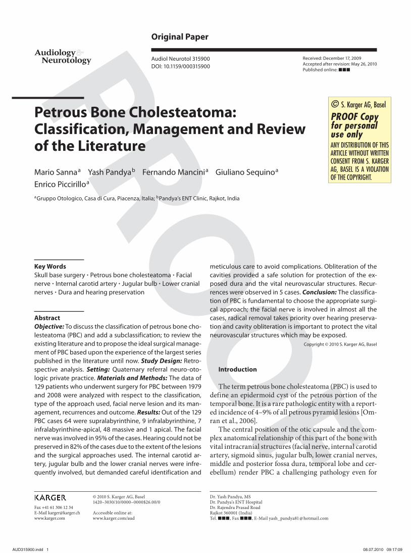
Fig. 1. a Diagrammatic representation of a supralabyrinthine PBC as viewed from the lateral aspect. The dotted ovoid area represents the site of the lesion and the arrows represent the route of spread. Directions for the route of spread are given in table 1. b Coronal HRCT image of the temporal bone showing a supralabyrinthine PBC. The dural plate is eroded. The cochlea is intact.
a
b
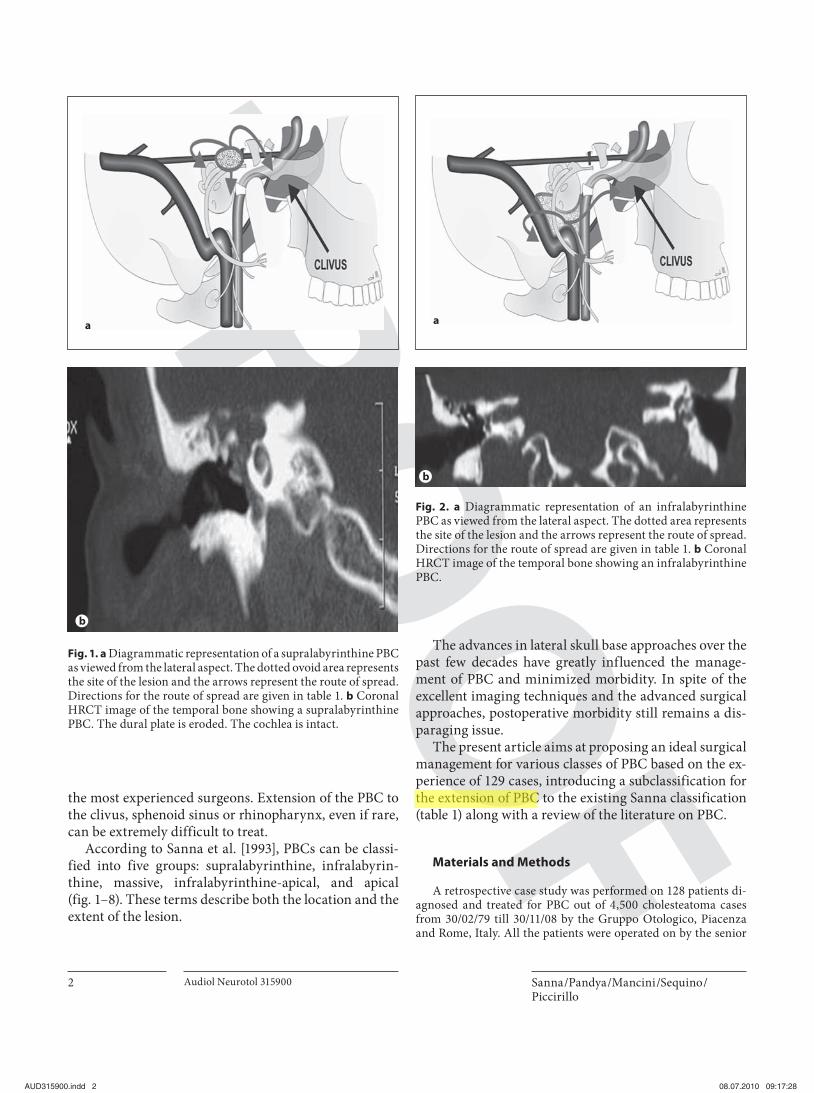
Fig. 2. a Diagrammatic representation of an infralabyrinthine PBC as viewed from the lateral aspect. The dotted area represents the site of the lesion and the arrows represent the route of spread. Directions for the route of spread are given in table 1. b Coronal HRCT image of the temporal bone showing an infralabyrinthine PBC.
Author Please rephrase this passage. I suppose, it should be ‘the extension of PBC pathology’ or something alike. Thank you.
PBC: Classification, Management, Review of the Literature
Audiol Neurotol 315900 3
author. There was 1 patient with bilateral PBC, hence the total number of cases was 129. There were 93 males and 35 females. The right side was affected in 74 patients whereas 55 patients had a left-sided lesion. All these patients underwent a thorough otoneu-rologic evaluation followed by pure-tone audiometric evaluation.
A high-resolution CT (HRCT) scan of the temporal bone (coro-nal and axial scans; bone window images with a width of 1–2 mm) was indicated in all patients with a history of chronic otorrhea, an-acusis, facial palsy, vertigo, or lower cranial nerve paralysis. A his-tory of previous ear surgery was another indication for a CT scan. A cerebral MRI (T 1 , T 2 -weighted images with gadolinium enhance-ment) was obtained in cases suspected of extratemporal spread on the CT scan till 1999, after which we got MRI in all cases.
All the lesions were classified according to Sanna et al. [1993] based on the topographic location and the extent of the choles-
teatoma on the CT scan. Depending on the class of the PBC, the management was planned. Between 1978 and 1985 the surgical approach evolved from open techniques to closed techniques ad-opted in most of the patients since then. Facial nerve function was graded preoperatively, immediately postoperatively and at 1 year postoperatively according to the House-Brackmann grading sys-tem.
Minimum follow-up was 1 year in 126 patients. Subsequently the patients underwent an annual radiological follow-up (CT scan and MRI) for detection of any recurrence for at least 5 years.
a
b
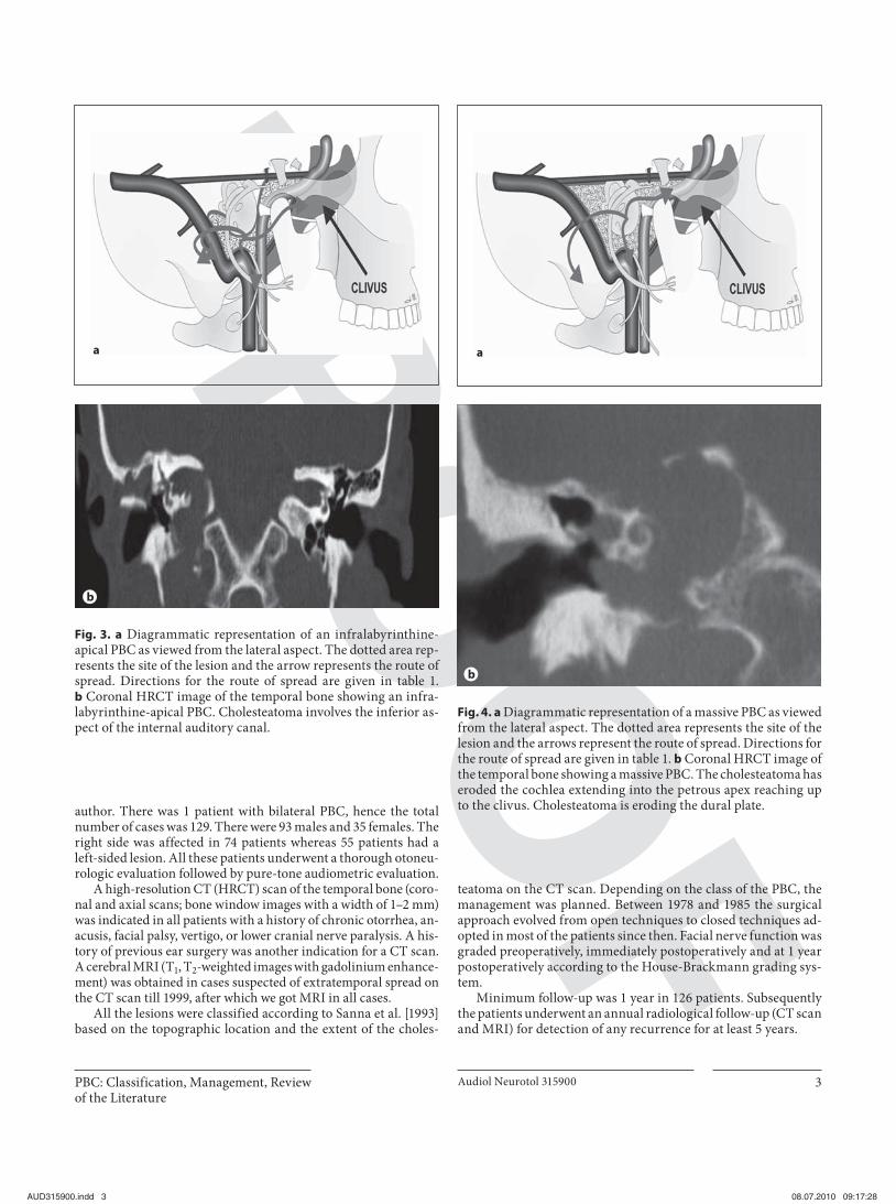
Fig. 3. a Diagrammatic representation of an infralabyrinthine-apical PBC as viewed from the lateral aspect. The dotted area rep-resents the site of the lesion and the arrow represents the route of spread. Directions for the route of spread are given in table 1. b Coronal HRCT image of the temporal bone showing an infra-labyrinthine-apical PBC. Cholesteatoma involves the inferior as-pect of the internal auditory canal.
a
b
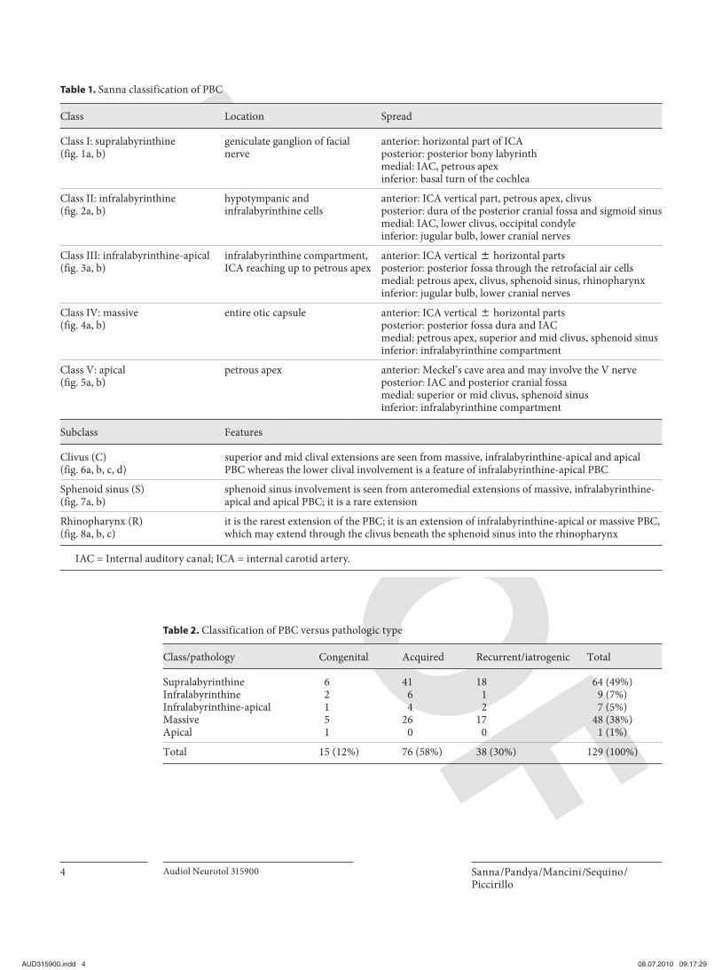
Fig. 4. a Diagrammatic representation of a massive PBC as viewed from the lateral aspect. The dotted area represents the site of the lesion and the arrows represent the route of spread. Directions for the route of spread are given in table 1. b Coronal HRCT image of the temporal bone showing a massive PBC. The cholesteatoma has eroded the cochlea extending into the petrous apex reaching up to the clivus. Cholesteatoma is eroding the dural plate.
anterior: horizontal part of ICA posterior: posterior bony labyrinthmedial: IAC, petrous apexinferior: basal turn of the cochlea
Class II: infralabyrinthine (fig. 2a, b)
hypotympanic andinfralabyrinthine cells
anterior: ICA vertical part, petrous apex, clivusposterior: dura of the posterior cranial fossa and sigmoid sinus medial: IAC, lower clivus, occipital condyleinferior: jugular bulb, lower cranial nerves
Class III: infralabyrinthine-apical (fig. 3a, b)
infralabyrinthine compartment, ICA reaching up to petrous apex
anterior: ICA vertical 8 horizontal partsposterior: posterior fossa through the retrofacial air cells medial: petrous apex, clivus, sphenoid sinus, rhinopharynxinferior: jugular bulb, lower cranial nerves
Class IV: massive (fig. 4a, b)
entire otic capsule anterior: ICA vertical 8 horizontal partsposterior: posterior fossa dura and IACmedial: petrous apex, superior and mid clivus, sphenoid sinus inferior: infralabyrinthine compartment
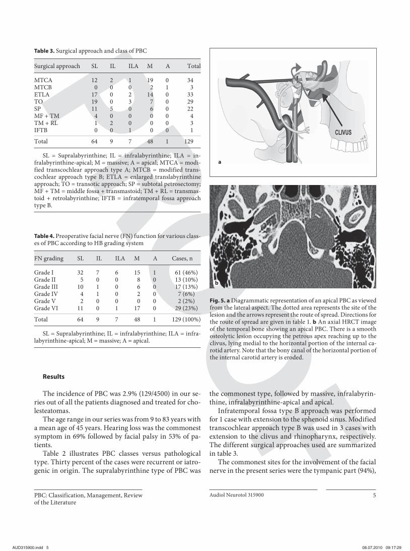
Class V: apical (fig. 5a, b)
petrous apex anterior: Meckel’s cave area and may involve the V nerve posterior: IAC and posterior cranial fossa medial: superior or mid clivus, sphenoid sinusinferior: infralabyrinthine compartment
Subclass Features
Clivus (C) (fig. 6a, b, c, d)
superior and mid clival extensions are seen from massive, infralabyrinthine-apical and apical PBC whereas the lower clival involvement is a feature of infralabyrinthine-apical PBC
Sphenoid sinus (S) (fig. 7a, b)
sphenoid sinus involvement is seen from anteromedial extensions of massive, infralabyrinthine-apical and apical PBC; it is a rare extension
Rhinopharynx (R) (fig. 8a, b, c)
it is the rarest extension of the PBC; it is an extension of infralabyrinthine-apical or massive PBC, which may extend through the clivus beneath the sphenoid sinus into the rhinopharynx
I AC = Internal auditory canal; ICA = internal carotid artery.
Table 2. C lassification of PBC versus pathologic type
Class/pathology Congenital Acquired Recurrent/iatrogenic Total
PBC: Classification, Management, Review of the Literature
Audiol Neurotol 315900 5
Results
The incidence of PBC was 2.9% (129/4500) in our se-ries out of all the patients diagnosed and treated for cho-lesteatomas.
The age range in our series was from 9 to 83 years with a mean age of 45 years. Hearing loss was the commonest symptom in 69% followed by facial palsy in 53% of pa-tients.
Table 2 illustrates PBC classes versus pathological type. Thirty percent of the cases were recurrent or iatro-genic in origin. The supralabyrinthine type of PBC was
the commonest type, followed by massive, infralabyrin-thine, infralabyrinthine-apical and apical.
Infratemporal fossa type B approach was performed for 1 case with extension to the sphenoid sinus. Modified transcochlear approach type B was used in 3 cases with extension to the clivus and rhinopharynx, respectively. The different surgical approaches used are summarized in table 3 .
The commonest sites for the involvement of the facial nerve in the present series were the tympanic part (94%),
a
b
Fig. 5. a Diagrammatic representation of an apical PBC as viewed from the lateral aspect. The dotted area represents the site of the lesion and the arrows represent the route of spread. Directions for the route of spread are given in table 1. b An axial HRCT image of the temporal bone showing an apical PBC. There is a smooth osteolytic lesion occupying the petrous apex reaching up to the clivus, lying medial to the horizontal portion of the internal ca-rotid artery. Note that the bony canal of the horizontal portion of the internal carotid artery is eroded.
geniculate ganglion (84%) and the labyrinthine portion (69%). The facial nerve was involved in multiple segments in most of the patients. Table 4 presents the preoperative distribution of facial nerve function in various types of PBC. Facial nerve decompression was performed in 55 cases, which was the commonest form of management of the facial nerve. It had to be frequently performed even in cases with grade I preoperative facial nerve function. 6/29 patients with preoperative grade VI palsy did not under-go any treatment for the facial nerve because of the long
duration of facial palsy of 1 3 years. Sural nerve grafting was the next common form of treatment for facial nerve lesions (22 cases). End-to-end anastomosis was done in 7 cases and facial-hypoglossal anastomosis was performed in 2 cases. A total number of 10/61 cases with grade I pre-operative function developed various grades of facial pa-resis. Out of these 10 cases 5 had grade II and 4 cases had grade III facial nerve function, and 1 patient with an api-cal PBC had postoperative grade VI facial palsy at 1-year follow-up. Later this patient was operated for facial-hypo-
a
b
c
d
Fig. 6. a Diagrammatic representation of massive PBC with clival extension as seen from the superior aspect (see text). The cholesteatoma involves the petrous apex and superior and mid clivus. b Axial CT scan showing clival extension of a massive PBC. Note the radical mastoid cavity; this patient had previous surgery 15 years prior to presentation. c Diagrammatic representation of extension of massive PBC to clivus and occipital con-dyle as viewed from the superior aspect. d Coronal CT scan showing extension of massive PBC to clivus and occipital condyle.
PBC: Classification, Management, Review of the Literature
Audiol Neurotol 315900 7
glossal anastomosis, following which facial nerve func-tion improved to grade IV ( table 5 ). Out of 128 patients 1 patient died of carcinoma of the colon, and 1 case of bi-lateral PBC was lost to follow-up.
Intraoperative complications included cerebrospinal fluid (CSF) leak in 24 cases (19%), accidental opening up of the jugular bulb and sigmoid sinus in 1 patient each ( ! 1%). There were no perioperative deaths.
Postoperatively, 82% of the patients had a dead ear (106/129 patients), which was a sequela to the surgical ap-
proaches chosen for the radical removal of the disease. Postoperative complications are listed in table 6 . Facial palsy was noted in 10/61 (18%) cases who had been clas-sified as grade I preoperatively.
Out of the 129 cases 126 had a minimum follow-up of 1 year. HRCT of the temporal bone and MRI with fat sup-pression were performed on follow-up. Recurrence was observed in 5 cases ( table 7 ).
Review of the Literature
A review of the literature on PBC was done using a PubMed database search. Inclusion criteria comprised case series describing cholesteatomas involving the pe-trous bone that had more than 10 cases. The following
a
b
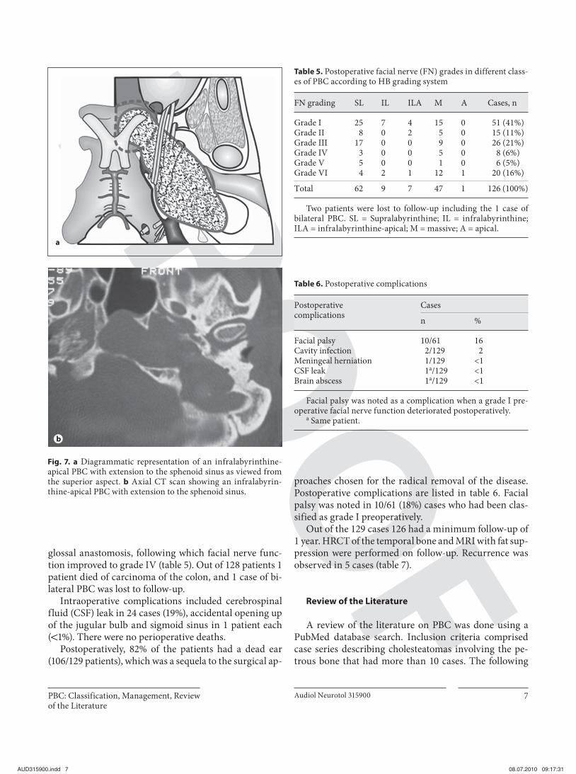
Fig. 7. a Diagrammatic representation of an infralabyrinthine-apical PBC with extension to the sphenoid sinus as viewed from the superior aspect. b Axial CT scan showing an infralabyrin-thine-apical PBC with extension to the sphenoid sinus.
Table 5. P ostoperative facial nerve (FN) grades in different class-es of PBC according to HB grading system
FN grading SL IL ILA M A Cases, n
Grade I 25 7 4 15 0 51 (41%)Grade II 8 0 2 5 0 15 (11%)Grade III 17 0 0 9 0 26 (21%)Grade IV 3 0 0 5 0 8 (6%)Grade V 5 0 0 1 0 6 (5%)Grade VI 4 2 1 12 1 20 (16%)
Total 62 9 7 47 1 126 (100%)
T wo patients were lost to follow-up including the 1 case ofbilateral PBC. SL = Supralabyrinthine; IL = infralabyrinthine; ILA = infralabyrinthine-apical; M = massive; A = apical.
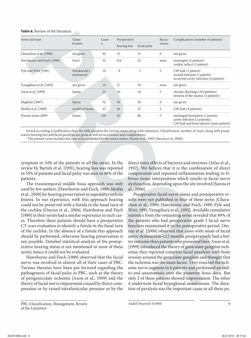
aspects were analyzed: nomenclature, classification, management protocols, preoperative and postoperative facial nerve function, surgical approaches used, hearing preservation, complications and recurrences. There were seven case series in the English literature that met our inclusion criteria, with a total of 221 cases ( table 8 ). In-cluding our own series of 129 cases, the total number of cases was 350. Bartels [1991] presented a review of 80 cas-es that were mentioned in the literature till 1991; we tried to compare the review of this case series and the one done by him. Part of this series has already been published by the senior author [Omran et al., 2006; Sanna et al., 1993]; we included those cases in our series.
Hearing loss was present in almost 70% of these pa-tients on presentation. Facial palsy was the presenting
ba
c
Table 7. R ecurrences and their management
Class Primary surgery
Revision surgery
Supralabyrinthine RPME SP with obliterationInfralabyrinthine SP SP with obliterationInfralabyrinthine SP SP with obliterationSupralabyrinthine MF + TM MTCAMassive SP MTCA
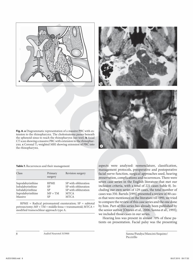
Fig. 8. a Diagrammatic representation of a massive PBC with ex-tension to the rhinopharynx. The cholesteatoma passes beneath the sphenoid sinus to reach the rhinopharynx (see text). b Axial CT scan showing a massive PBC with extension to the rhinophar-ynx. c Coronal T 1 -weighted MRI showing extension of PBC into the rhinopharynx.
PBC: Classification, Management, Review of the Literature
Audiol Neurotol 315900 9
symptom in 54% of the patients in all the series. In the review by Bartels et al. [1991], hearing loss was reported in 55% of patients and facial palsy was seen in 46% of the patients.
The transtemporal middle fossa approach was only used by few authors [Hawthorne and Fisch, 1989; Moffat et al., 2008] for hearing preservation in supralabyrinthine lesions. In our experience, with this approach hearing could not be preserved with a fistula in the basal turn of the cochlea [Omran et al., 2006]. Hawthorne and Fisch [1989] in their series had a similar experience in such cas-es. Therefore these patients should have a preoperative CT scan evaluation to identify a fistula in the basal turn of the cochlea. In the absence of a fistula this approach should be performed, otherwise hearing preservation is not possible. Detailed statistical analysis of the postop-erative hearing status is not mentioned in most of these series; hence it could not be evaluated.
Hawthorne and Fisch [1989] observed that the facial nerve was involved in almost all of their cases of PBC. Various theories have been put forward regarding the pathogenesis of facial palsy in PBC, such as the theory of perigeniculate ischemia [Axon et al., 1999] and the theory of facial nerve impairment caused by direct com-pression or by raised intrafunicular pressure or by the
direct toxic effects of bacteria and enzymes [Atlas et al., 1992]. We believe that it is the combination of direct compression and repeated inflammation leading to fi-brous tissue interposition which results in facial nerve dysfunction, depending upon the site involved [Sanna et al., 2006].
Preoperative facial nerve status and postoperative re-sults were not published in four of these series [Chara-chon et al., 1989; Hawthorne and Fisch, 1989; Pyle and Wiet, 1991; Yanagihara et al., 1992]. Available cumulative statistics from the remaining series revealed that 89% of the patients who had preoperative grade I facial nerve function maintained it in the postoperative period. Om-ran et al. [2006] observed that cases with onset of facial nerve dysfunction ! 12 months preoperatively had a bet-ter outcome than patients who presented later. Axon et al. [1999] introduced the theory of geniculate ganglion isch-emia; they reported complete facial paralysis with bony erosion around the geniculate ganglion and thought that the ischemia was the main factor. They resected the isch-emic nerve segment in 6 patients and performed an end-to-end anastomosis over the posterior fossa dura. But only 2 of these patients showed improvement. The other 4 underwent facial-hypoglossal anastomosis. The dura-tion of paralysis was the important cause in all these pa-
Table 8. R eview of the literature
Series and year Classi-fication
Casesn
P reoperative Recur-rences
Complications (number of patients)
hearing lo ss facial palsy
Charachon et al. [1989] not given 42 35 15 6 not given
Hawthorne and Fisch [1989] Fisch 33 NA 22 none meningitis (2 patients)cardiac infarct (1 patient)
Sor ted according to publication date, the table presents the various series along with references. Classification, number of cases, along with preop-erative hearing loss and facial paralysis are given as well as recurrences and complications.
a The present series includes the case series published by the senior author [Sanna et al., 1993; Omran et al., 2006].
tients; all these patients had paralysis of 1 12 months [Omran et al., 2006].
The cumulative incidence of intracranial complica-tions was 2%. It included 6 patients with CSF leak, 2 cas-es of meningitis and 1 case of brain abscess. Bartels [1991] in his review reported a 6% rate of intracranial infections. Meningitis was reported in 2 cases by Hawthorne and Fisch [1989], and 1 of their patients died of otogenic men-ingitis in an open cavity. Pyle and Wiet [1991] published a comparison of an open technique and cavity oblitera-tion. Cavity obliteration had the advantage of obviating the need for postoperative draining and of protecting vi-tal intracranial structures (internal carotid artery, dura mater, facial nerve, jugular bulb, etc.) that could be ex-posed.
There was no perioperative or immediately postopera-tive mortality as compared to the 3% death rate in the review by Bartels [1991]. There is no mention of choles-teatomas with extension into the petrous bone, particu-larly into the sphenoid sinus and the rhinopharynx, and their management. No definite management protocols are mentioned.
Our series of 129 PBC cases (as far as we know) is the largest series published until now in the English litera-ture. The present article focuses on the nomenclature and presents management guidelines based on the classifica-tion of PBC.
Discussion
The classification of PBC is of paramount importance as it gives information regarding the anatomical position and the extent of the disease. The subclassification pro-posed by us in this article aims at preoperatively diagnos-ing the extension of PBC beyond the temporal bone (usu-ally clivus, sphenoid sinus and rhinopharynx). This helps to plan the surgical approach, which is important to clear the disease from these areas.
The choice of surgical approach has evolved from rad-ical petromastoid exenteration with marsupialization of the cavity to closed and obliterative techniques following complete eradication. Decision making is the crucial as-pect of surgical management. It depends on several fac-tors, the most significant of which are the extent of the
Supralabyrinthine
SNHL
SP/ETLA/TOMF + TM
Hearingnormal
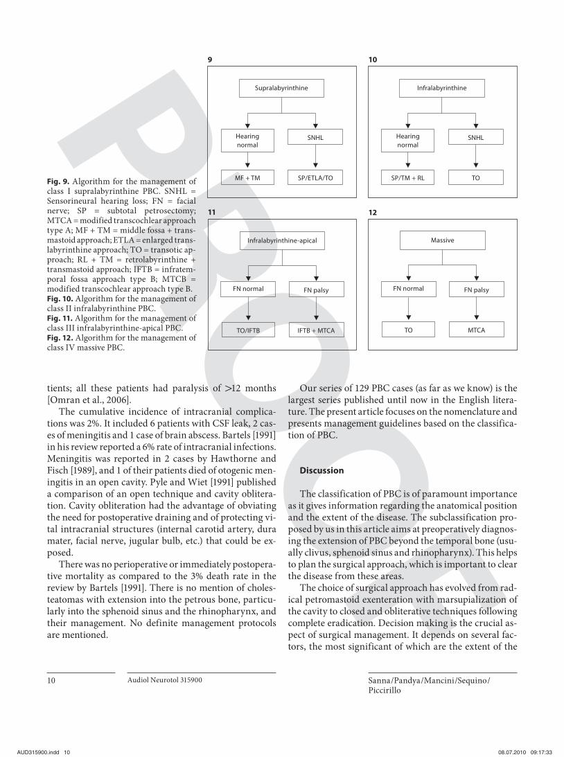
Fig. 9. Algorithm for the management of class I supralabyrinthine PBC. SNHL = Sensorineural hearing loss; FN = facial nerve; SP = subtotal petrosectomy;MTCA = modified transcochlear approach type A; MF + TM = middle fossa + trans-mastoid approach; ETLA = enlarged trans-labyrinthine approach; TO = transotic ap-proach; RL + TM = retrolabyrinthine + transmastoid approach; IFTB = infratem-poral fossa approach type B; MTCB = modified transcochlear approach type B. Fig. 10. Algorithm for the management of class II infralabyrinthine PBC. Fig. 11. Algorithm for the management of class III infralabyrinthine-apical PBC. Fig. 12. Algorithm for the management of class IV massive PBC.
PBC: Classification, Management, Review of the Literature
Audiol Neurotol 315900 11
disease and preoperative facial nerve function. The ap-proach is chosen depending upon the type of PBC and its extent, which should be determined according to the CT scan and MRI findings ( fig. 9–14 , algorithms 1–6).
Algorithms of PBC Management The main factors to be taken into consideration while
treating these lesions are as follows: (1) complete eradica-tion of the disease, (2) preservation of facial nerve func-tion, (3) prevention of CSF leak and meningitis, (4) cavity obliteration, and (5) hearing preservation whenever fea-sible.
In supralabyrinthine PBC ( fig. 9 ), if hearing is normal without any evidence of a fistula in the basal turn of the cochlea, we prefer a middle fossa approach which may be combined with a transmastoid approach depending on the extension of the disease. In the presence of sensori-neural hearing loss or CT evidence of a fistula in the bas-al turn of the cochlea we prefer a radical approach (subto-tal petrosectomy/enlarged translabyrinthine approach/transotic approach) with cavity obliteration ( fig. 15 ). In infralabyrinthine PBC ( fig. 10 ) bone conduction can be preserved with subtotal petrosectomy and blind sac clo-sure of the external auditory canal with cavity oblitera-tion. In very rare cases a transmastoid approach can be combined with a retrolabyrinthine approach to preserve bone conduction.
Hearing preservation is usually not possible in infra-labyrinthine-apical ( fig. 11 ) and massive PBC ( fig. 12 ), hence we use a transotic approach or a modified transco-chlear approach type A depending on preoperative facial nerve function. Modified transcochlear approaches pro-
vide excellent access to the petrous apex, clivus, sphenoid sinus and rhinopharynx depending upon the type used [Sanna et al., 1993]. The posterior rerouting of the facial nerve carries the disadvantage of postoperative facial pa-resis, which is attributed to the loss of blood supply from the deep petrosal artery near the geniculate ganglion; this can recover up to grade III of the HB grading system. Therefore we prefer a transotic approach when facial nerve function is normal preoperatively and a modified transcochlear approach when the patient presents with facial paresis. Facial nerve involvement carries the maxi-mum risk of morbidity as it not only has functional im-pairment but also a psychological effect.
Whenever the cholesteatoma involves the apical por-tion ( fig. 13 ) of the temporal bone or when it extends further to the clivus, sphenoid sinus or rhinopharynx ( fig. 14 ), an infratemporal fossa approach type B is incor-porated into the transotic approach or modified transco-chlear approach type A depending upon the preoperative status of the facial nerve. In selected cases with preopera-tively normal hearing, if the otic capsule is not involved, bone conduction can be preserved with infratemporal fossa approach type B.
Problems and Solutions Hearing Preservation Hearing preservation would be important in excep-
tional cases, i.e. bilateral PBCs or in a PBC of the only hearing ear. However, in the era of BAHA, cochlear im-plants and Vibrant Soundbridge even these cases can be treated successfully with hearing rehabilitation. Bartels [1991] pointed out the possibility of a cochlear implant in
Apical
FN normal + SNHL
IFTB + TOIFTB
Hearing normal+ FN normal
FN palsy + SNHL
IFTB + MTCA
Fig. 13. Algorithm for the management of class V apical PBC.
Extensions to clivus, sphenoid sinus, rhinopharynx
FN normal + SNHL
IFTB + TOIFTB
Hearing normal+ FN normal
FN palsy + SNHL
IFTB + MTCA
Fig. 14. Algorithm for the management of the extensions of PBC to clivus, sphenoid sinus and rhinopharynx.
ears with an accessible cochlear lumen even after labyrin-thectomy and hence the cochlea should not be destroyed beyond what is essential for the clearance of the disease. Another option is a BAHA if bone conduction is pre-served in the ipsilateral ear in cases of PBC in the only hearing ear. Linder et al. [2008] published results on Vi-brant Soundbridge in patients having undergone subtotal petrosectomy; this promises to be a good option for selec-tive patients with mixed hearing loss.
Facial Nerve Facial nerve lesions may vary from simple erosion of
the fallopian canal to total interruption of the nerve with fibrous tissue interposition. Management depends on three principal factors: preoperative status, degree of fa-cial nerve involvement and the extent of the lesion.
Decompression. If there is a compression of the nerve with preserved anatomical integrity, then decompression of the nerve should be performed.
Rerouting. When the lesion is present medial to the fa-cial nerve and complete control over the lesion is ham-pered due to the position of the nerve, then rerouting of the facial nerve is undertaken, which could be partial or complete.
End-to-End Anastomosis. If there is a discontinuity of the nerve or a fibrous tissue interposition, the affected segment should be excised and a tension-free end-to-end anastomosis should be performed.
Nerve Grafting. Whenever the nerve segment lost is long and a tension-free end-to-end anastomosis is not possible, continuity of the nerve can be restored using a nerve graft. We prefer a sural nerve for grafting pur-poses.
Facial-Hypoglossal Anastomosis. In patients with long duration of facial palsy ( 1 12 months) facial-hypoglossal anastomosis is indicated [Falcioni et al., 2003].
In cases where the preoperative facial nerve function is good, a surgeon can be optimistic about a favorable fa-cial nerve outcome depending upon the extension of the pathology.
Internal Carotid Artery The PBC may involve the internal carotid artery in the
vertical and/or the horizontal parts. In this situation a complete control over the artery is important prior to at-tempting its removal. A modified transcochlear approach type A/transotic approach is used for involvement of the vessel in the vertical part, whereas an infratemporal fossa type B/modified transcochlear approach type B is used for involvement of vertical and horizontal parts. In case of a lesion extending into the petrous apex, clivus, sphe-noid sinus and into the rhinopharynx it is important to perform a complete control of the internal carotid artery in order to mobilize the artery if necessary. PBC are less aggressive in terms of arterial involvement [Charachon et al., 1989; Hawthorne and Fisch, 1989; Bartels, 1991; Magliulo, 2007] and are easier to dissect as compared to other tumors (i.e. glomus jugulare). The internal carotid artery has a thick adventitia which resents the dissection of the matrix but it requires extreme caution and surgical skill to clear it.
Sigmoid Sinus and Jugular Bulb The involvement of the sigmoid sinus and the jugular
bulb presents a problem in matrix removal due to the thin wall and the fragility of these structures. In such cases it is important to control the internal jugular vein in the neck prior to the dissection of the matrix. The ligation of the internal jugular vein in the neck and the sigmoid si-nus packing (extraluminal and intraluminal) enables re-moval of the lateral wall of the dome of the jugular bulb and the sigmoid sinus to clear the matrix in the rare cas-es of accidental opening of the bulb. This maneuver also helps in preserving the IX, X, and XI cranial nerves. Dur-ing this procedure bleeding from the inferior petrosalsinus is controlled with Surgicel packing. In cases where its involvement is suspected it is always advisable to en-sure preoperatively the patency of the contralateral cere-bral vein.
Dura The matrix is often adherent to the dura of the middle
and posterior fossa. Bipolar coagulation of all the sus-
Fig. 15. Coronal HRCT suggestive of a fistula in the superior bas-al turn of the cochlea. In this case it is not possible to preserve hearing.
PBC: Classification, Management, Review of the Literature
Audiol Neurotol 315900 13
pected portions of the dura mater can be performed to destroy all the possible remnants of the matrix. We have been using bipolar coagulation in all cases to devitalize the epithelium, and also other authors [Axon et al., 1999] who have used the same technique agree with us. Bipolar-izing large areas of the dura does not lead to any dural necrosis if carefully performed. Long-term follow-up has shown that this maneuver is safe and adequate for com-plete control. There is a risk of opening the dura while removing the adherent matrix causing an intraoperative CSF leak.
CSF Leak CSF leaks resulting from dural tears do not need spe-
cial repair but can be swiftly managed by inserting free muscle plugs into the subarachnoid space through the de-fect and cavity obliteration with fat. CSF leak from the internal auditory canal can be treated by adopting a translabyrinthine approach.
Examination of Hidden Areas Once the removal of the disease has been achieved it
is useful to carry out an endoscopic examination of the cavity with a 30-degree rigid endoscope to visualize the hidden areas that might not be accessible to the micro-scope. In some cases epithelium missed by the conven-tional technique can be found on endoscopic examina-tion [Axon et al., 1999]. In our experience, if the approach is correct this technique is rarely required.
Residual Lesions or Recurrences After complete eradication of the disease, it is manda-
tory to obliterate the cavities with autologous abdominal fat. The major disadvantage of cavity obliteration is that the recurrence cannot be directly visualized and detect-ed. Therefore it is mandatory to follow up these patients radiologically. We perform a high-resolution CT scan and a cerebral MRI (T 1 , T 2 -weighted images with fat sup-pression) and gadolinium enhancement every year for at least 5 years.
Recurrent/iatrogenic cholesteatomas can be avoided if the surgeries for middle ear and mastoid cholesteatomas are meticulously performed. To achieve complete clear-ance we advocate that in the canal wall up (CWU) tech-nique for cholesteatoma the operation has to be staged: second stage surgery is mandatory (after 1 year) to avoid the risk of recurrence and future development of PBC, especially when we are dealing with children or young patients, whereas in the canal wall down (CWD) tech-nique it is mandatory to perform a good mastoidectomy
with saucerized margins, adequately exposed anterior and posterior epitympanum and with the facial ridge lowered till the level of the facial nerve. The cholesteato-ma matrix should be accurately removed from the middle ear cleft after it has been adequately exposed to prevent any recurrences. Following these principles a second stage is performed in cases in which cholesteatomas oc-cupy the oval window or the anterior epitympanum or infiltrate deeply into the hypotympanic cells. In cases with small cholesteatomas an alternative to the second stage can be a CT scan of the middle ear and temporal bone. In all cases of middle ear cholesteatomas treated either with the CWD or CWU technique, long-term fol-low-up is strongly recommended.
Conclusions
• The management of PBC requires thorough pretreat-ment evaluation, planning, execution and a meticu-lous radiological follow-up.
• Classification is fundamental in order to apply the ap-propriate surgical approach.
• The facial nerve requires special consideration as it gets involved in almost all the cases and is a corner-stone of management.
• Radical removal takes priority over hearing preserva-tion even in only hearing ears thanks to the possibil-ity of hearing rehabilitation (CI, BAHA and Sound-bridge).
• Cavity obliteration holds the key for a problem-free cavity.
• Regular follow-up is mandatory.
Acknowledgment
This study was supported by a grant from the Associazione Italiana Neuro-Otologica (A.I.N.OT).
References Atlas M, Moffat D, Hardy D: Petrous apex cho-lesteatoma: diagnostic and treatment dilem-mas. Laryngoscope 1992; 102: 1363–1368.
Axon P, Fergie N, Saeed S, Temple RH, Ramsden R: Petrosal cholesteatoma: management considerations for minimizing morbidity. Am J Otol 1999; 20: 505–510.
Bacciu A, Falcioni M, Pasanisi E, Di Lella F, Lau-da L, Flanagan S, Sanna M: Intracranial fa-cial nerve grafting after removal of vestibu-lar schwannoma. Am J Otolaryngol 2009; 30: 83–88.
Author, please note that all the highlighted references are not cited in the text (Bacciu et al. [2009], De Donato et al. [1993], Sanna et al. [1994, 2008], Glasscock [1969], Rosenberg et al. [1986], Jackler and Parker [1992], Selesnick et al. [1996], House and Histelberger [1976]). Please add citations to the text or delete the references from the list. Thank you.
Sanna /Pandya /Mancini /Sequino /Piccirillo
Audiol Neurotol 315900 14
Bartels LJ: Facial nerve and medially invasive pe-trous bone cholesteatomas. Ann Otol Rhinol Laryngol 1991; 100: 308–316.
Charachon R, Martin Ch, Gratacap B, Perron X: Temporal bone cholesteatoma; in Fisch U, Valavanis A, Yasargil MG (eds): Neurologi-cal Surgery of the Ear and the Skull Base. Berkeley, Kugler, 1989, pp 21–28.
De Donato G, Caylan R, Falconi M, Landolfi M, Titliz A, Russo A, Taibah A, Sanna M: Facial nerve management and results in petrous bone cholesteatomas surgery; in Nakano Y (ed): Cholesteatoma and Mastoid Surgery. Proc 4th Int Conf, Niigata, Sept 1992. Am-sterdam, Kugler, 1993, pp 500–506.
Falcioni M, Taibah A, Russo A, Piccirillo E, San-na M: Facial nerve grafting. Otol Neurotol 2003; 24: 486–489.
Glasscock ME 3rd: Middle fossa approach to the temporal bone: an otologic frontier. Arch Otolaryngol 1969; 90: 15–27.
Hawthorne M, Fisch U: The surgical manage-ment of supralabyrinthine and infralabyrin-thine-apical cholesteatoma of the temporal bone; in Fisch U, Valavanis A, Yasargil MG (eds): Neurological Surgery of the Ear and the Skull Base. Berkeley, Kugler, 1989, pp 11–19.
House W, Histelberger W: The transcochlear ap-proach to the skull base. Arch Otolaryngol 1976; 102: 334–342.
Jackler R, Parker D: Radiographic differential diagnosis of petrous apex lesions. Am J Otol 1992; 13: 561–574.
Linder T, Schlegel C, Demin N, van der Westhui-zen S: Active middle ear implant in patients undergoing subtotal petrosectomy: new ap-plication for Vibrant Soundbridge device and its implication for lateral cranium base surgery. Otol Neurotol 2008; 30: 41–47.
Moffat D, Jones S, Smith W: Petrous temporal bone cholesteatoma: a new classification and long-term surgical outcomes. Skull Base 2008; 18: 107–115.
Omran A, De Donato G, Piccirillo E, Leone O, Sanna M: Petrous bone cholesteatoma: man-agement and outcomes. Laryngoscope 2006; 116: 619–626.
Pyle GM, Wiet R: Petrous apex cholesteatoma: exteriorization versus subtotal petrosectomy with obliteration. Skull Base Surg 1991; 1: 97–105.
Rosenberg R, Hammerschlag P, Cohen N, Bergeron RT, Reede D: Cholesteatoma versus cholesterol granuloma of the petrous apex. Otolaryngol Head Neck Surg 1986; 94: 322–327.
Sanna M, Khrais T, Mancini F, Russo A, Taibah A: The Facial Nerve in Temporal Bone and Lateral Skull Base Microsurgery. Stuttgart, Thieme, 2006.
Sanna M, Mazzioni A, Saleh E, Taibah AK, Rus-so A: Lateral approaches to the median skull base through the petrous bone: the systemof the modified transcochlear approach. J Layngol Otol 1994; 108: 1036–1044.
Sanna M, Saleh E, Khrais T, Mancini F, Piazza P, Russo A, Taibah A: Atlas of Microsurgeryof the Lateral Skull Base, ed 2. Stuttgart, Thieme, 2008.
Sanna M, Zini C, Gamoletti R, Frau N, Taibah A, Russo A, Pasanici E: Petrous bone cholestea-toma. Skull Base Surg 1993; 3: 201–213.
Selesnick S, Abraham M, Carew JF: Rerouting of the intratemporal facial nerve: an analysis of the literature. Am J Otol 1996; 17: 793–805.
Yanagihara N, Nakamura K, Hatakeyama T: Surgical management of petrous apex cho-lesteatoma: a therapeutic scheme. Skull Base Surg 1992; 2: 22–27.
Author, please note that all the highlighted references are not cited in the text (Bacciu et al. [2009], De Donato et al. [1993], Sanna et al. [1994, 2008], Glasscock [1969], Rosenberg et al. [1986], Jackler and Parker [1992], Selesnick et al. [1996], House and Histelberger [1976]). Please add citations to the text or delete the references from the list. Thank you.