pH-dependent lipid packing, membrane permeability and fusion in phosphatidylcholine vesicles
S t e f a n o Massa r i , E l e o n o r a F o l e n a , V a l e r i a A m b r o s i n , G i a m p i e t r o Sch iavo a n d R a f f a e l e C o l o n n a
C.N.R. Unit for the Study of PI,ysiology of Mitochondria. Laboratory of Biophysics and Molecular Biology, Institute of General Pathology, Unirersity of Padova. Padova (Italy)
We have studied the rate of membrane fusion, the lipid dynamics and order and the membrane permeability of phosphatidylcholine vesicles as a function of pH. Acidification induced very different effects depending on the state of the bilayer. In liquid-crystalline hilayers, acidification decreased the rate of membrane fusion, the acyl chain motion and disorder and the rate of K + release, whereas in solid bilayers acidification increased the rate of membrane fusion, the lipid aryl chain disorder and the rate of K + release. These pH-dependent modifications are interpreted in terms of conformational a n d / o r packing changes of the phosphatidylcholine head group in the membrane. In solid Uilayers, these changes are not easily accommodated by the rigid structure, and the resulting stress leads to an unstable bilayer.
Introduction
In recent years the effect of pH changes on several membrane- related phenomena, such as binding of external molecules to the membrane, solute diffusion, membrane fusion, and distribution of lipid components in the bilayer has been the subject of considerable interest. Current interpretations of these pH-in- fluenced processes have considered almost exclusively the role of acidification on the polar head of negative phospholipids and phosphatidylethanolamine, or on the molecules interacting with the bilayer. Insufficient at- tention, however, has been paid to the effect of pH on the zwitterionic lipid phosphatidylcholine (PC), which was practically considered as an inert molecule.
Correspondence: S. Massari, Institute of General Pathology, Univer- sity of Padova, Via Trieste 75, 35121 Padova, Italy.
We present experimental evidence that acidification modifies the fusion capacity of PC bilayers, the lipid mobility and order, and the permeability properties of the membrane, presumably as a consequence of modi- fications of lipid packing. Molecular packing of the constituents of biological membranes is critical for several events such as receptor function [1], enzymatic activity [2], solute diffusion [3], segregation of mem- brane components [4], incorporation of proteins into membranes [5] or action of anesthetics [6]. Our results indicate that care should be taken in interpreting pH- dependent phenomena in PC-containing membranes.
Materials and Methods
Phospholipids, 1,6-diphenyl-l,3,5-hexatriene (DPH) and perylene were supplied by Sigma. in order to improve the purity, the lipids were precipitated from acetone as described by Massari and Colonna [7]. N-(7- Nitrobenz-2-oxa- 1,3-diazol-4-yl)dipalmitoylphosphatid- ylethanolamine (N-NBD-PE) and N-(lissamine Rho- damine B suffonyDdipalmitoylphosphatidylethanol- amine (N-Rh-PE) were synthetized in the laboratory of Dr. Montccucco (this Institute). 1-(4-Trimethylam- moniumphenyl)-6-phenyl- 1,3,5-hexatriene (TMA-DPH) was supplied by Molecular Probes, Inc.
Small unilamellar vesicles (SUV) were obtained by sonication in a medium containing 0,1 M KCI and I0
132
mM acetate-Tris. The details of the preparation are given in a previous publication [8]. Multilamellar vesi- cles were produced by hydrating and gently shaking a thin film of dry lipids with a buffered solution at a temperature above that of the lipid phase transition. Labeling of the vesicles was accomplished by adding a few microliters of DPH (in tetrahydrofuran) or TMA- DPH (in dimethylfonnamide) to a vortexed aqueous buffer and then by adding the phospholipid dispersion under stirring to a final l ipid/dye molar ratio of 200:1. Perylene in chloroform was added directly to a lipid solution in chloroform prior to evaporation to dryness, to a final l ipid/dye molar ratio of 200:1.
Measurements of turbidity changes, lipid mixing by N.~qBD-PE and N-Rh-PE energy transfer, electron microscopy of negatively stained samples, vesicle size and fluorescence anisotropy were performed as previ- ously described [7,8]. The excitation and emission wavelengths were 360 nm and 430 nm for DPH and TMA-DPH, and 412 nm and 470 nm for perylene, respectively.
K + release was measured electrometrically in a magnetically stirred cuvette thermostatted with a circu- lating water bath. The PC SUV were sonicated in 0.1 M KCI and 10 mM acetate-Tris (pH 7), stored for one week at 23°C in order to obtain larger vesicles [8], and finally dialyzed overnight at 4°C against 0.1 M choline- CI and 10 mM acetate-Tris (pH 7). The experiment was started by addition of 0.7 v.mol lipids to a sample cuvette (final volume 3 ml) containing 50 mM choline- CI, 0.1 M sucrose, 10 mM acetate-Tris and 50 v.M tetraphenylphosphonium, a lipophilic cation readily permeating the membrane. K + release was initiated by the addition of 1 /.tg of valinomycin.
Phospholipids hydrolysis was investigated according to Punzi et al. [9]. Samples of sonicated and unsoni- cated vesicles incubated at acidic and neutral pH were taken to dryness and redissolved in chloroform/ methanol (2:1, by volume). After bidimensional thin- layer chromatography the hydrolysis was quantified by phosphorus determination of lysophospholipids. Low levels of hydrolysis (below 0.2%) were detected in sonicated and unsonicated vesicles incubated at pH 7. Acidification did not increased significantly the phos- pholipids hydrolysis. Multilamellar vesicles directly produced at acidic pH showed percentages of hydroly- sis of 0.8, while after sonication the percentage of hydrolysis was 2.8.
R e s u l t s
Size increase of PC w'sicles Fig. 1 shows the kinetics of PC SUV dimensions
increase, as determined by the turbidity increase of the vesicle suspension. Acidification of a D M P C / D S P C SUV suspension equilibrated at pH 7 considerably
o b / p H 3 ,pH3
~: / / / pH 5 o /, ," pH 5.8 o~T / / p H , ~ i H 6./, ~ / / / ~ pH5 HEPES /~'~/"
HCI I 3rain I
Fig. 1. pH-dependent rate of absorbance increase of a DMPC/DSPC SUV suspension. The incubation medium contained 0.1 M KCI and 10 -3 M acetate-Tris adjusted to pH 7 (a) or pH 3 (b). Final volume 3 ml, 30°C, 334 nm. The experiments were started by the addition of DMPC/DSPC SUV (1 : I ratio) preequilibrated at 60°C at pH 7 (a) or pH 3 (b) (not shown). Where indicated, HCI or Hepes were added to give the final pH labeling each trace. Broken lines, no additions.
increased the rate of absorbance increase (Fig. la). Conversely, neutralization with Hepes buffer of a SUV suspension thoroughly equilibrated at acidic pH de- creased the rate of absorbance increase (Fig. lb). This effect appears to be related to the pH change rather than to the buffer per se, since the same experimental results were obtained when (i) larger amounts of buffer were present and (ii) the pH was readjusted by addi- tion of Tris instead ~f Hepes (not shown). By compar- ing the traces of l~igs, la and lb, it can be observed that the effect of acidification on the rate of ab- sorbance change was only partially reversed by restor- ing the pH to the initial value, indicating that the process is not completely reversible, it should be noted that the kinetics was measured at 30°C, the tempera- ture at which the process attains its maximum rate (see Fig. 2).
Fig. 2 shows the temperature-dependent rate of absorbance increa~,:e of DPPC and DMPC/DSPC SUV suspensions at neutral and acidic pH. For both systems the process was faster at pH 4, especially at lower temperatures. The SUV composed by two non-ideally miscible PC showed a higher increase than those com- posed by one single component.
In order to ascertain that the absorbance changes detected vesicle size increase and not aggregation, we studied the lipid mixing and analyzed the electron micrographs of negatively stained SUV. Lipid mixing and electron micrographs were performed with both pure and mixed PC systems with essentially the same results. In the following figures, however, experiments are shown for one system only, for the sake of brevity.
Fig. 3 shows an increased lipid mixing in D M P C / D S P C SUV when the pH is lowered. The lipid mixing was monitored by fluorescence intensity changes due to energy transfer between N-NBD-PE and N-Rh- PE probes [8].
I 00Fi DMPoC/DSPC pHt,
-° //
o.o2
PPC
10 20 30 40 50 Temperot ure,°C
Fig. 2, Temperature-dependent rate of absorbance increase of PC SUV suspensions at pH 7 and 4. The incubation medium contained 0.1 M KCI and t0 -'~ M acetate-Tris adjusted to pH 7 (closed symbols) or pH 4 (open symbols). Final volume 3 ml, 334 nm. The experiments were started by the addition of DMPC/DSPC SUV ( l : l ratio)or DPPC SUV preequilibrated at 60°C at the pH indi-
cated in the figure. Lipid concentration was 0,5 mM.
Fig, 4A shows the his tograms of the vesicle d i amete r dis tr ibut ion obta ined from electron micrographs. D P P C SUV incubated at 10°C for 1 h were t ransformed into larger vesicles in an acidic medium. Indeed, the aver- age d iameters were 28 and 36 nm at pH 7 and 4, respectively. The maximum error in the d iamete r mea- surements from the electron micrographs was ± 3 nm. Spontaneous fusion of PC SUV has been observed by
4C
o
==
r: 2c
o
pH4
Tempemture°C Fig, 3, Temperature-dependent lipid mixing of a DMPC/DSPC SUV suspension at pH 7 and 4. The percentage of lipid mixing was obtained by mixing 40 p.M of DMPC/DSPC SUV in I : l ratio containing H-NBD-PE and N-Rh-PE with 160 p.M of SUV devoid of the probes, and by measuring the fluorescence intensity increase as
previlmsly described [8].
133
e ~'~pH6 t 30
"-" ['IpH ~ l
~20 1(2
loo 2o n~.mmr, nm
Fig. 4. Histograms of the vesicle diameter obtained from micrographs of negatively stained samples. (A) 1 mM of DPPC SUV were incubated for 1 h at 10°C either at pH 7 (continuous lines) or at pH 4 (dotted lines). (B) DPPC SUV ! mM were incubated at 60°C for a
week either at pH 7 (continuous lines) or at pH 4 (dotted lines).
Lentz e t al. [10] to occur slowly when the vesicles are in the l iquid,crystal l ine state. Fig. 4B shows that DPPC SUV were t ransformed into a more he terogeneous populat ion of both small and intermediate-s ize vesicles when they were s tored at 60°C for a week at pH 7, with increase of the average d iamete r to 40 + 4 nm. The size increase process was inhibi ted when the SUV were s tored at 60°C for one week at pH 4, the average d iamete r be ing 32 + 4 nm.
Fig. 5 shows the pH dependence of the rate of absorbance increase of D M P C / D S P C SUV suspen- sions sonicated at pH 7.2. The rate of absorbance change became progressively faster as the pH was lowered below 7, reached a maximum at pH 2.5, and
O.0tE
"• O.01[
o ~ O.OO~
o \ \
I I I I i 1 i 1 2 3 4 5 6 7
pH Fig. 5. pH-dependent rate of absorbance increase of DMPC/DSPC SUV suspensions sonicated at pH 7. 0.25 mM DMPC/DSPC SUV in the 1 : t ratio were sonicated at pH 7. The DMPC/DSPC SUV were preequilibrated at 60°C after acidification to the appropriate DH
value. The rate of absorbance change was measured at 30eC,
134
o. o o
g = ~0~. o
~ A
p H / . ~ . DPPC
2'0 'O 20 'O ,' Temperot ure.°C
~ o H 7 B 0.3
[ DMPC/DSPC • ~
pH ~
02 g • ~
o
Fig. 6. Temperature-dependent DFH fluorescence anisotropy in PC SUV. 0.02 mM of DPPC (A). or DMPC/DSPC SUV in the 1 : I ratio (B), containing 0.5% DPH, were incubated at 50 ° C for 3 h at neutral or acidic pH. The fluorescence anisotropy was measured by starting from a temperature in which the bilayers were in the liquid-crystalline state and by lowering the sample temperature at a rate of 0.5 C*/min. The bars
indicate the standard error estimated in three different SUV preparations.
decreased as the pH was decreased further. The phos- phoric group is neutralized at pH values lower than 2.5, and the presence of a net positive charge on the vesicle surface provides sufficient repulsive electro- static energy to prevent close vesicle contact and mem- brane fusion.
Lipid dynamics and order The easiest and most frequently used method to
measure the physical state of the bilayers is steady-state fluorescence depolarization of the probe DPH, which predominantly reflects the mean structural order of membrane lipids [11]. Fig. 6 shows the temperature-de- pendent fluorescence anisotropy of DPH incorporated into DPPC and D M P C / D S P C SUV incubated at neu- tral or acidic pH. The midpoint temperature of the main lipid phase transition, T c, was progressively in- creased as the pH was lowered: in DPPC SUV, T c shifted by approx. 0.9 C ° at pH 4 and 7 C'* at pH 3. The anisotropy increased after acidification, when the bilay- ers are in the liquid-crystalline state, whereas it de- creased when the bilayers are in the solid state. The same results were obtained with multilayered DPPC vesicles, whose diameter is much larger than that of sonicated vesicles (not shown).
The anisotropy decrease in solid bilayers at acidic pH may be due either to a decreased order of the acyl chains, or to an increased energy transfer between segregated probe molecules. DPH could indeed prefer- entially partition into microdomains when lipid bilayers freeze at acidic pH. Moreover, it is possible that the lipid bilayer becomes more ordered in acidic conditions
and that the probe rotation results more from 'slipping' than from 'sticking' motion [12]. Finally DPH, which is predominantly located at and in the close vicinity of the center of the bilayer [13], has a certain amount of molecules oriented with their long axis perpendicular rather than parallel to the lipid acyl chain. According to Straume and Litman [14], the relative amount of molecules oriented in the two directions may change with temperature, acyl chain unsaturation and lipid packing of the bilayers. At present it is impossible to distinguish between shift in DPH distribution, changes in the rotational characteristics of the probe and en- hanced lipid disorder.
Two other fluorescent probes were used in order to further elucidate the physical changes accompanying the acidification: TMA-DPH and perylene. The amphi- pathic TMA-DPH molecule resides in the interfacial and head group regions of the membrane, and appears to exhibit only a unimodal distribution with maximum probability centered around the bilayer normal [14-17]. The use of TMA-DPH avoids therefore heterogeneous distribution of the probe in the bilayer, and minimizes segregation owing to electrostatic repulsion of the probe head group. Moreover, the probe is less sensitive to the entangling effect of nearby hydrocarbon chains since the phospholipid motion is slower at the interfa- cial rather than at the end-chain region. Perylene molecules preferentially align the plane of their ring system parallel to the lipid acyl chains. The rate of in-plane rotation is substantially faster than the ra*e of out-of-plane rotation, so that the steady-state fluores- cence anisotropy of the probe seems not to be greatly
~1- " .---. o ~ . I
g : I i T I I I I I -¢ n TMA_OPH /
e ~ o . . . . . . . . . . .
~-15_ -30 " ~ ~ ' m ~ : c
-/,5 L--
I I l I I I I c 10 20 30 /~0 50 60 70
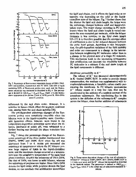
Temperoture.'C Fig. 7. Percentage of fluorescence anisotropy change of DPH, TMA- DPH and perylene, incorporated into PC SUV. 0.02 mM of lipids containing 0.5% of fluorescent probes were used, and the fluores- cence anisotropy was measured as described in Fig. 6. The percent- age is defined as 100(Apn 7- AvH4)/Avm, where A is the fluores- cence anisotropy of the probes. Panel A, DPH; panel B, TMA-DPH;
panel C. perylene.
influenced by the acyl chain order. However, it is sensitive to factors which affect the in-plane rotational rate, among them the acyl chain mobility [18].
The pH-dependent anisotropy changes of the fluo- rescent probes were completely reversible when the bilayers were in the liquid-crystalline state. Consider- able hysteresis effects, however, were found in the temperature-dependent anisotropy curve when the hi- layers, incubated at acidic pH, were subjected to a further heating run through the phase transition (not shown).
Fig. 7 shows the percentage change of the fluores- cence anisotropy of the three probes incorporated into DMPC, DPPC and DSPC SUV when the pH was decreased from 7 to 4. Acidic pH increased the anisotropy at temperatures where the PC bilayers pns- sess large regions of lipids in the liquid-crystalline state, and the maximum change occurred at a tempera- ture near the completion temperature of the main lipid phase transition. At pH 4 the anisotropy of TMA-DPH, like that of DPH, was lower in solid bilayers, whereas the anisotropy of perylene was higher. Since TMA-DPH detects the lipid order, while perylene detects the lipid motion, we can conclude that acidification immobilizes
135
the lipid acyl chains, and it affects the lipid order in an opposite way depending on the solid or the liquid- crystalline state of the bilayer. Fig. 7 further shows that the shorter the lipid acyl chain length, the larger were the anisotropy changes between solid and liquid-crys- talline state. The major change occurring in PC mem- branes when the lipid acyl chain length is varied con- cerns the area occupied per molecule, while the bilayer thickness is less sensitive to the lipid chain length [19-21]. It is therefore possible that the stronger effect of acidification is due to the reduced distance between the polar bead groups. According to this interpreta- tion, the pH-dependent variations of the lipid mobility and order are consequent to a change in the interac- tion between neighboring PC molecules rather than to a change in the physical state of a single PC molecule. This mechanism leads to the interesting consequence that acidification can decrease the miscibility between PC molecules in a mixture if the acyl chain length of the lipid components is different.
Membrane permeability to K + The release of K + was measured electrometrically
in K+-Ioaded DMPC SUV. In order to provide charge compensation, the medium was supplemented with re. traphenylpbosphonium, a lipopbilic cation readily per- meating the membrane. In PC bilayers spontaneous K + release occurs at a very low rate, that can b¢ accelerated by the addition of a catalytic amount of the ionophore valinomycin. The rate-limiting flux in this system is the diffusion of the valinomycin-K + complex across the bilayer, since further addition of valinomycin
~ 20
_g
"5
w P T o m p ° ~ l u r . ,
Fig. 8. Temperature-dependent changes of the rate of vatlnomycin- induced K + release in DMPC/DSPC SUV. DMPC/DSPC SUV in 1 : l ratio were used, and the K + release was measured as described in the Methods section. The percentage is defined as 100(Ypu4. a - Vpm)/Vpm, where V is the initial rate of valinomycin-mediated K +
release.
136
increases the K + release rate. Fig. 8 shows that, in the liquid-crystalline state, the kinetics of K + release medi- ated by valinomycin was inhibited as the pH was low- ered. The inhibition of K + release is in agreement with the observation that the conductance of liquid-crystal- line PC bilayers to electrolytes and to proton/hydrox- ide decreases when the external pH is lowered [22-25]. The rate of valinomycin-mediated K + transport is very sensitive to the fluidity of the lipid bilayer [26,27], and inhibition of the diffusion process at acidic pH may therefore be consequent to a lower fluidity. On the contrary, valinomycin-K + diffusion rate in gel state membranes was accelerated at acidic pH. The opposite effects induced by acidic pH in liquid-crystalline and solid membranes exclude the possibility that the per- meability changes are mediated by pH-dependent sur- face potential changes.
Discussion
Enhancement of the charge of the lipid polar head group generally causes the lipids to pack less closely in the bilayers and to melt at a lower temperature, due to electrostatic repulsion [29-35]. However, partial proto- nation of the phosphoric group of the zwitterionic PC molecules raises the midpoint temperature of the lipid phase transition, 7~:, instead of lowering it. The in- crease in T c suggests that the positively charged qua- ternary amine causes only a small electrostatic repul- sion [36]. In fact, the synthetic lipid HO-(CH2) 2- C(CH3) 3 esterified to dipalmitoylphosphatidic acid, with a non charged group which is otherwise similar in size and structure to choline, melts at a similar temper- ature as DPPC [36]. Likewise to the binding of hydro- gen ions, the binding of di- and trivalent cations to PC has been shown to cause a membrane rigidification as a consequence of the increased lipid packing [37-40]. It is extremely unlikely that fatty acid contamination in our PC preparations, either pre-existing or produced by PC hydrolysis during incubation at low pH, could account for the observed results. Three arguments strongly suggest that the role of fatty acid is negligible: (i) saturated fatty acids induce PC vesicle size enlarge- ment only above a 'critical' concentration, approx. 3-5% of total lipid concentration [28]. The fatty acid contamination of our acetone-treated PC preparations is below 0.2% i.e., well below this value (see Methods); (ii) the maximum rate of fatty acid-induced vesicle aize enlargement occurs 2-3 C ° below the vesicle transition temperature [28] while we obtain a different tempera- ture dependence both at neutral and acidic pH (Fig. 2); (iii) the rate of fatty acid-induced vesicle size enlarge- ment is inhibited at alkaline pH and remains constant below pH 6.5 [28] while the rate of vesicle size enlarge- ment described here is progressively increased as pH is lowered (Fig. 5).
pH-dependent properties of liquid-crystalline PC bilayers The fluorescent response of the probes DPH, TMA-
DPH and perylene indicates that, in the liquid-crystal- line state, PC molecules experience a reduction in the disorder and motion after acidification. Similarly, the decreased membrane permeability to K + and the inhi- bition of the fusion rate suggest higher compactness and stability of the bilayer under acidic conditions. The hydrogen ion binding to the PC head group may re- duce the lipid mobility either by decreasing the amount of gauche conformers in the acyl chain, or by closely packing the lipid chains. A closer chain approach is possible if the PC head group, which is predominant in determining the area per molecule, reduces its area: a decrease of the total vesicle surface area should be expected. PC SUV are, however, highly resistant to changes in the area per lipid molecule [41-46]. If the acyl-chain packing density remains unaltered in SUV upon acidification, the reduced acyl chain motion can be explained by a reduced extent and rate of trans- gauche isomerization in the chains.
pH-dependent properties of solid PC bilayers PC SUV show increased lipid disorder, permeability
and fusion capacity when they are frozen at acidic pH as compared to neut~al pH. The compactness of solid bilayers is therefore partially relieved under acidic con- ditions. Our experimental techniques, i.e., steady-state fluorescence anisotropy and solute diffusion rate mea- surements, yield only qualitative information on the lipid packing properties and are unable to properly analyze the details of the molecular interactions. Nev- ertheless, our experimental data can be rationalized with the hypothesis that a pH-dependent conforma- tional change of the PC polar group takes place in the solid bilayers. When PC bilayers incubated at acidic pH are in the solid state or contain domains of solid lipids, both the fusion rate and the response of the fluorescent dyes are not completely reversed after pH readjustraent at neutral value. These hysteresis effects are proLably due to a higher affinity of the solid lipids for hydrogen ions, which may be caused by a different head group configuration and packing in the mem- brane consequent to acidification. A conformational change of the PC head group may be induced, for instance, by a change of the hydrogen bond network arout,d the polar groups. The reduced hydration and the higher negative surface potential present in solid PC bilaycr [47] may further contribute to increase the proton affinity. A more extended conformation of the phosphorylcholine group has been observed after cation binding to liquid-crystalline PC [40,48-51].
In the liquid-crystalline state, there is considerable flexibility in the interplay between phosphorylcholine lattice and hydrocarbon matrix. In solid state bilayers, changes in the head group configuration and packing
are more difficult to accomodate by corresponding changes in the acyl chain configuration. If, for instance, the hydrogen ion binding reduces the area of the PC head group, the lipid molecules in the outer layer of the SUV membranes undergo a transition trom a trun- cated cone to a more eilindrical configuration, which does not properly match into curved bilayers. The resulting stress may be partially relieved by tormation or enlargement of structural defects between highly ordered lipid donmins. These boundary regions of mis- match in molecular packing cause a discontinuous ar- rangement in the bilaycr architecture which destabi- lizes the membrane stroctnre, These considerations explain the decreased order detected by the fluores- cence response of DPH and the increased K + perme- ability and fusion rate. Moreover, the effect of acidifi- cation is more relevant in fusing SUV composed of two non-ideally miscible PC: in these mixtures the struc- tural defects have been shown to be larger than in the mixtures of miscible PC [8,52].
In conclusion, our results indicate that acidification induces structural and dynamic changes in model mem- branes composed of PC. These changes are relevant at pH values not far from those occurring in vivo in some natural processes such as endosome acidification and steady state mainteinance of acidic pH in iysosomes. In this connection, experiments are under way to extend our approach to natural intracellular vesicles and to large unilamellar vesicles, whose curvature radius is closer to that of intraceilular vesicles. Although the major changes in the structure and function of natural membranes upon acidification involve negative phos- pholipids or phosphatidylethanolamine, the high con- tent of PC and its sensitivity to acidification can play an important role in modulating the lipid acyl chain mobility and order in the membrane domains, the membrane permeability, the surface properties of the bilayer, the miscibility properties of the lipid compo- nents and the interactions between proteins and sur- roundings lipids.
Acknowledgments
The authors wish to thank Mr. P. Veronese for his skilfull technical assistance, Dr, P. Bernardi for critical reading of the manuscript, and Dr. G,Viola from Fidia Research Laboratories, Abano T., Italy, for phospho- lipids hydrolysis determination. This research was sup- ported in part by the Ministero della Pubbiiea Istruzione and by ASSNE (Associazione Sviluppo Scienze Neurologiche).
References
1 Shinitzky, M. (1984) Biomcmbranes 12. 585-601. 2 Raison, J.R.. Lyons, J.M., Melhorn, RJ. and Keith, A,D, (1971)
J. Biol. Chem. 246, 4036-4040.
137
3 Block, M,C., Van der Neut-Knk, E.C.M., Van Deenen, L,L.M. and De Gier. J. (1975) Biochim. Biophys. Acta 406,1~17-196.
4 Jain, M.K. (1983) in Membrane Fluidity in Biology (Aloia, R.C., ed.), Vol. 1, pp. 1-37, Academic Press, New York.
5 ~'ain, M.K. and ~kim, D. (1987) Biochim, Biaphys. Aeta 906, 33-68.
6 Ueda. 1., Thasiro, C. and Arakana, K. (1977) Anesthesiology 46, 327-332.
7 Massari, S, and Co[onna, R. (1986a) Chem, Phys. Lipids 39, 203-220.
8 Massari, S. and Colonna, R, (1986b) Biochlm. Biophys. A¢ta 863, 264-276.
9 Punzi, L,, Todesco, S., Tnffano, G., Corona, R., Bigon, E. and Bruni, A. (1986) Rheumatol. Int, 6, 7-11.
l0 Lentz, B.R., Carpenter, T.J. and Alford, D.R. (1987) Biochem, istry 26, 5389-5397.
I1 Lakowicz. J.R., Prendergast, F.G. and Hogen, D. (1979) Bio- chemistry 18, 508-519.
39 Kataoka, R., Aruga, S., Mitaku, S., Kinosita, K. Jr. and Ikegami. A. (1985) Biophys. Chem. 21,277-2~q'.
40 Conti, J., Halladay, H.N. and Petetshcim. M. (19~7) Biochim. Biophys. Aeta 902. 53-64.
41 Johnson. S.M. and Buttress, N. (1973) Bi,xhim. Biophys Acta 307, 20-26
42 Aune, K.C., Ganaghgr, J.G., Gotto, A.M., Jr. and Morriset, J.D. (1977) Biochemistry 16, 2151-2155.
43 Ceuterick, F., Hermans, K., Desmedt, H., Niewenhuysen, P. and Clauwaert, J. 0979) Chem. Phys. Lett. 62, 341-343.
44 Cornell, B.A., Flechter, G.C., Middlehurst, J. and Separovic. F. (1981) Biochim. Biophys. Acta 642, 375-380.
45 Liehtenberg, D., Freire, E., Schmidt, C.F.. Barenholz, Y., gner, P.L. and Thompson, T.E. (1981) Biochemistry 20, 3L 3467.
46 Sun, S.-T., Milon, A., Tanaka, T., Ourisson, T. and Nakatam (1986) Biochim. Biophys. Act:: 860, 525-530.
47 Tatulian. S.A. (1987) Biochim. Biophys. Acta 901, 161-165. 48 Brown, M.F. and Seelig, J. (1977) Nature 269, 721-723. 49 Akutsu. H. and Seelig, J. (1981) Bioehemistt'y 20, 7366-7373. 50 Siiderman, D., Arvidson, G., Lindblom, G. and Fontell, K. (1
Eur. J. Biochem. 134, 309-314. 51 Altenbach. C. and Seelig, J. ~ 1984) Biochemistry 23, 3913-3 52 Nicolussi, A., Massari, S. and Colonna, R. (1982) Biochem