Pharm 201 Lecture 09, 2009 1 Understanding Sequence, Structure and Function Relationships and the Resulting Redundancy Pharm 201/Bioinformatics I Philip E. Bourne Department of Pharmacology, UCSD
Transcript
Pharm 201 Lecture 09, 2009 1
Understanding Sequence, Structure and Function Relationships and the
Resulting Redundancy
Pharm 201/Bioinformatics I
Philip E. BourneDepartment of Pharmacology, UCSD
Pharm 201 Lecture 09, 2009 2
Agenda
• Understand the relationship between sequence, structure and function. Consider specifically:– sequence-structure– structure-structure– structure-function
• Take home message: a non-redundant set of sequences is different than a non-redundant set of structures is different than a non-redundant set of functions
Pharm 201 Lecture 09, 2009 3
Why Bother?
• Biology:– A full understanding of a molecular system
comes from careful examination of the sequence-structure-function triad
– Each triad is then a component in a biological process
• Method:– Bioinformatics studies invariably start from a
non-redundant set of data to achieve appropriate statistical significance
Pharm 201 Lecture 09, 2009 4
Background – RMSD Defined
Protein A
a1
a2
a3
a4
Protein B
b1
b2
b3
b4
RMSD = Sqrt (1/N Σ | di|2)i=1
i=N
aN bN
d1d1
d2
d3
d4
Represents the overall distancebetween two proteins usually averaged over their Calpha atoms denoted here a and b
Thus RMSD is the square root of the sum of the squares of the distances between all Calpha atoms
Rule of thumb:1-2 Å RMSD the proteins are close<6 Å RMSD they are likely related
Note: Assumes you know residuescorrespondences
Pharm 201 Lecture 09, 2009 5
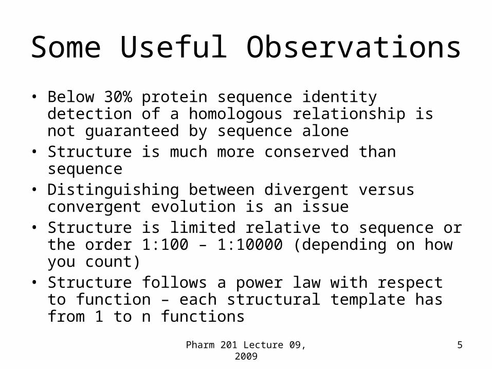
Some Useful Observations
• Below 30% protein sequence identity detection of a homologous relationship is not guaranteed by sequence alone
• Structure is much more conserved than sequence• Distinguishing between divergent versus convergent
evolution is an issue• Structure is limited relative to sequence or the order
1:100 – 1:10000 (depending on how you count)• Structure follows a power law with respect to function –
each structural template has from 1 to n functions
Pharm 201 Lecture 09, 2009 6
Relationship Between Sequence and Structure
Pharm 201 Lecture 09, 2009 7
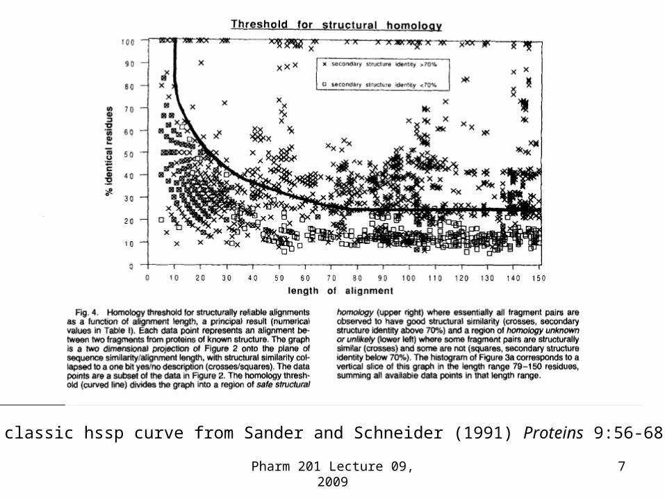
The classic hssp curve from Sander and Schneider (1991) Proteins 9:56-68
Random 1000 structurally similar PDB polypeptide chains from CE with z > 4.5 (% sequence identity vs alignment length)
Twilight Zone
Midnight Zone
Sequence vs Structure – Another Perspective
Alignment Length
% Seq. Id.
1HMP:A
Glycosyltransferase
1PIV:1Viral Capsid Protein
80 Residue Stretch (Yellow) with Over 40% Sequence Identity
There Are No Absolute Rules - Similar Sequences – Different Structures
10Pharm 201 Lecture 09, 2009
Pharm 201 Lecture 09, 2009 11
Given This Complex Relationship a Non-redundant Set of
Sequences Does not Imply a Non-redundant Set of Structures
Pharm 201 Lecture 09, 2009 12
Structure vs Structure
Pharm 201 Lecture 09, 2009 13
Structure Alignments using CE with z>4.0
Homology modelingis used here
The Russian Doll Effect
Structure Is Highly Redundant
Pharm 201 Lecture 09, 2009 14
We will be revisiting this in the next couple of lectures
• Specifically:– How do we capture this redundancy?– What systems are commonly used to express
this redundancy and what do they bring to our understanding of biology?
• For now consider what this means using the most popular structure classification scheme - SCOP
Pharm 201 Lecture 09, 2009 15
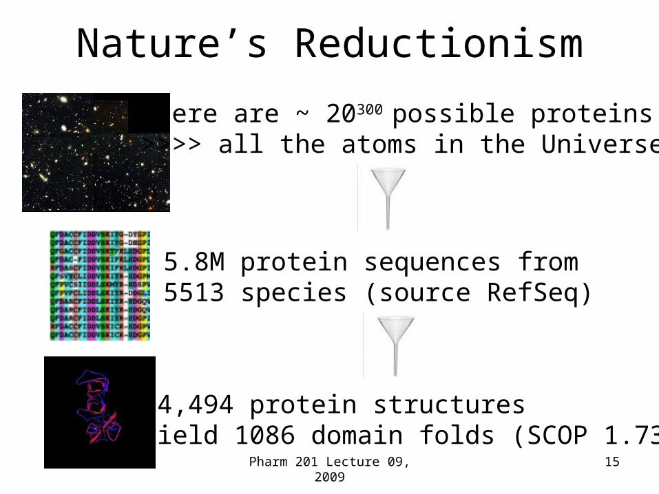
Nature’s Reductionism
There are ~ 20300 possible proteins>>>> all the atoms in the Universe
5.8M protein sequences from 5513 species (source RefSeq)
34,494 protein structures yield 1086 domain folds (SCOP 1.73)
Pharm 201 Lecture 09, 2009 16
The SCOP Hierarchy v1.73Based on 34494 Structures
7
1086
1777
3464
97178
This is remarkable!Explains the one fold many functions
Specific Examples From the SCOP
Hierarchy
17Pharm 201 Lecture 09, 2009
Pharm 201 Lecture 09, 2009 18
Protein Domains
• Definition– Compact,
spatially distinct– Fold in isolation– Recurrence
Pharm 201 Lecture 09, 2009 19
Structure vs Function
Pharm 201 Lecture 09, 2009 20
Some Basic Rules Governing Structure-Function Relationships …• The golden rule is there are no golden
rules – George Bernard Shaw
• Above 40% sequence identity sequences tend to have the same structure and function – But there are exceptions
• Structure and function tend to diverge at the same level of sequence identity
Pharm 201 Lecture 09, 2009 21
Structure vs Function
This is even more complicated than the relationship between sequence and structure and not as well understood
Pharm 201 Lecture 09, 2009 22
Complication Comes from One Structure Multiple Functions
• We saw this from GO already
• phosphoglucose isomerase acts as a neuroleukin, cytokine and a differentiation mediator as a monomer in the extracellular space and as a dimer in the cell involved in glucose metabolism
Pharm 201 Lecture 09, 2009 23
Consider an Example Relative to SCOP
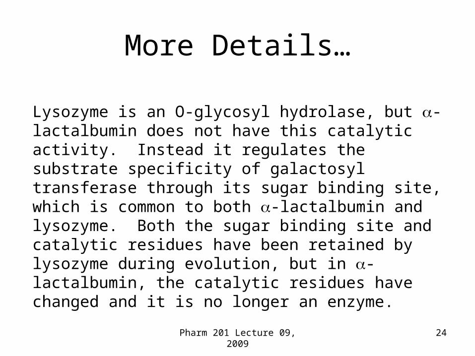
• lysozyme and alpha-lactalbumin:– Same class alpha+beta– Same superfamily – lysozyme-like– Same family C-type lysozyme– Same fold – lysozyme-like– different function at 40% sequence identity
Lysozyme is an O-glycosyl hydrolase, but -lactalbumin does not have this catalytic activity. Instead it regulates the substrate specificity of galactosyl transferase through its sugar binding site, which is common to both -lactalbumin and lysozyme. Both the sugar binding site and catalytic residues have been retained by lysozyme during evolution, but in -lactalbumin, the catalytic residues have changed and it is no longer an enzyme.
Pharm 201 Lecture 09, 2009 25
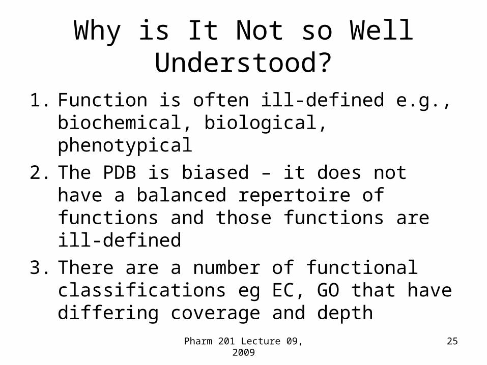
Why is It Not so Well Understood?
1. Function is often ill-defined e.g., biochemical, biological, phenotypical
2. The PDB is biased – it does not have a balanced repertoire of functions and those functions are ill-defined
3. There are a number of functional classifications eg EC, GO that have differing coverage and depth
Pharm 201 Lecture 09, 2009 26
Point 2 PDB Bias PDB vs Human Genome
EC – Hydrolases – Begins to Illustrate the Bias in the PDB
PDB
EnsemblHuman
GenomeAnnotation
2.5 Transferring alkyl or aryl groups over represented in PDB
2.4 Glycosyltransferases under represented in PDB
Xie and Bourne 2005 PLoS Comp. Biol. 1(3) e31 http://sg.rcsb.org
Pharm 201 Lecture 09, 2009 27
Structure vs Function Follows a Power Law Distribution
• Some folds are promiscuous and adopt many different functions - superfolds
Qian J, Luscombe NM, Gerstein M. JMB 2001313(4):673-81
Pharm 201 Lecture 09, 2009 28
Examples of Superfolds..1TIM
Pharm 201 Lecture 09, 2009 29
Examples of Superfolds
3ADK
1FXI
Pharm 201 Lecture 09, 2009 30
Specific Examples of the Relationship Between Structure
and Function
Pharm 201 Lecture 09, 2009 31
Same Structure and Function Low Sequence Identity
The globin fold is resilient to amino acid changes. V. stercoraria (bacterial) hemoglobin (left) and P. marinus (eukaryotic) hemoglobin (right) share just 8% sequence identity, but their overall fold and function is identical.
Pharm 201 Lecture 09, 2009 32
1fla1ymv 1pdo
Same Structure Different Function - Alpha/beta proteins characterized as different superfamilies
Pharm 201 Lecture 09, 2009 33
1flaFlavodoxin
Electron Transport
1pdoMannose Transporter
1ymvCheY
Signal Transduction
Less than 15% sequence identity
Example – Same Structure Different Function
Pharm 201 Lecture 09, 2009 34
Convergent Evolution
Subtilisin and chymotrypsin are both serine endopeptidases. They share no sequence identity, and their folds are unrelated. However, they have an identical, three-dimensionally conserved Ser-His-Asp catalytic triad, which catalyses peptide bond hydrolysis. These two enzymes are a classic example of convergent evolution.
•“Integrin-linked kinase” (Ilk) is a novel protein kinase fold with strong sequence similarity to known structures (Hannigan et al. 1996 Nature 379, 91-96)
•Aligns to Src kinases with BLAST e-value of 10-19 and 27% identity (alignment shown is to a known Src kinase structure)
•Several key residues are conserved, but residues important to catalysis, including catalytic Asp, are missing
•Recent experimental evidence suggests that Ilk lacks kinase activity (Lynch et al. 1999 Oncogene 18, 8024-8032)
Pharm 201 Lecture 09, 2009 36
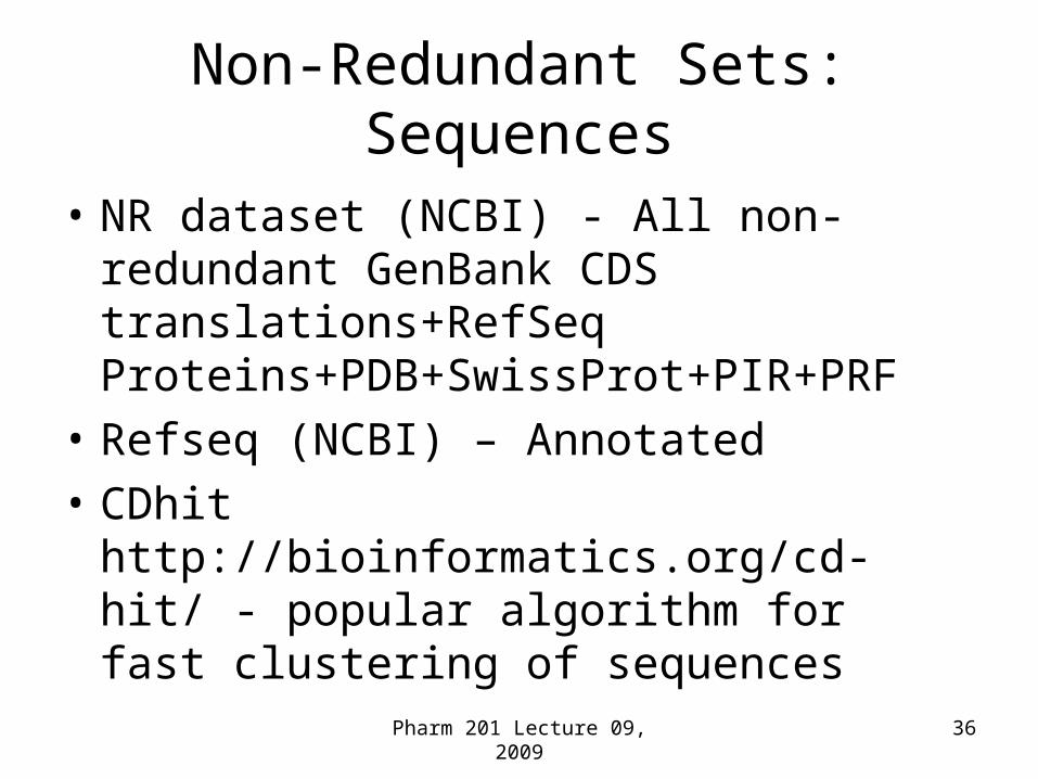
Non-Redundant Sets: Sequences
• NR dataset (NCBI) - All non-redundant GenBank CDS translations+RefSeq Proteins+PDB+SwissProt+PIR+PRF
• Refseq (NCBI) – Annotated
• CDhit http://bioinformatics.org/cd-hit/ - popular algorithm for fast clustering of sequences