134
PPAR Research Pharmacological and Toxicological Advances in PPAR-Related Medicines Guest Editors: Yuji Kamijo, Christopher J. Nicol, and Stefan E. H. Alexson

PPAR Research
Pharmacological and Toxicological Advances in PPAR-Related Medicines
Guest Editors: Yuji Kamijo, Christopher J. Nicol, and Stefan E. H. Alexson
Pharmacological and Toxicological Advances inPPAR-Related Medicines

PPAR Research
Pharmacological and Toxicological Advances inPPAR-Related Medicines
Guest Editors: Yuji Kamijo, Christopher J. Nicol,and Stefan E. H. Alexson

Copyright © 2012 Hindawi Publishing Corporation. All rights reserved.
This is a special issue published in “PPAR Research.” All articles are open access articles distributed under the Creative Commons Attri-bution License, which permits unrestricted use, distribution, and reproduction in any medium, provided the original work is properlycited.
Editorial Board
Khalid Al-Regaiey, USARozalyn M. Anderson, USAPaul Rodney Smith Baker, USAYaacov Barak, USAMarcin Baranowski, PolandJosep Bassaganya-Riera, USAAbdulbari Bener, QatarCarlos Bocos, SpainDaniela Bonofiglio, ItalySandra Brunelleschi, ItalyAntonio Brunetti, ItalyElke Burgermeister, GermanyNorm Buroker, USAMaria Paola Ceru, ItalyHyae Gyeong Cheon, KoreaAnnamaria Cimini, ItalySharon Cresci, USAMichael L. Cunningham, USASalvatore Cuzzocrea, ItalyPaul D. Drew, USAWilliam T. Festuccia, BrazilBrian N. Finck, USAPascal Froment, FranceYuchang Fu, USAAndrea Galli, ItalyConstantinos Giaginis, GreeceGeoff Girnun, USAHoward P. Glauert, USA
Youfei Guan, ChinaJames P. Hardwick, USASaswati Hazra, USAWeimin He, USAJaou-Chen Huang, USATom H. W. Huang, AustraliaN. Ishida, JapanUlrich Kintscher, GermanyJames Klaunig, USAJoshua K. Ko, ChinaCarolyn M. Komar, USABettina Konig, GermanyMarkus Peter Kummer, GermanyChristopher Lau, USABeata Lecka-Czernik, USAChih-Hao Lee, USATodd Leff, USAStephane Mandard, FranceHarry Martin, New ZealandAndrew J. McAinch, AustraliaJorg Mey, GermanyRaghavendra G. Mirmira, USAHiroyuki Miyachi, JapanKiyoto Motojima, JapanShaker A. Mousa, USAElisabetta Mueller, USALaszlo Nagy, HungaryMarcelo H. Napimoga, Brazil
Dipak Panigrahy, USAHemang Parikh, USAR. P. Phipps, USAD. Piomelli, USASuofu Qin, USAMike E. Robbins, USARuth Roberts, UKStephane Rocchi, FranceEnrique Saez, USAHerve Schohn, FranceHenrike Sell, GermanyLawrence Serfaty, FranceXu Shen, ChinaXing-Ming Shi, USATheodore J. Standiford, USAAlexander Staruschenko, USANguan Soon Tan, SingaporeSwasti Tiwari, IndiaVladimir Todorov, GermanyAntonella Trombetta, ItalyJohn P. Vanden Heuvel, USARaghu Vemuganti, USANanping Wang, ChinaRobert A. Winn, USAWei Xu, USAQinglin Yang, USATianxin Yang, USAWeiling Zhao, USA

Contents
Pharmacological and Toxicological Advances in PPAR-Related Medicines, Yuji Kamijo,Christopher J. Nicol, and Stefan E. H. AlexsonVolume 2012, Article ID 940964, 2 pages
PPAR Medicines and Human Disease: The ABCs of It All, Anthony J. Apostoli and Christopher J. B. NicolVolume 2012, Article ID 504918, 16 pages
The Key to Unlocking the Chemotherapeutic Potential of PPARγ Ligands: Having the RightCombination, Graham Skelhorne-Gross and Christopher J. B. NicolVolume 2012, Article ID 946943, 13 pages
Plasticizers May Activate Human Hepatic Peroxisome Proliferator-Activated Receptor α Less Than Thatof a Mouse but May Activate Constitutive Androstane Receptor in Liver, Yuki Ito, Toshiki Nakamura,Yukie Yanagiba, Doni Hikmat Ramdhan, Nozomi Yamagishi, Hisao Naito, Michihiro Kamijima,Frank J. Gonzalez, and Tamie NakajimaVolume 2012, Article ID 201284, 11 pages
Nutraceuticals as Ligands of PPARγ, Meera Penumetcha and Nalini SantanamVolume 2012, Article ID 858352, 7 pages
Idealized PPARγ-Based Therapies: Lessons from Bench and Bedside, Angelica Amorim Amatoand Francisco de Assis Rocha NevesVolume 2012, Article ID 978687, 9 pages
Peroxisome Proliferator-Activated Receptorα Agonists Differentially Regulate Inhibitor of DNA BindingExpression in Rodents and Human Cells, Marıa del Carmen Gonzalez, J. Christopher Corton, Nuria Acero,Dolores Munoz-Mingarro, Yolanda Quiros, Juan Jose Alvarez-Millan, Emilio Herrera,and Carlos BocosVolume 2012, Article ID 483536, 9 pages
Effects of PPARγ Ligands on Leukemia, Yoko Tabe, Marina Konopleva, Michael Andreeff,and Akimichi OhsakaVolume 2012, Article ID 483656, 8 pages
The Current Knowledge of the Role of PPAR in Hepatic Ischemia-Reperfusion Injury,M. Elias-Miro, M. B. Jimenez-Castro, M. Mendes-Braz, A. Casillas-Ramırez, and C. PeraltaVolume 2012, Article ID 802384, 14 pages
PPARα Activation Protects against Anti-Thy1 Nephritis by Suppressing Glomerular NF-κB Signaling,Koji Hashimoto, Yuji Kamijo, Takero Nakajima, Makoto Harada, Makoto Higuchi, Takashi Ehara,Hidekazu Shigematsu, and Toshifumi AoyamaVolume 2012, Article ID 976089, 11 pages
Global Gene Expression Profiling in PPAR-γ Agonist-Treated Kidneys in an Orthologous Rat Model ofHuman Autosomal Recessive Polycystic Kidney Disease, Daisuke Yoshihara, Masanori Kugita,Tamio Yamaguchi, Harold M. Aukema, Hiroki Kurahashi, Miwa Morita, Yoshiyuki Hiki,James P. Calvet, Darren P. Wallace, Takafumi Toyohara, Takaaki Abe, and Shizuko NagaoVolume 2012, Article ID 695898, 10 pages

Fatty Acid Accumulation and Resulting PPARα Activation in Fibroblasts due to Trifunctional ProteinDeficiency, Masato Wakabayashi, Yuji Kamijo, Takero Nakajima, Naoki Tanaka, Eiko Sugiyama,Tian Yangyang, Takefumi Kimura, and Toshifumi AoyamaVolume 2012, Article ID 371691, 7 pages
Hepatic Cerebroside Sulfotransferase Is Induced by PPARα Activation in Mice, Takefumi Kimura,Takero Nakajima, Yuji Kamijo, Naoki Tanaka, Lixuan Wang, Atsushi Hara, Eiko Sugiyama, Eiji Tanaka,Frank J. Gonzalez, and Toshifumi AoyamaVolume 2012, Article ID 174932, 10 pages
Hindawi Publishing CorporationPPAR ResearchVolume 2012, Article ID 940964, 2 pagesdoi:10.1155/2012/940964
Editorial
Pharmacological and Toxicological Advances inPPAR-Related Medicines
Yuji Kamijo,1 Christopher J. Nicol,2 and Stefan E. H. Alexson3
1 Department of Nephrology, Shinshu University School of Medicine, 3-1-1 Asahi, Matsumoto 390-8621, Japan2 Department of Pathology and Molecular Medicine, Cancer Biology and Genetics Division,Cancer Research Institute, and Department of Biomedical and Molecular Sciences (Pharmacology and Toxicology),Queen’s University, Kingston, ON, Canada K7L 3N6
3 Division of Clinical Chemistry, Department of Laboratory Medicine, Karolinska Institutet, Karolinska University Hospital,141 86 Stockholm, Sweden
Correspondence should be addressed to Yuji Kamijo, [email protected]
Received 13 August 2012; Accepted 13 August 2012
Copyright © 2012 Yuji Kamijo et al. This is an open access article distributed under the Creative Commons Attribution License,which permits unrestricted use, distribution, and reproduction in any medium, provided the original work is properly cited.
Peroxisome proliferator-activated receptors (PPARs) areinvolved in the pathophysiology of the various types ofdiseases. Many types of PPAR-related medicines developedand utilized clinically all over the world exert multiple effects,including regulation of hypolipidemic, antidiabetic, anti-inflammatory, antifibrotic, and antiproliferative pathways,with emerging potential benefits in other diseases. On theother hand, these medicines may also exert various toxicities,and some PPAR drugs are no longer in use clinically becauseof serious complications arising in some patients. Thus, theauthors here have focused on the benefits and risks of thesemedicines, and aim to clarify their therapeutic potential forappropriate clinical utilization. This special issue in PPARresearch includes 6 review articles and 6 research articles, asfollows.
Review Articles. The paper “The key to unlocking thechemotherapeutic potential of PPARγ ligands: Having the rightcombination” by G. Skelhorne-Gross and C. J. B. Nicol is areview of the vast in vitro, in vivo, and human clinical trialstudies, using chemotherapeutic combinations that includePPARγ activating drugs. This review article reveals the novelchemotherapeutic potential of PPARγ activating drugs, andprovides a guide for further basic and clinical research.This information is certainly useful for optimization ofchemotherapeutic interventions that will reduce the numberof cancer related deaths.
The paper “PPAR medicines and human disease: TheABCs of it all” by A. J. Apostoli and C. J. B. Nicol is areview article that summarizes the advances of knowledge
concerning effects of PPAR medicines on ATP-dependentbinding cassette (ABC) transporters based on in vitro, invivo, and human clinical trial studies. This review suggeststhe potential of PPAR-related medicines for controlling ABCtransporter activity at the transcriptional level, and discussestheir potential implications in human diseases with respectto cancer and atherosclerosis.
The paper “The current knowledge of the role of PPAR inhepatic ischemia-reperfusion injury” by M. Elias-Miro et al.is a review article concerning the roles of PPARs signalingpathways in hepatic ischemia reperfusion injury that is inher-ent to human liver transplantation and resection surgery. Ashortage of available healthy livers for organ transplantationcalls for the potential use of any available organ, including,for example, steatotic livers; however, steatotic livers aremore susceptible to ischemia-reperfusion injury. This paperreviews PPAR-signaling pathways, summarizes some of thelesser known functions of PPARs in liver regeneration, anddiscusses potential therapies based on PPAR regulation thatmay minimize the observed side effects in liver surgery. Thisreview emphasizes the need for further research into the rolesof PPARs in various liver conditions and surgical proceduresbefore being translated into treatment of human disease.
The paper “Effects of PPARγ ligands in leukemia” by Y.Tabe et al. is a review article that describes the antitumoradvances of PPARγ ligands, alone and in combination withretinoic acid receptor ligands in control of cell proliferation,differentiation, and apoptosis, and discusses their poten-tial therapeutic applications in hematological malignancies.

2 PPAR Research
Acute promyelocytic leukemia (APL, representing about 10%of AML patients) is unique among myeloid leukemias in thatit is sensitive to all-trans-retinoic acid (ATRA). However, anumber of APL patients relapse and develop ATRA resist-ance. This review article provides evidence on the conse-quences of the treatment with PPARγ ligands, in particu-lar the triterpenoid 2-cyano-3,12-dioxooleana-1,9-dien-28-oic acid (CDDO), on the epigenetic/transcriptional eventsinduced by retinoic acid in APL cells, and supports theclinical utility of ATRA/PPARγ-ligand combinations fortreating hematological malignancies.
The paper “Idealized PPARγ-based therapies: Lessons frombench and bedside” by A. A. Amato and F. de A. R. Neves isa review about the knowledge acquired regarding efficacyand safety issues by PPARγ ligands. This body of work isattractive since the interest for PPARγ modulation as astrategy to treat metabolic diseases has increased recently,due to better understanding of PPARγ action.
The paper “Nutraceuticals as ligands of PPARγ” by M.Penumetcha and N. Santanam reviews the transcriptionfactor PPARγ, which is the target for the thiazolidinediones,the first class of PPARγ agonist drugs used in the treatmentof diabetes. Due to the increased adverse effects related tothese drugs, newer safer drugs are being generated. Thisreview paper describes some of the dietary componentsthat have affinity for, and activate, PPARγ, as well as theirpharmacology and potential toxicology.
Research Articles. The paper “PPARα activation protectsagainst anti-Thy1 nephritis by suppressing glomerular NF-κBsignaling” by K. Hashimoto et al. is the first to demon-stratethe glomerular protective effects of treatment using arepresentative PPARα agonist, clofibrate, in rat mesan-gial proliferative glomerulonephritis model (MsPGN) anti-Thy1 nephritis. PPARα activation is known to exert anti-inflammatory effects in various cells and organs throughsuppression of NFκB signaling; however, its effect againstglomerulonephritis has remained obscure. Because MsPGNis one of the significant factors leading to chronic kidneydisease (CKD), the beneficial antinephritic effect of PPARαactivation may provide a novel treatment strategy againstCKD. Their findings may also be useful to create PPAR-basedtherapies to treat glomerular disease.
The paper “Hepatic cerebroside sulfotransferase is inducedby PPARα activation in mice” by T. Kimura et al. is thefirst to examine sulfatide levels and the expression ofenzymes related to sulfatide metabolism using wild-type(+/+), Ppara-heterozygous (+/−), and Ppara-null (−/−)mice given a control diet or one containing 0.1% fenofibrate,a typical PPARα activator. Recent studies have revealed aprotective role of serum sulfatides against arteriosclerosisand hypercoagulation. Their results suggest that PPARα acti-vation enhances hepatic sulfatide synthesis mainly throughcerebroside sulfotransferase (CST) induction. Accordingly,CST may be a novel PPARα target gene product candidatewith implications in disease prevention and treatment.
The paper “Fatty acid accumulation and resulting PPARαactivation in fibroblasts due to trifunctional protein deficiency”by M. Wakabayashi et al. demonstrates free fatty acid
accumulation, enhanced three acyl-CoA dehydrogenases,and PPARα activation in the fibroblasts from six patientswith mitochondrial trifunctional protein deficiency, who hadabnormalities in the second through fourth reactions in fattyacid β-oxidation system. These novel findings suggest thatthe fatty acid accumulation and resulting PPARα activationare major causes of the increase in the β-oxidation ability inthe patients’ fibroblasts, and that enhanced cell proliferationand increased oxidative stress relate to the development ofspecific clinical features. Additionally, significant suppressionof the PPARα activation by means of MK886 treatment mayprovide a new method of treating this deficiency.
In the paper “Global gene expression profiling in PPARγagonist-treated kidneys in an orthologous rat model of humanautosomal recessive polycystic kidney disease” by D. Yoshiharaet al., the authors explored the changes in gene expression byPioglitazone (PIO), a PPARγ agonist, using polycystic kidneydisease (PCK) rats. By analyzing globally, they successfullyfound that stearoyl-coenzyme A desaturase 1 (Scd1) washighly expressed in PCK kidneys, and PIO decreased itsexpression. Notably, they found that Scd1 plays a role inthe early cystogenesis, and this is the point where PIO mayintervene in the process of cystogenesis.
The paper “Plasticizers may activate human hepaticperoxisome proliferator-activated receptor α less than that of amouse but may activate constitutive androstane receptor (CAR)in liver” by Y. Ito et al. reported the species differences con-cerning activation of PPARα and CAR, which was inducedby the oral exposure with industrial PPARα ligands, includ-ing dibutyl phthalate, di(2-ethylhexyl)phthalate, and di(2-ethylhexyl)adipate, between wild-type mice and humanizedPPARα mice. These transcriptional species differences mightcause different hepatic toxicities between murine model andhuman cases. This information would be valuable for the riskassessment of PPARα-related medicines.
The paper “Peroxisome proliferator-activated receptor αagonists differentially regulate inhibitor of DNA bindingexpression in rodents and human cells” by M. del C. Gonzalezet al. reported rodent versus human species differences inthe regulatory manner of inhibitor of DNA binding (Id2)via PPARα agonists. Since Id2 protein is involved in celldifferentiation and proliferation, this finding may help tounderstand the species differences in toxicity of PPARαagonists.
Yuji KamijoChristopher J. Nicol
Stefan E. H. Alexson

Hindawi Publishing CorporationPPAR ResearchVolume 2012, Article ID 504918, 16 pagesdoi:10.1155/2012/504918
Review Article
PPAR Medicines and Human Disease: The ABCs of It All
Anthony J. Apostoli1, 2 and Christopher J. B. Nicol1, 2, 3
1 Department of Pathology and Molecular Medicine, Queen’s University, Kingston, ON, Canada K7L 3N62 Cancer Biology and Genetics Division, Cancer Research Institute, Queen’s University, Kingston, ON, Canada K7L 3N63 Department of Biomedical and Molecular Sciences, Queen’s University, Kingston, ON, Canada K7L 3N6
Correspondence should be addressed to Christopher J. B. Nicol, [email protected]
Received 24 February 2012; Revised 4 April 2012; Accepted 6 April 2012
Academic Editor: Yuji Kamijo
Copyright © 2012 A. J. Apostoli and C. J. B. Nicol. This is an open access article distributed under the Creative CommonsAttribution License, which permits unrestricted use, distribution, and reproduction in any medium, provided the original work isproperly cited.
ATP-dependent binding cassette (ABC) transporters are a family of transmembrane proteins that pump a variety of hydrophobiccompounds across cellular and subcellular barriers and are implicated in human diseases such as cancer and atherosclerosis.Inhibition of ABC transporter activity showed promise in early preclinical studies; however, the outcomes in clinical trials withthese agents have not been as encouraging. Peroxisome proliferator-activated receptors (PPARs) are ligand-activated transcriptionfactors that regulate genes involved in fat and glucose metabolism, and inflammation. Activation of PPAR signaling is also reportedto regulate ABC gene expression. This suggests the potential of PPAR medicines as a novel means of controlling ABC transporteractivity at the transcriptional level. This paper summarizes the advances made in understanding how PPAR medicines affectABC transporters, and the potential implications for impacting on human diseases, in particular with respect to cancer andatherosclerosis.
1. Introduction
Harnessing the energy released from adenosine triphosphate(ATP) hydrolysis, ATP-dependent binding cassette (ABC)transporters shuttle a wide range of substrates, includinglipids, metabolites, and xenobiotics, across biological mem-branes in order to maintain normal cell metabolism. Theyrepresent the largest family of transmembrane proteins inhumans, comprising 49 ABC genes, and are best reviewedelsewhere [1–3]. These genes are subdivided among sevensubfamilies (A-G) based on sequence and structural homol-ogy and are highly conserved among eukaryotic species,suggesting that most appeared early in metazoan evolution[4]. The proteins encoded by ABC genes consist of twodistinct domains: a transmembrane domain that recognizesspecific compounds and transports them across cellular andsubcellular barriers and a nucleotide-binding domain whereATP hydrolysis occurs to yield energy for substrate transport[5]. Typically, ABC proteins are unidirectional transportersexpressed at the cell membrane, which move hydrophobicmolecules internally for metabolic pathways, or externallyfor elimination from the cell and/or use by other tissuesand organs. Thus, ABC transporters play important roles in
a range of human physiologic, toxicologic, and pathologicfunctions. With respect to the latter, many preclinical reportsthat show promise in terms of regulating ABC transporters toovercome chemotherapeutic drug resistance in tumours, ormodify lipid homeostasis in order to reduce atheroscleroticrisk, have not achieved the same level of success in clinicaltrials.
Peroxisome proliferator-activated receptors (PPARs) areligand-activated transcription factors that regulate expres-sion of a plethora of genes involved in sugar and fatmetabolism, inflammation, and cancer [6–8]. Three PPARhomologs have been characterized—PPARα, PPARβ/δ, andPPARγ—each displaying a unique pattern of tissue-specificexpression that reflect their distinctive functions [9–11].Recently, there is mounting in vitro and in vivo evidencethat activation of PPARs may alter ABC protein expressionand/or function. Accordingly, this paper will summarizerecent developments in an emerging field where PPARmedicines, capable of modulating ABC transporter genesat the transcriptional level, may prove useful when suchmodulation provides novel therapeutic options for treatingcancer and atherosclerosis.
2 PPAR Research
2. PPARs and Their Ligands
As members of the nuclear receptor superfamily, PPARscontain a ligand-binding domain that recognizes and bindsspecific PPAR agonists, and a DNA-binding domain thatinteracts with specific peroxisome proliferator-response ele-ments (PPREs) within the genome [12]. PPARs are localizedto the nucleus and dimerize with retinoid X receptor (RXR)αto form complexes that bind to PPREs in the promoterregions of a broad range of target genes [13]. In its restingstate, the PPAR : RXRα complex associates with cell-specificcorepressor molecules that aid in the silencing of targetgene transcription. Ligand binding elicits a conformationalchange in PPAR that leads to the release of corepressors, andthe recruitment of coactivator molecules that promote targetgene transcriptional activity. Furthermore, ligand activationof PPARs may also repress signaling of some gene targetsthrough direct interaction with other transcription factors orcompetition for available coregulators [14].
PPARα is highly expressed in the liver, heart, kidney,skeletal muscle, and large intestine [15]. It is activated bythe “fibrate” class of drugs, such as bezafibrate, ciprofibrate,clofibrate, gemfibrozil, and fenofibrate, used to treat elevatedtriglycerides and low high-density lipoprotein (HDL) [16].PPARβ/δ is more ubiquitously expressed with highest levelsnoted within the large intestine and placenta [15]. Similarto other PPAR subtypes, it may also be activated by varioussaturated and unsaturated fatty acids [12]. Because less isunderstood about PPARβ/δ, fewer synthetic activators havebeen developed; however, emerging evidence supports thepotential therapeutic value of PPARβ/δ agonists, such asGW0742, GW501516, and MBX-8025, which remain to beclinically tested [17].
As a chief regulator of adipogenesis, PPARγ is abundantlyexpressed in adipose tissue [18], and like PPARα, is alsodetected in vascular and immune cells, as well as tissues suchas the colon, breast, and prostate [19, 20]. Synthetic agentsknown as thiazolidinediones (TZDs) like troglitazone, cigli-tazone, rosiglitazone, and pioglitazone are classic examplesof PPARγ activators [21]. In North America, rosiglitazoneand pioglitazone are still prescribed to treat type 2 diabeticpatients. However, there are reports suggesting increasedmyocardial infarction risk with rosiglitazone use and bladdercancer risk with long-term use of pioglitazone [22, 23]. Asa followup on the former, a safety review of rosiglitazoneby a panel of international experts deemed the availabledata inconclusive and requiring further study. In the lattercase, direct clinical evidence of this possible association isalso required. Despite the need for more evidence, thesedrugs remain FDA approved, albeit with warning updatesto package inserts clarifying the potential for risk [24, 25],and a Risk Evaluation and Mitigation Strategy (REMS) isin place to restrict access and distribution of rosiglitazone-containing medicines to those healthcare providers and theirpatients who confirm their awareness of the new warnings[26]. Nevertheless, the utility of these drugs remains valuablenot only for their ability to provide mechanistic insight intothe role of PPARγ-mediated target regulation, but also fortheir potential benefit in certain off-label uses.
Dual and pan PPAR ligands were also developed toenhance therapeutic potential via simultaneously activatingtwo or more PPAR isoforms. Examples include PPARα/γmodulators like tesaglitazar, muraglitazar, and aleglitazar,and the pan PPARα/(β/δ)/γ agonist chiglitazar [27].
The reported links between the above listed PPARmedicines and their in vitro and in vivo effects on ABCtransporters are summarized in Tables 1 and 2, respectively,and described in detail below in the context of several humandiseases.
3. Cancer
The goal of chemotherapy is to target rapidly dividing cells orderegulated signaling pathways to suppress tumour growth,and ultimately, cure cancer patients; however, one primaryroadblock to the success of chemotherapy is acquisition ofmultidrug resistance (MDR). A well-known cause of MDR isABC transporter-driven drug efflux from cancer cells instill-ing resistance to multiple agents [28]. The well-known ABCtransporters, P-glycoprotein (Pgp)/MDR1/ABCB1, mul-tidrug resistance protein (MRP)1/ABCC1, and breast cancer-resistance protein (BCRP)/MXR/ABCG2, are overexpressedin a variety of different human cancers and transport arange of chemotherapeutic drugs [4]. Pgp, an importantblood brain barrier component and regulator of intestinaldrug absorption, was the first ABC transporter to becharacterized in 1976 [29]. Its overexpression in tumours ofthe kidney, liver, colon, and breast correlates with chemore-sistance [30–32]. Substrates of Pgp include anthracyclines,vinca alkaloids, taxanes, camptothecins, mitoxantrone, andmethotrexate [33]. The second ABC gene discovered was themore ubiquitously expressed MRP1 [34], which transportsanthracyclines, vinca alkaloids, and etoposide, in additionto organic anions and glutathione conjugates [28]. Itsoverexpression confers chemotherapy resistance in prostate,lung, breast, and neuroblastoma cancer [35, 36]. Finally,BCRP is normally expressed in placenta and small intestine,as well as various stem cell populations [37, 38]. Severaldrug-resistant cell lines also contain elevated levels of thisABC transporter, which contributes to the efflux of sev-eral antitumour agents such as doxorubicin, daunorubicin,mitoxantrone, and topotecan [39–41].
In addition to MDR, other functions of ABC transportersin cancer are beginning to emerge, further implicating thesegenes as important targets of chemotherapy. For example,Pgp expression, devoid of ATP-dependent drug transport,suppresses cell death in the presence of apoptotic signals innormal and cancer cells [42–44]. Furthermore, Pgp knock-down reduced the migration and invasion potential of MCF7human breast cancer cells [45]. As a result of these studies,direct inhibition of ABC transporter activity has become anappealing undertaking for researchers in the development ofimproved cancer chemotherapeutics; however, several clini-cal trials using ABC inhibitors have proven unsuccessful [46].
Research has shown that PPAR activation inducesexpression of both mouse (Mdr1/Mdr1b/Abcb1b, Mdr2/Abcb4, and Mdr3/Mdr1a/Abcb1a) and human (MDR2/MDR3/ABCB4) homologs of Pgp, which efflux similar
PPAR Research 3
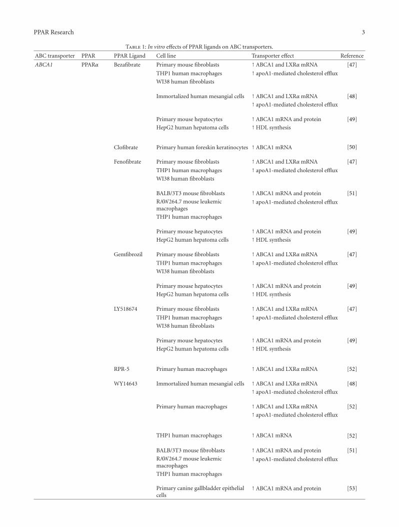
Table 1: In vitro effects of PPAR ligands on ABC transporters.
ABC transporter PPAR PPAR Ligand Cell line Transporter effect Reference
ABCA1 PPARα Bezafibrate Primary mouse fibroblasts ↑ ABCA1 and LXRα mRNA [47]
THP1 human macrophages ↑ apoA1-mediated cholesterol efflux
WI38 human fibroblasts
Immortalized human mesangial cells ↑ ABCA1 and LXRα mRNA [48]
↑ apoA1-mediated cholesterol efflux
Primary mouse hepatocytes ↑ ABCA1 mRNA and protein [49]
HepG2 human hepatoma cells ↑HDL synthesis
Clofibrate Primary human foreskin keratinocytes ↑ ABCA1 mRNA [50]
Fenofibrate Primary mouse fibroblasts ↑ ABCA1 and LXRα mRNA [47]
THP1 human macrophages ↑ apoA1-mediated cholesterol efflux
WI38 human fibroblasts
BALB/3T3 mouse fibroblasts ↑ ABCA1 mRNA and protein [51]
RAW264.7 mouse leukemicmacrophages
↑ apoA1-mediated cholesterol efflux
THP1 human macrophages
Primary mouse hepatocytes ↑ ABCA1 mRNA and protein [49]
HepG2 human hepatoma cells ↑HDL synthesis
Gemfibrozil Primary mouse fibroblasts ↑ ABCA1 and LXRα mRNA [47]
THP1 human macrophages ↑ apoA1-mediated cholesterol efflux
WI38 human fibroblasts
Primary mouse hepatocytes ↑ ABCA1 mRNA and protein [49]
HepG2 human hepatoma cells ↑HDL synthesis
LY518674 Primary mouse fibroblasts ↑ ABCA1 and LXRα mRNA [47]
THP1 human macrophages ↑ apoA1-mediated cholesterol efflux
WI38 human fibroblasts
Primary mouse hepatocytes ↑ ABCA1 mRNA and protein [49]
HepG2 human hepatoma cells ↑HDL synthesis
RPR-5 Primary human macrophages ↑ ABCA1 and LXRα mRNA [52]
WY14643 Immortalized human mesangial cells ↑ ABCA1 and LXRα mRNA [48]
↑ apoA1-mediated cholesterol efflux
Primary human macrophages ↑ ABCA1 and LXRα mRNA [52]
↑ apoA1-mediated cholesterol efflux
THP1 human macrophages ↑ ABCA1 mRNA [52]
BALB/3T3 mouse fibroblasts ↑ ABCA1 mRNA and protein [51]
RAW264.7 mouse leukemicmacrophages
↑ apoA1-mediated cholesterol efflux
THP1 human macrophages
Primary canine gallbladder epithelialcells
↑ ABCA1 mRNA and protein [53]
4 PPAR Research
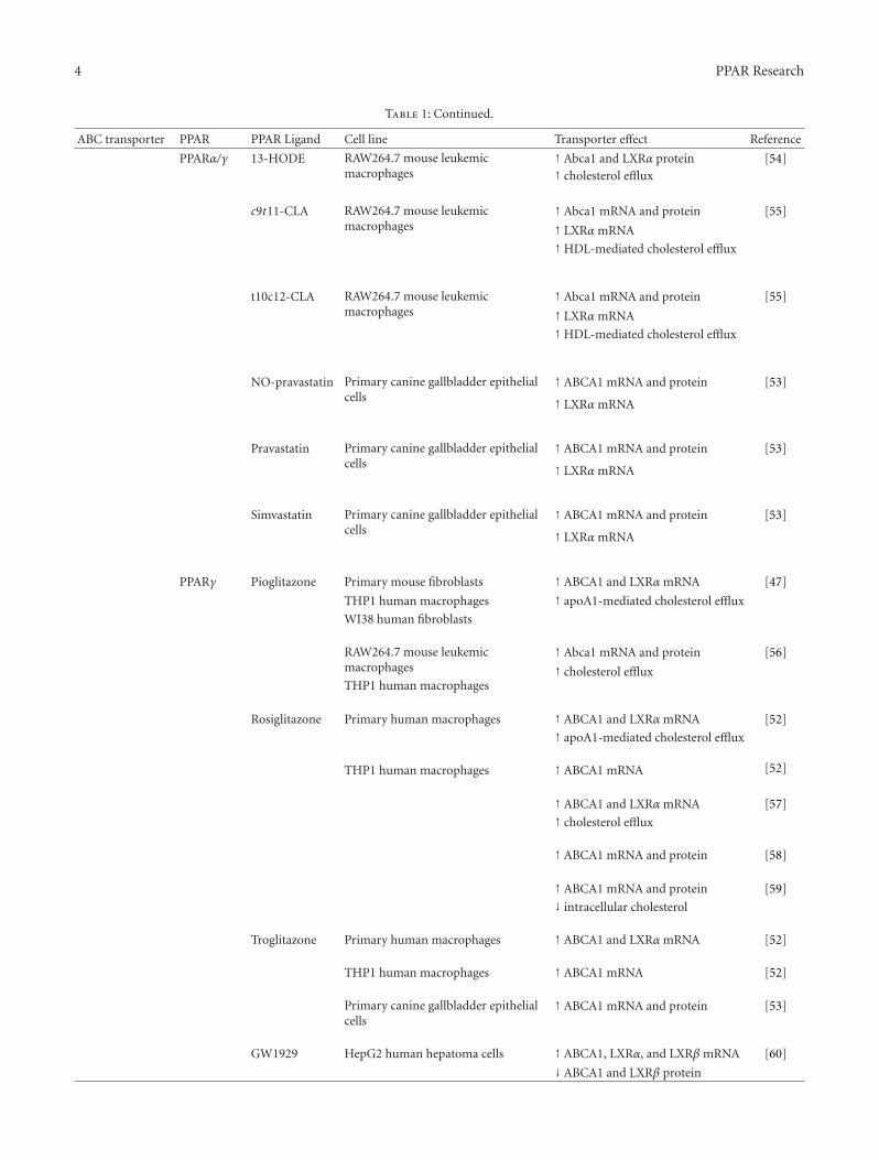
Table 1: Continued.
ABC transporter PPAR PPAR Ligand Cell line Transporter effect Reference
PPARα/γ 13-HODE RAW264.7 mouse leukemicmacrophages
↑ Abca1 and LXRα protein [54]↑ cholesterol efflux
c9t11-CLA RAW264.7 mouse leukemicmacrophages
↑ Abca1 mRNA and protein [55]
↑ LXRα mRNA
↑HDL-mediated cholesterol efflux
t10c12-CLA RAW264.7 mouse leukemicmacrophages
↑ Abca1 mRNA and protein [55]
↑ LXRα mRNA
↑HDL-mediated cholesterol efflux
NO-pravastatin Primary canine gallbladder epithelialcells
↑ ABCA1 mRNA and protein [53]
↑ LXRα mRNA
Pravastatin Primary canine gallbladder epithelialcells
↑ ABCA1 mRNA and protein [53]
↑ LXRα mRNA
Simvastatin Primary canine gallbladder epithelialcells
↑ ABCA1 mRNA and protein [53]
↑ LXRα mRNA
PPARγ Pioglitazone Primary mouse fibroblasts ↑ ABCA1 and LXRα mRNA [47]
THP1 human macrophages ↑ apoA1-mediated cholesterol efflux
WI38 human fibroblasts
RAW264.7 mouse leukemicmacrophages
↑ Abca1 mRNA and protein [56]
THP1 human macrophages↑ cholesterol efflux
Rosiglitazone Primary human macrophages ↑ ABCA1 and LXRα mRNA [52]
↑ apoA1-mediated cholesterol efflux
THP1 human macrophages ↑ ABCA1 mRNA [52]
↑ ABCA1 and LXRα mRNA [57]
↑ cholesterol efflux
↑ ABCA1 mRNA and protein [58]
↑ ABCA1 mRNA and protein [59]
↓ intracellular cholesterol
Troglitazone Primary human macrophages ↑ ABCA1 and LXRα mRNA [52]
THP1 human macrophages ↑ ABCA1 mRNA [52]
Primary canine gallbladder epithelialcells
↑ ABCA1 mRNA and protein [53]
GW1929 HepG2 human hepatoma cells ↑ ABCA1, LXRα, and LXRβ mRNA [60]
↓ ABCA1 and LXRβ protein

PPAR Research 5
Table 1: Continued.
ABC transporter PPAR PPAR Ligand Cell line Transporter effect Reference
GW7845 THP1 human macrophages ↑ ABCA1 mRNA [61]
Mycophenolicacid
HepG2 human hepatoma cells ↑ ABCA1 mRNA and protein [62]
↑ LXRα protein
Prostaglandin J2 Immortalized human mesangial cells ↑ ABCA1 and LXRα mRNA [48]
↑ apoA1-mediated cholesterolefflux
Primary human macrophages ↑ ABCA1 and LXRα mRNA [52]
Telmisartan RAW264.7 mouse leukemicmacrophages
↑ Abca1 mRNA [63]
↓macrophage proliferation
PPARβ/δ GW501516 Primary mouse fibroblasts ↑ ABCA1 and LXRα mRNA [47]
THP1 human macrophages ↑ apoA1-mediated cholesterolefflux
WI38 human fibroblasts
THP1 human macrophages ↑ ABCA1 mRNA [61]
1BR3N human fibroblasts ↑ apoA1-mediated cholesterolefflux
FHS74 human intestinal cells ↑ ABCA1 mRNA [61]
Primary human skeletal muscle cells ↑ ABCA1 mRNA [64]
Primary human foreskin keratinocytes ↑ ABCA1 mRNA [50]
ABCA12 PPARγ Ciglitazone Primary human foreskin keratinocytes ↑ ABCA12 mRNA and protein [65]
Troglitazone Primary human foreskin keratinocytes ↑ ABCA12 mRNA [65]
GI251929X Primary human foreskin keratinocytes ↑ ABCA12 mRNA [65]
PPARβ/δ Ceramide Primary human foreskin keratinocytes ↑ ABCA12 mRNA and protein [66]
GW610742 Primary human foreskin keratinocytes ↑ ABCA12 mRNA and protein [65]
Pgp/MDR1/ABCB1 PPARα Fenofibrate Pgp-overexpressing L-MDR1 porcinekidney epithelial cells
↓ calcein efflux [67]
PPARα/γ Simvastatin Pgp-overexpressing L-MDR1 porcinekidney epithelial cells
↓ calcein efflux [67]
PPARγ Rosiglitazone Doxorubicin-resistant P388 mouseleukemia cells
↓ calcein efflux [68]
Troglitazone Doxorubicin-resistant P388 mouseleukemia cells
↓ calcein efflux [68]
Doxorubicin-resistant K562 humanleukemia cells
↓ Pgp protein [69]
Doxorubicin-resistant MCF7 humanbreast cancer cells
↑ sensitivity to doxorubicin

6 PPAR Research
Table 1: Continued.
ABC transporter PPAR PPAR Ligand Cell line Transporter effect Reference
Vincristine-resistant SGC7901 humangastric cancer cells
↓ Pgp mRNA and protein [70]
↓ Rh123 efflux
↑ sensitivity to vincristine
MDR2/MDR3/ABCB4 PPARα Bezafibrate HepG2 human hepatoma cells ↑MDR2/MDR3 mRNA [71]
↑MDR2/MDR3 redistribution
↑MDR2/MDR3 mRNA [72]
↑MDR2/MDR3 redistribution
↑ phospholipid efflux
Ciprofibrate Primary mouse hepatocytes ↑Mdr2 mRNA [73]
WY14643 Primary mouse hepatocytes ↑Mdr2 mRNA [73]
MRP2/ABCC2 PPARγ Troglitazone Primary rat hepatocytes ↓Mrp2-associated bile efflux [74]
ABCG1 PPARα/γ 13-HODE RAW264.7 mouse leukemicmacrophages
↑ Abcg1 and LXRα protein [54]↑ cholesterol efflux
PPARγ Pioglitazone RAW264.7 mouse leukemicmacrophages
↑ ABCG1 mRNA and protein [56]
THP1 human macrophages↑ cholesterol efflux
Rosiglitazone THP1 human macrophages ↑ ABCG1 and LXRα mRNA [57]
↑ cholesterol efflux
Telmisartan RAW264.7 mouse leukemicmacrophages
↑ Abcg1 mRNA [63]
↓macrophage proliferation
BCRP/ABCG2 PPARα Clofibrate HCMEC/D3 human cerebralmicrovascular endothelial cells
↑ BCRP mRNA and protein [75]
↑mitoxantrone efflux
GW7647 HCMEC/D3 human cerebralmicrovascular endothelial cells
↑ BCRP mRNA and protein [75]
PPARγ Rosiglitazone Primary human dendritic cells ↑ BCRP mRNA and protein [76]
↑Hoescht efflux
↑mitoxantrone efflux
↑ sensitivity to mitoxantrone
BCRP-overexpressing MDCKII caninekidney epithelial cells
↓ PhA efflux [68]
HuH7 human hepatoma cells ↑ BCRP mRNA [68]
Troglitazone Primary human dendritic cells ↑ BCRP mRNA [76]
HuH7 human hepatoma cells ↑ BCRP mRNA [68]
Doxorubicin-resistant K562 humanleukemia cells
↓ BCRP protein [69]
Doxorubicin-resistant MCF7 humanbreast cancer cells
↑ sensitivity to doxorubicin

PPAR Research 7
Table 1: Continued.
ABC transporter PPAR PPAR Ligand Cell line Transporter effect Reference
GW7845 Primary human dendritic cells ↑ BCRP mRNA [76]
GW9662 Doxorubicin-resistant MCF7 humanbreast cancer cells
↓ BCRP protein [69]
chemotherapy substrates as MDR1 [33]. Fasting-inducedfatty acid release increased hepatic expression of Mdr2mRNA and protein, as well as activity, in wild-type but notPPARα-knockout mice [77]. Similar results were observedin ciprofibrate-treated mice [73]. Interestingly, the lattertrial demonstrated that elevated Mdr1 and Mdr3 mRNAexpression accompanied Mdr2 induction in liver; however, incultured mouse hepatocytes, only Mdr2 levels were elevatedby PPARα agonists suggesting that in vivo induction ofMdr1 and Mdr3 may be influenced by PPARα activationin surrounding tissue. Furthermore, both ciprofibrate andclofibrate increased hepatic expression of Mdr2 mRNA inCF1 mice. This was associated with increased Mdr2 redistri-bution into bile canaliculi and enhanced biliary phospholipidsecretion [78]. Similarly, in a chimeric mouse model withhumanized liver, bezafibrate increased hepatic MDR2/MDR3mRNA and protein, and promoted canalicular localiza-tion of the transporter [71]. Bezafibrate-treated HepG2human hepatocellular liver carcinoma cells also showedelevated expression of MDR2/MDR3 mRNA. Although therewas no subsequent change in protein levels, there wasa redistribution of the transporter into pseudocanaliculibetween cells, accompanied by enhanced apical localizationof phospholipids, which could be attenuated by PPARα-specific knockdown [72].
Several MRP1 homologs may also be upregulatedby PPARs, including MRP2/ABCC2, MRP3/ABCC3, andMRP4/ABCC4, which are known to transport substratesbelonging to a variety of chemotherapy drug classes [33].Although their normal physiological function remains elu-sive, it has been suggested that these transporters may playa role in MDR [79, 80]. Additionally, MRP4 expression mayplay a role in migration, as knockdown or pharmacologicalinhibition of this transporter appears to prevent humandendritic cell motility [81]. Moffit et al. examined the effectof clofibrate on hepatic transporters in mice. Following 10days of dosing, clofibrate upregulated hepatic expressionof Bcrp, Mrp3, and Mrp4 mRNA and protein in CD1mice. Similar findings for Mrp3 and Mrp4 were detectedin liver tissue isolated from clofibrate-treated wild-typeSV129 mice, while no changes were seen in liver from sim-ilarly treated PPARα-knockout mice [82]. Liver expressionof Mrp3 was also induced in C57BL mice treated withclofibrate, ciprofibrate, and diethylhexyl phthalate (DEHP)[83]. Maher et al. also reported the hepatic induction ofMrp3 and Mrp4 transcription in perfluorodecanoic-acid-(PFDA-) treated mice [84]. This was associated with elevatedserum levels of serum-conjugated bilirubin and bile acidsindicative of Mrp3- and Mrp4-specific hepatic efflux activity.These effects were attenuated in PPARα-knockout mice
treated with PFDA. Several putative PPRE sequences wereidentified upstream of the Mrp3 and Mrp4 promoters,providing further evidence that PPARα may directly regulatetranscription of these transporters in the liver.
Activation of PPARs may also induce expression of BCRP.PPARα agonists upregulate Bcrp transcription in mouseintestine [85]. Furthermore, PPARα-dependent activationinduces BCRP expression and efflux activity in humancerebral endothelial cells [75]. Here, transporter inductionis accompanied by binding of PPARα to a PPRE withinthe BCRP promoter. In human monocyte-derived dendriticcells, BCRP was directly induced by ligand-activated PPARγthrough three functional PPRE sequences located within thegene’s promoter [76]. This enhancement of BCRP activityelevated drug efflux and maintained intracellular low levelsof mitoxantrone, which could be reversed by addition ofa BCRP inhibitor. In doxorubicin-resistant MCF7 breastcancer and K562 human leukemia cell lines, troglitazonedownregulated expression of BCRP, and restored sensitivityto doxorubicin treatment [69]. Although troglitazone mayelicit effects that are PPARγ-dependent, it is also known tooperate via pathways that are independent of this nuclearreceptor [86]. Inhibition of PPARγ in untreated MCF7 cellsreduced BCRP expression indicating that the observed effectsof troglitazone were PPARγ-independent, and providingevidence that this TZD may suppress BCRP transcription inthese cells by indirectly antagonizing PPARγ itself.
In contrast to the studies previously outlined, a numberof reports indicate that PPAR activation may inhibit ABCtransporter expression and activity. Chen et al. observedthat troglitazone increased PPARγ activity and reversed Pgp-mediated chemoresistance in vincristine-resistant SGC7901human gastric cancer cells [70]. Furthermore, Rajkumarand Yamuna performed genetic expression analysis on adoxorubicin-resistant 143B human osteosarcoma cell lineand found increased expression of Pgp and Kruppel-likefactor 2 [91]. Given that the latter is a known suppressorof PPARγ expression [92], these findings may implicate thePPARγ pathway as a negative regulator of Pgp transcription.Wang et al. also demonstrated that tumour necrosis factor(TNF)α could partially reverse MDR by inducing PPARαand suppressing Pgp in an adriamycin-resistant cell linederived from HepG2 cells [93]. In another study, PPARαagonists downregulated Mrp1 expression in mouse intestine[85]. Hepatic expression of Mrp2 protein was reduced inmale Sprague-Dawley rats treated with the PPARα agonists,clofibrate, DEHP, and PFDA [89]. Furthermore, efflux of bileacids by Mrp2 may be suppressed by troglitazone in culturedrat hepatocytes [74]. Both rosiglitazone and troglitazoneinhibited BCRP function in BCRP-overexpressing MDCKII

8 PPAR Research
Table 2: In vivo effects of PPAR ligands on ABC transporters.
ABC transporter Ligand Receptor Model Transporter effect Reference
ABCA1 PPARα Fenofibrate Hypertriglyceridemicpatients
Differential HDL synthesis due toABCA1 variants
[87]
WY14643 SV129 mice ↑ Abca1 mRNA and protein inintestine
[88]
↓ intestinal absorption of cholesterol
PPARγ Telmisartan ApoE−/− C57BL mice ↑ Abca1 mRNA in aorta [63]
↓ atherosclerotic lesion size andnumber
Pgp/MDR1/ABCB1 PPARα Ciprofibrate SV129 mice ↑ hepatic Mdr1 & Mdr3 mRNA [73]
MDR2/MDR3/ABCB4 PPARα Bezafibrate CF1 mice ↑ hepatic Mdr2 mRNA [78]
↑ bile secretion of phospholipid
Humanized liver-uPA/SCID chimeric mice
↑ hepatic MDR2/MDR3 mRNA andprotein
[71]
↑ hepatic MDR2/MDR3 redistributioninto bile canaliculi
Ciprofibrate SV129 mice ↑ hepatic Mdr2 mRNA and protein [73]
↑ bile secretion of cholesterol andphospholipids
CF1 mice ↑ hepatic Mdr2 mRNA [78]
↑Mdr2 redistribution into bilecanaliculi
↑ bile secretion of phospholipid
Clofibrate CF1 mice ↑ hepatic Mdr2 mRNA [78]
↑Mdr2 redistribution into bilecanaliculi
↑ bile secretion of phospholipid
Fenofibrate CF1 mice ↑ hepatic Mdr2 mRNA [78]
Gemfibrozil CF1 mice ↑ hepatic Mdr2 mRNA [78]
MRP1/ABCC1 PPARα Ciprofibrate C57BL mice ↓ hepatic Mrp1 mRNA [83]
Clofibrate C57BL mice ↓ hepatic Mrp1 mRNA [83]
GW7647 C57BL mice ↓Mrp1 mRNA in small intestine [85]
WY14643 C57BL mice ↓Mrp1 mRNA in small intestine [85]
MRP2/ABCC2 PPARα Clofibrate Sprague-Dawley rats ↓ hepatic Mrp2 protein [89]
DEHP Sprague-Dawley rats ↓ hepatic Mrp2 protein [89]
PFDA Sprague-Dawley rats ↓ hepatic Mrp2 protein [89]
MRP3/ABCC3 PPARα Ciprofibrate C57BL mice ↑ hepatic Mrp3 mRNA [83]
Clofibrate C57BL mice ↑ hepatic Mrp3 mRNA [83]
CD1 mice ↑ hepatic Mrp3 mRNA and protein [82]SV129 mice

PPAR Research 9
Table 2: Continued.
ABC transporter Ligand Receptor Model Transporter effect Reference
DEHP C57BL mice ↑ hepatic Mrp3 mRNA [83]
PFDA C57BL mice ↑ hepatic Mrp3 mRNA [84]
↑ serum levels of bilirubin and bileacids
MRP4/ABCC4 PPARα Clofibrate CD1 mice ↑ hepatic Mrp4 mRNA and protein [82]
SV129 mice
PFDA C57BL mice ↑ hepatic Mrp3 mRNA [84]
↑ serum levels of bilirubin and bileacids
ABCG1 PPARα Fenofibrate Zucker diabetic fatty rats ↑ Abcg1 mRNA [90]
↑ HDL particle size
PPARγ Telmisartan ApoE−/− C57BL mice ↑ Abcg1 mRNA in aorta [63]
↓ atherosclerotic lesion size andnumber
BCRP/ABCG2 PPARα Clofibrate CD1 mice ↑ hepatic Bcrp mRNA and protein [82]
SV129 mice ↑ hepatic Bcrp mRNA [82]
GW7647 C57BL mice ↑ Bcrp mRNA in small intestine [85]
WY14643 C57BL mice ↑ Bcrp mRNA in small intestine [85]
canine kidney epithelial cells, but induced its transcrip-tion in the HuH7 human hepatoma cell line [68]. ThesePPARγ activators also decreased Pgp-mediated drug efflux indoxorubicin-resistant P388 mouse leukemia cells. Moreover,fenofibrate suppressed Mdr1 transport activity in L-MDR1porcine kidney epithelial cells [67]. Finally, in doxorubicin-resistant MCF7 and K562 cells, troglitazone downregulatedexpression of Pgp and reversed chemoresistance to doxoru-bicin [69]. However, among these studies it was not clarifiedif these activities were dependent on PPAR activation andsignaling.
From the laboratory perspective, the involvement of ABCtransporters in MDR and other cancer hallmarks necessitatethese genes as vital targets of chemotherapy, whereas theirprecise role in the clinical manifestation of cancer remainselusive. This is likely why clinical trials with Pgp inhibitorsfailed to reduce drug efflux and subsequent chemoresistance[94]. Regulation of ABC gene transcription by PPARs maybe another option, but primarily, a detailed understandingof the functional and clinical relevance of the entire ABCtransporter family in tumour samples and cell lines isobligatory. Future studies may identify new roles for ABCtransporters in cancer, which could be targeted by eitherpharmacological inhibition or regulation of PPARs. Mostof the evidence implies that PPARs are positive regulatorsof cancer-related ABC genes, indicating that transporterexpression can be suppressed by antagonizing PPARs. On theother hand, controversial findings have also been reported;therefore, improved understanding of the mechanism by
which PPARs regulate ABC genes is required. In particular,delineating the effects of PPAR-dependent and -independentsignaling on ABC gene transcription will determine theprecise link between PPARs and ABC transporters in cancerand may predict the success of PPAR ligand therapy inreversing MDR. Additional studies exploring the effect ofPPAR activation as an adjuvant to chemotherapy in a widerange of drug-resistant cancer cell lines may also proveinsightful.
4. Atherosclerosis
The atherosclerotic condition is characterized by the thick-ening of arterial vessels as a result of an accumulation ofoxidized low-density lipoproteins (LDL), and subsequently,cholesterol-laden macrophages as a consequence of a mal-adaptive immune response. The associated chronic inflam-mation and necrosis drives plaque formation and vesselhardening, which can invariably lead to coronary arterydisease (CAD)—the leading cause of death worldwide [95].Interestingly, recent evidence suggests that PPAR inductionof ABC transporter expression may improve lipid profilesthrough enhanced cholesterol cycling and excretion, andthus represents a promising avenue to prevent cardiovasculardisease progression.
As noted above, PPARα and PPARγ isoforms are alsoexpressed in immune cells, such as mature macrophages,where they regulate genes involved in inflammation, dif-ferentiation, and TNF-α/IFN-γ-mediated apoptosis [96–98].Expression of these two PPAR isoforms is also observed

10 PPAR Research
in macrophage foam cells that constitute atheroscleroticlesions [20, 99–101]. Recent studies suggest activating PPARsexerts antiatherosclerotic properties via improved choles-terol homeostasis through the regulation of specific ABCtransporters. ABCA1 is one such transporter that controlsapolipoprotein-A1- (apoA1-) mediated cholesterol efflux inmacrophages [102]. Another, ABCG1, also promotes thetransport of cholesterol from macrophages to HDL, althoughthe underlying mechanism remains unclear [103]. This effluxis a critical step in reverse cholesterol transport, a process thatallows for cholesterol displacement and excretion by the liver,and represents a protective modality against atheroscleroticrisk.
Activation of PPARγ stimulates apoA1-mediated choles-terol efflux from human and mouse macrophages andfoam cells through a signaling cascade that culminatesin ABCA1 induction [52, 57, 62]. This activity is medi-ated via PPARγ-dependent induction of liver X receptor(LXRα), an oxysterol-activated nuclear receptor, that triggersABCA1 transcription via interaction with specific responseelements in the ABCA1 promoter [104]. Although sev-eral putative PPRE sequences were initially identified inthe LXRα promoter [105], only one was confirmed as apreferential PPARγ binding site in macrophages [57]. Inaddition, specific ligands for PPARα, PPARβ/δ, and PPARγall increase LXRα and ABCA1 mRNA and protein andenhance apoA1-mediated lipid efflux and HDL synthesis inTHP1 macrophages, suggesting that non-PPRE-dependentregulatory mechanisms may be responsible for some of theseactivities [47, 51]. In a similar study, THP1 macrophagestreated with various PPAR ligands revealed that PPARβ/δactivation induced greater ABCA1 mRNA expression andapoA1-mediated cholesterol efflux compared to PPARα andPPARγ agonists [61]. Both rosiglitazone and pioglitazonetreatment of THP1 macrophages also stimulated cholesterolefflux and induced ABCA1 mRNA and protein expression,implicating a regulatory role for PPARγ [56, 58, 59]. Cor-respondingly, treatment of mouse RAW264.7 macrophage-derived foam cells with conjugated linoleic acid (CLA)isomers (c9t11-CLA and t10c12-CLA) or the hydroxylatedderivative of linoleic acid (13-HODE), known ligands of bothPPARα and PPARγ, decreased cholesterol accumulation,enhanced cholesterol clearance, and induced expression ofAbca1, and other genes involved in cholesterol homeostasis[54, 55]. Similarly, in other tissues, such as canine gallbladderepithelial cells, and human mesangial and skeletal musclecells, PPAR activators upregulate LXRα-mediated ABCA1transcription and prevent cholesterol accumulation [48, 53,64].
Another PPARγ activator, telmisartan, induced Abca1and Abcg1 expression in murine macrophages, and inthe aorta of ApoE-deficient mice, where it suppressedmacrophage proliferation and atherosclerotic progression[63]. It was also reported that the conditional deletionof PPARγ in macrophages led to decreased expressionof LXRα, Abcg1, and ApoE in mice [106]. This wasaccompanied by a significant reduction in cholesterolefflux from macrophages to HDL. Furthermore, granulocytemacrophage colony-stimulating factor (GM-CSF) knockout
mice showed reduced expression of PPARγ and Abcg1 inalveolar macrophages of the lung. Given that GM-CSF isa known positive regulator of PPARγ, reintroduction ofPPARγ in alveolar macrophages increased Abcg1 expressionand cholesterol efflux activity and decreased intracellularlipid content [107]. Consequently, PPARγ activation bypioglitazone induced cholesterol efflux activity and increasedABCG1 mRNA and protein in THP1 and RAW264.7macrophages [56]. Fenofibrate also stimulated Abcg1 tran-scription, which was associated with increased HDL particlesize, in Zucker diabetic fatty rats [90].
In the liver, ABCA1 is implicated in control of HDLsynthesis, which represents another means of protect-ing against atherosclerosis. HDLs are specialized carriermolecules in the blood that transport cholesterol fromperipheral tissues and cholesterol-laden macrophages to theliver for excretion [108]. This process is thought to bethe main mechanism underlying HDL’s antiatheroscleroticproperties [109]. Indeed, plasma HDL levels correspondinversely with cardiovascular risk [110]. Consequently,impaired ABCA1 activity is associated with low plasma HDL,which is linked to Tangier disease, familial HDL deficiency,and accelerated atherosclerosis [111]. Furthermore, Abcg1-overexpressing transgenic mice have greater plasma HDLlevels, improved cholesterol efflux from macrophages, andreduced atherosclerotic burden [112].
Several studies have demonstrated the ability of PPARsto regulate ABCA1 expression in the liver. In one study,PPAR activation with a variety of fibrates upregulated LXRαexpression coupled with enhanced ABCA1 transcriptionand HDL biosynthesis in HepG2 cells [49]. Of the fibratesused, fenofibrate and LY518674 acted exclusively throughPPARα, while bezafibrate and gemfibrozil preferred PPARγand PPARβ/δ, respectively, in addition to PPARα activity.Accordingly, antagonism of PPARγ in HepG2 cells blockedupregulation of ABCA1 mRNA and protein; however, PPARγactivation also reduced ABCA1 protein levels in this cell linedespite increased ABCA1 transcription [60]. In this model,activation of PPARγ caused the dissociation of LXRβ fromABCA1 at the cell membrane leading to increased ABCA1protein degradation. Subsequently, translocation of LXRβ tothe nucleus increased ABCA1 transcription via binding ofthis nuclear receptor to the promoter region of the ABCA1gene. Whether this affected HDL biosynthesis or cholesterolefflux from HepG2 cells remains to be seen.
Fasting-associated fatty acid release induces hepaticexpression of Abca1, Abcg5, and Abcg8 in wild-type butnot PPARα-null mice [77]. Although these ABC transportersare involved in hepatobiliary cholesterol transport, maximalcholesterol excretion from the liver was decreased by ∼50%after fasting. This raises the possibility of other PPARsand PPAR agonists playing a role in ABC transporter-mediated liver cholesterol efflux under normal conditions.More recently, a clinical trial examined the effect of fenofi-brate treatment on HDL subclass particle concentrationson patients with triglycerides ≥150 mg/dL [87]. Following3 weeks of therapy, stratification of participants by ABCA1polymorphism genotypes revealed two variants (R1587Kand R219K) that were associated with significant increases

PPAR Research 11
in small HDL particles. This suggests a synergism betweenABCA1 polymorphism and PPARα agonists.
One of the most intuitive ways to reduce the burden ofatherosclerosis is to regulate the uptake of dietary cholesterolat the intestine. In mice, intestinal expression of Abca1and Abcg8 is induced upon fasting [113]. Furthermore,normal mice maintained on a diet supplemented with aPPARα activator showed an increase in intestinal Abca1gene transcription and protein compared to PPARα-deficientmice, which showed no effect to treatment [88]. Thisincreased expression was associated with a reduction incholesterol absorption, as well as decreased plasma and livercholesterol concentrations.
Atherosclerotic heart disease is undoubtedly one of themost devastating diseases worldwide. While pharmacologicaland dietary interventions that lower LDL levels remainthe current treatment paradigm for atherosclerosis, theymay only decrease the incidence of cardiovascular eventsby ∼30% [109]. The literature indicates that induction ofABCA1 and ABCG1 expression by PPAR activation may playa role in preventing atherosclerosis by improving cholesterolhomeostasis and HDL synthesis. Moving forward, additionalstudies are required to address the clinical significance ofthese activities and to determine whether or not they arePPAR dependent. Clinical trials have begun to examine theeffect of some PPAR activators in atherosclerosis, yielding amixture of results. For example, fenofibrate treatment barelyincreased HDL levels and marginally lowered the incidenceof CAD in high-risk patients with type 2 diabetes [114, 115].In a similar study, gemfibrozil significantly reduced CAD, inpart, by elevating HDL [116]. Studies have also demonstratedthat TZDs promote the destabilization of atheroscleroticplaques in nondiabetic patients [117], while still othersreport that these PPAR activators may actually increase therisk of heart failure in type 2 diabetics [118]. Despite thesefindings, a better understanding of the pleiotropic effects ofPPARs and their role in atherosclerosis is required in orderto design and develop appropriate PPAR-based therapiesdevoid of detrimental effects.
5. Ichthyosis
Derived from the Greek ichthys for “fish,” ichthyosis refersto a group of dermatological disorders generally describedby severely dry, cracked, and flaky skin that is thought tobear resemblance to fish scales [119]. The main pathophys-iological feature of this disease is a failure of skin barrierpermeability, leading to a spectrum of conditions rangingfrom the most mild, such as the common ichthyosis vulgaris,to the most severe, such as Harlequin type ichthyosis,which is rare but fatal in newborns. Recently, mutationsin ABCA12, a keratinocyte lipid transporter, were shownto underlie the latter phenotype [120, 121]. Under normalconditions, ABCA12 facilitates the uptake of lipids intospecialized secretory granules, called lamellar bodies, withinkeratinocytes. These lipid-filled granules are then liberatedfrom the cell where they release their cargo to the outermostlayer of the epidermis, a requirement for normal formationof skin barrier permeability. On the other hand, ABCA12
deficiency prevents lipid loading into lamellar bodies, whichleads to abnormal development of the skin and strikinglyelevated rates of prenatal mortality [122].
While studies in this area are limited, they have demon-strated that ABCA12 may be regulated by PPARs, whichmay have important implications in Harlequin ichthyosis.Activation of PPARs promotes lamellar body secretion andimproved epidermal barrier permeability in mice [123].More recently, Jiang et al. demonstrated that ciglitazone,troglitazone, and the PPARβ/δ agonist, GW610742, inducedexpression of ABCA12 mRNA and protein in human ker-atinocytes [65]. Similarly, ceramide-induced transcription ofABCA12 was attenuated by siRNA knockdown of PPARβ/δ,indicating that this activity was dependent on PPARβ/δ [66].In a separate experiment, Jiang et al. also demonstrated thatclofibrate and the PPARβ/δ ligand, GW501516, increasedexpression of the ABCA1 cholesterol efflux pump in humankeratinocytes [50]. Given that these cells require cholesterolfor adequate formation of permeability barrier function[124], ABCA1 regulation by PPARs may also play animportant role in understanding the pathophysiology ofHarlequin ichthyosis. These findings implicate the potentialutility of PPAR ligands for the treatment of this disease,which should be further validated in vivo.
6. Conclusion
These studies describe compelling evidence for PPARmedicines in the regulation of ABC transporter expressionand function. Beyond their respective individual roles invarious human diseases, the overlap in tissue distributionand regulatory potential between PPARs and certain ABCtransporters make this emerging story an attractive fieldfor further research. They also provide an alternativeapproach when the targeting of ABC transporter genes inhuman cancer, atherosclerosis, or ichthyosis may suggesttherapeutic advantages for patients. In addition, targetingABC transporters at the transcriptional level may circumventissues previously identified during focused inhibition oftransporter activity. Furthermore, given the complex andmultistage etiology of cancer and atherosclerosis, dual/panPPAR modulators may prove especially useful in simul-taneously regulating multiple PPAR isoforms and ABCtransporters. For example, examining PPARα/γ agonists likealeglitazar, currently being assessed for cardiovascular safetyin Phase 3 clinical trials, for synergistic effects on multipleABC transporters may prove a fruitful area for future studies.Improving our understanding of the interactions betweenPPARs, their ligands, and ABC transporters will further aid indeveloping more targeted therapeutic strategies to mitigatethe burden of human disease on patients and the healthcaresystem.
References
[1] N. S. Wind and I. Holen, “Multidrug resistance in breastcancer: from in vitro models to clinical studies,” InternationalJournal of Breast Cancer, vol. 2011, Article ID 967419, 12pages, 2011.
12 PPAR Research
[2] A. J. Slot, S. V. Molinski, and S. P. Cole, “Mammalianmultidrug-resistance proteins (MRPs),” Essays in Biochem-istry, vol. 50, no. 1, pp. 179–207, 2011.
[3] R. G. Deeley, C. Westlake, and S. P. C. Cole, “Transmembranetransport of endo- and xenobiotics by mammalian ATP-binding cassette multidrug resistance proteins,” PhysiologicalReviews, vol. 86, no. 3, pp. 849–899, 2006.
[4] M. Dean, A. Rzhetsky, and R. Allikmets, “The human ATP-binding cassette (ABC) transporter superfamily,” GenomeResearch, vol. 11, no. 7, pp. 1156–1166, 2001.
[5] D. C. Rees, E. Johnson, and O. Lewinson, “ABC transporters:the power to change,” Nature Reviews Molecular Cell Biology,vol. 10, no. 3, pp. 218–227, 2009.
[6] J. P. Berger, T. E. Akiyama, and P. T. Meinke, “PPARs:therapeutic targets for metabolic disease,” Trends in Pharma-cological Sciences, vol. 26, no. 5, pp. 244–251, 2005.
[7] R. Kostadinova, W. Wahli, and L. Michalik, “PPARs indiseases: control mechanisms of inflammation,” CurrentMedicinal Chemistry, vol. 12, no. 25, pp. 2995–3009, 2005.
[8] J. M. Peters, Y. M. Shah, and F. J. Gonzalez, “The role ofperoxisome proliferator-activated receptors in carcinogenesisand chemoprevention,” Nature Reviews Cancer, vol. 12, pp.181–195, 2012.
[9] A. Montagner, G. Rando, G. Degueurce, N. Leuenberger,L. Michalik, and W. Wahli, “New insights into the role ofPPARs,” Prostaglandins Leukotrienes and Essential Fatty Acids,vol. 85, no. 5, pp. 235–243, 2011.
[10] Y. X. Wang, “PPARs: diverse regulators in energy metabolismand metabolic diseases,” Cell Research, vol. 20, no. 2, pp. 124–137, 2010.
[11] L. Michalik, J. Auwerx, J. P. Berger et al., “International unionof pharmacology. LXI. Peroxisome proliferator-activatedreceptors,” Pharmacological Reviews, vol. 58, no. 4, pp. 726–741, 2006.
[12] T. M. Willson, P. J. Brown, D. D. Sternbach, and B. R. Henke,“The PPARs: from orphan receptors to drug discovery,”Journal of Medicinal Chemistry, vol. 43, no. 4, pp. 527–550,2000.
[13] A. I. Shulman and D. J. Mangelsdorf, “Retinoid X receptorheterodimers in the metabolic syndrome,” New EnglandJournal of Medicine, vol. 353, no. 6, pp. 604–615, 2005.
[14] M. Ricote and C. K. Glass, “PPARs and molecular mecha-nisms of transrepression,” Biochimica et Biophysica Acta, vol.1771, no. 8, pp. 926–935, 2007.
[15] D. Auboeuf, J. Rieusset, L. Fajas et al., “Tissue distributionand quantification of the expression of mRNAs of peroxi-some proliferator-activated receptors and liver X receptor-α in humans: no alteration in adipose tissue of obese andNIDDM patients,” Diabetes, vol. 46, no. 8, pp. 1319–1327,1997.
[16] I. Issemann and S. Green, “Activation of a member ofthe steroid hormone receptor superfamily by peroxisomeproliferators,” Nature, vol. 347, no. 6294, pp. 645–650, 1990.
[17] C. Pirat, A. Farce, N. Lebegue et al., “Targeting peroxisomeproliferator-activated receptors (PPARs): development ofmodulators,” Journal of Medicinal Chemistry, vol. 55, no. 94,pp. 4027–4061, 2012.
[18] A. Chawla, E. J. Schwarz, D. D. Dimaculangan, and M. A.Lazar, “Peroxisome proliferator-activated receptor (PPAR)γ: adipose-predominant expression and induction early inadipocyte differentiation,” Endocrinology, vol. 135, no. 2, pp.798–800, 1994.
[19] J. Plutzky, “Inflammation in atherosclerosis and diabetesmellitus,” Reviews in Endocrine and Metabolic Disorders, vol.5, no. 3, pp. 255–259, 2004.
[20] M. Ricote, J. Huang, L. Fajas et al., “Expression of the per-oxisome proliferator-activated receptor γ (PPARγ) in humanatherosclerosis and regulation in macrophages by colonystimulating factors and oxidized low density lipoprotein,”Proceedings of the National Academy of Sciences of the UnitedStates of America, vol. 95, no. 13, pp. 7614–7619, 1998.
[21] B. M. Spiegelman, “PPAR-γ: adipogenic regulator and thia-zolidinedione receptor,” Diabetes, vol. 47, no. 4, pp. 507–514,1998.
[22] G. Daniel, “Risk of cardiovascular events and all-causemortality among commercially-insured patients treated withthiazolidinediones,” in Proceedings of the FDA Joint Meeting ofthe Endocrinologic and Metabolic Drugs Advisory Committeeand the Drug Safety and Risk Management Advisory Commit-tee, July 2010.
[23] C. Piccinni, D. Motola, G. Marchesini, and E. Poluzzi,“Assessing the association of pioglitazone use and bladdercancer through drug adverse event reporting,” Diabetes Care,vol. 34, no. 6, pp. 1369–1371, 2011.
[24] J. Woodcock, J. M. Sharfstein, and M. Hamburg, “Regulatoryaction on rosiglitazone by the U.S. Food and Drug Admin-istration,” New England Journal of Medicine, vol. 363, no. 16,pp. 1489–1491, 2010.
[25] “FDA Drug Safety Communication: Updated drug labelsfor pioglitazone-containing medicines,” http://www.fda.gov/Drugs/DrugSafety/ucm266555.htm.
[26] “Avandia (rosiglitazone): REMS—Risk of CardiovascularEvents,” http://www.fda.gov/Safety/MedWatch/SafetyInfor-mation/SafetyAlertsforHumanMedicalProducts/ucm226994.htm?utm source=fdaSearch&utm medium=website&utmterm=rosiglitazone&utm content=1%7D.
[27] E. Adeghate, A. Adem, M.Y. Hasan, K. Tekes, and H. Kalasz,“Medicinal chemistry and actions of dual and pan PPARmodulators,” The Open Medicinal Chemistry Journal, vol. 5,supplement 2, pp. 93–98, 2011.
[28] M. M. Gottesman, T. Fojo, and S. E. Bates, “Multidrugresistance in cancer: role of ATP-dependent transporters,”Nature Reviews Cancer, vol. 2, no. 1, pp. 48–58, 2002.
[29] R. L. Juliano and V. Ling, “A surface glycoprotein modulatingdrug permeability in Chinese hamster ovary cell mutants,”Biochimica et Biophysica Acta, vol. 455, no. 1, pp. 152–162,1976.
[30] S. V. Ambudkar, C. Kimchi-Sarfaty, Z. E. Sauna, and M. M.Gottesman, “P-glycoprotein: from genomics to mechanism,”Oncogene, vol. 22, no. 47, pp. 7468–7485, 2003.
[31] E. Mechetner, A. Kyshtoobayeva, S. Zonis et al., “Levels ofmultidrug resistance (MDR1) P-glycoprotein expression byhuman breast cancer correlate with in vitro resistance to taxoland doxorubicin,” Clinical Cancer Research, vol. 4, no. 2, pp.389–398, 1998.
[32] C. Atalay, I. D. Gurhan, C. Irkkan, and U. Gunduz, “Mul-tidrug resistance in locally advanced breast cancer,” TumorBiology, vol. 27, no. 6, pp. 309–318, 2006.
[33] J. I. Fletcher, M. Haber, M. J. Henderson, and M. D. Norris,“ABC transporters in cancer: more than just drug effluxpumps,” Nature Reviews Cancer, vol. 10, no. 2, pp. 147–156,2010.
[34] S. P. C. Cole, G. Bhardwaj, J. H. Gerlach et al., “Overexpres-sion of a transporter gene in a multidrug-resistant humanlung cancer cell line,” Science, vol. 258, no. 5088, pp. 1650–1654, 1992.

PPAR Research 13
[35] G. Szakacs, J. K. Paterson, J. A. Ludwig, C. Booth-Genthe,and M. M. Gottesman, “Targeting multidrug resistance incancer,” Nature Reviews Drug Discovery, vol. 5, no. 3, pp. 219–234, 2006.
[36] M. Munoz, M. Henderson, M. Haber, and M. Norris, “Role ofthe MRP1/ABCC1 multidrug transporter protein in cancer,”IUBMB Life, vol. 59, no. 12, pp. 752–757, 2007.
[37] M. Maliepaard, G. L. Scheffer, I. F. Faneyte et al., “Subcellularlocalization and distribution of the Breast Resistance ProteinTransporter in normal human tissues,” Cancer Research, vol.61, no. 8, pp. 3458–3464, 2001.
[38] S. Zhou, J. D. Schuetz, K. D. Bunting et al., “The ABCtransporter Bcrp1/ABCG2 is expressed in a wide varietyof stem cells and is a molecular determinant of the side-population phenotype,” Nature Medicine, vol. 7, no. 9, pp.1028–1034, 2001.
[39] K. Miyake, L. Mickley, T. Litman et al., “Molecular cloningof cDNAs which are highly overexpressed in mitoxantrone-resistant cells: demonstration of homology to ABC transportgenes,” Cancer Research, vol. 59, no. 1, pp. 8–13, 1999.
[40] J. W. Jonker, J. W. Smit, R. F. Brinkhuis et al., “Role ofbreast cancer resistance protein in the bioavailability andfetal penetration of topotecan,” Journal of the National CancerInstitute, vol. 92, no. 20, pp. 1651–1656, 2000.
[41] S. Kawabata, M. Oka, K. Shiozawa et al., “Breast cancer resis-tance protein directly confers SN-38 resistance of lung cancercells,” Biochemical and Biophysical Research Communications,vol. 280, no. 5, pp. 1216–1223, 2001.
[42] L. J. Robinson, W. K. Roberts, T. T. Ling, D. Lamming,S. S. Sternberg, and P. D. Roepe, “Human MDR 1 proteinoverexpression delays the apoptotic cascade in Chinesehamster ovary fibroblasts,” Biochemistry, vol. 36, no. 37, pp.11169–11178, 1997.
[43] M. J. Smyth, E. Krasovskis, V. R. Sutton, and R. W. John-stone, “The drug efflux protein, P-glycoprotein, additionallyprotects drug-resistant tumor cells from multiple forms ofcaspase-dependent apoptosis,” Proceedings of the NationalAcademy of Sciences of the United States of America, vol. 95,no. 12, pp. 7024–7029, 1998.
[44] K. M. Tainton, M. J. Smyth, J. T. Jackson et al., “Mutationalanalysis of P-glycoprotein: suppression of caspase activationin the absence of ATP-dependent drug efflux,” Cell Death andDifferentiation, vol. 11, no. 9, pp. 1028–1037, 2004.
[45] K. E. Miletti-Gonzalez, S. Chen, N. Muthukumaran etal., “The CD44 receptor interacts with P-glycoprotein topromote cell migration and invasion in cancer,” CancerResearch, vol. 65, no. 15, pp. 6660–6667, 2005.
[46] S. B. Kaye, “Reversal of drug resistance in ovarian cancer:where do we go from here?” Journal of Clinical Oncology, vol.26, no. 16, pp. 2616–2618, 2008.
[47] M. Ogata, M. Tsujita, M. A. Hossain et al., “On themechanism for PPAR agonists to enhance ABCA1 geneexpression,” Atherosclerosis, vol. 205, no. 2, pp. 413–419,2009.
[48] X. Z. Ruan, J. F. Moorhead, R. Fernando, D. C. Wheeler,S. H. Powis, and Z. Varghese, “PPAR agonists protectmesangial cells from interleukin 1β-induced intracellularlipid accumulation by activating the ABCA1 cholesterolefflux pathway,” Journal of the American Society of Nephrology,vol. 14, no. 3, pp. 593–600, 2003.
[49] M. A. Hossain, M. Tsujita, F. J. Gonzalez, and S. Yokoyama,“Effects of fibrate drugs on expression of ABCA1 andHDL biogenesis in hepatocytes,” Journal of CardiovascularPharmacology, vol. 51, no. 3, pp. 258–266, 2008.
[50] Y. J. Jiang, B. Lu, P. Kim, P. M. Elias, and K. R. Feingold,“Regulation of ABCA1 expression in human keratinocytesand murine epidermis,” Journal of Lipid Research, vol. 47, no.10, pp. 2248–2258, 2006.
[51] R. Arakawa, N. Tamehiro, T. Nishimaki-Mogami, K. Ueda,and S. Yokoyama, “Fenofibric acid, an active form offenofibrate, increases apolipoprotein A-I-mediated high-density lipoprotein biogenesis by enhancing transcriptionof ATP-binding cassette transporter A1 gene in a liverX receptor-dependent manner,” Arteriosclerosis, Thrombosis,and Vascular Biology, vol. 25, no. 6, pp. 1193–1197, 2005.
[52] G. Chinetti, S. Lestavel, V. Bocher et al., “PPAR-α andPPAR-γ activators induce cholesterol removal from humanmacrophage foam cells through stimulation of the ABCA1pathway,” Nature Medicine, vol. 7, no. 1, pp. 53–58, 2001.
[53] J. Lee, E. M. Hong, H. W. Byun et al., “The effect of PPARαand PPARγ ligands on inflammation and ABCA1 expressionin cultured gallbladder epithelial cells,” Digestive Diseases andSciences, vol. 53, no. 6, pp. 1707–1715, 2008.
[54] I. Kammerer, R. Ringseis, R. Biemann, G. Wen, and K.Eder, “13-hydroxy linoleic acid increases expression of thecholesterol transporters ABCA1, ABCG1 and SR-BI andstimulates apoA-I-dependent cholesterol efflux in RAW264.7macrophages,” Lipids in Health and Disease, vol. 10, article222, 2011.
[55] R. Ringseis, G. Wen, D. Saal, and K. Eder, “Conjugatedlinoleic acid isomers reduce cholesterol accumulation inacetylated LDL-induced mouse RAW264.7 macrophage-derived foam cells,” Lipids, vol. 43, no. 10, pp. 913–923, 2008.
[56] H. Ozasa, M. Ayaori, M. Iizuka et al. et al., “Pioglitazoneenhances cholesterol efflux from macrophages by increas-ing ABCA1/ABCG1 expressions via PPARgamma/LXRalphapathway: findings from in vitro and ex vivo studies,”Atherosclerosis, vol. 219, no. 1, pp. 141–150, 2011.
[57] A. Chawla, W. A. Boisvert, C. H. Lee et al., “A PPARγ-LXR-ABCA1 pathway in macrophages is involved in cholesterolefflux and atherogenesis,” Molecular Cell, vol. 7, no. 1, pp.161–171, 2001.
[58] G. Llaverias, D. Lacasa, M. Vinals et al., “Reduction of intra-cellular cholesterol accumulation in THP-1 macrophages bya combination of rosiglitazone and atorvastatin,” BiochemicalPharmacology, vol. 68, no. 1, pp. 155–163, 2004.
[59] G. Llaverias, A. Rebollo, J. Pou et al., “Effects of rosiglitazoneand atorvastatin on the expression of genes that controlcholesterol homeostasis in differentiating monocytes,” Bio-chemical Pharmacology, vol. 71, no. 5, pp. 605–614, 2006.
[60] D. A. Mogilenko, V. S. Shavva, E. B. Dizhe, S. V. Orlov, and A.P. Perevozchikov, “PPARγ activates ABCA1 gene transcrip-tion but reduces the level of ABCA1 protein in HepG2 cells,”Biochemical and Biophysical Research Communications, vol.402, no. 3, pp. 477–482, 2010.
[61] W. R. Oliver, J. L. Shenk, M. R. Snaith et al., “A selective per-oxisome proliferator-activated receptor δ agonist promotesreverse cholesterol transport,” Proceedings of the NationalAcademy of Sciences of the United States of America, vol. 98,no. 9, pp. 5306–5311, 2001.
[62] Y. Xu, F. Lai, Y. Wu et al., “Mycophenolic acid induces ATP-binding cassette transporter A1 (ABCA1) expression throughthe PPARgamma-LXRalpha-ABCA1 pathway,” Biochemicaland Biophysical Research Communications, vol. 414, no. 4, pp.779–782, 2011.
[63] T. Matsumura, H. Kinoshita, N. Ishii et al., “Telmisar-tan exerts antiatherosclerotic effects by activating perox-isome proliferator-activated receptor-γ in macrophages,”
14 PPAR Research
Arteriosclerosis, Thrombosis, and Vascular Biology, vol. 31, no.6, pp. 1268–1275, 2011.
[64] D. L. Sprecher, C. Massien, G. Pearce et al., “Triglyc-eride: high-density lipoprotein cholesterol effects in healthysubjects administered a peroxisome proliferator activatedreceptor δ agonist,” Arteriosclerosis, Thrombosis, and VascularBiology, vol. 27, no. 2, pp. 359–365, 2007.
[65] Y. J. Jiang, B. Lu, P. Kim et al., “PPAR and LXR activators reg-ulate ABCA12 expression in human keratinocytes,” Journal ofInvestigative Dermatology, vol. 128, no. 1, pp. 104–109, 2008.
[66] Y. J. Jiang, Y. Uchida, B. Lu et al., “Ceramide stimulatesABCA12 expression via peroxisome proliferator-activatedreceptor δ in human keratinocytes,” Journal of BiologicalChemistry, vol. 284, no. 28, pp. 18942–18952, 2009.
[67] M. Ehrhardt, H. Lindenmaier, J. Burhenne, W. E. Haefeli,and J. Weiss, “Influence of lipid lowering fibrates on P-glycoprotein activity in vitro,” Biochemical Pharmacology,vol. 67, no. 2, pp. 285–292, 2004.
[68] J. Weiss, A. Sauer, M. Herzog, R. H. Boger, W. E. Haefeli,and R. A. Benndorf, “Interaction of thiazolidinediones(glitazones) with the ATP-Binding cassette transporters P-glycoprotein and breast cancer resistance protein,” Pharma-cology, vol. 84, no. 5, pp. 264–270, 2009.
[69] G. F. Davies, B. H. J. Juurlink, and T. A. A. Harkness, “Trogli-tazone reverses the multiple drug resistance phenotype incancer cells,” Drug Design, Development and Therapy, no. 3,pp. 79–88, 2009.
[70] Q. Chen, J. Zhou, C. Jiang, and J. Chen, “Rever-sal of P-glycoprotein-mediated multidrug resistance inSGC7901/VCR cells by PPARγ activation by troglitazone,”Journal of Huazhong University of Science and Technology,Medical Science, vol. 30, no. 3, pp. 326–331, 2010.
[71] J. Shoda, K. Okada, Y. Inada et al., “Bezafibrate inducesmultidrug-resistance P-Glycoprotein 3 expression in culturedhuman hepatocytes and humanized livers of chimeric mice,”Hepatology Research, vol. 37, no. 7, pp. 548–556, 2007.
[72] J. Shoda, Y. Inada, A. Tsuji et al., “Bezafibrate stimulatescanalicular localization of NBD-labeled PC in HepG2 cells byPPARα-mediated redistribution of ABCB4,” Journal of LipidResearch, vol. 45, no. 10, pp. 1813–1825, 2004.
[73] T. Kok, V. W. Bloks, H. Wolters et al., “Peroxisomeproliferator-activated receptor α (PPARα)-mediated regu-lation of multidrug resistance 2 (Mdr2) expression andfunction in mice,” Biochemical Journal, vol. 369, no. 3, pp.539–547, 2003.
[74] T. L. Marion, C. H. Perry, I. R. L. S. Claire, W. Yue, and K.L. R. Brouwer, “Differential disposition of chenodeoxycholicacid versus taurocholic acid in response to acute troglitazoneexposure in rat hepatocytes,” Toxicological Sciences, vol. 120,no. 2, Article ID kfr014, pp. 371–380, 2011.
[75] M. T. Hoque, K. R. Robillard, and R. Bendayan, “Regulationof breast cancer resistant protein (BCRP) by peroxisomeproliferator-activated receptor Alpha (PPARalpha) in humanbrain microvessel endothelial cells,” Molecular Pharmacology,vol. 81, no. 4, pp. 598–609, 2012.
[76] I. Szatmari, G. Vamosi, P. Brazda et al., “Peroxisomeproliferator-activated receptor γ-regulated ABCG2 expres-sion confers cytoprotection to human dendritic cells,” Journalof Biological Chemistry, vol. 281, no. 33, pp. 23812–23823,2006.
[77] T. Kok, H. Wolters, V. W. Bloks et al., “Induction of hepaticABC transporter expression is part of the PPArα-mediatedfasting response in the mouse,” Gastroenterology, vol. 124, no.1, pp. 160–171, 2003.
[78] J. Chianale, V. Vollrath, A. M. Wielandt et al., “Fibratesinduce mdr2 gene expression and biliary phospholipidsecretion in the mouse,” Biochemical Journal, vol. 314, no. 3,pp. 781–786, 1996.
[79] K. Yoh, G. Ishii, T. Yokose et al., “Breast cancer resis-tance protein impacts clinical outcome in platinum-basedchemotherapy for advanced non-small cell lung cancer,”Clinical Cancer Research, vol. 10, no. 5, pp. 1691–1697, 2004.
[80] M. Kool, M. De Haas, G. L. Scheffer et al., “Analysis ofexpression of cMOAT (MRP2), MRP3, MRP4, and MRP5,homologues of the multidrug resistance-associated proteingene (MRP1), in human cancer cell lines,” Cancer Research,vol. 57, no. 16, pp. 3537–3547, 1997.
[81] R. Van De Ven, G. L. Seheffer, A. W. Reurs et al., “A role formultidrug resistance protein 4 (MRP4; ABCC4) in humandendritic cell migration,” Blood, vol. 112, no. 6, pp. 2353–2359, 2008.
[82] J. S. Moffit, L. M. Aleksunes, J. M. Maher, G. L. Scheffer,C. D. Klaassen, and J. E. Manautou, “Induction of hepatictransporters multidrug resistance-associated proteins (Mrp)3 and 4 by clofibrate is regulated by peroxisome proliferator-activated receptor α,” Journal of Pharmacology and Experi-mental Therapeutics, vol. 317, no. 2, pp. 537–545, 2006.
[83] J. M. Maher, X. Cheng, A. L. Slitt, M. Z. Dieter, andC. D. Klaassen, “Induction of the multidrug resistance-associated protein family of transporters by chemical acti-vators of receptor-mediated pathways in mouse liver,” DrugMetabolism and Disposition, vol. 33, no. 7, pp. 956–962, 2005.
[84] J. M. Maher, L. M. Aleksunes, M. Z. Dieter et al., “Nrf2-and PPARα-mediated regulation of hepatic mrp transportersafter exposure to perfluorooctanoic acid and perfluorode-canoic acid,” Toxicological Sciences, vol. 106, no. 2, pp. 319–328, 2008.
[85] T. Hirai, Y. Fukui, and K. Motojima, “PPARα agonistspositively and negatively regulate the expression of severalnutrient/drug transporters in mouse small intestine,” Biolog-ical and Pharmaceutical Bulletin, vol. 30, no. 11, pp. 2185–2190, 2007.
[86] S. Salomone, “Pleiotropic effects of glitazones: a double edgesword?” Front Pharmacol, vol. 2, article 14, 2011.
[87] M. Y. Tsai, J. M. Ordovas, N. Li et al., “Effect offenofibrate therapy and ABCA1 polymorphisms on high-density lipoprotein subclasses in the Genetics of LipidLowering Drugs and Diet Network,” Molecular Genetics andMetabolism, vol. 100, no. 2, pp. 118–122, 2010.
[88] B. L. Knight, D. D. Patel, S. M. Humphreys, D. Wiggins,and G. F. Gibbons, “Inhibition of cholesterol absorptionassociated with a PPARα -dependent increase in ABC bindingcassette transporter A1 in mice,” Journal of Lipid Research,vol. 44, no. 11, pp. 2049–2058, 2003.
[89] D. R. Johnson and C. D. Klaassen, “Regulation of ratmultidrug resistance protein 2 by classes of prototypicalmicrosomal enzyme inducers that activate distinct transcrip-tion pathways,” Toxicological Sciences, vol. 67, no. 2, pp. 182–189, 2002.
[90] J. Tanabe, N. Tamasawa, M. Yamashita et al., “Effects ofcombined PPARγ and PPARα agonist therapy on reversecholesterol transport in the Zucker diabetic fatty rat,”Diabetes, Obesity and Metabolism, vol. 10, no. 9, pp. 772–779,2008.
[91] T. Rajkumar and M. Yamuna, “Multiple pathways areinvolved in drug resistance to doxorubicin in an osteosar-coma cell line,” Anti-Cancer Drugs, vol. 19, no. 3, pp. 257–265, 2008.

PPAR Research 15
[92] S. L. Schober, K. Chay T, K. S. Schluns, L. Lefrancois, J. M.Leiden, and S. C. Jameson, “Expression of the transcriptionfactor lung Kruppel-like factor is regulated by cytokines andcorrelates with survival memory T cells in vitro and in vivo,”Journal of Immunology, vol. 163, no. 7, pp. 3662–3667, 1999.
[93] Q. Wang, X. P. Chen, S. Hai et al., “TNF-alpha inducedreversal of multidrug resistance in human hepatocellularcarcinoma cells,” Zhonghua Wai Ke Za Zhi, vol. 45, no. 9, pp.602–604, 2007.
[94] L. D. Cripe, H. Uno, E. M. Paietta et al., “Zosuquidar,a novel modulator of P-glycoprotein, does not improvethe outcome of older patients with newly diagnosed acutemyeloid leukemia: a randomized, placebo-controlled trial ofthe Eastern Cooperative Oncology Group 3999,” Blood, vol.116, no. 20, pp. 4077–4085, 2010.
[95] C. Weber and H. Noels, “Atherosclerosis: current pathogene-sis and therapeutic options,” Nature Medicine, vol. 17, no. 11,pp. 1410–1422, 2011.
[96] C. Jiang, A. T. Ting, and B. Seed, “PPAR-γ agonists inhibitproduction of monocyte inflammatory cytokines,” Nature,vol. 391, no. 6662, pp. 82–86, 1998.
[97] M. Ricote, A. C. Li, T. M. Willson, C. J. Kelly, and C. K.Glass, “The peroxisome proliferator-activated receptor-γ isa negative regulator of macrophage activation,” Nature, vol.391, no. 6662, pp. 79–82, 1998.
[98] G. Chinetti, S. Griglio, M. Antonucci et al., “Activation ofproliferator-activated receptors α and γ induces apoptosis ofhuman monocyte-derived macrophages,” Journal of Biologi-cal Chemistry, vol. 273, no. 40, pp. 25573–25580, 1998.
[99] P. Tontonoz, L. Nagy, J. G. A. Alvarez, V. A. Thomazy,and R. M. Evans, “PPARγ promotes monocyte/macrophagedifferentiation and uptake of oxidized LDL,” Cell, vol. 93, no.2, pp. 241–252, 1998.
[100] N. Marx, G. Sukhova, C. Murphy, P. Libby, and J.Plutzky, “Macrophages in human atheroma contain PPARγ:differentiation-dependent peroxisomal proliferator-activatedreceptor γ (PPARγ) expression and reduction of MMP-9activity through PPARγ activation in mononuclear phago-cytes in vitro,” American Journal of Pathology, vol. 153, no.1, pp. 17–23, 1998.
[101] G. Chinetti, F. G. Gbaguidi, S. Griglio et al., “CLA-1/SR-BI is expressed in atherosclerotic lesion macrophages andregulated by activators of peroxisome proliferator-activatedreceptors,” Circulation, vol. 101, no. 20, pp. 2411–2417, 2000.
[102] R. M. Lawn, D. P. Wade, M. R. Garvin et al., “TheTangier disease gene product ABC1 controls the cellularapolipoprotein-mediated lipid removal pathway,” Journal ofClinical Investigation, vol. 104, no. 8, pp. R25–R31, 1999.
[103] N. Wang, D. Lan, W. Chen, F. Matsuura, and A. R. Tall, “ATP-binding cassette transporters G1 and G4 mediate cellularcholesterol efflux to high-density lipoproteins,” Proceedingsof the National Academy of Sciences of the United States ofAmerica, vol. 101, no. 26, pp. 9774–9779, 2004.
[104] P. Costet, Y. Luo, N. Wang, and A. R. Tall, “Sterol-dependenttransactivation of the ABC1 promoter by the liver X recep-tor/retinoid X receptor,” Journal of Biological Chemistry, vol.275, no. 36, pp. 28240–28245, 2000.
[105] K. A. R. Tobin, H. H. Steineger, S. Albert et al., “Cross-talkbetween fatty acid and cholesterol metabolism mediated byliver X receptor-α,” Molecular Endocrinology, vol. 14, no. 5,pp. 741–752, 2000.
[106] T. E. Akiyama, S. Sakai, G. Lambert et al., “Conditionaldisruption of the peroxisome proliferator-activated receptorγ gene in mice results in lowered expression of ABCA1,
ABCG1, and apoE in macrophages and reduced cholesterolefflux,” Molecular and Cellular Biology, vol. 22, no. 8, pp.2607–2619, 2002.
[107] A. Malur, A. D. Baker, A. J. McCoy et al., “Restoration ofPPARγ reverses lipid accumulation in alveolar macrophagesof GM-CSF knockout mice,” American Journal of Physiology,vol. 300, no. 1, pp. L73–L80, 2011.
[108] C. J. Fielding and P. E. Fielding, “Molecular physiology ofreverse cholesterol transport,” Journal of Lipid Research, vol.36, no. 2, pp. 211–228, 1995.
[109] A. R. Tall, “Cholesterol efflux pathways and other potentialmechanisms involved in the athero-protective effect of highdensity lipoproteins,” Journal of Internal Medicine, vol. 263,no. 3, pp. 256–273, 2008.
[110] H. B. Brewer, “High-density lipoproteins: a new potentialtherapeutic target for the prevention of cardiovascular dis-ease,” Arteriosclerosis, Thrombosis, and Vascular Biology, vol.24, no. 3, pp. 387–391, 2004.
[111] S. Rust, M. Rosier, H. Funke et al., “Tangier disease is causedby mutations in the gene encoding ATP-binding cassettetransporter 1,” Nature Genetics, vol. 22, no. 4, pp. 352–355,1999.
[112] M. J. Price and P. K. Shah, “New strategies in managingand preventing atherosclerosis: focus on HDL,” Reviews inCardiovascular Medicine, vol. 3, no. 3, pp. 129–137, 2002.
[113] H. M. van den Bosch, M. Bunger, P. J. de Groot, J. van derMeijde, G. J. E. J. Hooiveld, and M. Muller, “Gene expressionof transporters and phase I/II metabolic enzymes in murinesmall intestine during fasting,” BMC Genomics, vol. 8, article267, 2007.
[114] P. J. Barter and K. A. Rye, “Is there a role for fibrates in themanagement of dyslipidemia in the metabolic syndrome?”Arteriosclerosis, Thrombosis, and Vascular Biology, vol. 28, no.1, pp. 39–46, 2008.
[115] A. Keech, R. J. Simes, P. Barter et al., “Effects of long-termfenofibrate therapy on cardiovascular events in 9795 peoplewith type 2 diabetes mellitus (the FIELD study): randomisedcontrolled trial,” The Lancet, vol. 366, no. 9500, pp. 1849–1861, 2005.
[116] S. J. Robins, D. Collins, J. T. Wittes et al., “Relation ofgemfibrozil treatment and lipid levels with major coronaryevents. VA-HIT: a randomized controlled trial,” Journal ofthe American Medical Association, vol. 285, no. 12, pp. 1585–1591, 2001.
[117] K. A. M. Jandeleit-Dahm, A. Calkin, C. Tikellis, and M.Thomas, “Direct antiatherosclerotic effects of PPAR ago-nists,” Current Opinion in Lipidology, vol. 20, no. 1, pp. 24–29,2009.
[118] P. D. Home, S. J. Pocock, H. Beck-Nielsen et al., “Rosigli-tazone evaluated for cardiovascular outcomes in oral agentcombination therapy for type 2 diabetes (RECORD): amulticentre, randomised, open-label trial,” The Lancet, vol.373, no. 9681, pp. 2125–2135, 2009.
[119] M. Schmuth, R. Gruber, P. M. Elias, and M. L. Williams,“Ichthyosis update: towards a function-driven model ofpathogenesis of the disorders of cornification and the roleof corneocyte proteins in these disorders,” Advances inDermatology, vol. 23, pp. 231–256, 2007.
[120] M. Akiyama, Y. Sugiyama-Nakagiri, K. Sakai et al., “Muta-tions in lipid transporter ABCA12 in harlequin ichthyosisand functional recovery by corrective gene transfer,” Journalof Clinical Investigation, vol. 115, no. 7, pp. 1777–1784, 2005.
[121] D. P. Kelsell, E. E. Norgett, H. Unsworth et al., “Mutationsin ABCA12 underlie the severe congenital skin disease
16 PPAR Research
harlequin ichthyosis,” American Journal of Human Genetics,vol. 76, no. 5, pp. 794–803, 2005.
[122] M. Akiyama, “Pathomechanisms of harlequin ichthyosisand ABCA transporters in human diseases,” Archives ofDermatology, vol. 142, no. 7, pp. 914–918, 2006.
[123] M. Q. Man, E. H. Choi, M. Schmuth et al., “Basis forimproved permeability barrier homeostasis induced by PPARand LXR activators: liposensors stimulate lipid synthesis,lamellar body secretion, and post-secretory lipid processing,”Journal of Investigative Dermatology, vol. 126, no. 2, pp. 386–392, 2006.
[124] N. Y. Schurer and P. M. Elias, “The biochemistry andfunction of stratum corneum lipids.,” Advances in lipidresearch, vol. 24, pp. 27–56, 1991.

Hindawi Publishing CorporationPPAR ResearchVolume 2012, Article ID 946943, 13 pagesdoi:10.1155/2012/946943
Review Article
The Key to Unlocking the Chemotherapeutic Potential ofPPARγ Ligands: Having the Right Combination
Graham Skelhorne-Gross1, 2 and Christopher J. B. Nicol1, 2, 3
1 Department of Pathology and Molecular Medicine, Queen’s University, Kingston, ON, Canada K7L 3N62 Cancer Biology and Genetics Division, Cancer Research Institute, Queen’s University, Kingston, ON, Canada K7L 3N63 Department of Biomedical and Molecular Sciences (Pharmacology and Toxicology), Queen’s University, Kingston,ON, Canada K7L 3N6
Correspondence should be addressed to Christopher J. B. Nicol, [email protected]
Received 3 February 2012; Accepted 14 March 2012
Academic Editor: Yuji Kamijo
Copyright © 2012 G. Skelhorne-Gross and C. J. B. Nicol. This is an open access article distributed under the Creative CommonsAttribution License, which permits unrestricted use, distribution, and reproduction in any medium, provided the original work isproperly cited.
Despite extensive preclinical evidence that peroxisome proliferator-activated receptor (PPAR)γ activation protects againsttumourigenesis, results from a few clinical trials using PPARγ ligands as monotherapy show modest success. In spite of this, severalgroups reported exciting results with therapeutic regimens that combine PPARγ ligands with other compounds: chemotherapeuticagents, retinoid x receptor (RXR)α agonists, statins, or cell-to-cell signaling molecules in preclinical cancer models and humantrials. Here we have compiled an extensive review, consolidating the existing literature, which overwhelmingly supports a beneficialeffect of treating with PPARγ ligands in combination with existing chemotherapies versus their monotherapy in cancer. There aremany examples in which combination therapy resulted in synergistic/additive effects on apoptosis, differentiation, and the ability toreduce cell growth and tumour burden. There are also studies that indicate that PPARγ ligand pretreatment overcomes resistanceand reduces toxicities. Several mechanisms are explored to explain these protective effects. This paper highlights each of thesestudies that, collectively, make a very strong case for the use of PPARγ ligands in combination with other agents in the treatmentand management of several cancers.
1. Introduction
Cancer is the leading cause of death worldwide, with theprojected number of associated deaths continuing to rise toan estimated 13.1 million people by 2030 [1]. For any giventumour, a concerted evaluation of type, stage, location, andsize at the time of diagnosis influence the selection of oneor more available treatment interventions, including surgery,radiotherapy, chemotherapy, or combinations as appropri-ate. Accordingly, improved understanding of how chemo-therapeutic interventions can be optimized will assist withcancer prevention, as well as treatment and care of cancerpatients.
Though many single agent treatments of solid or hema-tologic tumours are effective, they often select for resis-tant cells, and ultimately recurrent tumours, which nolonger respond to the initial therapy [2]. To minimize the
development of resistance, researchers and clinicians haveexpanded the use of combination drug therapies for sometime. This approach favours combining individual classicchemotherapeutic agents aimed at forming new optimizedregimens with additive/synergistic protective effects [3–5].Of course, these combinations must also be chosen wisely toavoid similar synergism in toxicity. To achieve maximalchemotherapeutic potential and satisfy the previous condi-tions, many groups have explored combinations of tradi-tional chemotherapies with the growing arsenal of targetedpathway-specific drugs [6], including those that activatean emerging target peroxisome proliferator-activated receptor(PPAR)γ. This paper is a review of the vast in vitro, in vivo,and human clinical trial studies, irrespective of cancer type,using chemotherapeutic combinations that include PPARγ-activating drugs. The aims are to evaluate the novel chemo-therapeutic potential of PPARγ-activating drugs and provide
2 PPAR Research
a guide for further basic and clinical research, in order tooptimize chemotherapeutic interventions that will reducethe number of cancer-related deaths worldwide.
PPARγ is a candidate tumour suppressor gene and mem-ber of the nuclear receptor superfamily [7]. The gene encodestwo isoforms, PPARγ1 and PPARγ2, derived from alternativesplicing, which are preferentially expressed in nonadipogeniccell types and cells committed to the adipocyte lineage, res-pectively [8–10]. PPARγ normally associates with the ret-inoid X receptor (RXR)α and the resulting PPARγ; RXRαcomplex recognizes direct-repeat- (DR-) 1 motifs, referredto as peroxisome proliferator response elements (PPREs), inthe promoters of target genes [11]. Complexed PPARγ is acti-vated by ligands which include synthetic thiazolidinedionessuch as the gold standard activator rosiglitazone (ROSI) [12],used widely for >10 years to treat and prevent type II diabetes[13], as well as pioglitazone (PIO), troglitazone (TRO),ciglitazone (CIG), and many natural fatty acids and fatty acidmetabolites, such as linoleic acid and signaling molecules like15-deoxy-D12,14-prostaglandin J2(15d-PGJ2) [14].
PPARγ ligands are reported to exert antitumourigenicproperties in vitro and to induce tumour growth arrest orshrinkage in murine in vivo models [15–19]. Based on this,a few clinical trials have been performed to evaluate theeffectiveness of PPARγ ligands in human cancer. In the mostsuccessful of these trials, three patients with advanced unre-sectable myxoid and pleiomorphic liposarcoma were treatedwith TRO. Serial biopsies revealed increased lipid accumula-tion, indicative of adipocyte differentiation, and a 2- to 4-folddecrease in the percentage of cells expressing the Ki-67 anti-gen, a marker of proliferation [20]. Unfortunately, furthermonotherapy trials using PPARγ ligands on more commonepithelial-based cancers have not been as fortuitous. In sep-arate phase II clinical trials, 22 women with refractory breastcancer and 25 patients with advanced colorectal cancer, res-pectively, treated with TRO experienced no objective tumourresponses [21, 22]. Similarly, ROSI treatment did not prolongtime to disease progression compared to placebo in 106 menwith prostate carcinoma [23] or affect proliferation in breasttumours during a short pilot study [24].
Despite the limited success as a monotherapy, PPARγagonists have shown tremendous potential for clinical utilitywhen combined with traditional chemotherapeutics, RXRαligands, statins, and cellular signaling molecules. Substan-tial evidence suggests that activating PPARγ synergisticallyenhances the protective effects of these agents, reduces theirinherent toxicity, and even, in some cases, overcomesresistance. A summary of the preclinical and clinical workcombining PPARγ ligands with various other compounds isprovided in Tables 1 and 2, respectively. Extensive literaturesearches were performed using the US Library of Medicineand National Institute of Health’s http://www.ncbi.nlm.nih.gov/pubmed/ for papers using treatment regimens thatcombined PPARγ agonists with other therapeutic agents.Any errors by omission are unintentional.
2. Chemotherapeutic Agents
2.1. Platinum Compounds. Platinum-based compounds havebeen widely used as chemotherapeutics since the 1970s to
treat cancers of the breast, lung, ovary, testis, head, and neck[25]. These agents exert their cytotoxic effects by cross-linking DNA, which impairs DNA transcription and repli-cation [26]. This damages cells which invoke DNA repairmechanisms and, when those fail, apoptosis [27]. Cisplatin,the first such compound available, is an extremely effectivechemotherapeutic, although dosing is limited due to theassociated risk of nephrotoxicity [28, 29]. Second and thirdgeneration drugs, carboplatin and oxaliplatin, are less dam-aging to kidneys but are associated with severe neuropathies[30]. PPARγ ligands in combination with platinum-basedcompounds have increased therapeutic efficacy, overcomeresistance, and decreased toxicity in multiple cancer models.
Several cancer cell lines, including A549, Calu1, H23,H596, and H1650 non-small-cell lung cancer (NSCLC),Mosher colon cancer, and OVCA420, OVCA429, and ES2ovarian cancer cells have demonstrated the synergy of com-bination treatment with platinum-based compounds andtherapeutic doses of ROSI. These cells exhibited greatergrowth reduction, G2-M arrest, and increased apoptosiswhen treated with the combination than either agent, ROSIor chemotherapeutic, alone. In vivo xenograft mouse modelsusing A549 lung cancer cells also suggest synergy, as lowdoses of ROSI and carboplatin reduced xenografted tumoursto one-third the size of tumours from monotherapy controls[31]. In a separate study, ROSI pretreatment resulted in max-imum reduction in mammary tumour volume when com-bined with cisplatin compared to treatment with cisplatinalone. The mammary tumours from cotreated mice alsoexhibited more glandular structures suggesting improveddifferentiation, an indication of less aggressive tumourswhich, clinically, would have a better prognosis [32]. Inter-estingly, another study, using TRO in combination withcisplatin in A549 and H522 non-small-cell lung cancer cells,found synergistic effects when TRO treatment followed cis-platin treatment but not vice versa, suggesting that thebeneficial effects of PPARγ activation might depend on thesequence of drug administration [33]. The combinationalregimen may also be effective to treat malignant pleuralmesothelioma as TRO and cisplatin have an additive effect onEHMES-10 cells in vitro as well as tumour growth reductionand overall survival in xenograft mouse models, compared toeither agent singularly in an animal model [34].
Many tumours, including ovarian and non-small-celllung, that are initially responsive to platinum-based com-pounds eventually develop resistance [35]. The accruingresistant tumours grow unabated and are associated withpoor prognosis [36]. Resistant tumours use multiple survivalstrategies including altered drug-uptake pathways, whichprevent platinums from reaching DNA, or decreased DNAdamage recognition and apoptosis network signaling [26].
Interestingly, combination treatment with PPARγ activa-tors may be able to overcome this resistance. In one study,mice with EGFR- and K-Ras-driven lung adenocarcinomas,a model of platinum-resistant lung cancer, were treated withcarboplatin, ROSI, or both. Neither monotherapy reducedtumour burden; however, combination therapy resulted in80% reduction in tumour volume [57]. Microarray analysisfrom a separate study revealed that ROSI treatment reduces
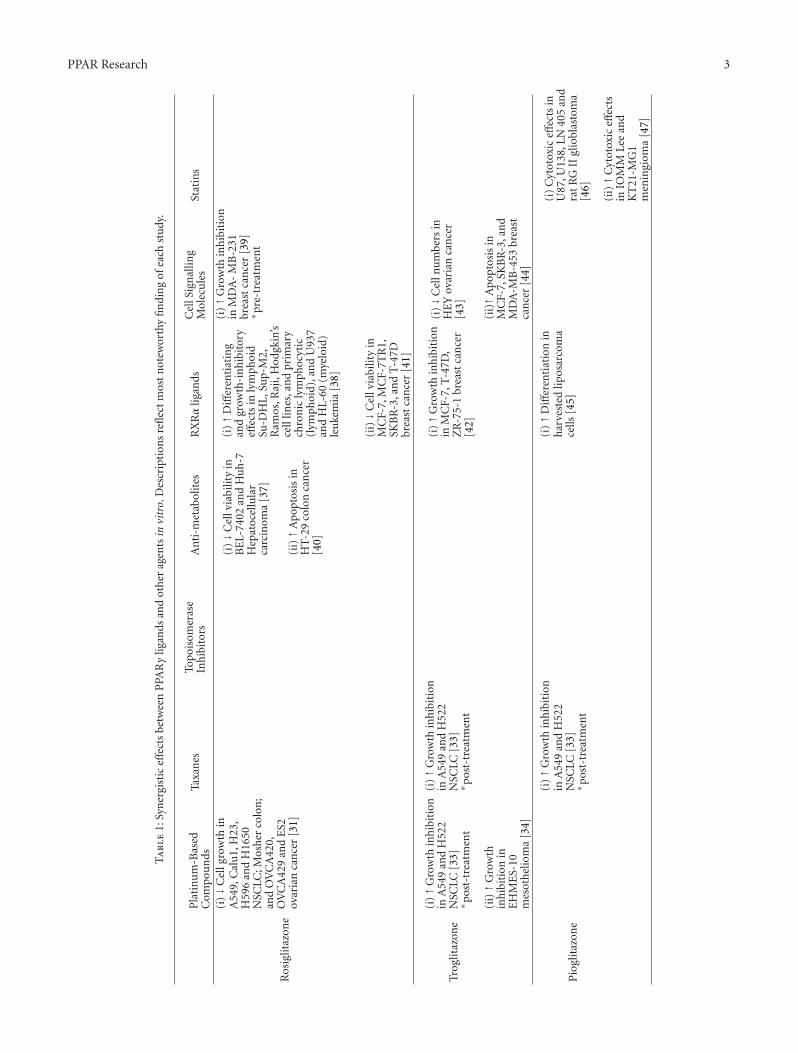
PPAR Research 3
Ta
ble
1:Sy
ner
gist
iceff
ects
betw
een
PPA
Rγ
ligan
dsan
dot
her
agen
tsin
vitr
o.D
escr
ipti
ons
refl
ect
mos
tn
otew
orth
yfi
ndi
ng
ofea
chst
udy
.
Pla
tin
um
-Bas
edC
ompo
un
dsTa
xan
esTo
pois
omer
ase
Inh
ibit
ors
An
ti-m
etab
olit
esR
XRα
ligan
dsC
ellS
ign
allin
gM
olec
ule
sSt
atin
s
Ros
iglit
azon
e
(i)↓C
ellg
row
thin
A54
9,C
alu
1,H
23,
H59
6an
dH
1650
NSC
LC;M
osh
erco
lon
;an
dO
VC
A42
0,O
VC
A42
9an
dE
S2ov
aria
nca
nce
r[3
1]
(i)↓C
ellv
iabi
lity
inB
EL-
7402
and
Hu
h-7
Hep
atoc
ellu
lar
carc
inom
a[3
7]
(i)↑D
iffer
enti
atin
gan
dgr
owth
-in
hib
itor
yeff
ects
inly
mph
oid
Su-D
HL
,Su
p-M
2,R
amos
,Raj
i,H
odgk
in’s
cell
lines
,an
dpr
imar
ych
ron
icly
mph
ocyt
ic(l
ymph
oid)
,an
dU
937
and
HL-
60(m
yelo
id)
leu
kem
ia[3
8]
(i)↑G
row
thin
hib
itio
nin
MD
A-
MB
-231
brea
stca
nce
r[3
9]∗ p
re-t
reat
men
t
(ii)↑A
popt
osis
inH
T-2
9co
lon
can
cer
[40]
(ii)↓C
ellv
iabi
lity
inM
CF-
7,M
CF-
7TR
1,SK
BR
-3,a
nd
T-4
7Dbr
east
can
cer
[41]
Trog
litaz
one
(i)↑G
row
thin
hib
itio
nin
A54
9an
dH
522
NSC
LC[3
3]∗ p
ost-
trea
tmen
t
(i)↑G
row
thin
hib
itio
nin
A54
9an
dH
522
NSC
LC[3
3]∗ p
ost-
trea
tmen
t
(i)↑G
row
thin
hib
itio
nin
MC
F-7,
T-4
7D,
ZR
-75-
1br
east
can
cer
[42]
(i)↓C
elln
um
bers
inH
EY
ovar
ian
can
cer
[43]
(ii)↑G
row
thin
hib
itio
nin
EH
ME
S-10
mes
oth
elio
ma
[34]
(ii)↑A
pop
tosi
sin
MC
F-7,
SKB
R-3
,an
dM
DA
-MB
-453
brea
stca
nce
r[4
4]
Pio
glit
azon
e
(i)↑G
row
thin
hib
itio
nin
A54
9an
dH
522
NSC
LC[3
3]∗ p
ost-
trea
tmen
t
(i)↑D
iffer
enti
atio
nin
har
vest
edlip
osar
com
ace
lls[4
5]
(i)
Cyt
otox
iceff
ects
inU
87,U
138,
LN40
5an
dra
tR
GII
glio
blas
tom
a[4
6]
(ii)↑C
ytot
oxic
effec
tsin
IOM
MLe
ean
dK
T21
-MG
1m
enin
giom
a[4
7]

4 PPAR Research
Ta
ble
1:C
onti
nu
ed.
Pla
tin
um
-Bas
edC
ompo
un
dsTa
xan
esTo
pois
omer
ase
Inh
ibit
ors
An
ti-m
etab
olit
esR
XRα
ligan
dsC
ellS
ign
allin
gM
olec
ule
sSt
atin
s
Cig
litaz
one
(i)↑G
row
thin
hib
itio
nin
ZR
-75-
1an
dT
-47D
brea
st;a
nd
Cal
u-6
lun
gca
nce
r[4
8]
(i)↓P
rolif
erat
ion
inH
EY
ovar
ian
can
cer
[43]
(i)↑C
ytot
oxic
and
cyto
stat
iceff
ects
inPa
nc
02an
dM
IAPa
Ca-
2pa
ncr
eati
c;C
-26
colo
n;a
nd
EM
T6
and
MD
A-M
B-3
61br
east
can
cer
[49]
(ii)
Apo
ptos
isin
AN
BL6
and
8226
mu
ltip
lem
yelo
ma
[50]
;C
6,U
87M
Gan
dGL2
61gl
iobl
asto
ma
[51]
,an
dG
361
mel
anom
a[5
0,52
]
RS5
444
(i)↑A
nti
prol
ifer
ativ
eeff
ects
inD
RO
90-1
and
AR
O81
anap
last
icth
yroi
dca
rcin
oma
[53]
15d-
PG
J2
(i)↑C
ytot
oxic
ity
inA
549
and
H46
0N
SCLC
[54]
(i)↑C
ytot
oxic
ity
inC
ak-2
ren
alce
llca
rcin
oma
[55]
(i)↑D
iffer
enti
atin
gan
dgr
owth
-in
hib
itor
yeff
ects
inSu
-DH
L,
Sup-
M2,
Ram
os,R
aji,
Hod
gkin
’sce
lllin
es,
and
prim
ary
chro
nic
lym
phoc
ytic
(lym
phoi
d),a
nd
U93
7an
dH
L-60
(mye
loid
)le
uke
mia
[38]
(i)↑G
row
thin
hib
itio
nin
HE
Yov
aria
nca
nce
r[4
3]
(ii)↑A
popt
osis
inA
NB
L6an
d82
26m
ult
iple
mye
lom
a[5
1]
LY29
3111
(i)↑C
ytot
oxic
ity
inM
CF-
7,M
CF-
7/ad
ran
dSK
BR
-3br
east
;H
460
lun
g;SW
480
and
RT
4co
lon
;an
dH
T11
97bl
adde
rca
nce
r[6
]
Lin
olei
cA
cid
(i)↑A
popt
osis
and↓
prol
ifer
atio
nin
MC
F-7,
MD
A-M
B-2
31,
MD
A-M
B-4
68,T
-47D
and
SKB
R-3
brea
stca
nce
r[5
6]
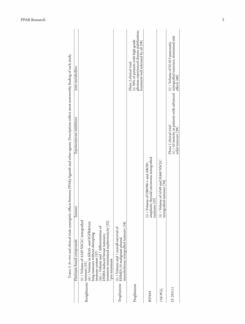
PPAR Research 5
Ta
ble
2:In
vivo
and
clin
ical
tria
lssy
ner
gist
iceff
ects
betw
een
PPA
Rγ
ligan
dsan
dot
her
agen
ts.D
escr
ipti
ons
refl
ect
mos
tn
otew
orth
yfi
ndi
ng
ofea
chst
udy
.
Pla
tin
um
-bas
edco
mpo
un
dsTa
xan
esTo
pois
omer
ase
inh
ibit
ors
An
ti-m
etab
olit
es
Ros
iglit
azon
e(i
)↓V
olu
me
ofA
549
NSC
LCxe
nog
raft
edtu
mou
rs[3
1](i
i)↓V
olu
me
inK
RA
S-an
dE
GFR
driv
enlu
ng
tum
ours
wit
hou
tdi
sru
ptin
gim
mu
ne
syst
em[5
7](i
ii)↓V
olu
me
and↑d
iffer
enti
atio
nof
DM
BA
-in
duce
dbr
east
tum
ours
;tr
eatm
ent
min
imiz
edn
eph
roto
xici
ty[3
2]∗ p
re-t
reat
men
t
Trog
litaz
one
(i)↓V
olu
me
and↑o
vera
llsu
rviv
alof
EH
ME
S-10
mal
ign
ant
pleu
ral
mes
oth
elio
ma
xen
ogra
fted
tum
ours
[34]
Pio
glit
azon
eP
hase
2cl
inic
altr
ial:
(i)
30%
ofpa
tien
tsw
ith
hig
hgr
ade
glio
mas
expe
rien
ced
dise
ase
stab
iliza
tion
;tr
eatm
ent
wel
ltol
erat
edby
all[
58]
RS5
444
(i)↓V
olu
me
ofD
RO
90-1
and
AR
O81
anap
last
icth
yroi
dca
rcin
oma
xen
ogra
fted
tum
ours
[53]
15d-
PG
J 2(i
)↓V
olu
me
ofA
549
and
H46
0N
SCLC
xen
ogra
fted
tum
ours
[54]
LY29
3111
Pha
se1
clin
ical
tria
l:(i
)↓G
Ito
xici
tyin
pati
ents
wit
had
van
ced
solid
tum
ours
[59]
(i)↓V
olu
me
ofS2
-013
pan
crea
tic
xen
ogra
fted
tum
ours
;min
imiz
edsi
deeff
ects
[60]

6 PPAR Research
expression of five members of the metallothionein genefamily [31]: metal-binding proteins that play a crucial rolein platinum-drug resistance by sequestering platinum com-pounds outside the cell [61].
In addition to developed resistance, platinum-basedcompounds are associated with several morbidities, includ-ing nephrotoxicity, myelosuppression, and GI complications[26]. Given this, and the potential for an additional drug,in this case a PPARγ ligand, to exacerbate the inherenttoxicity of platinums, the authors of the aforementioned lungadenocarcinoma study conducted extensive toxicologicalanalysis on their treated mice. Fortunately, compared tomonotherapy, combination therapy did not decrease mark-ers of immune function, white blood cell counts, or hema-tocrit, and BUN and creatinine levels, indicative of kidneydamage, were similarly unaffected [57].
Nephrotoxicity, experienced by 28–36% of patients aftera single injection of cisplatin [62], may be, in part, exacer-bated by TNF-α, a well-known mediator of inflammation[40]. Interestingly, PPARγ activators reduce inflammatoryresponses [63, 64]. Therefore, Tikoo et al. used a DMBA-induced murine breast cancer model to evaluate the ability ofROSI to decrease nephrotoxicity. They found that ROSI pre-treatment significantly decreased circulating BUN, creatinineand TNF-α, and minimized tubular damage, suggesting thatPPARγ activation ameliorated the nephrotoxicity associatedwith cisplatin treatment [32]. If this holds true in humans,ROSI treatment may allow physicians to use platinum-basedcompounds at higher, previously toxic, doses that may conferadditional therapeutic benefit.
2.2. Taxanes. Taxanes, including paclitaxel and docetaxel, arecommonly used chemotherapy agents for a large array ofcancers which include ovarian, lung, head and neck, eso-phageal, breast, prostrate, and gastric cancers. Taxanes exerttheir effects by binding and immobilizing microtubuleswhich prevents cell division [65]. There are multiple sideeffects associated with taxanes including reduced hematocrit,neuropathy, and myalgias/arthralgias [66].
A novel high-affinity PPARγ agonist, and thiazolidine-dione derivative, RS5444, demonstrated additive antiprolif-erative activity on DRO90-1 and ARO81 anaplastic thyroidcarcinoma cells, a particularly aggressive and dedifferentiatedcancer [67]. RS5444 did not induce apoptosis by itself; how-ever, when combined with paclitaxel, the apoptotic fractionof cells doubled. Using IC25 values experimentally derivedfrom in vitro experiments, the group found that combinationtreatment with RS5444 and paclitaxel significantly reducedxenograft tumour volumes compared to either monotherapyalone [53].
Non-small-cell lung cancer is a leading cause of deathfrom malignant disease in industrialized nations with a5-year survival rate of approximately 15% [68, 69]. Noveltherapeutic regimens involving PPARγ activators and tra-ditional chemotherapeutics have shown some promise thatthey may someday improve this rate. An in vitro study indi-cated synergy between multiple PPARγ ligands (TRO andPIO) and paclitaxel in A549, H522 non-small-cell lung
cancer cells that was dependent upon treatment order, withpaclitaxel preceding TRO treatment [33]. Another groupconfirmed the synergistic effect of combining PPARγ acti-vation with, this time, docetaxel. In this study, 15d-PJ2
increased cytotoxicity in A549 and H460 cells in vitro.Extending this, they found that 15d-PJ2 and docetaxelreduced A549 and H460 xenografted tumour volumes by72%, nearly double the effect of docetaxel alone [54].
2.3. Topoisomerase Inhibitors. Both classes of topoisomeraseinhibitors, type I (including irinotecan) and type II, workby binding and incapacitating topoisomerases: enzymes thatare critical for DNA supercoiling and strain relief [70]. Ulti-mately, this binding prevents movement of the DNA repli-cation fork which induces stress responses that can lead toapoptosis or the involvement of DNA damage repair mech-anisms [71]. A topoisomerase I inhibitor, irinotecan, hasdemonstrated activity against a vast range of cancers [72]but is associated with significant GI toxicity and myelosup-pression [73]. Budman and Calabro have shown synergisticcytotoxic increases in a variety of cell lines (MCF-7, MCF-7/adr, and SK-BR-3 breast cancer; H460 lung cancer; SW480and RT4 colon cancer; HT1197 bladder cancer) betweenirinotecan and the PPARγ ligand LY293111 at clinicallyattainable doses [6], prompting human studies with this drugcombination. To date, a phase I clinical trial has established adosing schedule that minimized adverse GI events associatedwith LY293111 and irinotecan [59]. Another topoisomeraseI inhibitor, camptothecin, enhanced the cytotoxicity of 15d-PGJ2 in Cak-2 renal cell carcinoma cells. Interestingly, theauthors did not find synergy when 15d-PGJ2 was combinedwith other chemotherapeutics including doxorubicin, 5-FU,and cisplatin [55]. This synergism may allow clinicians toreduce the dose of topoisomerase inhibiting agents andthereby reduce associated toxicity, by combining treatmentwith PPARγ ligands.
2.4. Antimetabolites. Antimetabolites, including 5-Fluorou-racil (5-FU), methotrexate, and others, are structurally sim-ilar compounds to vitamins, amino acids, or nucleic acidprecursors which become incorporated into cellular macro-molecules with disastrous consequences for cells such asinhibition of cell growth and division [74]. They have beenused to treat several types of cancer including leukemia,breast, and ovarian but have been associated with myelosup-pression, dermatitis, and diarrhea [75]. A phase II clinicaltrial was undertaken to evaluate the role of capecitabine,a precursor to 5-FU, in combination with PIO to treatrecurrent high-grade gliomas. Only 29% of patients experi-enced disease stabilization after three months; however, theregimen was well tolerated by patients indicating potentialfor future therapeutic utility [58].
Hepatocellular carcinoma (HCC) and colorectal tum-ours are among the leading forms of cancer contributing tocancer-related deaths [69, 76]. HCC usually requires chemo-therapy because tumours are often surgically unresectabledue to advanced stage at diagnosis [77]. Treatment of bothdiseases often involves 5-FU; however, patients often respond

PPAR Research 7
poorly as tumours develop multiple drug resistance [78–80]due to multiple mechanisms including increased drug efflux[81]. Interestingly, PPARγ may regulate ABC transporters,key proteins involved in drug efflux [82]. Accordingly, activa-tion of PPARγ with ROSI, in combination with 5-FU treat-ment, has been evaluated in HCC and colon cancer. ROSItreatment decreased cell viability in two HCC cell lines (BEL-7402 and Huh-7) by 4- and 2-fold, respectively, compared totreatment with 5-FU alone. The authors also used siRNA toshow that this effect was dependent on PPARγ [37]. Anothergroup evaluated ROSI treatment with 5-FU in HT-29 coloncancer cells and found that ROSI treatment, at a low dose thatdid not affect proliferation or cell growth, enhanced 5-FU-induced apoptosis. Again, this effect was PPARγ dependentas it was ameliorated by the PPARγ antagonist GW9662 [83].
Another antimetabolite, gemcitabine, is a useful chemo-therapeutic that arrests cell growth in multiple ways includ-ing incorporation into DNA and impeding cell division [84].Gemcitabine is standard therapy for pancreatic cancer, adisease with a strikingly poor prognosis as most patientsdie within six months of diagnosis [85]. Gemcitabine onlymodestly prolongs survival but is useful as a palliative agentfor several cancer-related morbidities. Hennig et al. evaluatedthe ability of the PPARγ activator LY293111 to enhance theactivity of gemcitabine in an orthotopic pancreatic cancermodel. Consistent with previous models, both gemcitabineand LY293111 significantly inhibited tumour growth andreduced the incidence of liver metastasis; however, the com-bination was more effective than either therapy alone. Fur-thermore, combination treatment maintained stable bodyweights, relieved tumour-induced cachexia, and decreasedincidence of bowel obstruction [60]. This suggests that thiscombination may be effective, to not only treat aggressivepancreatic adenocarcinomas but also relieve side effectsassociated with monotherapy [86].
2.5. RXRα Ligands. The PPARγ binding partner, RXRα, isalso a member of the nuclear receptor superfamily. RXR hasthree subtypes (α, β, and γ), which are activated by retinoids,a group of vitamin A analogues. After ligand binding, RXRαis able to modulate gene expression by binding retinoidX receptor responsive elements (RXREs), present in thepromoter regions of target genes. Similar to PPARγ, RXRαactivation profoundly affects multiple cellular activities thatare pertinent to cancer including cellular growth, differenti-ation, apoptosis, and morphogenesis [87, 88].
Given this, multiple groups have investigated the com-bined use of PPARγ and RXRα ligands. The first report, fromTontonoz et al., indicated that simultaneous treatment ofliposarcoma cells, selected from freshly harvested tumours,with both RXRα- and PPARγ-specific ligands, synergisticallystimulated differentiation. Additionally, the authors showedthat PPARγ is highly expressed in the major histological typesof liposarcoma, suggesting that PPARγ-targeting agents,especially combined with RXRα ligands, may be usefultherapy for human liposarcoma [45].
Since that time, beneficial effects have been reportedfor several types of malignancies, including hematological,
breast, and lung cancer, for the combined treatment ofPPARγ ligands and retinoids. Konopleva et al. reported thatPPARγ is expressed in lymphoid (Su-DHL, Sup-M2, Ramos,Raji, Hodgkin’s cell lines, and primary chronic lymphocyticleukemia) and myeloid (U937 and HL-60) cell lines, severalof which undergo apoptosis when treated with PPARγligands including ROSI and 15d-PGJ2. The apoptotic effectsof PPARγ ligands were enhanced when combined with anRXRα agonist, LG100268, as reflected by mitochondrialdepolarization and caspase activation [38]. Similarly, Rayet al. showed that PPARγ is expressed in ANBL6 and 8226human multiple myeloma cell lines and that PPARγ ligandsinduce apoptosis, an effect which is enhanced by combina-tion with 9-cis retinoic acid, a ligand of RXRα [51]. Elstneret al. found that PPARγ ligands were potentiated by RXRαligands in multiple breast cancer cells (MCF-7, T-47D, ZR-75-) [42], work that was later confirmed by multiple groups[41, 56] including one study that also found protective effectsin Calu-6 lung cancer cells [48]. One of these studies showedthat combination treatment with ROSI and the RXRα ligand9-cis retinoic acid inhibited cell viability in MCF-7, MCF-7TR1, SKBR-3, and T-47D breast cancer cells but did notaffect MCF-10a normal immortalized breast epithelial cells.This exciting work suggests that the cytotoxic effect maybespecific to cancer cells. Mehta et al. took this approach intomouse models and found that the combination of LG10068,an RXRα-specific ligand, and TRO completely inhibiteddevelopment of mammary tumours at micromolar concen-trations in a DMBA-induced breast tumourigenesis model[17].
Many potential mechanisms are postulated to explain thesynergistic protective effects of PPARγ and RXRα ligands.The protective effect at the whole body level may, in part, bemediated at the transcriptional level by the ability of PPARγand RXRα ligands to inhibit aromatase secretion [89],enhance expression of glutathione S-transferases (GSTs)[90], or downregulate expression of matrix metalloprotein-ases (MMPs) [52]. Aromatase catalyzes the rate-limiting stepin estrogen biosynthesis [91], which drives breast tumourige-nesis by stimulating proliferation of breast tumour cells [92].GSTs have multiple functions including the detoxificationof several xenobiotics and carcinogens [93]. MMPs degradeextracellular matrix proteins carving pathways for migratingcancer cells and releasing sequestered growth factors [94].Combined RXRα ligand and CIG treatment decreases cellgrowth and the invasive potential in G361 melanoma andU87MG glioblastoma cells by decreasing expression ofmatrix metalloproteinases [52].
Other groups have theorized that the synergistic effectsof PPARγ and RXRα ligands may not be directly related totranscriptional effects. Ligands of PPARγ and RXRα recruitdifferent subsets of transcriptional coactivators [95]; there-fore, cotreatment may increase transcription as there is lesscompetition [96]. Additionally, PPARγ activity may enhanceproteosome inhibitors, allowing for RXRα accumulation andthe enhancement of PPARγ : RXRα-mediated transcription[97]. Collectively, this work suggests that combining agentsthat activate both PPARγ and RXRα could synergisticallyenhance the protective effects of either agent alone.

8 PPAR Research
3. Cell Signaling Molecules
Protective synergy with PPARγ ligands is not exclusive totraditional chemotherapeutic agents or RXRα ligands. Thereare a few reports of PPARγ activators combining with normalcell signaling molecules, including tumour necrosis factor(TNF)α, tumour necrosis factor-related apoptosis-inducingligand (TRAIL), and Heregulin to confer an additive orsynergistic protective effect. TNFα is a cytokine, chieflyproduced by activated macrophages, that is involved insystemic inflammation, and leads to tumour regression [98,99]. TRAIL, a member of the TNF family, induces apoptosisby binding receptors and recruiting the Fas-associated deathdomain and caspase-8, triggering apoptosis [100]. Heregulinis a soluble secreted growth factor that activates severalclassic tumourigenic signal transduction pathways includingPI3K/Akt, Ras/MAPK, and JNK [101].
Based on evidence that ROSI upregulates p53 and p21,Mody et al. examined the ability of ROSI pretreatment tosensitize MDA-MB-231 breast cancer cells to therapies thatact on these apoptosis/cell death pathways, such as TNFα.ROSI pretreatment dramatically increased TNFα-mediatedgrowth inhibition by 9-fold versus control TNFα or ROSIalone. The authors also performed microarray analysis toevaluate genetic changes associated with ROSI treatment[39]. This may be a valuable tool to predict other agentswhich synergize with PPARγ ligand activity based on sharedpathway utilization.
Partridge and Barnes evaluated the ability of multiplePPARγ ligands (CIG, TRO, and 15d-PGJ2) to enhance theefficacy of TRAIL in a drug-resistant ovarian cancer cell line.Drug resistance is a serious problem in ovarian cancer, espe-cially in advanced disease, where survival rates fall to 10–30%[102]. The combined treatment with CIG and TRAIL syn-ergistically reduced proliferation in multiple cell lines, mostnotably the paclitaxel-resistant HEY ovarian cancer sub-clone. TRO treatment showed no effect on proliferationon its own; however, when combined with TRAIL, thatreduced cell numbers in etoposide-, pemetrexed-, cisplatin-,docetaxel-, and gemcitabine-resistant cell lines. Similarly,15d-PGJ2 treatment inhibited growth in all cell lines, espe-cially the HEY cell line which was developed by the authors[43].
Park et al. showed that Heregulin, which paradoxicallydrives tumourigenesis [103, 104], synergistically increasesTRO-mediated breast cancer cell apoptosis and necrosis invitro [44]. In light of previous reports that Heregulin playsa causal role in Tamoxifen- and Gefitinib-resistant breastcancer [105], Park’s work provides evidence that combina-tion therapy with Heregulin and PPARγ-activators may bea novel strategy for the treatment of resistant or refractorybreast cancer [44].
4. Statins
Statins are another important class of drugs acting as inhi-bitors of 3-hydroxy-3-methylglutarylcoenzyme A (HMG-CoA) reductase, a critical rate-limiting enzyme in choles-terol biosynthesis. Statins are commonly used to manage
hypercholesterolemia and cardiovascular diseases and aresome of the most frequently prescribed therapeutics forelderly patients. Recently, statins were evaluated for theirprotective effects in cancer and showed antiproliferative andpro-apoptotic effects in vitro [106–108]. Incubation withlovastatin and CIG for 48hrs exerted additive cytotoxic andcytostatic effects in multiple cancer cell lines (Panc 02 andMIA PaCa-2 pancreatic cancer, C-26 colon cancer, and EMT6and MDA-MB-361 breast cancer) compared to either treat-ment alone [49, 50]. Further experiments on human U87,U138, LN 405, and rat RG II glioblastoma cells indicatedcytotoxic synergy after 48- and 144-hour treatments withPIO and a variety of statins [46]. Additionally, treatment oftwo meningioma cell lines (IOMM-Lee and KT21-MG1)with PIO and statins showed significant synergistic cytotoxiceffects [47]. It was also suggested that statins may signalthrough the transcription factor sterol response element-binding protein (SREBP) to encourage PPARγ-mediatedupregulation of PTEN [109]. This evidence suggests yetanother class of drugs that, combined with PPARγ-ligands,show synergistic protective effects in cancer.
5. Areas Needing More Work
The majority of literature in the field supports the viewthat combination cancer therapy with PPARγ ligands andchemotherapeutic agents produce beneficial effects. How-ever, this trend is not universal. Multiple groups haveevaluated combinations of chemotherapeutic cocktails thatinclude PPARγ ligands and found no synergism. For exam-ple, Yamamoto et al. reported that the synergistic toxic effectsof 15d-PGJ2 in renal cell carcinoma were specific to its com-bination therapy with camptothecin, and not evident withtwo common chemotherapeutic agents: 5-fluorouracin andcisplatin [55]. Tapia-Perez’s group also found that synergismin glioblastoma cells depends on the combination, this timethe PPARγ ligand, as PIO + statin treatment produced asignificant cytotoxic effect although the same was not true forROSI + statin [46] Clearly, more work is needed to establishwhich combinations will be effective in which diseases, workthat will be further complicated by factors such as doseand treatment timing (pre-, post-, cotreatment, etc.). Inter-estingly, the same combination regimen may not alwaysbe effective, even within the same disease, as Elstner et al.reported that only three (MCF-7, MDA-MB-231, and ZR-75-1) of the eight (MCF-7, BT20, BT474, MDA-MB-231,MDA-MB-436, SKBR3, T-47D, ZR-75-1) breast cancer celllines they evaluated were sensitive to combinations of thePPARγ ligand TRO and RXRα ligand 9-cis retinoic acid.Interestingly, the sensitive cell lines all express high levelsof the apoptosis protein bcl-2 [42]. This underscores theimportance of work to evaluate the molecular mechanismsby which combination therapies exert their effects so that,someday, clinicians and researchers may predict treatmentefficacy using molecular signatures. Most notably, extensiveliterature searches did not reveal reports of PPARγ lig-ands impeding the therapeutic efficacy of chemotherapeuticagents.
PPAR Research 9
Synthetic PPARγ ligands are generally well-tolerated andnontoxic; however, multiple groups have reported adversecardiovascular events associated with PPARγ ligands, includ-ing myocardial hypertrophy and congestive heart failure dueto plasma volume expansion and edema, in humans andanimal models [110–112]. To address this problem, the FDAconvened leading experts in 2010 to carry out more researchto definitively show whether PPARγ ligands are associatedwith increased cardiovascular risk. The committee observedno significant difference in acute myocardial infarction andacute heart failure between patients treated with ROSI orPIO versus matched control cases and recommended thatfurther studies be performed to address this issue [113, 114].While these studies are in progress, research should continueto evaluate PPARγ ligands for their efficacy and mechanismsof action given their well-documented protective effects inmany diseases, including, but not limited to, cancer. Further-more, a better understanding of the mechanisms by whichactivation of PPARγ-dependent signaling stops tumourigen-esis may provide the basis for future development of moreefficacious drugs to prevent and/or reduce cancer-relateddeaths.
6. Discussion
The studies summarized previously, and in Tables 1 and 2,suggest that the combination of PPARγ ligands plus standardchemotherapeutic agents, RXRα agonists, statins, and certaincellular signaling molecules holds promise as a novel therapyfor several types of malignancy. In general, combined use oftwo or more therapeutic compounds is often advantageousbecause of the potential to use lower clinical doses of each,which decreases nonspecific toxicity. However, here wereport several examples of synergistic/additive interactionsbetween agents that activate PPARγ as well as reductions intoxicity and the ability to overcome resistance. The resultshere are largely preclinical, with a select few regimens beingevaluated in human subjects and, even in those cases,early clinical trials which, naturally, focused largely ontoxicity as opposed to efficacy. However, the volume of pre-clinical evidence suggests that a breakthrough in the clinicalapplication of combination therapy with PPARγ agonists isvery possible. Moving forward, studies should continue toevaluate mechanisms by which these regimens induce theirtherapeutic effects as this will ultimately lead to identificationof patient populations with high probabilities of therapeuticefficacy. In summary, the types of combination therapydescribed here are promising strategies for the chemopre-vention, management, and/or treatment of several types ofcancer.
References
[1] World Health Organization, “Cancer Fact Sheet #297,” 2012,http://www.who.int/mediacentre/factsheets/fs297/en/.
[2] D. Hanahan and R. A. Weinberg, “Hallmarks of cancer: thenext generation,” Cell, vol. 144, no. 5, pp. 646–674, 2011.
[3] D. R. Budman, A. Calabro, L. G. Wang et al., “Synergismof cytotoxic effects of vinorelbine and paclitaxel in vitro,”Cancer Investigation, vol. 18, no. 8, pp. 695–701, 2000.
[4] T. C. Chou, R. J. Motzer, Y. Tong, and G. J. Bosl, “Comput-erized quantitation of synergism and antagonism of taxol,topotecan, and cisplatin against human teratocarcinoma cellgrowth: a rational approach to clinical protocol design,” Jour-nal of the National Cancer Institute, vol. 86, no. 20, pp. 1517–1524, 1994.
[5] A. A. Adjei, I. I. Budihardjo II, E. K. Rowinsky et al., “Cyto-toxic synergy between pyrazoloacridine (NSC 366140) andcisplatin in vitro: inhibition of platinum-DNA adductremoval,” Clinical Cancer Research, vol. 3, no. 5, pp. 761–770,1997.
[6] D. R. Budman and A. Calabro, “Studies of synergistic andantagonistic combinations of conventional cytotoxic agentswith the multiple eicosanoid pathway modulator LY 293111,”Anti-Cancer Drugs, vol. 15, no. 9, pp. 877–881, 2004.
[7] S. A. Kliewer, B. M. Forman, B. Blumberg et al., “Differentialexpression and activation of a family of murine peroxisomeproliferator-activated receptors,” Proceedings of the NationalAcademy of Sciences of the United States of America, vol. 91,no. 15, pp. 7355–7359, 1994.
[8] O. Braissant, F. Foufelle, C. Scotto, M. Dauca, and W. Wahli,“Differential expression of peroxisome proliferator-activatedreceptors (PPARs): tissue distribution of PPAR-α,-β, and -γin the adult rat,” Endocrinology, vol. 137, no. 1, pp. 354–366,1996.
[9] S. Jain, S. Pulikuri, Y. Zhu et al., “Differential expression ofthe peroxisome proliferator-activated receptor γ (PPARγ)and its coactivators steroid receptor coactivator-1 and PPAR-binding protein PBP in the brown fat, urinary bladder, colon,and breast of the mouse,” The American Journal of Pathology,vol. 153, pp. 349–354, 1998.
[10] L. Michalik, J. Auwerx, J. P. Berger et al., “International unionof pharmacology. LXI. Peroxisome proliferator-activatedreceptors,” Pharmacological Reviews, vol. 58, no. 4, pp. 726–741, 2006.
[11] S. A. Kliewer, H. E. Xu, M. H. Lambert, and T. M. Willson,“Peroxisome proliferator-activated receptors: from genes tophysiology,” Recent Progress in Hormone Research, vol. 56, pp.239–263, 2001.
[12] T. M. Larsen, S. Toubro, and A. Astrup, “PPARγ agonists inthe treatment of type II diabetes: is increased fatness com-mensurate with long-term efficacy?” International Journal ofObesity and Related Metabolic Disorders, vol. 27, pp. 147–161,2003.
[13] C. Rosak, E. Standl, T. Reblin, H. Stammer, and D. K. Seidel,“Rosiglitazone is effective and well-tolerated in a range oftherapeutic regimens during daily practice in patients withtype 2 diabetes,” International Journal of Clinical Practice, vol.60, no. 9, pp. 1040–1047, 2006.
[14] N. Kubota, Y. Terauchi, H. Miki et al., “PPARγ mediateshigh-fat diet-induced adipocyte hypertrophy and insulinresistance,” Molecular Cell, vol. 4, no. 4, pp. 597–609, 1999.
[15] E. Mueller, P. Sarraf, P. Tontonoz et al., “Terminal differentia-tion of human breast cancer through PPARγ,” Molecular Cell,vol. 1, no. 3, pp. 465–470, 1998.
[16] E. Elstner, C. Muller, K. Koshizuka et al., “Ligands for per-oxisome proliferator-activated receptory and retinoic acidreceptor inhibit growth and induce apoptosis of humanbreast cancer cells in vitro and in BNX mice,” Proceedings ofthe National Academy of Sciences of the United States of Ameri-ca, vol. 95, no. 15, pp. 8806–8811, 1998.
[17] R. G. Mehta, E. Williamson, M. K. Patel, and H. P. Koeffler,“A ligand of peroxisome proliferator-activated receptor γ,
10 PPAR Research
retinoids, and prevention of preneoplastic mammary lesions,”Journal of the National Cancer Institute, vol. 92, no. 5,pp. 418–423, 2000.
[18] G. M. Pighetti, W. Novosad, C. Nicholson et al., “Therapeutictreatment of DMBA-induced mammary tumors with PPARligands,” Anticancer Research, vol. 21, no. 2A, pp. 825–830,2001.
[19] N. Suh, Y. Wang, C. R. Williams et al., “A new ligand forthe peroxisome proliferator-activated receptor-γ (PPAR-γ),GW7845, inhibits rat mammary carcinogenesis,” CancerResearch, vol. 59, no. 22, pp. 5671–5673, 1999.
[20] G. D. Demetri, C. D. M. Fletcher, E. Mueller et al., “Inductionof solid tumor differentiation by the peroxisome proliferator-activated receptor-γ ligand troglitazone in patients withliposarcoma,” Proceedings of the National Academy of Sciencesof the United States of America, vol. 96, no. 7, pp. 3951–3956,1999.
[21] H. J. Burstein, G. D. Demetri, E. Mueller, P. Sarraf, B. M.Spiegelman, and E. P. Winer, “Use of the peroxisome proli-ferator-activated receptor (PPAR) γ ligand troglitazone astreatment for refractory breast cancer: a phase II study,”Breast Cancer Research and Treatment, vol. 79, no. 3, pp. 391–397, 2003.
[22] M. H. Kulke, G. D. Demetri, N. E. Sharpless et al., “A phaseII study of troglitazone, an activator of the PPARγ receptor,in patients with chemotherapy-resistant metastatic colorectalcancer,” Cancer Journal, vol. 8, no. 5, pp. 395–399, 2002.
[23] M. R. Smith, J. Manola, D. S. Kaufman et al., “Rosiglitazoneversus placebo for men with prostate carcinoma and a risingserum prostate-specific antigen level after radical prostatec-tomy and/or radiation therapy,” Cancer, vol. 101, no. 7, pp.1569–1574, 2004.
[24] L. D. Yee, N. Williams, P. Wen et al., “Pilot study of rosiglita-zone therapy in women with breast cancer: effects of short-term therapy on tumor tissue and serum markers,” ClinicalCancer Research, vol. 13, no. 1, pp. 246–252, 2007.
[25] R. A. Alderden and T. W. Hambley, “The discovery anddevelopment of cisplatin,” Journal of Chemical Education, vol.83, no. 5, pp. 724–728, 2006.
[26] L. Kelland, “The resurgence of platinum-based cancer chem-otherapy,” Nature Reviews Cancer, vol. 7, no. 8, pp. 573–584,2007.
[27] Z. H. Siddik, “Cisplatin: mode of cytotoxic action and mol-ecular basis of resistance,” Oncogene, vol. 22, no. 47, pp.7265–7279, 2003.
[28] J. Sastry and S. J. Kellie, “Severe neurotoxicity, ototoxicityand nephrotoxicity following high-dose cisplatin and amifos-tine,” Pediatric Hematology and Oncology, vol. 22, no. 5, pp.441–445, 2005.
[29] I. Arany and R. L. Safirstein, “Cisplatin nephrotoxicity,”Seminars in Nephrology, vol. 23, no. 5, pp. 460–464, 2003.
[30] R. J. Cersosimo, “Oxaliplatin-associated neuropathy: areview,” Annals of Pharmacotherapy, vol. 39, no. 1, pp. 128–135, 2005.
[31] G. D. Girnun, E. Naseri, S. B. Vafai et al., “Synergy betweenPPARγ ligands and platinum-based drugs in cancer,” CancerCell, vol. 11, no. 5, pp. 395–406, 2007.
[32] K. Tikoo, P. Kumar, and J. Gupta, “Rosiglitazone synergizesanticancer activity of cisplatin and reduces its nephrotoxicityin 7, 12-dimethyl benzaanthracene (DMBA) induced breastcancer rats,” BMC Cancer, vol. 9, article 107, 2009.
[33] R. C. Reddy, A. Srirangam, K. Reddy et al., “Chemothera-peutic drugs induce PPAR-γ expression and show sequence-specific synergy with PPAR-γ ligands in inhibition of
non-small cell lung cancer,” Neoplasia, vol. 10, no. 6, pp. 597–603, 2008.
[34] N. Hamaguchi, H. Hamada, S. Miyoshi et al., “In vitro and invivo therapeutic efficacy of the PPAR-γ agonist troglitazonein combination with cisplatin against malignant pleuralmesothelioma cell growth,” Cancer Science, vol. 101, no. 9,pp. 1955–1964, 2010.
[35] S. A. Cannistra, “Cancer of the ovary,” The New England Jour-nal of Medicine, vol. 351, no. 24, pp. 2519–2529, 2004.
[36] D. A. Eberhard, B. E. Johnson, L. C. Amler et al., “Mutationsin the epidermal growth factor receptor and in KRAS arepredictive and prognostic indicators in patients with non-small-cell lung cancer treated with chemotherapy alone andin combination with erlotinib,” Journal of Clinical Oncology,vol. 23, no. 25, pp. 5900–5909, 2005.
[37] L. Q. Cao, X. L. Wang, Q. Wang et al., “Rosiglitazone sen-sitizes hepatocellular carcinoma cell lines to 5-fluorouracilantitumor activity through activation of the PPARγ signalingpathway,” Acta Pharmacologica Sinica, vol. 30, no. 9, pp.1316–1322, 2009.
[38] M. Konopleva, E. Elstner, T. J. McQueen et al., “Peroxisomeproliferator-activated receptor and retinoid X receptor lig-ands are potent inducers of differentiation and apoptosis inleukemias,” Molecular Cancer Therapeutics, vol. 3, no. 10, pp.1249–1262, 2004.
[39] M. Mody, N. Dharker, M. Bloomston et al., “Rosiglitazonesensitizes MDA-MB-231 breast cancer cells to anti-tumoureffects of tumour necrosis factor-α, CH11 and CYC202,”Endocrine-Related Cancer, vol. 14, no. 2, pp. 305–315, 2007.
[40] B. Zhang, G. Ramesh, C. C. Norbury, and W. B. Reeves, “Cis-platin-induced nephrotoxicity is mediated by tumor necrosisfactor-α produced by renal parenchymal cells,” Kidney Inter-national, vol. 72, no. 1, pp. 37–44, 2007.
[41] D. Bonofiglio, E. Cione, H. Qi et al., “Combined low dosesof PPARγ and RXR ligands trigger an intrinsic apoptoticpathway in human breast cancer cells,” The American Journalof Pathology, vol. 175, no. 3, pp. 1270–1280, 2009.
[42] E. Elstner, E. A. Williamson, C. Zang et al., “Novel thera-peutic approach: ligands for PPARγ and retinoid receptorsinduce apoptosis in bcl-2-positive human breast cancer cells,”Breast Cancer Research and Treatment, vol. 74, no. 2, pp. 155–165, 2002.
[43] K. Brautigam, J. Biernath-Wupping, D. O. Bauerschlag et al.,“Combined treatment with TRAIL and PPARγ ligands over-comes chemoresistance of ovarian cancer cell lines,” Journalof Cancer Research and Clinical Oncology, vol. 137, no. 5, pp.875–886, 2011.
[44] B. H. Park, S. B. Lee, D. B. Stolz, Y. J. Lee, and B. C. Lee,“Synergistic interactions between heregulin and peroxisomeproliferator-activated receptor-γ (PPARγ) agonist in breastcancer cells,” Journal of Biological Chemistry, vol. 286, no. 22,pp. 20087–20099, 2011.
[45] P. Tontonoz, S. Singer, B. M. Forman et al., “Terminal differ-entiation of human liposarcoma cells induced by ligands forperoxisome proliferator-activated receptor γ and the retinoidX receptor,” Proceedings of the National Academy of Sciences ofthe United States of America, vol. 94, no. 1, pp. 237–241, 1997.
[46] J. H. Tapia-Perez, E. Kirches, C. Mawrin, R. Firsching, andT. Schneider, “Cytotoxic effect of different statins and thiazo-lidinediones on malignant glioma cells,” Cancer ChemotherPharmacol, vol. 67, no. 5, pp. 1193–1201, 2011.
[47] S. Gehring, J. H. Tapia-Perez, E. Kirches et al., “Cytotoxiceffects of statins and thiazolidinediones on meningioma
PPAR Research 11
cells,” Journal of Neuro-Oncology, vol. 102, no. 3, pp. 383–393,2011.
[48] S. Y. James, F. Lin, S. K. Kolluri, M. I. Dawson, and X. K.Zhang, “Regulation of retinoic acid receptor β expressionby peroxisome proliferator-activated receptor γ ligands incancer cells,” Cancer Research, vol. 63, no. 13, pp. 3531–3538,2003.
[49] P. Mrowka, E. Glodkowska, D. Nowis et al., “Ciglitazone,an agonist of peroxisome proliferator-activated receptor γ,exerts potentiated cytostatic/cytotoxic effects against tumorcells when combined with lovastatin,” International Journalof Oncology, vol. 32, pp. 249–255, 2008.
[50] A. Papi, L. Tatenhorst, D. Terwel et al., “PPARγ and RXRγligands act synergistically as potent antineoplastic agents invitro and in vivo glioma models,” Journal of Neurochemistry,vol. 109, pp. 1779–1790, 2009.
[51] D. M. Ray, S. H. Bernstein, and R. P. Phipps, “Human multi-ple myeloma cells express peroxisome proliferator-activatedreceptor γ and undergo apoptosis upon exposure to PPARγligands,” Clinical Immunology, vol. 113, pp. 203–213, 2004.
[52] A. Papi, P. Rocchi, A. M. Ferreri, F. Guerra, and M.Orlandi, “Enhanced effects of PPARγ ligands and RXRselective retinoids in combination to inhibit migration andinvasiveness in cancer cells,” Oncology Reports, vol. 21, pp.1083–1089, 2009.
[53] J. A. Copland, L. A. Marlow, S. Kurakata et al., “Novel high-affinity PPARγ agonist alone and in combination with pacli-taxel inhibits human anaplastic thyroid carcinoma tumorgrowth via p21WAF1/CIP1,” Oncogene, vol. 25, no. 16, pp.2304–2317, 2006.
[54] S. V. Fulzele, A. Chatterjee, M. S. Shaik, T. Jackson, N. Ichite,and M. Singh, “15-Deoxy-Δ12,14-prostaglandin J2 enhancesdocetaxel anti-tumor activity against A549 and H460 non-small-cell lung cancer cell lines and xenograft tumors,” Anti-Cancer Drugs, vol. 18, no. 1, pp. 65–78, 2007.
[55] Y. Yamamoto, M. Fujita, H. Koma et al., “15-Deoxy-Δ12,14-prostaglandin J2 enhanced the anti-tumor activity ofcamptothecin against renal cell carcinoma independentlyof topoisomerase-II and PPARγ pathways,” Biochemical andBiophysical Research Communications, vol. 410, no. 3, pp.563–567, 2011.
[56] D. L. Crowe and R. A. Chandraratna, “A retinoid X receptor(RXR)-selective retinoid reveals that RXR-α is potentiallya therapeutic target in breast cancer cell lines, and that itpotentiates antiproliferative and apoptotic responses to per-oxisome proliferator-activated receptor ligands,” Breast Can-cer Research, vol. 6, no. 5, pp. R546–R555, 2004.
[57] G. D. Girnun, L. Chen, J. Silvaggi et al., “Regression of drug-resistant lung cancer by the combination of rosiglitazone andcarboplatin,” Clinical Cancer Research, vol. 14, no. 20, pp.6478–6486, 2008.
[58] P. Hau, L. Kunz-Schughart, U. Bogdahn et al., “Low-dosechemotherapy in combination with COX-2 inhibitors andPPAR-γ agonists in recurrent high-grade gliomas—a phaseII study,” Oncology, vol. 73, no. 1-2, pp. 21–25, 2008.
[59] T. Baetz, E. Eisenhauer, L. Siu et al., “A phase I study oforal LY293111 given daily in combination with irinotecan inpatients with solid tumours,” Investigational New Drugs, vol.25, no. 3, pp. 217–225, 2007.
[60] R. Hennig, J. Ventura, R. Segersvard et al., “LY293111improves efficacy of gemcitabine therapy on pancreatic can-cer in a fluorescent orthotopic model in athymic mice,” Neo-plasia, vol. 7, no. 4, pp. 417–425, 2005.
[61] M. Knipp, “Metallothioneins and platinum(II) anti-tumorcompounds,” Current Medicinal Chemistry, vol. 16, no. 5, pp.522–537, 2009.
[62] D. Lebwohl and R. Canetta, “Clinical development of plat-inum complexes in cancer therapy: an historical perspectiveand an update,” European Journal of Cancer, vol. 34, no. 10,pp. 1522–1534, 1998.
[63] C. Jiang, A. T. Ting, and B. Seed, “PPAR-γ agonists inhibitproduction of monocyte inflammatory cytokines,” Nature,vol. 391, no. 6662, pp. 82–86, 1998.
[64] S. Cuzzocrea, B. Pisano, L. Dugo et al., “Rosiglitazone, aligand of the peroxisome proliferator-activated receptor-γ,reduces acute inflammation,” European Journal of Pharma-cology, vol. 483, no. 1, pp. 79–93, 2004.
[65] E. K. Rowinsky, N. Onetto, R. M. Canetta, and S. G. Arbuck,“Taxol: the first of the taxanes, an important new class ofantitumor agents,” Seminars in Oncology, vol. 19, no. 6, pp.646–662, 1992.
[66] L. B. Michaud, V. Valero, and G. Hortobagyi, “Risks andbenefits of taxanes in breast and ovarian cancer,” Drug Safety,vol. 23, no. 5, pp. 401–428, 2000.
[67] S. I. Sherman, “Thyroid carcinoma,” The Lancet, vol. 361, no.9356, pp. 501–511, 2003.
[68] P. Gargiullo, P. A. Wingo, R. J. Coates, and T. D. Thompson,“Recent trends in mortality rates for four major cancers, bysex and race/ethnicity—United States,” Morbidity and Mor-tality Weekly Report, vol. 51, pp. 49–53, 2002.
[69] Canadian Cancer Society, “Canadian cancer statistics,” 2011,http://www.cancer.ca/Canada-wide/About%20cancer/Can-cer%20statistics.aspx?sc lang=en.
[70] J. C. Wang, “Cellular roles of DNA topoisomerases: a molec-ular perspective,” Nature Reviews Molecular Cell Biology, vol.3, no. 6, pp. 430–440, 2002.
[71] P. Heisig, “Type II topoisomerases—inhibitors, repair mech-anisms and mutations,” Mutagenesis, vol. 24, no. 6, pp. 465–469, 2009.
[72] L. S. Rosen, “Irinotecan in lymphoma, leukemia, and breast,pancreatic, ovarian, and small-cell lung cancers,” Oncology,vol. 12, no. 8, pp. 103–109, 1998.
[73] D. Sandmeier, P. Chaubert, and H. Bouzourene, “Irinotecan-induced colitis,” International Journal of Surgical Pathology,vol. 13, no. 2, pp. 215–218, 2005.
[74] D. B. Longley, D. P. Harkin, and P. G. Johnston, “5-Fluor-ouracil: mechanisms of action and clinical strategies,” NatureReviews Cancer, vol. 3, no. 5, pp. 330–338, 2003.
[75] F. G. A. Jansman, D. T. Sleijfer, J. C. de Graaf, J. L. L. M.Coenen, and J. R. B. J. Brouwers, “Management of chemo-therapy-induced adverse effects in the treatment of colorectalcancer,” Drug Safety, vol. 24, no. 5, pp. 353–367, 2001.
[76] American Cancer Society, “American cancer statistics,” 2011,http://www.cancer.org/Cancer/BreastCancer/OverviewGuide/breast-cancer-overview-key-statistics.
[77] S.-W. Tong, Y.-X. Yang, H.-D. Hu et al., “Proteomic investi-gation of 5-fluorouracil resistance in a human hepatocellularcarcinoma cell line,” Journal of Cellular Biochemsitry, vol. 113,pp. 1671–1680, 2012.
[78] A. F. Sobrero, C. Aschele, and J. R. Bertino, “Fluorouracilin colorectal cancer—a tale of two drugs: implications forbiochemical modulation,” Journal of Clinical Oncology, vol.15, no. 1, pp. 368–381, 1997.
[79] I. Chau and D. Cunningham, “Chemotherapy in colorectalcancer: new options and new challenges,” British MedicalBulletin, vol. 64, pp. 159–180, 2002.

12 PPAR Research
[80] J. D. Schwartz, M. Schwartz, J. Mandeli, and M. Sung,“Neoadjuvant and adjuvant therapy for resectable hepatocel-lular carcinoma: review of the randomised clinical trials,” TheLancet Oncology, vol. 3, no. 10, pp. 593–603, 2002.
[81] F. Borel, R. Han, A. Visser et al., “Adenosine triphosphate-binding cassette transporter genes up-regulation in untreatedhepatocellular carcinoma is mediated by cellular microR-NAs,” Hepatology, vol. 55, no. 3, pp. 821–832, 2012.
[82] T. E. Akiyama, S. Sakai, G. Lambert et al., “Conditional dis-ruption of the peroxisome proliferator-activated receptorγ gene in mice results in lowered expression of ABCA1,ABCG1, and apoE in macrophages and reduced cholesterolefflux,” Molecular and Cellular Biology, vol. 22, no. 8, pp.2607–2619, 2002.
[83] Y. Q. Zhang, X. Q. Tang, L. Sun et al., “Rosiglitazoneenhances fluorouracil-induced apoptosis of HT-29 cells byactivating peroxisome proliferator-activated receptor γ,”World Journal of Gastroenterology, vol. 13, no. 10, pp. 1534–1540, 2007.
[84] E. Mini, S. Nobili, B. Caciagli, I. Landini, and T. Mazzei,“Cellular pharmacology of gemcitabine,” Annals of Oncology,vol. 17, supplement 5, pp. v7–v12, 2006.
[85] D. Goldstein, S. Carroll, M. Apte, and G. Keogh, “Modernmanagement of pancreatic carcinoma,” Internal MedicineJournal, vol. 34, no. 8, pp. 475–481, 2004.
[86] X. Z. Ding, M. S. Talamonti, R. H. Bell Jr., and T. E. Adrian, “Anovel anti-pancreatic cancer agent, LY293111,” Anti-CancerDrugs, vol. 16, no. 5, pp. 467–473, 2005.
[87] P. Chambon, “A decade of molecular biology of retinoic acidreceptors,” The FASEB Journal, vol. 10, no. 9, pp. 940–954,1996.
[88] S. A. Kliewer, K. Umesono, D. J. Noonan, R. A. Heyman,and R. M. Evans, “Convergence of 9-cis retinoic acid andperoxisome proliferator signalling pathways through het-erodimer formation of their receptors,” Nature, vol. 358, no.6389, pp. 771–774, 1992.
[89] G. L. Rubin, J. H. Duong, C. D. Clyne et al., “Ligands for theperoxisomal proliferator-activated receptor γ and the reti-noid X receptor inhibit aromatase cytochrome P450 (CYP19)expression mediated by promoter II in human breast adi-pose,” Endocrinology, vol. 143, no. 8, pp. 2863–2871, 2002.
[90] E. Y. Park, I. J. Cho, and S. G. Kim, “Transactivation of thePPAR-responsive enhancer module in chemopreventive glu-tathione S-transferase gene by the peroxisome proliferator-activated receptor-γ and retinoid X receptor heterodimer,”Cancer Research, vol. 64, no. 10, pp. 3701–3713, 2004.
[91] E. R. Simpson, M. S. Mahendroo, G. D. Means et al., “Aro-matase cytochrome P450, the enzyme responsible for estro-gen biosynthesis,” Endocrine Reviews, vol. 15, no. 3, pp. 342–355, 1994.
[92] W. Yue, J. P. Wang, C. J. Hamilton, L. M. Demers, and R. J.Santen, “In situ aromatization enhances breast tumor estra-diol levels and cellular proliferation,” Cancer Research, vol.58, no. 5, pp. 927–932, 1998.
[93] A. E. Salinas and M. G. Wong, “Glutathione S-transferases—a review,” Current Medicinal Chemistry, vol. 6, no. 4, pp. 279–309, 1999.
[94] P. van Lint and C. Libert, “Chemokine and cytokine process-ing by matrix metalloproteinases and its effect on leukocytemigration and inflammation,” Journal of Leukocyte Biology,vol. 82, no. 6, pp. 1375–1381, 2007.
[95] W. Yang, C. Rachez, and L. P. Freedman, “Discrete roles forperoxisome proliferator-activated receptor γ and retinoid X
receptor in recruiting nuclear receptor coactivators,” Molecu-lar and Cellular Biology, vol. 20, no. 21, pp. 8008–8017, 2000.
[96] M. Shimizu and H. Moriwaki, “Synergistic effects of PPARγligands and retinoids in cancer treatment,” PPAR Research,vol. 2005, Article ID 181047, 10 pages, 2008.
[97] W. C. Tsao, H. M. Wu, K. H. Chi, Y. H. Chang, and W. W.Lin, “Proteasome inhibitors induce peroxisome proliferator-activated receptor transactivation through RXR accumula-tion and a protein kinase C-dependent pathway,” Experimen-tal Cell Research, vol. 304, no. 1, pp. 234–243, 2005.
[98] E. A. Carswell, L. J. Old, R. L. Kassel, S. Green, N. Fiore,and B. Williamson, “An endotoxin-induced serum factorthat causes necrosis of tumors,” Proceedings of the NationalAcademy of Sciences of the United States of America, vol. 72,pp. 3666–3670, 1975.
[99] R. M. Locksley, N. Killeen, and M. J. Lenardo, “The TNFand TNF receptor superfamilies: integrating mammalianbiology,” Cell, vol. 104, no. 4, pp. 487–501, 2001.
[100] J. L. Bodmer, N. Holler, S. Reynard et al., “TRAIL receptor-2signals apoptosis through FADD and caspase-8,” Nature CellBiology, vol. 2, no. 4, pp. 241–243, 2000.
[101] X. F. Le, C. R. Varela, and R. C. Bast Jr., “Heregulin-inducedapoptosis,” Apoptosis, vol. 7, no. 6, pp. 483–491, 2002.
[102] E. E. Partridge and M. N. Barnes, “Epithelial ovarian cancer:prevention, diagnosis, and treatment,” Ca-A Cancer Journalfor Clinicians, vol. 49, no. 5, pp. 297–320, 1999.
[103] R. Lupu, M. Cardillo, C. Cho et al., “The significance ofheregulin in breast cancer tumor progression and drug resis-tance,” Breast Cancer Research and Treatment, vol. 38, no. 1,pp. 57–66, 1996.
[104] R. Vadlamudi, L. Adam, B. Tseng, L. Costa, and R. Kumar,“Transcriptional up-regulation of paxillin expression byheregulin in human breast cancer cells,” Cancer Research, vol.59, no. 12, pp. 2843–2846, 1999.
[105] I. R. Hutcheson, J. M. Knowlden, S. E. Hiscox et al., “Hereg-ulin β1 drives gefitinib-resistant growth and invasion intamoxifen-resistant MCF-7 breast cancer cells,” Breast CancerResearch, vol. 9, no. 4, article R50, 2007.
[106] M. Jakobisiak and J. Golab, “Potential antitumor effects ofstatins (Review),” International Journal of Oncology, vol. 23,no. 4, pp. 1055–1069, 2003.
[107] M. S. Katz, “Therapy insight: potential of statins for cancerchemoprevention and therapy,” Nature Clinical PracticeOncology, vol. 2, pp. 82–89, 2005.
[108] G. Fritz, “HMG-CoA reductase inhibitors (statins) as anti-cancer drugs (review),” International journal of oncology., vol.27, no. 5, pp. 1401–1409, 2005.
[109] R. E. Teresi, S. M. Planchon, K. A. Waite, and C. Eng, “Reg-ulation of the PTEN promoter by statins and SREBP,” HumanMolecular Genetics, vol. 17, no. 7, pp. 919–928, 2008.
[110] K. Arakawa, T. Ishihara, M. Aoto, M. Inamasu, K. Kitamura,and A. Saito, “An antidiabetic thiazolidinedione induceseccentric cardiac hypertrophy by cardiac volume overload inrats,” Clinical and Experimental Pharmacology and Physiol-ogy, vol. 31, pp. 8–13, 2004.
[111] R. W. Nesto, D. Bell, R. O. Bonow et al., “Thiazolidinedioneuse, fluid retention, and congestive heart failure: a consensusstatement from the American Heart Association and Amer-ican Diabetes Association,” Circulation, vol. 108, no. 23, pp.2941–2948, 2003.
[112] J. El-Hage, “Peroxisome proliferator-activated receptor(PPAR) agonists: preclinical and clinical cardiac safety con-siderations,” in Proceedings of the Drug Information AssociatedAnnual Meeting, Philadelphia, Pa, USA, 2006.

PPAR Research 13
[113] G. Daniel, “Risk of cardiovascular events and all-cause mor-tality among commercially-insured patients treated withthiazolidinediones,” in Proceedings of the FDA Joint Meeting ofthe Endocrinologic and Metabolic Drugs Advisory Committeeand the Drug Safety and Risk Management Advisory Commit-tee, July 2010.
[114] J. Woodcock, J. M. Sharfstein, and M. Hamburg, “Regulatoryaction on rosiglitazone by the U.S. Food and Drug Adminis-tration,” The New England Journal of Medicine, vol. 363, no.16, pp. 1489–1491, 2010.
Hindawi Publishing CorporationPPAR ResearchVolume 2012, Article ID 201284, 11 pagesdoi:10.1155/2012/201284
Research Article
Plasticizers May Activate Human Hepatic PeroxisomeProliferator-Activated Receptor α Less Than That of a Mousebut May Activate Constitutive Androstane Receptor in Liver
Yuki Ito,1, 2 Toshiki Nakamura,2 Yukie Yanagiba,2
Doni Hikmat Ramdhan,2 Nozomi Yamagishi,2 Hisao Naito,2 Michihiro Kamijima,1
Frank J. Gonzalez,3 and Tamie Nakajima2
1 Department of Occupational and Environmental Health, Nagoya City University Graduate School of Medical Sciences,Kawasumi 1, Mizuho-cho, Mizuho-ku, Nagoya 467-8601, Japan
2 Department of Occupational and Environmental Health, Nagoya University Graduate School of Medicine, Nagoya 466-8550, Japan3 Laboratory of Metabolism, National Cancer Institute, National Institutes of Health, Bethesda, MD 20892, USA
Correspondence should be addressed to Yuki Ito, [email protected]
Received 31 January 2012; Revised 19 March 2012; Accepted 21 March 2012
Academic Editor: Yuji Kamijo
Copyright © 2012 Yuki Ito et al. This is an open access article distributed under the Creative Commons Attribution License, whichpermits unrestricted use, distribution, and reproduction in any medium, provided the original work is properly cited.
Dibutylphthalate (DBP), di(2-ethylhexyl)phthalate (DEHP), and di(2-ethylhexyl)adipate (DEHA) are used as plasticizers. Theirmetabolites activate peroxisome proliferator-activated receptor (PPAR) α, which may be related to their toxicities. However, speciesdifferences in the receptor functions between rodents and human make it difficult to precisely extrapolate their toxicity fromanimal studies to human. In this paper, we compared the species differences in the activation of mouse and human hepatic PPARαby these plasticizers using wild-type (mPPARα) and humanized PPARα (hPPARα) mice. At 12 weeks old, each genotyped malemouse was classified into three groups, and fed daily for 2 weeks per os with corn oil (vehicle control), 2.5 or 5.0 mmol/kg DBP(696, 1392 mg/kg), DEHP (977, 1953 mg/kg), and DEHA (926, 1853 mg/kg), respectively. Generally, hepatic PPARα of mPPARαmice was more strongly activated than that of hPPARα mice when several target genes involving β-oxidation of fatty acids wereevaluated. Interestingly, all plasticizers also activated hepatic constitutive androstane receptor (CAR) more in hPPARα mice thanin mPPARα mice. Taken together, these plasticizers activated mouse and human hepatic PPARα as well as CAR. The activation ofPPARα was stronger in mPPARα mice than in hPPARα mice, while the opposite was true of CAR.
1. Introduction
Dibutylphthalate (DBP), di(2-ethylhexyl)phthalate (DEHP),and di(2-ethylhexyl)adipate (DEHA) are used as represen-tative industrial plasticizers, though the use of the first twoconsiderably decreased recently. These chemicals are in-volved in peroxisome proliferations, similar to endogenousfatty acids, exogenous fibrates, and thiazolidinediones [1–4]. Once most plasticizers are taken into the body, theyare metabolized by lipase in several organs such as liverand small intestine, and their metabolites, especially mono-carboxylic acids, activate peroxisome proliferator-activatedreceptor alpha (PPARα), and influence the receptor-related
lipid metabolism, anti-inflammation, glucose metabolism,and ketogenesis [5].
Peroxisome proliferators (PPs) cause hepatocarcinogen-esis in rodents, and PPARα is involved in the mode of action[6]. However, the lower expression of PPARα in human liver[7] and ligand affinity for the agonists [2, 3] has been dis-cussed within the context of how the risk of these chemicals isextrapolated to human from the animal data [8]. Indeed, theInternational Agency for Research on Cancer downgradedthe DEHP carcinogenicity potential from 2B to 3, whichproduced some conflicting views over the past decade [9–13], but then restored the potential to the 2B grade in 2011[14]. In addition, recent results showed that not only mouse
2 PPAR Research
but also human PPARα was eventually activated by severalactivators, such as trichloroacetic acid [15] or perfluorooc-tanoic acid [16], with species differences in PPARα-relatedgene activation [17]. These results further complicated therisk assessment of peroxisome proliferators.
PPARα-humanized (hPPARα) mice, so-calledhPPARαTet-OFF, that express human PPARα only in the liverof PPARα-null mice were recently established [18]. Thismouse line expresses human PPARα considerably higherthan mouse PPARα in wild-type mice and is a useful toolto elucidate the former function: 0.1, 0.3 mg/kg b.w. ofammonium perfluorooctanoate-activated mouse PPARα,but not human PPARα, suggesting that the activation ofthe latter may be weaker than the former [16]. In contrast,when 0.1% Wy-14,643 (which is estimated at about 100 ∼130 mg/kg b.w.) was administered to wild-type and hPPARαmice, the functional activations of the target genes such asmitochondrial and peroxisomal β-oxidation enzymes werealmost the same or slightly less in the latter than in theformer [18–20]. Taken together, the activation of humanPPARα may be weaker than that of mouse PPARα. However,it is doubtful whether the findings are always similar to theother peroxisome proliferators such as DEHP.
Constitutive androstane receptor (CAR) is a repre-sentative transcriptional regulator for drug-metabolizingenzymes such as cytochrome P450 (CYP), UDP-glucurono-syl transferase (UGT), or sulfotransferase and activated byxenobiotic ligand phenobarbital (PB) or 1,4-bis [2-(3,5-dichloropyridyloxyl)] benzene (TCPOBOP) [21–23]. Manyperoxisome proliferators such as DEHP [24] or PFOA [25]are also xenobiotic ligands or activators. On the other hand,CAR plays an important role in lipid homeostasis because ofthe interactive action with PPARα and inhibition of PPARα-related oxidation of fatty acids [26]. Indeed, TCPOBOPtreatment increased serum triglyceride (TG) [27] because ofdownregulation of β-oxidation and upregulation of fatty acidsynthesis. However, there is no report whether other phtha-lates such as DBP and adipates activate CAR and influencelipid homeostasis. It is important to examine whether thesephthalates act on CAR because CAR activation is related withliver toxicity, such as modulation of acetaminophen-inducedhepatotoxicity [28] or PB-induced liver tumor development[29, 30].
In this study, we selected three plasticizers currentlyused worldwide, DBP, DEHP, and DEHA, to determinethe differences among hepatic mouse and human PPARαand CAR activation in response to these plasticizers usingtwo PPARα mouse lines, wild-type (mPPARα) and hPPARαmice. We also investigated how both receptor activationsinfluence plasma and liver TG levels for detection of func-tional changes in hepatic PPARα and CAR by treatment ofplasticizers.
2. Materials and Methods
2.1. Chemicals. Standard grades of DEHP (≥99.5%), DEHA(≥99.0%), and DBP (≥99.5%) were purchased from WakoPure Chemical Industries (Osaka, Japan).
2.2. Experimental Animals. This study was conductedaccording to the Guidelines for Animal Experiments ofThe Nagoya University Animal Center. Two genotyped malemice with a Sv/129 genetic background, hPPARα [18] andwild-type mPPARα, were used to identify respective PPARαfunctions in the lipid metabolism. All mice were housedin a temperature- and light-controlled environment (25◦C,12 h light/dark cycle) and maintained on stock rodent chowand tap water ad libitum. At 12 weeks old, each genotypedmouse was classified into three groups: one group was treatedwith corn oil daily for two weeks by gavage (vehicle controlgroup); the other two were treated with 2.5 or 5.0 mmol/kgDEHP (977, 1953 mg/kg), DEHA (926, 1853 mg/kg), or DBP(696, 1392 mg/kg), for two weeks. No significant differenceswere observed in the body weight at the start of the threeplasticizer treatments (data not shown). On the next day afterthe last dose (18–20 hours later), all the mice were killed bydecapitation, and the blood and livers were removed. Theliver samples were stored at−80◦C until use; as for the blood,after centrifuging at 3,500 g for 10 min, the plasma was storedat −80◦C until use.
2.3. Nuclear Fraction. A nuclear fraction was extracted froma part of the frozen liver using a CelLytic NuCLEAR Extrac-tion Kit (SIGMA, Tokyo, Japan).
2.4. Analysis of Protein Concentrations. Each tissue washomogenized with a three-fold volume of 10 mM phosphatebuffer (pH 7.4) containing 0.25 M sucrose. Protein concen-trations of the homogenate samples were measured using aProtein Assay Kit (Bio-Rad, Tokyo, Japan).
2.5. Lipid Concentrations in Plasma and Liver. Lipid fromliver was extracted using the method of Folch et al. [31]. TGin the liver and plasma measured using a TG-IE kit (Wako,Osaka, Japan).
2.6. Histopathological Analysis. The organs fixed in 10%neutral buffered formalin were embedded in paraffin andsliced into 2 μm sections. Tissue sections of the livers werestained with hematoxylin and eosin and examined undera light microscope using the BZ-8000 (Keyence Corpora-tion, Osaka, Japan). Histopathological findings were scoredaccording to the degree of lipid accumulation and necrosiswith inflammatory cell infiltration.
2.7. Real-Time Quantitative PCR. Total RNA was isolatedusing RNeasy Mini Kit (QIAGEN, Tokyo, Japan). Com-plementary DNA (cDNA) was synthesized from 1 μg oftotal RNA using Oligo(dT)20 primer. RNA quantity andquality were checked by a GeneQuant II RNA/DNA Calcu-lator (Pharmacia Biotech, Framingham, MA). Primers weredesigned using Primer Express software (Applied Biosys-tems) based on the sequence of the respective GI number,as shown in the Supplemental Table available online atdoi:10.1155/2012/201284. As for MTP and Cyp4a14, primerswere used elsewhere [26, 32]. These mRNA levels weremonitored by the ABI PRISM 7000 Sequence Detection

PPAR Research 3
system (Applied Biosystems, Foster City, CA), as describedpreviously [16, 33, 34].
2.8. Western Blotting. Western blotting was conducted bythe method described previously [35]. Briefly, the sam-ples for electrophoresis adjusted to 10 μg protein in liverhomogenates of nuclear fraction were subjected to 10%SDS-PAGE and transferred to the nitrocellulose membranes.After blocking with 3% skim milk, each membrane wasincubated with the primary antibody, followed by incubationwith alkaline phosphatase-conjugated goat anti-rabbit IgG(Jackson Immuno Research, West Grove, PA). The primarypolyclonal antibody was prepared using purified medium-chain acyl-CoA dehydrogenase (MCAD) [36], keto-acyl-CoA thiolase (PT) [37], very long-chain acyl-CoA dehydro-genase (VLCAD) [38], and peroxisomal bifunctional protein(PH) [39]. These antibodies were already used elsewhere[15]. The primary polyclonal antibodies of PPARα werepurchased from Santa Cruz Biotechnology, Inc. (CA). Eachband was quantified using densitometry, the Lane & SpotAnalyzer version 5.0 (ATTO Corporation, Tokyo, Japan) asdescribed elsewhere [16, 33, 35]. Each band was normalizedto the respective level of glyceraldehyde-3-phosphate dehy-drogenase.
2.9. Electrophoretic Mobility Shift Assay (EMSA). The fol-lowing oligonucleotides, synthesized by Sigma Aldrich Japan(Tokyo, Japan), were used as probes based on the sequenceof DR-4 nuclear-receptor-(NR-) binding sites reported byKim et al. [40]: NR-1 probe, 5′-biotin-TCTGTACTT-TCCTGACCTT-3′; NR-2 probe, 5′-biotin-TCAACTTGA-CTGACACC-3′. LightShift Chemiluminescent EMSA kit(Pierce Biotechnology, Rockford) was used with a slightmodification. Sample mixture contained nuclear extract(4 μg), 0.2 mg/mL poly (dI-dC), 5% glycerol, 0.1% NP-40,5 mM MgCl2, 0.2 mM EDTA, 2% Ficol (400), 47 mg/mLtransfer RNA, and 2 μM biotin-labeled double-strandedoligonucleotide. The reaction samples were resolved on non-denaturing electrophoresis (4% acrylamide) and transferredto a positively charged nylon membrane (Roche Diagnostics,Mannheim, Germany). Constitutive androstane receptor(CAR)-NR-1 and CAR-NR-2 complexes were detected witha Chemiluminescent Nucleic Acid Detection Module (PierceBiotechnology) and visualized using a Lumi Vision PRO HSII (Aisin Seiki Co., Ltd., Japan).
2.10. Statistical Analysis. Comparisons were made using thetwo-way analysis of variance (ANOVA) and the Tukey-Kramer HSD post hoc test. A logarithmic transformationwas applied to MTP-mRNA before statistical analysis. Valuesof P < 0.05 were considered to indicate statistical signifi-cance.
3. Results
3.1. Body and Liver Weights. No significant differences wereobserved in body weight after the treatments (Table 1).Exposure to 2.5 (low-dose) and 5.0 mmnol/kg (high-dose)
DEHP and DEHA increased both liver weight and liver/bodyweight ratio only in mPPARα mice, but high-dose DBPincreased only the absolute liver weights (Table 1). In con-trast, treatment with any plasticizer failed to influence eitherthe liver weight or the liver/body ratio in hPPARα mice.
3.2. TG in the Plasma and Liver. The plasma TG level inmPPARα control mice was similar to that in hPPARα controls(Table 1). High-dose DEHA increased plasma TG levels inhPPARα mice, but not in mPPARα mice. In contrast, theother plasticizers did not influence the levels. In each of thecontrol mice, hepatic TG levels were significantly greater inhPPARα mice than in the mPPARα mice (Table 1). High-dose DEHP and DEHA decreased the levels in the liverof mPPARα mice. High-dose DEHP increased the levels inhPPARα mice, whereas DEHA did not. DBP did not influencethe TG levels in both genotyped mice. Thus, the TG decreasedue to the accelerated lipid metabolism was seen in mPPARαmice treated with DEHP or DEHA. In contrast, hepatic TGaccumulation was seen in DEHP-treated hPPARα mice.
3.3. Histopathological Changes. In the control animals, noobvious differences in the scores of lipid accumulation,inflammatory and necrotic cell infiltrations were observedin the liver between both genotyped mice (Figure 1, scoresnot shown). As mentioned above, hepatic TG levels weregreater in hPPARα controls than mPPARα controls; howeverno obvious histopathological differences in lipid accumu-lation were found between the two genotyped mice. Thehepatocellular enlargements were prominently observed inmPPARα mice of the high-dose DEHP group and slightlyin those of high-dose DEHA and DBP groups. Cytoplasmicvacuoles due to lipid accumulation were seen in hPPARαmice exposed to the three plasticizers, though the changeswere not dose dependent. A focal necrosis with inflammatorycells was seen in two of five hPPARα mice exposed to high-dose DEHP, all animals exposed to high-dose DEHA andthree of five animals exposed to low-dose DEHA. Moderateeosinophilic cytoplasm which may result from the increasein peroxisome or mitochondria was observed in all mPPARαmice treated with high-dose DEHP; however, the finding wasminimal in those on the low dose. In contrast, only two of fiveanimals on high-dose DBP and DEHA exhibited minimalor mild eosinophilic cytoplasm, respectively. Taken together,popular histopathological changes caused by peroxisomeproliferators such as liver enlargement and eosinophiliccytoplasm were prominent in mPPARα mice treated withhigh-dose DEHP. On the other hand, focal necrosis was seenmainly in hPPARα mice exposed to high-dose DEHA.
3.4. PPARα and Target Genes. Low-dose DBP significantlyincreased PH- and PT-mRNA levels (2.7-fold and 2.0-fold,resp.) in mPPARα mice (Figure 2), whereas low-dose DEHPand DEHA did not. In high-dose groups, all plasticizersincreased hepatic peroxisomal PH- and PT-mRNA inmPPARα mice, while DBP alone induced PT-mRNA inhPPARα mice. The increases were greatest in DEHP-treatedmPPARα mice (7.1-fold and 4.1-fold, resp.), and those by

4 PPAR Research
Table 1: Body, liver weights and TG levels after treatment with plasticizers for 2 weeks.
B.W. Liver weight Liver weight/ B.W. (%) Plasma TG Liver TG
mPParα
Control 23.9± 0.91 0.88± 0.11 3.68± 0.38 79.4± 16.3 14.8± 1.53
DBP 2.5 25.9± 2.05 1.08± 0.13 4.14± 0.17 89.9± 24.8 12.5± 2.76
DBP 5.0 26.7± 2.01 1.20± 0.10∗ 4.49± 0.40 113.9± 40.4 11.4± 1.68
DEHP 2.5 22.1± 1.82 1.13± 0.11∗ 5.09± 0.24∗ 82.6± 13.8 11.6± 1.56
DEHP 5.0 22.9± 0.92 1.26± 0.06∗ 5.54± 0.33∗ 84.0± 24.5 6.8± 0.90∗
DEHA 2.5 25.9± 0.85 1.20± 0.07∗ 4.63± 0.22∗ 136.9± 15.9 11.4± 0.90
DEHA 5.0 24.2± 1.81 1.28± 0.18∗ 5.27± 0.35∗ 119.5± 36.3 7.5± 1.76∗
hPParα
Control 22.7± 2.20 1.04± 0.06 4.59± 0.25 97.0± 23.6 24.4± 5.51#
DBP 2.5 25.0± 2.32 1.07± 0.08 4.29± 0.18 127.0± 35.0 22.6± 4.66
DBP 5.0 23.1± 4.51 1.05± 0.28 4.76± 0.29 95.1± 26.0 31.9± 19.31
DEHP 2.5 23.8± 2.58 1.12± 0.17 4.69± 0.25 111.5± 28.0 20.6± 4.66
DEHP 5.0 21.6± 2.58 1.03± 0.17 4.52± 0.37 67.8± 35.0 30.9± 4.24∗
DEHA 2.5 24.9± 1.03 1.12± 0.08 4.48± 0.14 142.3± 59.9 23.1± 1.98
DEHA 5.0 24.7± 2.94 1.23± 0.17 4.98± 0.25 176.0± 41.0∗ 28.4± 2.73
B.W: body weight.Each value represents mean ± S.D. ∗Significantly different from respective controls (P < 0.05). #Significantly different from mPPARα controls (P < 0.05).
Control
DBP 5
DEHP 5
DEHA 5
m PPAR hPPAR
Figure 1: Histopathological changes in livers in mPPARα andhPPARα mice treated with control, high-dose DBP, DEHP, andDEHP for 2 weeks. Hepatocellular enlargements were prominentlyobserved in mPPARα mice of DEHP group and slightly in those ofDEHA and DBP. Moderate eosinophilic cytoplasm was observedin mPPARα mice treated with DEHP. Cytoplasmic vacuoles dueto lipid accumulation were seen in hPPARα mice exposed to threeplasticizers. Each scale bar indicates 50 μm.
DBP and DEHA treatments were almost the same (2.6-fold,2.5-fold and 3.0-fold, 2.9-fold, resp.). All plasticizers atlow dose did not influence hepatic mitochondrial MCAD-and VLCAD-mRNA levels. High-dose DEHP, however,increased both mRNA levels only in mPPARα mice, but onlymarginally (1.8-fold and 1.4-fold, resp.).
All plasticizers at low dose increased PH and PT proteinin the liver of both genotyped mice except PH in DEHA-treated hPPARα mice and PT in DBP-treated mPPARα mice(Figures 3(a) and 3(b)). All plasticizers at high dose alsoincreased PH and PT protein in the livers of both mPPARαand hPPARα mice. The inductions of PH were slightlystronger in mPPARα exposed to DBP and DEHP (DBP, 5.9-fold; DEHP, 6.0-fold; DEHA, 5.3-fold) than in hPPARα mice(3.9-fold, 1.9-fold, 5.1-fold, resp.). The increases of PT byDEHP or DEHA treatments were also stronger in mPPARα(2.8-fold and 1.8-fold, resp.) than in hPPARα mice (1.3-foldand 1.4-fold, resp.), although those by DBP were almost thesame in both mPPARα and hPPARα mice.
In mitochondrial enzymes, three plasticizers at anydoses increased hepatic VLCAD protein expressions in bothmPPARα and hPPARα mice. The inductions appeared to bestronger in mPPARα mice exposed to DEHP and DEHA(DBP: 2.6-fold, DEHP: 5.4-fold, DEHA: 5.4-fold) than incorresponding hPPARα mice (2.3-fold, 1.4-fold, 1.5-fold,resp.), similar to peroxisomal enzyme PH. High-dose DEHPand DEHA increased hepatic MCAD levels in mPPARα andhPPARα mice, and in hPPARα mice, respectively, whereasDBP did not affect the levels in either mPPARα mice orhPPARα mice.
Low- and high-dose DEHA, DEHP, and DBP also in-creased hepatic Cyp4a14, a microsomal enzyme involvedin ω-oxidation of many plasticizers, expressions only inmPPARα mice but not in hPPARα mice (Figure 2). Induc-tions in the former mice were 23-fold, 62-fold, and 21-foldat high-dose DBP, DEHP, and DEHA, respectively.

PPAR Research 5
DBP DEHP DEHA
m PPAR m PPAR m PPARhPPAR hPPAR hPPAR
89
1011
76543210
MCAD
∗
(a)
DBP DEHP DEHA
m PPAR m PPAR m PPARhPPAR hPPAR hPPAR
89
1011
76543210
VLCAD
∗
(b)
DBP DEHP DEHA
m PPAR m PPAR m PPARhPPAR hPPAR hPPAR
89
1011
76543210
PH
∗
∗ ∗∗
(c)
DBP DEHP DEHA
m PPAR m PPAR m PPARhPPAR hPPAR hPPAR
89
1011
76543210
PT
∗∗ ∗
∗∗
(d)
DBP DEHP DEHA
m PPAR m PPAR m PPARhPPAR hPPAR hPPAR
800
700
600
500
400
300
2
0
PPAR
# # #
(e)
DBP DEHP DEHA
m PPAR m PPAR m PPARhPPAR hPPAR hPPAR
Cyp4a14
120
100
80
60
40
20
0
∗∗∗
∗∗∗
∗
(f)
DBP DEHP DEHA
m PPAR m PPAR m PPARhPPAR hPPAR hPPAR
25
20
15
10
5
0
FAS
∗
∗
∗
∗
(g)
DBP DEHP DEHA
m PPAR m PPAR m PPARhPPAR hPPAR hPPAR
MTP6
5
4
3
2
1
0
∗
∗
∗
∗
∗
(h)
Figure 2: mRNA expressions of hepatic PPARα and its related genes in duplicate analyses. Expressions of mRNA were analyzed byquantitative real-time PCR. Each mRNA was normalized to the level of GAPDH-mRNA expression in the same preparation, and mean ofcontrol in mPPARα mice was assigned a value of 1.0. White, gray, and black columns represent control values, 2.5 mM- and 5.0 mM-treatedgroup, respectively. Each column and bar represents mean ± S.D., respectively. A logarithmic transformation was applied to MTP-mRNAbefore statistical analysis. ∗Significantly different from respective controls (P < 0.05). #Significantly different among genotypes (P < 0.05).

6 PPAR Research
DBP
DEHP
DEHA
DBP
DEHP
DEHA
DBP
DEHP
DEHA
DBPDEHPDEHA
DBP
DEHP
DEHA
MC
AD
VLC
AD
PH
PT
PPA
R
ControlControl 2.5 mM2.5 mM 5 mM5 mM ControlControl 2.5 mM2.5 mM 5 mM5 mM
ControlControl 2.5 mM2.5 mM 5 mM5 mM ControlControl 2.5 mM2.5 mM 5 mM5 mM
ControlControl 2.5 mM2.5 mM 5 mM5 mM
m PPAR hPPAR m PPAR hPPAR
m PPAR hPPAR m PPAR hPPAR
m PPAR hPPAR
(a)
MCAD
DBP DEHP DEHA DBP DEHP DEHA
DBP DEHP DEHADBP DEHP DEHA
DBP DEHP DEHA
m PPAR m PPAR m PPARhPPAR hPPAR hPPAR
∗
8
7
6
5
4
3
2
1
0
VLCAD
m PPAR m PPAR m PPARhPPAR hPPAR hPPAR
8
7
6
5
4
3
2
1
0
m PPAR m PPAR m PPARhPPAR hPPAR hPPAR
8
7
6
5
4
3
2
1
0
PH
m PPAR m PPAR m PPARhPPAR hPPAR hPPAR
8
7
6
5
4
3
2
1
0
PT
m PPAR m PPAR m PPARhPPAR hPPAR hPPAR
8
7
6
5
4
3
2
1
0
PPAR
∗ ∗
∗
∗
∗∗
∗∗
∗∗
∗
∗ ∗ ∗
∗ ∗
∗ ∗∗ ∗ ∗ ∗
∗ ∗
∗ ∗
∗∗
∗∗
∗∗
∗∗
∗ ∗
## #
(b)
Figure 3: (a) Western blotting analysis of hepatic PPARα and related genes. All mice from each treatment and genotype were examinedacross two gels, one of which is shown here. (b) Western blotting analysis of hepatic PPARα and related genes. Each band was quantified bydensitometric analysis as described in Materials and Methods, and mean strength of control in mPPARα mice was assigned a value of 1.0.White, gray, and black columns represent control values, 2.5 mM- and 5.0 mM-treated group, respectively. Each column and bar representsmean ± S.D., respectively. ∗Significantly different from respective controls (P < 0.05). #Significantly different among genotypes (P < 0.05).

PPAR Research 7
In the control group, the expression of PPARα was signif-icantly greater in hPPARα mice than in mPPARα mice eitherin the mRNA (540-fold) or protein (about 3-fold) levels(Figures 2, 3(a), and 3(b)). No treatments elevated mouseand human PPARα-mRNAs. High-dose DEHP increasedonly PPARα protein expression in hPPARα mice, but othertreatments did not.
Low- and high-dose DEHA and high-dose DEHP sig-nificantly increased FAS-mRNA to 4.4-fold and 14.7-fold,and 5.8-fold in mPPARα mice, respectively (Figure 2). Low-dose DEHP also increased it to 14.9-fold in hPPARα mice.However, DBP treatment did not influence FAS-mRNA inboth genotype mice. We also measured MTP-mRNA levelsin the liver: low- and high-dose DBP and DEHP increasedthe mRNA to 8.8-fold and 13.5-fold, and 18.8-fold and 11.8-fold, respectively, in hPPARα mice but not in mPPARα mice.Similarly, high-dose DEHA increased MTP-mRNAs (8.5-fold) only in hPPARα mice.
Collectively, inductions of peroxisomal, mitochondrial,and microsomal enzymes involved in β-oxidation werestronger in mPPARα mice than in hPPARα mice treated withplasticizers in terms of mRNA levels, whereas transporterenzyme was induced only in hPPARα mice exposed toplasticizers.
3.5. CAR and Target Gene. Low- and high-dose DEHA andhigh-dose DEHP and DBP decreased CAR-mRNA levels inmPPARα mice, but the levels in hPPARα mice were notaffected at any dose (Figure 4(a)). In contrast, high-doseDEHP strongly induced typical CAR target gene, Cyp2b10-mRNA, in hPPARα mice (48.3-fold). Low- and high-doseDEHA induced Cyp2b10-mRNA levels in hPPARα mice(31.2-fold and 24.5-fold, resp.). The high-dose DEHA alsoelevated the mRNA levels in mPPARα mice (9.2-fold), butonly marginally compared with those in hPPARα mice. Incontrast, DBP did not influence the levels in both genotypedmice.
The treatments with all plasticizers dramatically inducedNR-1 (Figure 4(b) A) and NR-2 (Figure 4(b) B) DNA-binding activity of hepatic CAR in hPPARαmice at high dose.The high-dose DEHP also induced NR-2-binding activity inmPPARα mice, but DBP or DEHA did not. The activitiesin hPPARα mice were strongest in the DEHP-treated group,followed by the DEHA- and DBP-treated group.
In summary, plasticizers, especially in DEHP or DEHA,bind to hepatic CAR and markedly induce CAR-target genemainly in hPPARα mice.
4. Discussion
The present study clearly shows that three plasticizers(DEHP, DEHA, and DBP) significantly activated mousehepatic PPARα in mPPARα mice, but the activation ofhuman hepatic PPARα in hPPARα mice was weaker than thatof the former mouse line even at the high-dose exposure,especially in peroxisomal β- orω-oxidation. Among the threeplasticizers, DEHP is the strongest from the standpoint ofPPARα-mediated gene responses. These results are consistent
with in vitro studies [3, 4] which demonstrated that mono(2-ethylhexyl) phthalate (MEHP) activated mouse PPARα atlower concentrations and exhibited a stronger response thanthose of human PPARα [4], and MEHP activated mouse andhuman PPARα at a lower concentration than the respectivemonoesters of DBP and DEHA [3, 4]. Interestingly, thesespecies differences in PPARα activation were most prominentin microsomal PPARα-target gene, Cyp4a14, followed bymitochondrial (MCAD, VLCAD) or peroxisomal enzymes(PH, PT). Notably, all the plasticizers also activated CARpreferentially in hPPARα mice. The activation was alsostronger in DEHP than DEHA judging from the target gene(Cyp2b10) as well as the DNA-binding (NR-1 and 2) activityanalysis.
As mentioned above, DEHP and DEHA activated PPARαand CAR preferentially in mPPARα and hPPARα mice,respectively. Our finding is very similar to the fact that DEHPinduced Cyp2b10 more strongly in the livers of PPARα-null mice than mPPARα ones [24, 41]. Although the reasonwhy CAR induction was stronger in hPPARα mice than inmPPARα mice remains unclear, it is likely that CAR is moreeasily activated when the function of PPARα is weak, as withhuman PPARα in hPPARa mice [15] or lack of PPARα inPparα-null mice [41]. CAR was reported to crosstalk withPPARα and suppress its related gene expressions such asCyp4a14 and carnitine palmitoyltransferase 1α in the liverof mice [26, 27]. It is of interest that DEHP activated bothreceptors more than DEHA. However, the chemical formof the activator for each receptor may be different; sinceMEHP did not induce Cyp2b10 in JWZ-CAR cell line [42],the parent substance itself may be an activator of CAR. Noreport on DEHA indicated that either the parent substanceitself or the metabolite(s) is a preferential activator for CAR.In the present study, DBP also induced binding activity ofCAR in hPPARα mice but did not increase Cyp2b10-mRNAin that strain, though DBP has been reported to activate CARin the liver of rats [43]. Interestingly, the CAR2 splice variantof human CAR is activated by DEHP [44], which suggeststhat human CAR may also play an important role in DEHPtoxicity. Taken together, CAR-mediated effects by plasticizersshould be noted as a novel aspect of their toxicities to providea new rationale to evaluate toxicity correctly.
Species differences of mouse and human PPARα acti-vation by Wy-14,643 have been investigated using mPPARαand hPPARα mice fed 0.1% Wy-14,643-containing feed for2 weeks ad libitum [18], at a dose roughly estimated tobe 0.3 ∼ 0.4 mmol/kg/day. This dose significantly inducedperoxisomal and mitochondrial fatty acid-metabolizingenzymes such as acyl-CoA oxidase, VLCAD, and MCAD,followed by a similar decrease in serum triglycerides inboth mouse lines. Even a lower dose of Wy-14,643 thanthe plasticizers used in this study was presumed to activatemouse and human PPARα to a similar extent along withdecreased plasma TG levels. This result suggests that theremay not be a species difference in the activation by Wy-14,643. Since all plasticizers induced PPARα-related enzymesinvolved in β- or ω-oxidation in mPPARα mice but none ofthem influenced the plasma TG level, the PPARα activationby Wy-14,643 is not coincident with the present study from

8 PPAR Research
CAR
DBP DEHADEHP
5
4
3
2
1
0m PPAR m PPAR m PPARhPPAR hPPAR hPPAR
DBP DEHADEHP
m PPAR m PPAR m PPARhPPAR hPPAR hPPAR
Cyp2b10100
80
60
40
20
0
∗ ∗ ∗ ∗
∗
∗
∗ ∗
(a)
m PPAR hPPAR
LaneNuclearProbe
1++
2++
3++
4++
5++
6++
7++
8++
9
+−
m PPAR hPPAR
1++
2++
3++
4++
5++
6++
7++
8++
9
+−
A B
(b)
Figure 4: (a) Effects on hepatic expressions of CAR and Cyp2b10-mRNA levels. Each mRNA level was normalized to the level of GAPDHmRNA in the same preparation, and the mean of the control group in wild-type (mPPARα) mice was assigned a value of 1.0. White, gray andblack columns represent control values, 2.5 mM- and 5.0 mM-treated group, respectively. Values are expressed as mean ± S.D. ∗Significantlydifferent from respective control group (P < 0.05). (b) Electrophoresis mobility shift assays of CAR-NR-1 (A) and CAR-NR-2 (B) complexesin liver nuclear fraction from control or treated-mPPARα (wild-type) and hPPARα mice. Lanes 1 and 5, control of wild-type, respectively;lanes 2 and 6, wild-type and hPPARαmice treated with 5.0 mM DBP, respectively; lanes 3 and lane 7, wild-type and hPPARαmice treated with5.0 mM DEHP, respectively; lanes 4 and lane 8, wild-type and hPPARα mice treated with 5.0 mM DEHA, respectively; lane 9, oligonucleotidefor NR-1 or NR-2 only. Arrows indicate the shifted CAR-NR complex.
the standpoint of PPARα-target gene induction as well asplasma TG levels.
DEHP was the strongest inducer of PPARα-related β-oxidation enzymes in mPPARα mice among the threechemicals. It was also the strongest activator for CAR inboth mPPARα and hPPARα mice in our study. However, Wy-14,643 did not activate CAR [41]. In this regard, the effectof Wy-14,643 on the nuclear receptors is different from thatof DEHP. TCPOBOP, a CAR potent agonist, was suggestedto cause an accumulation of serum TG [26, 27], whereasthe PPARα agonist Wy-14,643 decreased it. These oppositeactions by CAR and PPARα in TG homeostasis [45] mayreflect the plasma TG unchanged by DEHP, because DEHPinduced both PPARα and CAR. In contrast, the hPPARαmice exposed to high-dose DEHA had elevated plasma TG.In these mice, MTP-mRNA, which was involved in thetransport of TG from liver to blood, was induced and maypartly be the reason for the increased plasma TG, eventhough CAR was also induced by DEHA treatment.
As for TG levels in livers, the high dose of DEHP orDEHA decreased the levels in mPPARα mice, whereas DEHPincreased the levels in hPPARα mice. The increase in hPPARα
mice, as different from that in mPPARα mice, may beascribed to the weaker inductions of enzymes involved inβ- and ω-oxidation in hPPARα mice than in mPPARα mice.MEHP increased TG in hepatocyte culture of guinea pigbecause of the weak induction of β-oxidation and lauric acidhydroxylation, whereas it decreased TG in rat hepatocytesdue to the significant induction of these enzymes [46]. Thedegree of β-oxidation-related enzyme inductions by DEHPwas comparable between mice and rats [34]. Taken together,the difference in mouse and human PPPARα functionspresumably produced the different effects of DEHP or DEHAon hepatic TG accumulation between mPPARα and hPPARαmice.
In the present study, we only investigated the effects ofthree kinds of plasticizers on the lipid metabolism and didnot investigate DEHP- or DEHA-caused tumors in relationto PPARα. CAR is thought to mediate the hepatocarcino-genic effects of xenobiotics [29], suggesting that it maycontribute to the PPARα-independent hepatocarcinogenesisobserved in PPARα-null mice following chronic DEHPexposure [35]. DEHP at a 1150 mg/kg dose for 4 daysinduced CAR and Cyp2b10-mRNAs only in PPARα-null

PPAR Research 9
mice, and 200 mg/kg DEHP induced them in both wild-typeand PPARα-null mice [41]. The induced rate was greater inthe latter than the former mice, suggesting that PPARα-nullmice are more susceptible to DEHP-induced CAR signalingcompared to that of mPPARα mice. DEHP activated notonly PPARα but also CAR, though Wy-14,643 did notactivate CAR [41]. This different signaling suggests that themolecular mechanism of carcinogenicity in phthalates maynot always be the same as that of Wy-14,643.
Finally, hepatic mRNAs of cell cycle-related genes suchas cyclin D1, protooncogene such as c-jun, and apoptosis-related gene Bax, were measured using mPPARα and hPPARαmice exposed to the plasticizers, but these mRNA levels didnot increase in both genotyped mice; instead, decreases ofcell cycle-related genes were observed in both genotypedmice (unpublished data), which is not consistent with thecase of Wy-14,643 [19]. These results again suggest thatDEHP-induced molecular signalings are not always the sameas those by Wy-14,643. The reason for this is unclear, but theweaker affinity of DBP, DEHP, and DEHA for human andmouse PPARα than Wy-14,643 may be a possible explanation[4].
In conclusion, these plasticizers activated not only mouseand human hepatic PPARα but also CAR, and the activationof PPARα was stronger in mPPARα mice than in hPPARαmice, while that of CAR was the opposite. Thus, DEHP isnot only a PPARα agonist but also a CAR activator, whichmay trigger each function.
Abbreviations
ANOVA: Analysis of varianceCAR: Constitutive androstane receptorCV: Central veinDBP: DibutylphthalateDEHP: Di(2-ethylhexyl)phthalateDEHA: Di(2-ethylhexyl)adipateDGAT: Diacylglycerol acyltransferaseEMSA: Electrophoretic mobility shift assayhPPARα: Humanized PPARα mouseMCAD: Medium-chain acyl-CoA dehydrogenaseMEHP: Mono(2-ethylhexyl)phthalatemPPARα: Wild-type mouseMTP: Microsomal triacylglycerol transfer proteinNR: DR-4 nuclear receptor binding sitePB: PhenobarbitalPH: Peroxisomal bifunctional proteinPPARα: Peroxisome proliferator-activated receptor αPT: Keto-acyl-CoA thiolaseTG: TriglycerideVLCAD: Very long-chain acyl-CoA dehydrogenase.
Acknowledgment
This study was supported in part by Grants-in-Aid for Sci-entific Research from the Japan Society for the Promotion ofScience (B. 14370121, 17390169), Food Safety Commission,Japan (1002), and Health and Labour Sciences Research
Grants from Research on Food Safety of the Ministry ofHealth, Labour and Welfare in Japan.
Conflict of Interests
The authors declare that they have no conflict of interests.
References
[1] F. J. Gonzalez, J. M. Peters, and R. C. Cattley, “Mechanism ofaction of the nongenotoxic peroxisome proliferators: role ofthe peroxisome proliferator-activated receptor,” Journal of theNational Cancer Institute, vol. 90, no. 22, pp. 1702–1709, 1998.
[2] E. K. Maloney and D. J. Waxman, “trans-Activation of PPAR-alpha and PPARgamma by structurally diverse environmentalchemicals,” Toxicology and Appllied Pharmacology, vol. 161,no. 2, pp. 209–218, 1999.
[3] C. H. Hurst and D. J. Waxman, “Activation of PPARα andPPARγ by environmental phthalate monoesters,” ToxicologicalSciences, vol. 74, no. 2, pp. 297–308, 2003.
[4] M. T. Bility, J. T. Thompson, R. H. McKee et al., “Activationof mouse and human peroxisome proliferator-activated recep-tors (PPARs) by phthalate monoesters,” Toxicological Sciences,vol. 82, no. 1, pp. 170–182, 2004.
[5] S. Mandard, M. Muller, and S. Kersten, “Peroxisome pro-liferator-activated receptor α target genes,” Cellular and Molec-ular Life Sciences, vol. 61, no. 4, pp. 393–416, 2004.
[6] J. E. Klaunig, M. A. Babich, K. P. Baetcke et al., “PPARαagonist-induced rodent tumors: modes of action and humanrelevance,” Critical Reviews in Toxicology, vol. 33, no. 6, pp.655–780, 2003.
[7] C. N. A. Palmer, M. H. Hsu, K. J. Griffin, J. L. Raucy, andE. F. Johnson, “Peroxisome proliferator activated receptor-αexpression in human liver,” Molecular Pharmacology, vol. 53,no. 1, pp. 14–22, 1998.
[8] I. Rusyn and J. C. Corton, “Mechanistic considerations forhuman relevance of cancer hazard of di(2-ethylhexyl) phtha-late,” Mutation Research, vol. 750, no. 2, pp. 141–158, 2012.
[9] R. L. Melnick, “Is peroxisome proliferation an obligatory pre-cursor step in the carcinogenicity of di(2-ethylhexyl)phthalate(DEHP)?” Environmental Health Perspectives, vol. 109, no. 5,pp. 437–442, 2001.
[10] K. Z. Guyton, W. A. Chiu, T. F. Bateson et al., “A reexaminationof the PPAR-α activation mode of action as a basis for assessinghuman cancer risks of environmental contaminants,” Environ-mental Health Perspectives, vol. 117, no. 11, pp. 1664–1672,2009.
[11] R. L. Melnick, C. Brody, J. DiGangi, and J. Huff, “TheIARC evaluation of DEHP excludes key papers demonstratingcarcinogenic effects,” International Journal of Occupational andEnvironmental Health, vol. 9, no. 4, pp. 400–402, 2003.
[12] R. L. Melnick, “The IARC evaluation of di(2-ethylhex-yl)phthalate (DEHP): a flawed decision based on an untestedhypothesis,” International Journal of Occupational and Envi-ronmental Health, vol. 8, no. 3, pp. 284–286, 2002.
[13] J. Huff, “IARC and the DEHP quagmire,” International Journalof Occupational and Environmental Health, vol. 9, no. 4, pp.402–404, 2003.
[14] Y. Grosse, R. Baan, B. Secretan-Lauby et al., “Carcinogen-icity of chemicals in industrial and consumer products,food contaminants and flavourings, and water chlorinationbyproducts.,” The lancet oncology, vol. 12, no. 4, pp. 328–329,2011.

10 PPAR Research
[15] D. H. Ramdhan, M. Kamijima, D. Wang et al., “Differentialresponse to trichloroethylene-induced hepatosteatosis in wild-type and PPARα-humanized mice,” Environmental HealthPerspectives, vol. 118, no. 11, pp. 1557–1563, 2010.
[16] T. Nakamura, Y. Ito, Y. Yanagiba et al., “Microgram-orderammonium perfluorooctanoate may activate mouse per-oxisome proliferator-activated receptor α, but not humanPPARα,” Toxicology, vol. 265, no. 1-2, pp. 27–33, 2009.
[17] Y. M. Shah, K. Morimura, Q. Yang, T. Tanabe, M. Takagi, andF. J. Gonzalez, “Peroxisome proliferator-activated receptor αregulates a microRNA-mediated signaling cascade responsiblefor hepatocellular proliferation,” Molecular and Cellular Biol-ogy, vol. 27, no. 12, pp. 4238–4247, 2007.
[18] C. Cheung, T. E. Akiyama, J. M. Ward et al., “Diminishedhepatocellular proliferation in mice humanized for the nuclearreceptor peroxisome proliferator-activated receptor α,” CancerResearch, vol. 64, no. 11, pp. 3849–3854, 2004.
[19] K. Morimura, C. Cheung, J. M. Ward, J. K. Reddy, and F. J.Gonzalez, “Differential susceptibility of mice humanized forperoxisome proliferator-activated receptor α to Wy-14,643-induced liver tumorigenesis,” Carcinogenesis, vol. 27, no. 5, pp.1074–1080, 2006.
[20] C. E. Perrone, L. Shao, and G. M. Williams, “Effect ofrodent hepatocarcinogenic peroxisome proliferators on fattyacyl-CoA oxidase, DNA synthesis, and apoptosis in culturedhuman and rat hepatocytes,” Toxicology and Applied Pharma-cology, vol. 150, no. 2, pp. 277–286, 1998.
[21] I. Tzameli, P. Pissios, E. G. Schuetz, and D. D. Moore,“The xenobiotic compound 1,4-bis[2-(3,5-dichloropyridylox-y)]benzene is an agonist ligand for the nuclear receptor CAR,”Molecular and Cellular Biology, vol. 20, no. 9, pp. 2951–2958,2000.
[22] I. Zelko and M. Negishi, “Phenobarbital-elicited activationof nuclear receptor CAR in induction of cytochrome P450genes,” Biochemical and Biophysical Research Communications,vol. 277, no. 1, pp. 1–6, 2000.
[23] P. Honkakoski, I. Zelko, T. Sueyoshi, and M. Negishi, “Thenuclear orphan receptor CAR-retinoid X receptor heterodimeractivates the phenobarbital-responsive enhancer module ofthe CYP2B gene,” Molecular and Cellular Biology, vol. 18, no.10, pp. 5652–5658, 1998.
[24] A. Eveillard, L. Mselli-Lakhal, A. Mogha et al., “Di-(2-ethylhexyl)-phthalate (DEHP) activates the constitutiveandrostane receptor (CAR): a novel signalling pathway sensi-tive to phthalates,” Biochemical Pharmacology, vol. 77, no. 11,pp. 1735–1746, 2009.
[25] X. Cheng and C. D. Klaassen, “Perfluorocarboxylic acidsinduce cytochrome P450 enzymes in mouse liver through acti-vation of PPAR-α and CAR transcription factors,” ToxicologicalSciences, vol. 106, no. 1, pp. 29–36, 2008.
[26] J. M. Maglich, D. C. Lobe, and J. T. Moore, “The nuclear recep-tor CAR (NR1I3) regulates serum triglyceride levels underconditions of metabolic stress,” Journal of Lipid Research, vol.50, no. 3, pp. 439–445, 2009.
[27] T. Rezen, V. Tamasi, A. Lovgren-Sandblom, I. Bjorkhem, U. A.Meyer, and D. Rozman, “Effect of CAR activation on selectedmetabolic pathways in normal and hyperlipidemic mouselivers,” BMC Genomics, vol. 10, article 384, 2009.
[28] J. Zhang, W. Huang, S. S. Chua, P. Wei, and D. D. Moore,“Modulation of acetaminophen-induced hepatotoxicity by thexenobiotic receptor CAR,” Science, vol. 298, no. 5592, pp. 422–424, 2002.
[29] W. Huang, J. Zhang, M. Washington et al., “Xenobiotic stressinduces hepatomegaly and liver tumors via the nuclear recep-tor constitutive androstane receptor,” Molecular Endocrinol-ogy, vol. 19, no. 6, pp. 1646–1653, 2005.
[30] Y. Yamamoto, R. Moore, T. L. Goldsworthy, M. Negishi, andR. R. Maronpot, “The orphan nuclear receptor constitutiveactive/androstane receptor is essential for liver tumor promo-tion by phenobarbital in mice,” Cancer Research, vol. 64, no.20, pp. 7197–7200, 2004.
[31] J. Folch, M. Lees, and G. H. Sloane Stanley, “A simple methodfor the isolation and purification of total lipides from animaltissues,” The Journal of Biological Chemistry, vol. 226, no. 1, pp.497–509, 1957.
[32] C. Ameen, U. Edvardsson, A. Ljungberg et al., “Activationof peroxisome proliferator-activated receptor alpha increasesthe expression and activity of microsomal triglyceride transferprotein in the liver,” The Journal of Biological Chemistry, vol.280, no. 2, pp. 1224–1229, 2005.
[33] Y. Yanagiba, Y. Ito, M. Kamijima, F. J. Gonzalez, and T. Naka-jima, “Octachlorostyrene induces cytochrome P450, UDP-glucuronosyltransferase, and sulfotransferase via the aryl hy-drocarbon receptor and constitutive androstane receptor,”Toxicological Sciences, vol. 111, no. 1, pp. 19–26, 2009.
[34] Y. Ito, O. Yamanoshita, Y. Kurata, M. Kamijima, T. Aoyama,and T. Nakajima, “Induction of peroxisome proliferator-activated receptor alpha (PPARα)-related enzymes by di(2-ethylhexyl) phthalate (DEHP) treatment in mice and rats, butnot marmosets,” Archives of Toxicology, vol. 81, no. 3, pp. 219–226, 2007.
[35] Y. Ito, O. Yamanoshita, N. Asaeda et al., “Di(2-ethylhex-yl)phthalate induces hepatic tumorigenesis through a peroxi-some proliferator-activated receptor α-independent pathway,”Journal of Occupational Health, vol. 49, no. 3, pp. 172–182,2007.
[36] S. Furuta, S. Mayazawa, and T. Hashimoto, “Purification andproperties of rat liver Acyl-CoA dehydrogenases and electrontransfer flavoprotein,” Journal of Biochemistry, vol. 90, no. 6,pp. 1739–1750, 1981.
[37] S. Miyazawa, T. Osumi, and T. Hashimoto, “The presence of anew 3-oxoacyl-CoA thiolase in rat liver peroxisomes,” Euro-pean Journal of Biochemistry, vol. 103, no. 3, pp. 589–596,1980.
[38] K. Izai, Y. Uchida, T. Orii, S. Yamamoto, and T. Hashimoto,“Novel fatty acid β-oxidation enzymes in rat liver mito-chondria: I. Purification and properties of very-long-chainacyl-coenzyme A dehydrogenase,” The Journal of BiologicalChemistry, vol. 267, no. 2, pp. 1027–1033, 1992.
[39] T. Osumi and T. Hashimoto, “Purification and properties ofmitochondrial and peroxisomal 3-hydroxyacyl-CoA dehydro-genase from rat liver,” Archives of Biochemistry and Biophysics,vol. 203, no. 1, pp. 372–383, 1980.
[40] J. Kim, G. Min, and B. Kemper, “Chromatin assemblyenhances binding to the CYP2B1 phenobarbital-responsiveunit (PBRU) of nuclear factor-1, which binds simultaneouslywith constitutive androstane receptor (CAR)/retinoid X recep-tor (RXR) and enhances CAR/RXR-mediated activation of thePBRU,” The Journal of Biological Chemistry, vol. 276, no. 10,pp. 7559–7567, 2001.
[41] H. Ren, L. M. Aleksunes, C. Wood et al., “Characterizationof peroxisome proliferator-activated receptor α—independenteffects of PPARα activators in the rodent liver: di-(2-eth-ylhexyl) phthalate also activates the constitutive-activatedreceptor,” Toxicological Sciences, vol. 113, no. 1, pp. 45–59,2010.

PPAR Research 11
[42] A. Eveillard, F. Lasserre, M. de Tayrac et al., “Identificationof potential mechanisms of toxicity after di-(2-ethylhexyl)-phthalate (DEHP) adult exposure in the liver using a systemsbiology approach,” Toxicology and Applied Pharmacology, vol.236, no. 3, pp. 282–292, 2009.
[43] M. E. Wyde, S. E. Kirwan, F. Zhang et al., “Di-n-butyl phtha-late activates constitutive androstane receptor and pregnane Xreceptor and enhances the expression of steroid-metabolizingenzymes in the liver of rat fetuses,” Toxicological Sciences, vol.86, no. 2, pp. 281–290, 2005.
[44] J. G. DeKeyser, M. C. Stagliano, S. S. Auerbach, K. S.Prabhu, A. D. Jones, and C. J. Omiecinski, “Di(2-ethylhexyl)phthalate is a highly potent agonist for the human constitutiveandrostane receptor splice variant CAR2,” Molecular Pharma-cology, vol. 75, no. 5, pp. 1005–1013, 2009.
[45] C. Wu, R. Gilroy, R. Taylor et al., “Alteration of hepatic nuclearreceptor-mediated signaling pathways in HCV patients withand without a history of alcohol drinking,” Hepatology, vol.54, no. 6, pp. 1966–1974, 2011.
[46] H. A. A. M. Dirven, P. H. H. van den Broek, M. C. E.Peeters et al., “Effects of the peroxisome proliferator mono(2-ethylhexyl)phthalate in primary hepatocyte cultures derivedfrom rat, guinea pig, rabbit and monkey. Relationship betweeninterspecies differences in biotransformation and peroxisomeproliferating potencies,” Biochemical Pharmacology, vol. 45,no. 12, pp. 2425–2434, 1993.
Hindawi Publishing CorporationPPAR ResearchVolume 2012, Article ID 858352, 7 pagesdoi:10.1155/2012/858352
Review Article
Nutraceuticals as Ligands of PPARγ
Meera Penumetcha1 and Nalini Santanam2
1 Division of Nutrition, BFLSON and Health Professions, Urban Life Building, 140 Decatur Street, Suite 862,Atlanta, GA 30303, USA
2 Department of Pharmacology, Physiology and Toxicology, Joan C Edwards School of Medicine, Marshall University,One John Marshall Drive, Huntington, WV 25755, USA
Correspondence should be addressed to Nalini Santanam, [email protected]
Received 27 February 2012; Revised 10 April 2012; Accepted 13 April 2012
Academic Editor: Christopher J. Nicol
Copyright © 2012 M. Penumetcha and N. Santanam. This is an open access article distributed under the Creative CommonsAttribution License, which permits unrestricted use, distribution, and reproduction in any medium, provided the original work isproperly cited.
Peroxisome proliferator-activated receptors (PPARs) are ligand-activated nuclear receptors that respond to several exogenous andendogenous ligands by modulating genes related to lipid, glucose, and insulin homeostasis. PPARγ, expressed in adipose tissue andliver, regulates lipid storage and glucose metabolism and is the target of type 2 diabetes drugs, thiazolidinediones (TZDs). Due tohigh levels of toxicity associated with the first generation TZDs, troglitazone (Rezulin), rosiglitazone (Avandia), and pioglitazone(Actos), there is a renewed search for newer PPAR drugs that exhibit better efficacy but lesser toxicity. In recent years, there has beena definite increase in the consumption of dietary supplements among diabetics, due to the possible health benefits associated withthese nutraceutical components. With this impetus, investigations into alternative natural ligands of PPARs has also risen. Thisreview highlights some of the dietary compounds (dietary lipids, isoflavones, and other flavanoids) that bind and transactivatePPARγ. A better understanding of the physiological effects of this PPAR activation by nutraceuticals and the availability of high-throughput technologies should lead to the discovery of less toxic alternatives to the PPAR drugs currently on the market.
1. Introduction
Peroxisome proliferator-activated receptor gamma (PPARγ),or NRIC3, is a ligand-activated transcription factor thatbelongs to the superfamily of nuclear receptors. PPARγplays an important role in glucose and lipid homeostasis,inflammation, and adipocyte differentiation [1]. There arethree known isoforms of PPARs: PPARα, PPARγ, andPPARβ/δ, each with different tissue specificity and physiolog-ical function [2]. All three isoforms share common molecularstructure and functional domains similar to other nuclearreceptor superfamilies consisting of the following: a distinctN-terminal ligand-independent transcriptional activationdomain (AF-1), a DNA binding domain, the hinge region,and the ligand-binding domain which contains the ligand-dependent transcriptional activation domain (AF-2). Uponligand binding, PPARγ forms a heterodimer with the retinoicacid receptor (RXR) and controls the expression of genes thathave PPAR response elements (PPRE). This transcriptionfactor is further regulated by commonly known coactivator
proteins such as CBP/p300, the SRC family, TRAP 220,and corepressors such as SMART, NCoR, and RIP140 [1].Two isoforms of PPARγ have been identified (PPARγ1 andPPARγ2), with a wide tissue distribution among variousanimal species [3].
Over the past two decades, there has been a flurryof research investigating the physiological significance ofPPARγ activation. It is now generally accepted that bothligand dependent and independent activation of PPARγmediate multiple metabolic pathways in the immune sys-tem [4], cardiovascular system [5], and the adipose tissue[6], thus modulating genes related to inflammation, lipidmetabolism and adipogenesis. Most of these physiologicalfunctions of PPARγ were revealed because of the discoveryof thiazolidinediones (TZD). These drugs are high affinityligands of PPARγ with insulin sensitizing effects and usedin the treatment of type 2 diabetes [7]. The identificationof PPARγ as the molecular target of glitazones such aspioglitazone (TZD), came from seminal work by Kliewer etal. [8], Kletzien et al. [9], and Graves et al. [10]. Troglitazone
2 PPAR Research
(Rezulin), rosiglitazone (Avandia), and pioglitazone (Actos)were the three originally approved TZD drugs for diabetes.Increased hepatic toxicity, edema, and cardiovascular riskassociated with the use of the TZD drugs lead to thewithdrawal of troglitazone (Rezulin) from the market andblack box warnings on the other two available drugs [11].Although these drugs are known PPARγ agonists, it isstill not clear if the toxicities associated with these drugsare due to their interactions with the PPARγ receptor. Anew generation of PPARγ drugs with equivalent insulinsensitizing activity like TZDs, but with lower toxicity, hasbeen in development since the withdrawal of the earlierTZDs. These include (i) non-TZD like PPARγ agonists, (ii)PPAR α/γ dual agonists, (iii) PPAR pan agonists, (iv) PPARγantagonists, and (iv) selective PPARγ modulating drugs(SPPARγM) [12, 13]. These newer agonists seem to have sim-ilar or better insulin sensitizing effects as compared to TZDs(rosiglitazone). Still, several of these new drugs exhibit someform of toxicity [14]. Yet, SPPARγM are purported to be lesstoxic because they are designed based on the ligand selectiveregulation of receptor function [13, 15–17]. Recent studiesindicate that SPPARγM are mechanistically distinct from theTZDs in that these drugs interact at a site that is differentthan the AF-2 region, thus altering subsequent coregulatorbinding and resulting in favorable cellular responses [18].The search will continue until better alternative drugs tothe currently available TZDs with equal or greater beneficialeffects, but fewer adverse effects are identified.
2. Natural Ligands of PPARγ
Although there is a renewed interest in identification ofsynthetic PPARγ modulators for the treatment of type 2diabetes, developing known dietary components (nutraceu-ticals) that bind and activate PPARγ with more efficacyand safety, while promoting health benefits has become anabsolute necessity [19]. The term nutraceutical is defined asany food (fruits, vegetables, nuts, tea, etc.) or part (extract)of a food, such as a dietary supplement that has a medicalor health benefit including the prevention and treatment ofdisease [20]. However, there is no consensus on the definitionor the regulation of nutraceuticals among scientists [21].The majority of nutraceuticals are of plant origin. Thus,nutraceuticals are “pills” that contain concentrated formsof presumed bioactive phytochemicals extracted from theoriginal food item (e.g., genistein from soy). Because of theirplant origin, these compounds are considered safe and arepopular among consumers. This review will elaborate onsome of the currently well-known dietary constituents thatact as PPARγ ligands, with a demonstrated ability to bindto and activate PPARγ. The subsequent biological responsesthat result from this activation is not the focus of thisreview. For the purposes of this review, any isolated dietarycomponent used in cell based or animal studies is considereda nutraceutical. Dietary components that act as ligands ofPPARγ include dietary lipids such as n-3 and n-6 fatty acidsand their derivatives, isoflavones and flavonoids. Table 1provides a partial list of dietary PPARγ ligands.
2.1. Exogenous and Endogenous Lipid Derivatives. The major-ity of available research has focused on understandingthe physiological significance of the interactions betweendietary lipids and their derivatives with PPARs [25, 32–38]. Dietary fats and oils are major sources of theseligands, which include both n-3 and n-6 lipids and theiroxidized counterparts. Elegant structure-function studieshave determined the binding efficiency of the dietary lipidswith PPARs [25, 39–42] by comparing them to syntheticdrugs (TZD). Though dietary lipids similar to syntheticligands were able to bind to the ligand binding domain andcause conformational changes to activate the receptor, theyare considered as weak PPARγ ligands because of their lowphysiological concentrations. One must keep in mind thatmost of the studies determining the binding efficiency of thenutraceuticals have been performed in either cell-free or cell-based systems. The specificity of the dietary compounds toact as ligands for PPARγ was determined by a lack of responsewhen cells were either pretreated with a known antagonist ofPPARγ or with constructs that lacked PPAR ligand bindingdomain. However, in cell based systems it is conceivablethat a metabolite of the parent compound, not the parentcompound itself, might be mediating the response throughinteractions with PPARγ. For example, 13-HODE (oxidizedn-6 lipid), a known agonist of PPARγ, could be convertedinto 13-Ox-HODE prior to interacting with PPARγ.
2.1.1. Exogenous Lipids: Dietary Lipids. Many studies havedemonstrated that nonesterified unsaturated fatty acids arebetter ligands of PPARγ as compared to saturated fattyacids [43]. Although unoxidized unsaturated fatty acids arepresent in abundance in vivo, evidence suggests that theyare weak activators of PPARγ. However, there is compellingevidence that oxidized unsaturated fatty acids are potentligands compared to their unoxidized counterparts. UsingNMR spectroscopy, Itoh and colleagues [39] studied thecrystal structure of PPARγ bound fatty acids. They deter-mined that fatty acids that bound covalently to the receptorwere strong activators of PPARγ and the binding was alsodependent on the polar nature of the lipid. Furthermore,using a dual luciferase reporter system, they demonstratedthat the oxidized forms of the docosahexaenoic acid (DHA),a dietary n-3 fatty acid, 4-hydroxy docosahexaenoic acid(4-HDHA), and 4-oxo docosahexaenoic acid (4-oxoDHA)were potent ligands (EC50 values of 3.7 μM and 0.4 μM) ascompared to DHA (>10 μM). Fatty acids that are modifiedby oxidation or nitration can originate in the diet or canbe generated in vivo. Research from our laboratory [44]and by others [45, 46], has shown that dietary oxidizedlipids are absorbed by the intestine and incorporated intolipoproteins and tissues. A study by Ringseis et al. [47]showed increased PPARγ DNA binding in the intestinal cellsof pigs fed oxidized (heated) sunflower oil compared to pigsfed unoxidized oil. Even though it was not possible to identifythe specific ligands that bound to PPARγ, the findings fromthis study are important because they demonstrated thatdietary oxidized fats were able to increase PPARγ interactionswith the DNA, even though this activation of PPARγ was not

PPAR Research 3
Table 1: Potential dietary PPARγ ligands.
Ligand Binding affinity Type of assay Reference
Linoleic acid Ki > 1 μM
Competitive radio-labeled binding assay
[22]
Nitrolinoleic acid Ki = 133 nM
9-Hydroxyoctadecadienoic acid(9-HODE)
13-Hydroxyoctadecadienoic acid(13-HODE)
9/10-NO2-linoleic acid IC50 = 0.6 μM Scintillation proximity [23]
12-NO2-linoleic acid IC50 = 0.41 μMCompetitive binding assay
13-NO2-linoleic acid IC50 = 0.44 μM
Azelaoyl phosphatidylcholine (inoxidized LDL)
40 nm Radiolabeled binding assay [24]
Docosahexaenoic acid (DHA) EC50 > 10 μmDual luciferase reporter system
[25]
4-Hydroxy docosahexaenoic acid(4-HDHA)
EC50 = 3.7 μm
4-Oxodocosahexaenoic acid(4-oxo-DHA)
EC50 = 0.4 μM
Conjugated linoleic acid isomers(CLA)
IC50 = 3.2–7.4 μM Competitive scintillation proximity assays [26]
Isoflavones:
Genistein Ki = 5.7 μM Membrane-bound competitive PPARγ binding assay [27]
Daidzein20 μM Luciferase reporter assay in 3T3-L1 cells [28]
EC50 = 73 μM Luciferase reporter assay in HeLa cells [29]
Equol 20 μM Luciferase reporter assay in 3T3-L1 cells [28]
Biochanin AEC50 = 3.7 μm Luciferase reporter assay in HeLa cells [29]
EC50 < 1 μM Luciferase reporter assay in HepG2 cells [29]
Flavonoids:
Psi-baptigenin EC50 = 2.9 μMTranscriptional factor activity assay in ThP-1 cells
[30]
Hesperidin EC50 = 6.6 μM
Quercetin (from dill, bay leaves,and oregano)
EC50 = 2.8 μM Ligand screening assay [31]
2′-Hydroxy chalcone (cinnamon inpolymeric form)
EC50 = 3.8 μM Ligand screening assay [31]
Rosmarinic acid (marjoram) EC50 = 16 μM Ligand screening assay [31]
associated with concomitant NFκB mediated inflammation.Seminal work by Schopfer et al. [22] has shown thatnitrolinoleic acid (LNO2), which acts as a PPARγ ligand, ispresent in the plasma of healthy humans and has a Ki of133 nM as compared to a Ki of >1000 nM for linoleic acid.Additionally, it was capable of promoting adipogenesis andglucose uptake in the 3T3-L1 cell model. Another group ofisomers of linoleic acid, conjugated linoleic acid (CLA), ispresent in dairy products and can also be produced in vivoby commensal bacteria. Based on competitive scintillationproximity assays, various CLA isomers had IC50 values of3.2–7.4 μM for PPARγ, but had IC50 values in the nM range(140–260 nM) for PPARα [26, 48]. This suggests that CLAisomers are stronger activators of PPARα as compared toPPARγ. However, in the past few years there has been aflurry of research investigating the role of CLA isomersin experimental colitis [49] because PPARγ is abundantlyexpressed in this tissue, and it appears that the protective
effects of CLA isomers are due to the activation of PPARγ.Future investigations should consider if these protectiveeffects are being partially mediated by other PPAR isotypes.
2.1.2. Endogenous Lipids. The identification of an endoge-nous physiological ligand for PPARγ has been problematic,possibly due to its low abundance. Even though it hasbeen well established that endogenous ligand-mediatedactivation of PPARγ leads to adipocyte differentiation, theidentification of this ligand has not yet materialized. Isthere any evidence that ligands of PPARγ are generated invivo? Yes, since there are endogenous enzymes that generatelipid ligands that interact with PPARs. 12/15 lipoxygenase-derived oxidized fatty acids such as 13-HODE, 12-HETE, and15-HETE have been shown to activate PPARγ in vascularsmooth muscle cells [50, 51]. In addition, ligands such as9-HODE, 13-HODE [52], and 1-O-hexadecyl-2-Azelaoyl-sn-glycero-3-phosphocholine (AZ-PC) [24], derived from
4 PPAR Research
oxidized LDL, have also been shown to activate PPARγin cell based studies. Similarly, ligands such as 15-deoxy-Δ,12,14-prostaglandin J2 (PGJ2) generated by the actionof cyclooxygenase (COX) on arachidonic acid (n20 : 4) areexcellent activators of PPARγ [53] but due to their low in vivoabundance are considered as weak ligands.
2.2. Dietary Isoflavones. The primary dietary sources ofisoflavones that are used as supplements are extracted fromlegumes, especially soybeans. The isoflavones in soy aremainly daidzein, genistein, and glycitein. After hydrolysisin the gastrointestinal tract, isoflavones are further mod-ified by intestinal microflora. Thus, the metabolites ofisoflavones that end up in the circulation depend on thetype of microflora that inhabits the intestine. Equol andO-desmethylangolensin (ODMA) are the most commonmetabolites of daidzein. Several studies have shown thatgenestein activates PPARγ at micromolar concentration [54,55] but inhibits adipogenesis in 3T3-L1 adipocytes [56],primary human adipocytes [57], and in animal models[58, 59]. This antiadipogenic effect of genestein is attributedto mechanisms beyond PPARγ activation. For example,downregulation of adipocyte-specific genes such as C/EBPαand β, PPARγ, SREBP-1, and HSL has been reported [60].A study by Dang et al. demonstrated that genistein hasconcentration-dependent effects on progenitor cells, that is,genistein can act as an agonist of the estrogen receptor atlower concentrations (<1 μM) but become a PPARγ agonistat higher concentrations (>1 μM) in mesenchymal progeni-tor cells, thus promoting either osteogenesis or adipogenesis,respectively [27]. Moreover, a role for the estrogen receptorcannot be overlooked because genistein down regulatedERα and ERβ in an animal study of ovariectomized mice[61]. Daidzein and its metabolite equol activated PPARγ[28] in luciferase reporter assays utilizing several cell typesand promoted adipogenesis in 3T3-L1 cells at much lowerconcentrations (10–100 μm) than genistein [29].
2.3. Other Dietary Constituents. Fruits and vegetables arerich in flavonoids. By screening a natural product library,Salam and colleagues [30] identified two flavonoids, Ψ-baptigenin (EC50 = 2.9 μM) and hesperidin (EC50 =6.6 μM) as strong agonists of PPARγ. Furthermore, theseflavonoids promoted a strong induction of PPARγ in THP-1 cells which was abolished by treatment with the PPARγantagonist GW9662. Interestingly, in a recent study [62],healthy humans who ingested a supplement of Red Cloverhad detectable levels of Ψ-baptigenin in their plasma, thusmaking this a plausible physiological ligand of PPARγ. Thebiological effects of these natural PPARγ agonists needfurther investigation. Other dietary components that havebeen studied are epigallocatechin gallate (EGCG, from greentea) and resveratrol (abundant in grapes, wine, and peanuts).Once again, there are very few studies that demonstrate thePPARγ binding ability of these compounds. Because of theirability to reduce lipid accumulation [63] by altering PPARγexpression [64], these agents are presumptive ligands ofPPARγ. In an extensive review on culinary herbs and spices,
Jungbauer and Medjakovic [31] identified components ofherbs and spices such as cinnamon, oregano, and marjoramwith PPARγ binding affinities between 2.8 and 23.7 μM.Interestingly, most of these components seem to be very weaktransactivators of PPARγ.
In summary, it is obvious that dietary components canbind and activate PPAR gamma. What is lacking, however, isthe delineation of the metabolic effects that are specific to thisPPAR gamma activation. Thus, future efforts should focus onstudy methodologies and techniques that can demonstratea cause and effect relationship between nutraceutical activa-tion of PPAR gamma and its physiological function.
3. Toxicology of Nutraceuticals
Nutraceuticals are increasingly being used as nutritionalsupplements in treatment of diseases. Due to the plant originof these supplements they are considered safe for humanconsumption. However, the levels of the active substanceconsumed vary when taken as a whole food, as comparedto a nutritional supplement [65, 66]. Very few studies havereported on long-term effects of nutrition supplements inhumans. High consumption of lipids is associated with highrisk of cardiovascular disease, diabetes, obesity, and cancer[67, 68]. Higher consumption of flavonoid supplements canalter the physiological levels of iron, vitamins, and othernutrients [66]. Flavonoids also interact with cytochromeP450 enzymes thus altering pharmacodynamics and phar-macokinetics of various drugs [69–71]. Similar to reports onTZDs, some of the flavonoids such as genestein have beenassociated with increased cancer risk [72–75]. Therefore,unless safety profiles of these nutraceutical supplements inhumans are available, caution should be used in their long-term use as PPAR modulators.
4. Conclusions
The study of nutraceuticals as PPAR ligands is in its infancy.Newer insights into the role of PPARs in physiology andpathophysiology will help design better therapeutics. Futurestudies utilizing both high throughput screening technol-ogy and tissue specific metabolic profiling should identifynutraceuticals that modulate PPARγ activity. Subsequentcell culture and animal studies followed by rigorous clinicaltrials should then be able to establish the pharmacologicaland toxicological profiles of these nutraceuticals and theirpotential in influencing human health.
Acknowledgments
The authors wish to acknowledge the financial support ofNIH Grants RO1 HL074239 (NS) and P20RR016477-09S2(NS). The authors thank Dr. Nadya Merchant and Ms. LisaDaniels for proofreading our paper.

PPAR Research 5
References
[1] P. Tontonoz and B. M. Spiegelman, “Fat and beyond: thediverse biology of PPARγ,” Annual Review of Biochemistry, vol.77, pp. 289–312, 2008.
[2] R. M. Evans, G. D. Barish, and Y. X. Wang, “PPARs and thecomplex journey to obesity,” Nature Medicine, vol. 10, no. 4,pp. 355–361, 2004.
[3] S. Heikkinen, J. Auwerx, and C. A. Argmann, “PPARγ inhuman and mouse physiology,” Biochimica et Biophysica Acta,vol. 1771, no. 8, pp. 999–1013, 2007.
[4] C. K. Glass and K. Saijo, “Nuclear receptor transrepressionpathways that regulate inflammation in macrophages and Tcells,” Nature Reviews Immunology, vol. 10, no. 5, pp. 365–376,2010.
[5] P. C. Stafylas, P. A. Sarafidis, and A. N. Lasaridis, “Thecontroversial effects of thiazolidinediones on cardiovascularmorbidity and mortality,” International Journal of Cardiology,vol. 131, no. 3, pp. 298–304, 2009.
[6] M. Kawai and C. J. Rosen, “PPARγ: a circadian transcriptionfactor in adipogenesis and osteogenesis,” Nature ReviewsEndocrinology, vol. 6, no. 11, pp. 629–636, 2010.
[7] J. M. Lehmann, L. B. Moore, T. A. Smith-Oliver, W. O.Wilkison, T. M. Willson, and S. A. Kliewer, “An antidiabeticthiazolidinedione is a high affinity ligand for peroxisomeproliferator-activated receptor γ (PPARγ),” The Journal ofBiological Chemistry, vol. 270, no. 22, pp. 12953–12956, 1995.
[8] S. A. Kliewer, K. Umesono, D. J. Noonan, R. A. Heyman, andR. M. Evans, “Convergence of 9-cis retinoic acid and perox-isome proliferator signalling pathways through heterodimerformation of their receptors,” Nature, vol. 358, no. 6389, pp.771–774, 1992.
[9] R. F. Kletzien, L. A. Foellmi, P. K. W. Harris, B. M. Wyse, and S.D. Clarke, “Adipocyte fatty acid-binding protein: regulation ofgene expression in vivo and in vitro by an insulin-sensitizingagent,” Molecular Pharmacology, vol. 42, no. 4, pp. 558–562,1992.
[10] R. A. Graves, P. Tontonoz, and B. M. Spiegelman, “Analysisof a tissue-specific enhancer: ARF6 regulates adipogenic geneexpression,” Molecular and Cellular Biology, vol. 12, no. 3, pp.1202–1208, 1992.
[11] M. A. Peraza, A. D. Burdick, H. E. Marin, F. J. Gonzalez,and J. M. Peters, “The toxicology of ligands for peroxi-some proliferator-activated receptors (PPAR),” ToxicologicalSciences, vol. 90, no. 2, pp. 269–295, 2006.
[12] C. Pirat, A. Farce, N. Lebegue et al., “Targeting peroxi-some proliferator-activated receptors (PPARs): developmentof modulators,” Journal of Medicinal Chemistry, vol. 55, no. 9,pp. 4027–4061, 2012.
[13] F. L. Dunn, L. S. Higgins, J. Fredrickson, and A. M. Depaoli,“Selective modulation of PPARγ activity can lower plasma glu-cose without typical thiazolidinedione side-effects in patientswith type 2 diabetes,” Journal of Diabetes and its Complications,vol. 25, no. 3, pp. 151–158, 2011.
[14] B. G. Shearer and A. N. Billin, “The next generation of PPARdrugs: do we have the tools to find them?” Biochimica etBiophysica Acta, vol. 1771, no. 8, pp. 1082–1093, 2007.
[15] B. L. Balint and L. Nagy, “Selective modulators of PPARactivity as new therapeutic tools in metabolic diseases,”Endocrine, Metabolic and Immune Disorders, vol. 6, no. 1, pp.33–43, 2006.
[16] P. L. Feldman, M. H. Lambert, and B. R. Henke, “PPARmodulators and PPAR pan agonists for metabolic diseases: thenext generation of drugs targeting peroxisome proliferator-activated receptors?” Current Topics in Medicinal Chemistry,vol. 8, no. 9, pp. 728–749, 2008.
[17] L. S. Higgins and A. M. Depaoli, “Selective peroxisomeproliferator-activated receptor γ (PPARγ) modulation as astrategy for safer therapeutic PPARγ activation,” AmericanJournal of Clinical Nutrition, vol. 91, no. 1, pp. 267S–272S,2010.
[18] J. B. Bruning, M. J. Chalmers, S. Prasad et al., “Partial agonistsactivate PPARγ using a helix 12 independent m echanism,”Structure, vol. 15, no. 10, pp. 1258–1271, 2007.
[19] T. H. W. Huang, A. W. Teoh, B. L. Lin, D. S. H. Lin,and B. Roufogalis, “The role of herbal PPAR modulators inthe treatment of cardiometabolic syndrome,” PharmacologicalResearch, vol. 60, no. 3, pp. 195–206, 2009.
[20] A. de Silva and P. Lanerolle, “Nutraceuticals: concepts andcontroversies,” Ceylon Medical Journal, vol. 56, no. 4, pp. 171–173, 2011.
[21] S. H. Zeisel, “Regulation of ‘nutraceuticals’,” Science, vol. 285,no. 5435, pp. 1853–1855, 1999.
[22] F. J. Schopfer, Y. Lin, P. R. S. Baker et al., “Nitrolinoleic acid:an endogenous peroxisome proliferator-activated receptor γligand,” Proceedings of the National Academy of Sciences of theUnited States of America, vol. 102, no. 7, pp. 2340–2345, 2005.
[23] R. L. Alexander, M. W. Wright, M. J. Gorczynski et al.,“Differential potencies of naturally occurring regioisomers ofnitrolinoleic acid in PPARγ activation,” Biochemistry, vol. 48,no. 2, pp. 492–498, 2009.
[24] S. S. Davies, A. V. Pontsler, G. K. Marathe et al., “Oxidizedalkyl phospholipids are specific, high affinity peroxisomeproliferator-activated receptor γ ligands and agonists,” TheJournal of Biological Chemistry, vol. 276, no. 19, pp. 16015–16023, 2001.
[25] T. Itoh and K. Yamamoto, “Peroxisome proliferator activatedreceptor γ and oxidized docosahexaenoic acids as new class ofligand,” Naunyn-Schmiedeberg’s Archives of Pharmacology, vol.377, no. 4–6, pp. 541–547, 2008.
[26] M. A. Belury, S. Y. Moya-Camarena, M. Lu, L. Shi, L. M.Leesnitzer, and S. G. Blanchard, “Conjugated linoleic acid isan activator and ligand for peroxisome proliferator-activatedreceptor-gamma (PPARγ),” Nutrition Research, vol. 22, no. 7,pp. 817–824, 2002.
[27] Z. C. Dang, V. Audinot, S. E. Papapoulos, J. A. Boutin, and C.W. G. M. Lowik, “Peroxisome proliferator-activated receptorγ (PPARγ) as a molecular target for the soy phytoestrogengenistein,” The Journal of Biological Chemistry, vol. 278, no. 2,pp. 962–967, 2003.
[28] K. W. Cho, O. H. Lee, W. J. Banz, N. Moustaid-Moussa, N. F.Shay, and Y. C. Kim, “Daidzein and the daidzein metabolite,equol, enhance adipocyte differentiation and PPARγ tran-scriptional activity,” Journal of Nutritional Biochemistry, vol.21, no. 9, pp. 841–847, 2010.
[29] P. Shen, M. H. Liu, T. Y. Ng, Y. H. Chan, and E. L. Yong, “Dif-ferential effects of isoflavones, from Astragalus membranaceusand Pueraria thomsonii, on the activation of PPARα, PPARγ,and adipocyte differentiation in vitro,” Journal of Nutrition,vol. 136, no. 4, pp. 899–905, 2006.
[30] N. K. Salam, T. H. W. Huang, B. P. Kota, M. S. Kim, Y.Li, and D. E. Hibbs, “Novel PPAR-gamma agonists identifiedfrom a natural product library: a virtual screening, induced-fit docking and biological assay study,” Chemical Biology andDrug Design, vol. 71, no. 1, pp. 57–70, 2008.
6 PPAR Research
[31] A. Jungbauer and S. Medjakovic, “Anti-inflammatory proper-ties of culinary herbs and spices that ameliorate the effects ofmetabolic syndrome,” Maturitas, vol. 71, no. 3, pp. 227–239,2012.
[32] J. Fei, C. Cook, M. Gillespie, B. Yu, K. Fullen, and N.Santanam, “Atherogenic omega-6 lipids modulate PPAR-EGR-1 crosstalk in vascular cells,” PPAR Research, vol. 2011, ArticleID 753917, 11 pages, 2011.
[33] M. Garelnabi, K. Selvarajan, D. Litvinov, N. Santanam,and S. Parthasarathy, “Dietary oxidized linoleic acid lowerstriglycerides via APOA5/APOClll dependent mechanisms,”Atherosclerosis, vol. 199, no. 2, pp. 304–309, 2008.
[34] J. Barlic and P. M. Murphy, “An oxidized lipid-peroxisomeproliferator-activated receptor gamma-chemokine pathway inthe regulation of macrophage-vascular smooth muscle celladhesion,” Trends in Cardiovascular Medicine, vol. 17, no. 8,pp. 269–274, 2007.
[35] T. Kita and H. Arai, “Oxidized fatty acid and PPAR-gamma,”Nippon Rinsho, vol. 59, pp. 823–830, 2001.
[36] W. Siess and G. Tigyi, “Thrombogenic and atherogenic activi-ties of lysophosphatidic acid,” Journal of Cellular Biochemistry,vol. 92, no. 6, pp. 1086–1094, 2004.
[37] K. Uchida, “Lipid peroxidation and redox-sensitive signalingpathways,” Current Atherosclerosis Reports, vol. 9, no. 3, pp.216–221, 2007.
[38] G. Wolf, “The role of oxidized low-density lipoprotein inthe activation of peroxisome proliferator-activated receptor γ:implications for atherosclerosis,” Nutrition Reviews, vol. 57,no. 3, pp. 88–91, 1999.
[39] T. Itoh, L. Fairall, K. Amin et al., “Structural basis for theactivation of PPARγ by oxidized fatty acids,” Nature Structuraland Molecular Biology, vol. 15, no. 9, pp. 924–931, 2008.
[40] Y. Li, J. Zhang, F. J. Schopfer et al., “Molecular recognitionof nitrated fatty acids by PPARγ,” Nature Structural andMolecular Biology, vol. 15, no. 8, pp. 865–867, 2008.
[41] T. Waku, T. Shiraki, T. Oyama et al., “Structural insightinto PPARγ activation through covalent modification withendogenous fatty acids,” Journal of Molecular Biology, vol. 385,no. 1, pp. 188–199, 2009.
[42] H. Yokoi, H. Mizukami, A. Nagatsu, H. Tanabe, and M.Inoue, “Hydroxy monounsaturated fatty acids as agonists forperoxisome proliferator-activated receptors,” Biological andPharmaceutical Bulletin, vol. 33, no. 5, pp. 854–861, 2010.
[43] H. E. Xu, M. H. Lambert, V. G. Montana et al., “Molecularrecognition of fatty acids by peroxisome proliferator-activatedreceptors,” Molecular Cell, vol. 3, no. 3, pp. 397–403, 1999.
[44] M. Penumetcha, N. Khan, and S. Parthasarathy, “Dietaryoxidized fatty acids: an atherogenic risk?” Journal of LipidResearch, vol. 41, no. 9, pp. 1473–1480, 2000.
[45] I. Staprans, D. A. Hardman, X. M. Pan, and K. R. Feingold,“Effect of oxidized lipids in the diet on oxidized lipid levelsin postprandial serum chylomicrons of diabetic patients,”Diabetes Care, vol. 22, no. 2, pp. 300–306, 1999.
[46] I. Staprans, X. M. Pan, J. H. Rapp, and K. R. Feingold,“Oxidized cholesterol in the diet is a source of oxidizedlipoproteins in human serum,” Journal of Lipid Research, vol.44, no. 4, pp. 705–715, 2003.
[47] R. Ringseis, N. Piwek, and K. Eder, “Oxidized fat inducesoxidative stress but has no effect on NF-κB-mediated proin-flammatory gene transcription in porcine intestinal epithelialcells,” Inflammation Research, vol. 56, no. 3, pp. 118–125, 2007.
[48] S. Y. Moya-Camarena, J. P. Vanden Heuvel, S. G. Blanchard, L.A. Leesnitzer, and M. A. Belury, “Conjugated linoleic acid is
a potent naturally occurring ligand and activator of PPARα,”Journal of Lipid Research, vol. 40, no. 8, pp. 1426–1433, 1999.
[49] J. Bassaganya-Riera and R. Hontecillas, “Dietary conjugatedlinoleic acid and n-3 polyunsaturated fatty acids in inflamma-tory bowel disease,” Current Opinion in Clinical Nutrition andMetabolic Care, vol. 13, no. 5, pp. 569–573, 2010.
[50] R. Limor, O. Sharon, E. Knoll, A. Many, G. Weisinger, andN. Stern, “Lipoxygenase-derived metabolites are regulators ofperoxisome proliferator-activated receptor γ-2 expression inhuman vascular smooth muscle cells,” American Journal ofHypertension, vol. 21, no. 2, pp. 219–223, 2008.
[51] J. T. Huang, J. S. Welch, M. Ricote et al., “Interleukin-4-dependent production of PPAR-γ ligands in macrophages by12/15-lipoxygenase,” Nature, vol. 400, no. 6742, pp. 378–382,1999.
[52] L. Nagy, P. Tontonoz, J. G. A. Alvarez, H. Chen, and R. M.Evans, “Oxidized LDL regulates macrophage gene expressionthrough ligand activation of PPARγ,” Cell, vol. 93, no. 2, pp.229–240, 1998.
[53] B. M. Forman, J. Chen, and R. M. Evans, “Hypolipidemicdrugs, polyunsaturated fatty acids, and eicosanoids are ligandsfor peroxisome proliferator-activated receptors α and δ,”Proceedings of the National Academy of Sciences of the UnitedStates of America, vol. 94, no. 9, pp. 4312–4317, 1997.
[54] S. Medjakovic, M. Mueller, and A. Jungbauer, “Potentialhealth-modulating effects of isoflavones and metabolites viaactivation of PPAR and AhR,” Nutrients, vol. 2, no. 3, pp. 241–279, 2010.
[55] B. K. Chacko, R. T. Chandler, T. L. D’Alessandro et al., “Anti-inflammatory effects of isoflavones are dependent on flow andhuman endothelial cell PPARγ,” Journal of Nutrition, vol. 137,no. 2, pp. 351–356, 2007.
[56] P. Phrakonkham, S. Viengchareun, C. Belloir, M. Lombes,Y. Artur, and M. C. Canivenc-Lavier, “Dietary xenoestrogensdifferentially impair 3T3-L1 preadipocyte differentiation andpersistently affect leptin synthesis,” Journal of Steroid Biochem-istry and Molecular Biology, vol. 110, no. 1-2, pp. 95–103, 2008.
[57] H. J. Park, M. A. Della-Fera, D. B. Hausman, S. Rayalam, S.Ambati, and C. A. Baile, “Genistein inhibits differentiation ofprimary human adipocytes,” Journal of Nutritional Biochem-istry, vol. 20, no. 2, pp. 140–148, 2009.
[58] M. Penza, C. Montani, A. Romani et al., “Genistein affectsadipose tissue deposition in a dose-dependent and gender-specific manner,” Endocrinology, vol. 147, no. 12, pp. 5740–5751, 2006.
[59] A. Naaz, S. Yellayi, M. A. Zakroczymski et al., “The soyisoflavone genistein decreases adipose deposition in mice,”Endocrinology, vol. 144, no. 8, pp. 3315–3320, 2003.
[60] F. Zhang, B. E. Lavan, and F. M. Gregoire, “Selective modu-lators of PPAR-γ activity: molecular aspects related to obesityand side-effects,” PPAR Research, vol. 2007, Article ID 32696,2007.
[61] H. K. Kim, C. Nelson-Dooley, M. A. Della-Fera et al.,“Genistein decreases food intake, body weight, and fat padweight and causes adipose tissue apoptosis in ovariectomizedfemale mice,” Journal of Nutrition, vol. 136, no. 2, pp. 409–414,2006.
[62] R. Maul and S. E. Kulling, “Absorption of red cloverisoflavones in human subjects: results from a pilot study,”British Journal of Nutrition, vol. 103, no. 11, pp. 1569–1572,2010.
PPAR Research 7
[63] M. S. Lee, C. T. Kim, and Y. Kim, “Green tea (-)-epigallocatechin-3-gallate reduces body weight with regula-tion of multiple genes expression in adipose tissue of diet-induced obese mice,” Annals of Nutrition and Metabolism, vol.54, no. 2, pp. 151–157, 2009.
[64] C. Y. Chan, L. Wei, F. Castro-Munozledo, and W. L. Koo, “(-)-Epigallocatechin-3-gallate blocks 3T3-L1 adipose conversionby inhibition of cell proliferation and suppression of adiposephenotype expression,” Life Sciences, vol. 89, no. 21-22, pp.779–785, 2011.
[65] S. Egert and G. Rimbach, “Which sources of flavonoids:complex diets or dietary supplements?” Advances in Nutrition,vol. 2, no. 1, pp. 8–14, 2011.
[66] Z. Kyselova, “Toxicological aspects of the use of phenolic com-pounds in disease prevention,” Interdisciplinary Toxicology,vol. 4, no. 4, pp. 173–183, 2011.
[67] L. Hooper, C. D. Summerbell, R. Thompson et al., “Reducedor modified dietary fat for preventing cardiovascular disease,”Cochrane Database of Systematic Reviews, no. 7, Article IDCD002137, 2011.
[68] J. Salas-Salvado, M. A. Martinez-Gonzalez, M. Bullo, and E.Ros, “The role of diet in the prevention of type 2 diabetes,”Nutrition, Metabolism and Cardiovascular Diseases, vol. 21,supplement 2, pp. B32–B48, 2011.
[69] E. J. Choi and T. Kim, “Daidzein modulatesinduction of hepatic CYP1A1, 1B1, and AhR by 7,12-dimethylbenz[a]anthracene in mice,” Archives of PharmacalResearch, vol. 31, no. 9, pp. 1115–1119, 2008.
[70] F. A. Simmen, C. P. Mercado, A. M. Zavacki et al., “Soy proteindiet alters expression of hepatic genes regulating fatty acidand thyroid hormone metabolism in the male rat,” Journal ofNutritional Biochemistry, vol. 21, no. 11, pp. 1106–1113, 2010.
[71] R. Cermak, “Effect of dietary flavonoids on pathways involvedin drug metabolism,” Expert Opinion on Drug Metabolism andToxicology, vol. 4, no. 1, pp. 17–35, 2008.
[72] R. Govindarajan, L. Ratnasinghe, D. L. Simmons et al.,“Thiazolidinediones and the risk of lung, prostate, and coloncancer in patients with diabetes,” Journal of Clinical Oncology,vol. 25, no. 12, pp. 1476–1481, 2007.
[73] C. Koro, S. Barrett, and N. Qizilbash, “Cancer risks in thia-zolidinedione users compared to other anti-diabetic agents,”Pharmacoepidemiology and Drug Safety, vol. 16, no. 5, pp. 485–492, 2007.
[74] M. E. Ramos-Nino, C. D. MacLean, and B. Littenberg,“Association between cancer prevalence and use of thiazo-lidinediones: results from the Vermont diabetes informationsystem,” BMC Medicine, vol. 5, article 17, 2007.
[75] S. Andres, K. Abraham, K. E. Appel, and A. Lampen, “Risksand benefits of dietary isoflavones for cancer,” Critical Reviewsin Toxicology, vol. 41, no. 6, pp. 463–506, 2011.

Hindawi Publishing CorporationPPAR ResearchVolume 2012, Article ID 978687, 9 pagesdoi:10.1155/2012/978687
Review Article
Idealized PPARγ-Based Therapies:Lessons from Bench and Bedside
Angelica Amorim Amato and Francisco de Assis Rocha Neves
Laboratorio de Farmacologia Molecular, Departamento de Ciencias Farmaceuticas, Faculdade de Ciencias da Saude,Universidade de Brasılia, Campus Universitario Darcy Ribeiro, Brasılia, CEP 70910-900, Brazil
Correspondence should be addressed to Angelica Amorim Amato, [email protected]
Received 6 February 2012; Accepted 23 April 2012
Academic Editor: Christopher J. Nicol
Copyright © 2012 A. A. Amato and F. de Assis Rocha Neves. This is an open access article distributed under the CreativeCommons Attribution License, which permits unrestricted use, distribution, and reproduction in any medium, provided theoriginal work is properly cited.
The incidence of type 2 (T2D) diabetes and other chronic conditions associated with insulin resistance is increasing at analarming rate, underscoring the need for effective and safe therapeutic strategies. Peroxisome-proliferator-activated receptorgamma (PPARγ) has emerged as a critical regulator of glucose homeostasis, lipid homeostasis, and vascular inflammation.Currently marketed drugs targeting this receptor, the thiazolidinediones (TZDs), have proven benefits on insulin resistance andhyperglycemia associated with T2D. Unfortunately, they have been associated with long-term unfavorable effects on health, suchas weight gain, plasma volume expansion, bone loss, cardiovascular toxicity, and possibly cancer, and these safety concerns haveled to reduced interest for many PPARγ ligands. However, over the last years, data from human genetic studies, animal models,and studies with ligands have increased our understanding of PPARγ’s actions and provided important insights into how liganddevelopment strategies could be optimized to increase effectiveness and safety of PPARγ-based therapies.
1. Introduction
Peroxisome-proliferator-activated receptors (PPARs) aremembers of the nuclear receptor superfamily that het-erodimerize with retinoid X receptors (RXRs) to modulatethe transcription of target genes. They are activated by fattyacids [1] and are thus considered lipid sensors involvedin the transcriptional regulation of energy metabolism [1].Three isotypes of PPAR have been identified so far, namely,PPARα, PPARβ/δ, and PPARγ, each with a distinct pattern oftissue distribution and with unique physiological functions[2]. Briefly, PPARα is found in the liver, kidney, heart, andmuscle and is implicated in the uptake and oxidation of fattyacids and lipoprotein metabolism. PPARβ/δ is expressedin most cell types and plays an important role in lipidmetabolism and cell differentiation and growth. PPARγactions are mediated by two isoforms, PPARγ1, which hasa wide tissue expression, and PPARγ2, highly expressedin adipose tissue and considered the master regulator ofadipocyte differentiation and function. It is noteworthy that
PPARs are also expressed in macrophages, in which they arekey modulators of the inflammatory response [3].
Consistent with their significance in metabolism phys-iology, this subfamily of nuclear receptors is an importanttarget in metabolic disease. This is evidenced by the fact thatPPARα is the molecular target for the lipid-lowering fibratedrugs and PPARγ is the target for the insulin-sensitizingTZDs. In fact, the identification of the lipid sensor PPARγas a key regulator of glucose metabolism came from thediscovery that TZDs are potent agonists for this receptor [4].TZDs increase insulin action in diverse animal models ofinsulin resistance and also in patients with T2D. However,the molecular basis of improved insulin sensitivity byactivation of this “pro-obesogenic” receptor is incompletelyunderstood [5], especially considering that obesity and T2Ddo not represent states of PPARγ deficiency. Insights fromtissue-specific animal knockout models of PPARγ and alsofrom ligand studies suggest there are at least two plausiblemechanisms [6]. Activation of PPARγ in adipose tissueimproves its ability to store lipids, reducing lipotoxicity
2 PPAR Research
in muscle and liver. Also, PPARγ agonists modulate thesynthesis and release of a number of signaling moleculesfrom the adipocytes and macrophages resident in the adiposetissue, with significant metabolic effects in other tissues [2].There is also evidence that PPARγ activation outside theadipose tissue is important for the insulin-sensitizing actionsof TZDs [7–9].
Despite their metabolic benefits, TZDs may have clini-cally significant adverse effects, such as increased body weight[6, 10], fluid retention [11], increased risk of heart failure[11], bone loss [12], increased risk of myocardial infarction[13], and a potential link with bladder cancer [14, 15].Because of the concerns on cardiovascular toxicity, rosiglita-zone has been withdrawn in many countries worldwide, anddue to concerns over its possible association with bladdercancer, pioglitazone has been suspended in some Europeancountries.
These safety issues regarding TZDs have raised a numberof questions. Firstly, what are the mechanisms underlyingthese unfavorable effects? Is PPARγ still an attractive phar-macological target to treat metabolic disease? What are thetools to find safe and effective PPARγ ligands? Over thelast years, basic research and clinical studies have providedmany insights into how PPARγ-based therapies could beoptimized.
2. What Are the Basis of TZDs’ Adverse Events?
Three TZDs have been approved for the treatment ofinsulin resistance associated with T2D over the last 15 years:troglitazone (which was discontinued in 1998), rosiglita-zone, and pioglitazone (which have been discontinued insome countries and restricted in others). Although theyare effective agents for the treatment of T2D, their useis associated with a number of adverse events. Some ofthem are considered common to the TZD class of drugs,whereas others are unique to individual TZDs. The latterare best characterized by idiosyncratic hepatotoxicity asso-ciated specifically with troglitazone [16], which was thereason for its discontinuance. Well-established class adverseeffects include fluid retention, increased risk of congestiveheart failure, weight gain and bone loss. The mechanismsunderlying some of these unfavorable effects have beendefined, but those of many others remain to be defined, asis the case of increased risk of myocardial infarction seenwith rosiglitazone treatment [13] or the possible associationbetween bladder cancer and pioglitazone [14, 15].
2.1. Fluid Retention. TZD treatment is consistently asso-ciated with body fluid expansion, which is accompaniedby hemodilution, peripheral edema, and the potential toincrease the risk of congestive heart failure [11, 17]. Themechanisms underlying fluid retention are not completelydefined, although PPARγ action in modulating sodiumtransport in the collecting duct (CD) in both animal models[18, 19] and humans [20] seems to be involved. PPARγ ismainly expressed in CD [21, 22] and CD-specific PPARγknockout in mice reduces fluid retention induced by TZDs
[18, 19]. Moreover, activation of PPARγ in CD cells results inincreased expression of epithelium sodium channel (ENaC)[18, 19] and enhances apical localization of the β-subunitof the ENaC in cortical CD cells [23], which in turnincrease sodium and fluid reabsorption. In addition, TZDsincrease the activity of the ENaC and Na-K-ATPase system,independent of the increase in ENaC expression [24, 25].There are also data to suggest that ENaC-independentmechanisms might be involved, since amiloride, an inhibitorof ENaC, fails to prevent TZD-induced fluid retention [24,25]. Accordingly, aquaporin-2 has been also implicated inthis phenomenon [26].
Plasma volume expansion secondary to renal fluidreabsorption results in increase luminal pressure in themicrovasculature, which in turn leads to a rise in pressuregradient across the microvessel wall and hence in fluid fluxto the interstitial compartment [3]. This is considered asthe main mechanism of formation of peripheral edema,although a direct action of TZDs in endothelium cells toincrease vascular permeability, mediated by PPARγ, has alsobeen implicated [27–29].
In addition to peripheral edema, renal fluid retentionby TZDs is associated with the potential to increase cardiacload and precipitate or exacerbate congestive heart failure[30–32]. This has been the rationale to contraindicate TZDtreatment in patients with class III or IV heart failureaccording to the criteria of the New York Heart Association[17]. Despite the propensity to precipitate congestive heartfailure, there has been an intense debate over the possibilityof direct cardiotoxicity of TZDs, especially of rosiglitazone,as will be discussed later.
2.2. Weight Gain. Increases in body weight are seen withall TZDs in both animal studies including rodents andnonrodents [6] and clinical studies [10, 11]. This effecthas been traditionally ascribed to increased adipogenesisand fluid retention resulting from PPARγ activation byTZDs in adipose tissue and collecting duct cells, respectively.Moreover, it has been recently suggested that TZDs mightinfluence energy balance by activating PPARγ in the centralnervous system (CNS) and inducing increased food intake[33, 34].
Increased body fat mass has been classically associatedwith insulin resistance and cardiovascular disease, and henceweight gain is considered unfavorable in the treatment ofT2D patients, in whom overweight or obesity is alreadyfrequent. However, increased adipogenesis with TZD treat-ment is associated with fat redistribution characterized byan increase in subcutaneous adipose tissue and concomitantdecrease in visceral adipose tissue [35, 36]. Because of theunfavorable effect of visceral fat on insulin sensitivity, thisredistribution of fat by TZDs is generally considered asbeneficial in spite of increased body adiposity [37].
Despite the correlation between increased insulin sensi-tivity and adipogenesis and fat redistribution by TZD treat-ment, the need for increased adipogenesis to the antidiabeticeffect of these drugs has been questioned. A substantial partof the insulin-sensitizing effect of TZDs has been ascribed to
PPAR Research 3
their ability to induce adipocyte expression of adiponectinand reduce the expression other adipokines, which impairinsulin action in peripheral tissues [2]. In addition, manyPPARγ ligands with partial agonist activity have been shownto dissociate adipogenesis and weight gain from the insulinsensitizing effects [38], as will be discussed later.
Weight gain with TZD treatment has also been correlatedwith increased food intake for some years, at least inmurine models [39]. Only recently, however, their effects onfeeding have been dissociated from PPARγ activation on theadipose tissue. Complimentary reports by two independentresearch groups have suggested that PPARγ action in theCNS mediates its effects on food intake and energy balance[33, 34]. Ryan et al. showed that either acute or chronicactivation of PPARγ by TZD treatment or hypothalamicoverexpression of PPARγ, respectively, resulted in hyperpha-gia, positive energy balance, and weight gain. Conversely,inhibition of endogenous brain PPARγ action led to theopposite effects [33]. Lu et al. demonstrated that neuron-specific PPARγ knockout mice exhibited reduced foodintake, increased energy expenditure during high-fat diet,resulting in reduced weight gain. Moreover, these animalswere resistant to rosiglitazone-induced increase in feedingand weight gain [34].
2.3. Myocardial Infarction and Cardiovascular Mortality.Increased risk of congestive heart failure with TZD treatmenthas been traditionally associated with the propensity of thesedrugs to induce plasma volume expansion and increasedcardiac load. However, the role of PPARγ in the heart hasbeen controversial. Some animal studies have suggested thatthe direct action of PPARγ on the heart could be beneficial,since TZDs improve cardiac performance [40, 41], decreasecardiac hypertrophy [42–44], and may also have beneficialeffects on left ventricular remodeling and function afterischemic injury [45, 46]. Other studies, in contrast, havesuggested that TZDs induce cardiac hypertrophy in rodentmodels of diabetes [47, 48], although increased cardiac masscould not be attributed directly to PPARγ actions on theheart. Indeed, there are data to suggest that cardiac hyper-trophy seen with TZDs may involve PPARγ-dependent andindependent pathways, since cardiomyocyte-specific PPARγ-knockout mice were shown to develop cardiac hypertrophyand treatment of both wild-type and knockout mice withrosiglitazone also induced cardiac hypertrophy [49].
Clinical studies not primarily designed to address definitecardiovascular outcomes have also suggested no adverseeffects of TZDs on cardiac performance or even a trendtoward beneficial effects [40, 50]. Despite these potentialfavorable effects, in 2007 a meta-analysis indicated a sig-nificant increased risk for myocardial infarction and car-diovascular mortality in patients treated with rosiglitazone[13] and initiated concerns about the drug’s cardiovascularsafety. Since then, there has been no randomized controlledcardiovascular outcome trial sufficiently powered to confirmor refute these data [51–53]. Other meta-analyses conductedsubsequently have either confirmed the initial findings orbeen inconclusive [54, 55], but none has refuted that
rosiglitazone is associated with increased myocardial infarc-tion risk. Moreover, the meta-analysis published in 2007was updated in 2010 using alternative analysis to includetrials with no cardiovascular events and confirmed theprevious data that rosiglitazone increases risk for myocardialinfarction [56].
The concerns regarding rosiglitazone’s cardiovascularsafety have raised the question of whether pioglitazonetreatment is associated with a similar risk, since the mech-anisms underlying increased risk for myocardial infarctionwith rosiglitazone have not been defined and it is there-fore not known whether they are specific to this drugor represent a class effect. The Prospective PioglitazoneClinical Trial in Macrovascular Events (PROACTIVE trial)was a large randomized controlled trial designed to addresscardiovascular outcomes that showed a benefit only inprespecified endpoints of death, myocardial infarction, andstroke [30]. It did not show statistically significant benefitsin primary outcome, a broad composite of cardiovascularevents. Smaller studies have similarly found that pioglitazoneis not associated with increased cardiovascular risk otherthan the potential of exacerbation of congestive heart failure[57–59], whereas others have even suggested cardiovascularbenefit [60].
Collectively, these data have raised two important ques-tions. Firstly, what are the potential mechanisms underlyingthe cardiovascular adverse effects associated with rosigli-tazone treatment? Further, what explains the differencesbetween rosiglitazone and pioglitazone with respect tocardiovascular hazards? These questions remain unanswered,although conceivable mechanisms have been suggested.Clinical studies have shown that pioglitazone and rosigli-tazone have different effects on lipid profiles. Rosiglitazonetreatment increases low-density lipoprotein cholesterol levelsand triglyceride levels [61], whereas pioglitazone reducestriglyceride levels and induces greater increases in high-density lipoprotein cholesterol levels [61]. In addition, thepattern of modulation of gene expression seems to bedifferent when comparing both TZDs [62–64]. In a murinemodel of diabetes, rosiglitazone upregulated the expressionof a matrix metalloproteinase gene in the heart, whichencodes an enzyme implicated in plaque rupture [64].
2.4. Bone Loss and Increased Fracture Risk. Several clinicalstudies have linked both rosiglitazone and pioglitazonetreatment to small but significant decreases in bone min-eral density and increased fracture risk [12, 65–71], mostfrequently in women. Preclinical in vivo studies have greatlycontributed to elucidate the mechanisms underlying thisunfavorable effect. Treatment of mice with rosiglitazonesuppresses osteoblast differentiation and increases marrowadipocytes [72], possibly by activating PPARγ in bonemarrow stromal cells and diverting them from the osteoblastlineage into the adipocyte lineage [73]. Marrow insulingrowth factor system may also be involved, since it is akey modulator of osteoblast differentiation and proliferation,and activation of PPARγ by rosiglitazone downregulatessome components of this system [74]. Moreover, PPARγ
4 PPAR Research
activation in hematopoietic precursors of the monocytic-macrophage lineage increases osteoclastogenesis and boneresorption [75].
2.5. Carcinogenesis. Concerns regarding the effect of TZDson carcinogenesis are not recent; in 2005, pioglitazone andfive of six dual PPARα/γ agonists were listed as havingcarcinogenic activity in rat bladder, and this has been [76]the rationale for FDA’s official requirement, since 2006, that2-year rodent carcinogenicity studies with PPAR ligands areconducted before clinical trials [77]. These concerns havebeen intensified recently, after the publication of observa-tional clinical studies linking pioglitazone to bladder cancerrisk [14, 15]. In contrast, there have been no preclinicaland clinical data linking PPARα agonist to this type orcancer [78], neither there have been clinical data linkingrosiglitazone to this type of cancer, although in a recent studyrosiglitazone enhanced bladder tumors in rats pretreatedwith a bladder carcinogen [79].
Data from animal studies assessing the effects of PPARligands on tumorigenesis have been controversial. Somerodent studies have suggested that PPAR ligands maypotentiate the development of diverse types of tumors,such as transitional cell carcinomas of the urothelium,hemangiosarcomas, liposarcomas, and sarcomatous tumorsat various sites, whereas other animal studies have indicateda protective effect. These differences have been attributed to anumber of factors, including ligand specificity (selective acti-vation of PPARγ versus activation of other PPAR isotypes),the animal model (rodent versus non-rodent), and cancertype [3]. This issue is further complicated by data fromin vitro studies suggesting the antiproliferation propertiesof PPARγ ligands [80]. Hence, the mechanisms underlyingtumor formation are not established, and although thetumor types mentioned have been shown to express PPARγit still discussed whether these effects are receptor dependentor -independent.
In particular, urothelium carcinomas have been associ-ated with pioglitazone and some dual PPARα/δ agonists indifferent strains of rats (Sprague-Dawley, Fisher, Wistar).In these models, cellular hypertrophy has been an earlyfinding in the bladder urothelium [81] although these effectshave not been established as PPARγ-dependent. In addition,there are data to suggest that these compounds may resultin the production of cytotoxic urinary solids that couldinduce regenerative proliferation in the urothelium in rats[82]. However, this effect is not seen in mice and is notlikely to occur in primates [82]. The significance of thesefindings to humans is not clear, but recent observations havelinked pioglitazone to bladder cancer. An interim analysisof an ongoing 10-year observational study with diabeticpatients has not indicated a significant risk of bladdercancer with pioglitazone treatment for a median durationof 2 years. However, this risk was significantly increased inpatients with longest duration of drug exposure or highestcumulative drug dose [14]. Further, data from the AdverseEvent Reporting System of the FDA and the French Agencyfor the Safety of Health Products indicated a significantly
increased risk of bladder cancer with pioglitazone treat-ment [15]. Pioglitazone was then withdrawn in Franceand Germany, and regulatory agencies in other countrieshave recommended that the drug should not be used inpatients with active bladder cancer [83]. Notwithstanding,in a cohort study of 252,467 patients with a followupof less than 6 years, pioglitazone was not associated withincreased risk of cancer at various sites, including prostate,female breast, lung/bronchitis, endometrium, colon, pan-creas, kidney/renal pelvis, rectum, and also of non-Hodgkinlymphoma and melanoma [84].
3. A Historical Perspective on the Concept ofSafety and Efficacy of PPARγ Ligands
The identification of PPARs as key regulators of diverseaspects of energy homeostasis has made them attractivepharmacological targets to treat metabolic diseases such aslipid disorders (drugs targeting PPARα or -δ), T2D (drugstargeting PPARγ), and obesity (drugs targeting PPARδ).
Initial strategies of ligand design aimed to develop potentfull agonists or ligands acting on different PPAR isotypesto broaden their therapeutic effects. With respect to drugstargeting PPARγ, the clinical problems observed with thefull agonists TZDs, as well as data from human geneticstudies, animal knockout models, and preclinical and in vitrostudies with ligands with different pharmacologic properties,have provided important insights into optimization of drugdesign strategies.
3.1. PPARγ Ligand Specificity. The possibility to targetmultiple risk factors associated with the metabolic syndromeby designing drugs with agonistic properties for more thanone isotype of PPAR seemed very promising in the lightof the diverse physiologic roles of this subfamily of nuclearreceptors. Based on this rationale, some dual and pan-PPAR agonists were developed and some dual PPARα/γagonists were evaluated in clinical trials, including muragli-tazar, tesaglitazar, ragaglitazar, MK-767, and imiglitazar [3].Failure with these ligands is probably best exemplified by thefirst PPARα/γ agonist, muraglitazar, which showed beneficialeffects on glucose control and lipid levels of diabetic patientsbut was associated with a significantly increased risk ofmajor cardiovascular events in a review of data from phase2 and 3 clinical trials [85]. Other dual PPARα/γ agonistsevaluated in clinical trials were also discontinued due tosafety concerns [3]. It should be noted, however, that thereason for development discontinuation of these drugs wasalways compound specific, and therefore it is not clear if theiradverse effects are a class effects or are unrelated to PPARactivation.
It is also noteworthy that the TZDs pioglitazone androsiglitazone, although classically considered selectivePPARγ ligands [4, 86], show weak agonist activity in bothPPARα [87] and PPARδ [87, 88]. In fact, the favorable effectsof pioglitazone on lipid profile accounted for its agonistproperties on PPARα [89, 90]. As discussed before, althoughthere are no data to attribute developmental failures with
PPAR Research 5
dual PPARα/γ agonists to PPAR-dependent mechanisms, theproperties of pioglitazone and rosiglitazone to activate bothisotypes should be carefully considered.
3.2. Full versus Partial PPAR Agonists and Selective PPARγModulation. PPARγ agonists can be grouped into full ago-nists, classically represented by the TZDs, and partial agoniststhat, at saturating concentrations, result in lower levels ofreceptor activation than that of a full agonists. The interestfor compounds with partial agonist activity comes frombetter understanding of PPARγ function with data fromanimal and human genetic studies and also from studieswith ligands. The minor Ala allele of the human PPARγ2polymorphism Pro12Ala [91] results in reduced bindingaffinity for responsive elements and reduced transcriptionalactivity [92, 93]. Clinically, this allele has been associatedwith improved insulin sensitivity and reduced risk of T2D[94–96] and seems to be associated with increased weight[97]. In addition, mice with germline heterozygous deletionof the gene encoding PPARγ resulting in reduced PPARγactivity exhibited increased insulin sensitivity as comparedto wild-type mice [98] and were also resistant to high-fatdiet-induced obesity and insulin resistance [99]. Collectively,these findings suggest that milder degrees of PPARγ activa-tion, rather than its full activation, might be a better strategyto improve insulin sensitivity while preventing unfavorableeffects of PPARγ action [100]. Based on this concept, partialPPARγ agonists are viewed as a strategy to maintain the ben-efits of PPARγ activation and at the same time reduce dose-dependent side effects observed with the full agonists, such asweight gain and plasma volume expansion. Indeed, in animalmodels and clinical studies many compounds with weakagonist activity minimize these unfavorable effects withoutloss of the insulin-sensitizing and antidiabetic activity [101].Due to their ability to discriminate between the actions ofPPARγ in different tissues, these compounds are also referredto as selective PPARγ modulators (SPPARγM) [101].
The molecular basis of the effects of SPPARγM is incom-pletely understood, but their effects probably stem from theirdistinct binding mode in the receptor’s ligand binding pocketand differential recruitment of transcriptional cofactors[102], which can explain the different patterns of geneexpression compared to that of full agonists [38]. However,the pattern of action of these ligands raise an importantquestion: if the insulin-sensitizing and antidiabetic activityof PPARγ is closely correlated with their ability to activatePPARγ-induced transcription [86], why would ligands withweak agonist activity retain the favorable effects on glucosehomeostasis, comparably to full agonists? Poor understand-ing of the mechanisms involved in the effects of partialPPARγ actions may have been one of the reasons for thereduced interest in these compounds in clinical trials in spiteof their favorable effects in in vitro and preclinical studies.
A recent study by Choi et al. [103] greatly contributed toclarify important aspects of PPARγ action. This work showedthat obesity-related inflammation activates cyclin-dependentkinase 5 (Cdk5) in the adipose tissue, which phosphorylatesPPARγ at the serine residue at position 273 and results
in dysregulation of a subset of PPARγ target genes, withreduced expression of genes with favorable metabolic effects,notably insulin sensitivity. They also showed that bothfull and weak agonists inhibit PPARγ phosphorylation byCdk5 comparably. Moreover, this inhibition appears to bedissociated from classical receptor activation and is wellcorrelated to the anti-diabetic effects of PPARγ ligands.These data suggest the rationale behind the action of theseligands and may not only renew interest for partial PPARγligands that have been already characterized in vitro andpreclinically, but also be viewed as the basis for developingnew PPARγ ligands. It is important to note that these dataalso raise important questions. Firstly, how does Cdk5-mediated phosphorylation of PPARγ lead to dysregulation ofa subset PPARγ target genes? Further, how can the bindingof a ligand to PPARγ inhibit S273 phosphorylation yetdissociate this effect from general transcriptional activity?
Based on the concept that the transcriptional effects ofPPARγ ligands can be separated from the effects which resultin insulin sensitization, in a subsequent work, Choi et al.[104] described a novel high-affinity synthetic PPARγ lig-and (SR1664) completely devoid of classical transcriptionalagonism but with full blocking activity of Cdk5-medidatedphosphorylation. Treatment of wild-type mice with obesityand insulin resistance induced by high-fat and high-sugardiet with this ligand resulted in improvement of insulinsensitivity but in a nonstatistically significant reduction inglucose levels. As expected, in cell-based assays SR1664antagonized transcriptional activity of PPARγ induced byrosiglitazone. Collectively, these data might indicate that aslight degree of partial agonism should be desirable for thebenefits of PPARγ-based therapies.
4. Concluding Remarks
In the light of current knowledge regarding PPARγ action,optimized ligands would be those with mild agonistic activ-ity, potent phosphorylation-inhibiting activity, and tissue-specific actions. With this profile, it might be possible tolower the risk of side effects while achieving maximal efficacyin treating insulin resistance. An important question iswhether it would be cost-effective to search for new ligandswith these features, since there are safe drugs currentlyavailable to treat T2D. The answer is probably yes, sincemetformin is the only marketed drug to treat insulinresistance, an important physiopathological component ofthe disease. Moreover, insulin resistance is associated withconditions other than T2D, such as obesity, cancer, andcardiovascular disease, and therefore new insulin-sensitizingagents could potentially have extensive clinical indications.
Conflict of Interests
The authors declare no conflict of interests.
Acknowledgment
This work was supported by Grants from Decanato dePesquisa e Pos-Graduacao, from University of Brasılia,
6 PPAR Research
Brazil (Edital 05/2012). Molecular Pharmacology Labora-tory Group is supported by MCT/CNPq/CT-Infra/CT-Grant620195/2008-8 and 485811/2011-1.
References
[1] A. Chawla, J. J. Repa, R. M. Evans, and D. J. Mangelsdorf,“Nuclear receptors and lipid physiology: opening the x-files,”Science, vol. 294, no. 5548, pp. 1866–1870, 2001.
[2] R. M. Evans, G. D. Barish, and Y. X. Wang, “PPARs and thecomplex journey to obesity,” Nature Medicine, vol. 10, no. 4,pp. 355–361, 2004.
[3] V. Zoete, A. Grosdidier, and O. Michielin, “Peroxisomeproliferator-activated receptor structures: ligand specificity,molecular switch and interactions with regulators,” Biochim-ica et Biophysica Acta, vol. 1771, no. 8, pp. 915–925, 2007.
[4] J. M. Lehmann, L. B. Moore, T. A. Smith-Oliver, W. O.Wilkison, T. M. Willson, and S. A. Kliewer, “An antidiabeticthiazolidinedione is a high affinity ligand for peroxisomeproliferator-activated receptor γ (PPARγ),” Journal of Biolog-ical Chemistry, vol. 270, no. 22, pp. 12953–12956, 1995.
[5] P. Tontonoz and B. M. Spiegelman, “Fat and beyond: thediverse biology of PPARγ,” Annual Review of Biochemistry,vol. 77, pp. 289–312, 2008.
[6] R. K. Semple, V. K. K. Chatterjee, and S. O’Rahilly, “PPARγand human metabolic disease,” Journal of Clinical Investiga-tion, vol. 116, no. 3, pp. 581–589, 2006.
[7] C. F. Burant, S. Sreenan, K. I. Hirano et al., “Troglitazoneaction is independent of adipose tissue,” Journal of ClinicalInvestigation, vol. 100, no. 11, pp. 2900–2908, 1997.
[8] O. Gavrilova, M. Haluzik, K. Matsusue et al., “Liver peroxi-some proliferator-activated receptor γ contributes to hepaticsteatosis, triglyceride clearance, and regulation of body fatmass,” Journal of Biological Chemistry, vol. 278, no. 36, pp.34268–34276, 2003.
[9] J. K. Kim, J. J. Fillmore, O. Gavrilova et al., “Differentialeffects of rosiglitazone on skeletal muscle and liver insulinresistance in A-ZIP/F-1 fatless mice,” Diabetes, vol. 52, no. 6,pp. 1311–1318, 2003.
[10] M. A. Khan, J. V. S. Peter, and J. L. Xue, “A prospective, ran-domized comparison of the metabolic effects of pioglitazoneor rosiglitazone in patients with type 2 diabetes who werepreviously treated with troglitazone,” Diabetes Care, vol. 25,no. 4, pp. 708–711, 2002.
[11] H. Yki-Jarvinen, “Thiazolidinediones,” The New EnglandJournal of Medicine, vol. 351, no. 11, pp. 1106–1118, 2004.
[12] A. Grey, “Skeletal consequences of thiazolidinedione ther-apy,” Osteoporosis International, vol. 19, no. 2, pp. 129–137,2008.
[13] S. E. Nissen and K. Wolski, “Effect of rosiglitazone on therisk of myocardial infarction and death from cardiovascularcauses,” The New England Journal of Medicine, vol. 356, no.24, pp. 2457–2471, 2007.
[14] J. D. Lewis, A. Ferrara, T. Peng et al., “Risk of bladder canceramong diabetic patients treated with pioglitazone: interimreport of a longitudinal cohort study,” Diabetes Care, vol. 34,no. 4, pp. 916–922, 2011.
[15] C. Piccinni, D. Motola, G. Marchesini, and E. Poluzzi,“Assessing the association of pioglitazone use and bladdercancer through drug adverse event reporting,” Diabetes Care,vol. 34, no. 6, pp. 1369–1371, 2011.
[16] P. B. Watkins and R. W. Whitcomb, “Hepatic dysfunctionassociated with troglitazone,” The New England Journal ofMedicine, vol. 338, no. 13, pp. 916–917, 1998.
[17] R. W. Nesto, D. Bell, R. O. Bonow et al., “Thiazolidinedioneuse, fluid retention, and congestive heart failure: a consensusstatement from the American Heart Association and Amer-ican Diabetes Association,” Diabetes Care, vol. 27, no. 1, pp.256–263, 2004.
[18] H. Zhang, A. Zhang, D. E. Kohan, R. D. Nelson, F.J. Gonzalez, and T. Yang, “Collecting duct-specific dele-tion of peroxisome proliferator-activated receptor γ blocksthiazolidinedione-induced fluid retention,” Proceedings of theNational Academy of Sciences of the United States of America,vol. 102, no. 26, pp. 9406–9411, 2005.
[19] Y. Guan, C. Hao, D. R. Cha et al., “Thiazolidinediones expandbody fluid volume through PPARγ stimulation of ENaC-mediated renal salt absorption,” Nature Medicine, vol. 11, no.8, pp. 861–866, 2005.
[20] A. Zanchi, A. Chiolero, M. Maillard, J. Nussberger, H.R. Brunner, and M. Burnier, “Effects of the peroxisomalproliferator-activated receptor-γ agonist pioglitazone onrenal and hormonal responses to salt in healthy men,” Journalof Clinical Endocrinology and Metabolism, vol. 89, no. 3, pp.1140–1145, 2004.
[21] Y. Guan, Y. Zhang, L. Davis, and M. D. Breyer, “Expression ofperoxisome proliferator-activated receptors in urinary tractof rabbits and humans,” American Journal of Physiology, vol.273, no. 6, pp. F1013–F1022, 1997.
[22] T. Yang, D. E. Michele, J. Park et al., “Expression ofperoxisomal proliferator-activated receptors and retinoid Xreceptors in the kidney,” American Journal of Physiology, vol.277, no. 6, pp. F966–F973, 1999.
[23] G. Hong, A. Lockhart, B. Davis et al., “PPAR gammaactivation enhances cell surface ENaCalpha via up-regulationof SGK1 in human collecting duct cells,” The FASEB Journal,vol. 17, no. 13, pp. 1966–1968, 2003.
[24] L. Chen, B. Yang, J. A. McNulty et al., “GI262570, aperoxisome proliferator-activated receptor γ agonist, changeselectrolytes and water reabsorption from the distal nephronin rats,” Journal of Pharmacology and Experimental Therapeu-tics, vol. 312, no. 2, pp. 718–725, 2005.
[25] B. Yang, L. G. Clifton, J. A. McNulty, L. Chen, K. K. Brown,and P. G. Baer, “Effects of a PPARγ agonist, GI262570, onrenal filtration fraction and nitric oxide level in consciousrats,” Journal of Cardiovascular Pharmacology, vol. 42, no. 3,pp. 436–441, 2003.
[26] S. Tiwari, E. R. Blasi, J. R. Heyen, A. D. McHarg, and C.M. Ecelbarger, “Time course of AQP-2 and ENaC regulationin the kidney in response to PPAR agonists associated withmarked edema in rats,” Pharmacological Research, vol. 57, no.5, pp. 383–392, 2008.
[27] K. Yamakawa, M. Hosoi, H. Koyama et al., “Peroxisomeproliferator-activated receptor-γ agonists increase vascularendothelial growth factor expression in human vascularsmooth muscle cells,” Biochemical and Biophysical ResearchCommunications, vol. 271, no. 3, pp. 571–574, 2000.
[28] T. Baba, K. Shimada, S. Neugebauer, D. Yamada, S.Hashimoto, and T. Watanabe, “The oral insulin sensi-tizer, thiazolidinedione, increases plasma vascular indothelialgrowth factor in type 2 diabetic patients,” Diabetes Care, vol.24, no. 5, pp. 953–954, 2001.
[29] A. B. Walker, E. K. Naderali, P. D. Chattington, R. E. Buck-ingham, and G. Williams, “Differential vasoactive effectsof the insulin sensitizers rosiglitazone (BRL 49653) and
PPAR Research 7
troglitazone on human small arteries in vitro,” Diabetes, vol.47, no. 5, pp. 810–814, 1998.
[30] J. A. Dormandi, B. Charbonnel, D. J. Eckland et al., “Sec-ondary prevention of macrovascular events in patients withtype 2 diabetes in the PROactive Study (PROspective piogli-tAzone Clinical Trial in macroVascular Events): a randomisedcontrolled trial,” The Lancet, vol. 366, no. 9493, pp. 1279–1289, 2005.
[31] H. C. Gerstein, S. Yusuf, J. Bosch et al., “Effect of rosiglitazoneon the frequency of diabetes in patients with impairedglucose tolerance or impaired fasting glucose: a randomisedcontrolled trial,” The Lancet, vol. 368, no. 9541, pp. 1096–1105, 2006.
[32] A. Benbow, M. Stewart, and G. Yeoman, “Thiazolidinedionesfor type 2 diabetes. All glitazones may exacerbate heartfailure,” British Medical Journal, vol. 322, no. 7280, p. 236,2001.
[33] K. K. Ryan, B. Li, B. E. Grayson, E. K. Matter, S. C. Woods,and R. J. Seeley, “A role for central nervous system PPAR-γ inthe regulation of energy balance,” Nature Medicine, vol. 17,no. 5, pp. 623–626, 2011.
[34] M. Lu, D. A. Sarruf, S. Talukdar et al., “Brain PPAR-γpromotes obesity and is required for the insuling-sensitizingeffect of thiazolidinediones,” Nature Medicine, vol. 17, no. 5,pp. 618–622, 2011.
[35] I. E. Kelly, T. S. Han, K. Walsh, and M. E. J. Lean, “Effects of athiazolidinedione compound on body fat and fat distributionof patients with type 2 diabetes,” Diabetes Care, vol. 22, no. 3,article 536, 1999.
[36] T. Nakamura, T. Funahashi, S. Yamashita et al., “Thiazo-lidinedione derivative improves fat distribution and multiplerisk factors in subjects with visceral fat accumulation—double-blind placebo-controlled trial,” Diabetes Research andClinical Practice, vol. 54, no. 3, pp. 181–190, 2001.
[37] Y. Miyazaki, A. Mahankali, M. Matsuda et al., “Effectof pioglitazone on abdominal fat distribution and insulinsensitivity in type 2 diabetic patients,” Journal of ClinicalEndocrinology and Metabolism, vol. 87, no. 6, pp. 2784–2791,2002.
[38] J. P. Berger, A. E. Petro, K. L. Macnaul et al., “Dis-tinct properties and advantages of a novel peroxisomeproliferator-activated protein γ selective modulator,” Molec-ular Endocrinology, vol. 17, no. 4, pp. 662–676, 2003.
[39] P. J. Larsen, P. B. Jensen, R. V. Sørensen et al., “Differentialinfluences of peroxisome proliferator-activated receptorsγand -α on food intake and energy homeostasis,” Diabetes, vol.52, no. 9, pp. 2249–2259, 2003.
[40] M. N. Ghazzi, J. E. Perez, T. K. Antonucci et al., “Cardiacand glycemic benefits of troglitazone treatment in NIDDM,”Diabetes, vol. 46, no. 3, pp. 433–439, 1997.
[41] M. Shimoyama, K. Ogino, Y. Tanaka, T. Ikeda, and I.Hisatome, “Hemodynamic basis for the acute cardiac effectsof troglitazone in isolated perfused rat hearts,” Diabetes, vol.48, no. 3, pp. 609–615, 1999.
[42] M. Asakawa, H. Takano, T. Nagai et al., “Peroxisomeproliferator-activated receptor γ plays a critical role ininhibition of cardiac hypertrophy in vitro and in vivo,”Circulation, vol. 105, no. 10, pp. 1240–1246, 2002.
[43] K. Yamamoto, R. Ohki, R. T. Lee, U. Ikeda, and K.Shimada, “Peroxisome proliferator-activated receptor γ acti-vators inhibit cardiac hypertrophy in cardiac myocytes,”Circulation, vol. 104, no. 14, pp. 1670–1675, 2001.
[44] T. Tsuji, K. Mizushige, T. Noma et al., “Pioglitazone improvesleft ventricular diastolic function and decreases collagen
accumulation in prediabetic stage of a type II diabetic rat,”Journal of Cardiovascular Pharmacology, vol. 38, no. 6, pp.868–874, 2001.
[45] T. L. Yue, J. Chen, W. Bao et al., “In vivo myocardialprotection from ischemia/reperfusion injury by the peroxi-some proliferator-activated receptor-γ agonist rosiglitazone,”Circulation, vol. 104, no. 21, pp. 2588–2594, 2001.
[46] T. Shiomi, H. Tsutsui, S. Hayashidani et al., “Pioglitazone, aperoxisome proliferator-activated receptor-γ agonist, attenu-ates left ventricular remodeling and failure after experimentalmyocardial infarction,” Circulation, vol. 106, no. 24, pp.3126–3132, 2002.
[47] L. C. Pickavance, M. Tadayyon, P. S. Widdowson, R. E.Buckingham, and J. P. H. Wilding, “Therapeutic index forrosiglitazone in dietary obese rats: separation of efficacy andhaemodilution,” British Journal of Pharmacology, vol. 128, no.7, pp. 1570–1576, 1999.
[48] K. Arakawa, T. Ishihara, M. Aoto, M. Inamasu, K. Kitamura,and A. Saito, “An antidiabetic thiazolidinedione induceseccentric cardiac hypertrophy by cardiac volume overload inrats,” Clinical and Experimental Pharmacology and Physiol-ogy, vol. 31, no. 1-2, pp. 8–13, 2004.
[49] S. Z. Duan, C. Y. Ivashchenko, M. W. Russell, D. S. Milstone,and R. M. Mortensen, “Cardiomyocyte-specffic knockoutand agonist of peroxisome proliferator-activated receptor-γ both induce cardiac hypertrophy in mice,” CirculationResearch, vol. 97, no. 4, pp. 372–379, 2005.
[50] M. S. J. Sutton, M. Rendell, P. Dandona et al., “A comparisonof the effects of rosiglitazone and glyburide on cardiovascularfunction and glycemic control in patients with type 2diabetes,” Diabetes care, vol. 25, no. 11, pp. 2058–2064, 2002.
[51] P. D. Home, S. J. Pocock, H. Beck-Nielsen et al., “Rosigli-tazone evaluated for cardiovascular outcomes in oral agentcombination therapy for type 2 diabetes (RECORD): amulticentre, randomised, open-label trial,” The Lancet, vol.373, no. 9681, pp. 2125–2135, 2009.
[52] B. M. Psaty and R. L. Prentice, “Variation in event rates intrials of patients with type 2 diabetes,” Journal of the AmericanMedical Association, vol. 302, no. 15, pp. 1698–1700, 2009.
[53] S. E. Nissen, “Setting the RECORD straight,” Journal of theAmerican Medical Association, vol. 303, no. 12, pp. 1194–1195, 2010.
[54] S. Singh, Y. K. Loke, and C. D. Furberg, “Long-term riskof cardiovascular events with rosiglitazone: a meta-analysis,”Journal of the American Medical Association, vol. 298, no. 10,pp. 1189–1195, 2007.
[55] G. A. Diamond, L. Bax, and S. Kaul, “Uncertain effectsof rosiglitazone on the risk for myocardial infarction andcardiovascular death,” Annals of Internal Medicine, vol. 147,no. 8, pp. 578–581, 2007.
[56] S. E. Nissen and K. Wolski, “Rosiglitazone revisited: anupdated meta-analysis of risk for myocardial infarction andcardiovascular mortality,” Archives of Internal Medicine, vol.170, no. 14, pp. 1191–1201, 2010.
[57] D. J. Graham, R. Ouellet-Hellstrom, T. E. Macurdy et al.,“Risk of acute myocardial infarction, stroke, heart failure,and death in elderly medicare patients treated with rosigli-tazone or pioglitazone,” Journal of the American MedicalAssociation, vol. 304, no. 4, pp. 411–418, 2010.
[58] D. N. Juurlink, T. Gomes, L. L. Lipscombe, P. C. Austin,J. E. Hux, and M. M. Mamdani, “Adverse cardiovascularevents during treatment with pioglitazone and rosiglitazone:population based cohort study,” British Medical Journal, vol.339, Article ID b2942, 2009.
8 PPAR Research
[59] E. Mannucci, M. Monami, C. Lamanna, G. F. Gensini, andN. Marchionni, “Pioglitazone and cardiovascular risk. Acomprehensive meta-analysis of randomized clinical trials,”Diabetes, Obesity and Metabolism, vol. 10, no. 12, pp. 1221–1238, 2008.
[60] A. M. Lincoff, K. Wolski, S. J. Nicholls, and S. E. Nissen,“Pioglitazone and risk of cardiovascular events in patientswith type 2 diabetes mellitus: a meta-analysis of randomizedtrials,” Journal of the American Medical Association, vol. 298,no. 10, pp. 1180–1188, 2007.
[61] R. B. Goldberg, D. M. Kendall, M. A. Deeg et al., “Acomparison of lipid and glycemic effects of pioglitazoneand rosiglitazone in patients with type 2 diabetes anddyslipidemia,” Diabetes Care, vol. 28, no. 7, pp. 1547–1554,2005.
[62] A. Hsiao, D. S. Worrall, J. M. Olefsky, and S. Subramaniam,“Variance-modeled posterior inference of microarray data:detecting gene-expression changes in 3T3-L1 adipocytes,”Bioinformatics, vol. 20, no. 17, pp. 3108–3127, 2004.
[63] L. Guo, L. Zhang, Y. Sun et al., “Differences in hepatotoxicityand gene expression profiles by anti-diabetic PPAR γ agonistson rat primary hepatocytes and human HepG2 cells,”Molecular Diversity, vol. 10, no. 3, pp. 349–360, 2006.
[64] K. D. Wilson, Z. Li, R. Wagner et al., “Transcriptomealteration in the diabetic heart by rosiglitazone: implicationsfor cardiovascular mortality,” PLoS ONE, vol. 3, no. 7, ArticleID e2609, 2008.
[65] A. V. Schwartz, D. E. Sellmeyer, E. Vittinghoff et al., “Thi-azolidinedione use and bone loss in older diabetic adults,”Journal of Clinical Endocrinology and Metabolism, vol. 91, no.9, pp. 3349–3354, 2006.
[66] A. Grey, M. Bolland, G. Gamble et al., “The peroxi-some proliferator-activated receptor-γ agonist rosiglitazonedecreases bone formation and bone mineral density inhealthy postmenopausal women: a randomized, controlledtrial,” Journal of Clinical Endocrinology and Metabolism, vol.92, no. 4, pp. 1305–1310, 2007.
[67] S. Yaturu, B. Bryant, and S. K. Jain, “Thiazolidinedionetreatment decreases bone mineral density in type 2 diabeticmen,” Diabetes Care, vol. 30, no. 6, pp. 1574–1576, 2007.
[68] http://www.fda.gov/MedWatch/index.html.[69] C. Meier, M. E. Kraenzlin, M. Bodmer, S. S. Jick, H. Jick, and
C. R. Meier, “Use of thiazolidinediones and fracture risk,”Archives of Internal Medicine, vol. 168, no. 8, pp. 820–825,2008.
[70] Z. A. Habib, S. L. Havstad, K. Wells, G. Divine, M.Pladevall, and L. K. Williams, “Thiazolidinedione use and thelongitudinal risk of fractures in patients with type 2 diabetesmellitus,” Journal of Clinical Endocrinology and Metabolism,vol. 95, no. 2, pp. 592–600, 2010.
[71] D. Bilik, L. N. McEwen, M. B. Brown et al., “Thiazolidine-diones and fractures: evidence from translating research intoaction for diabetes,” Journal of Clinical Endocrinology andMetabolism, vol. 95, no. 10, pp. 4560–4565, 2010.
[72] A. A. Ali, R. S. Weinstein, S. A. Stewart, A. M. Parfitt, S. C.Manolagas, and R. L. Jilka, “Rosiglitazone causes bone lossin mice by suppressing osteoblast differentiation and boneformation,” Endocrinology, vol. 146, no. 3, pp. 1226–1235,2005.
[73] S. O. Rzonca, L. J. Suva, D. Gaddy, D. C. Montague, andB. Lecka-Czernik, “Bone is a target for the antidiabeticcompound rosiglitazone,” Endocrinology, vol. 145, no. 1, pp.401–406, 2004.
[74] B. Lecka-Czernik, C. Ackert-Bicknell, M. L. Adamo et al.,“Activation of peroxisome proliferator-activated receptor γ(PPARγ) by rosiglitazone suppresses components of theinsulin-like growth factor regulatory system in vitro and invivo,” Endocrinology, vol. 148, no. 2, pp. 903–911, 2007.
[75] Y. Wan, L. W. Chong, and R. M. Evans, “PPAR-γ regulatesosteoclastogenesis in mice,” Nature Medicine, vol. 13, no. 12,pp. 1496–1503, 2007.
[76] J. El Hage, 2005, http://www.fda.gov/ohrms/dockets/ac/05/slides/2005-4169S2 02 02-FDA-ElHage.ppt.
[77] J. El Hage, “Clinical and preclinical safety assessments forPPAR agonists,” 2006, http://www.fda.gov/.
[78] J. E. Klaunig, M. A. Babich, K. P. Baetcke et al., “PPARαagonist-induced rodent tumors: modes of action and humanrelevance,” Critical Reviews in Toxicology, vol. 33, no. 6, pp.655–780, 2003.
[79] R. A. Lubet, S. M. Fischer, V. E. Steele, M. M. Juliana, R.Desmond, and C. J. Grubbs, “Rosiglitazone, a PPAR gammaagonist: potent promoter of hydroxybutyl(butyl)nitrosam-ine-induced urinary bladder cancers,” International Journalof Cancer, vol. 123, no. 10, pp. 2254–2259, 2008.
[80] M. A. K. Rumi, S. Ishihara, H. Kazumori, Y. Kadowaki, and Y.Kinoshita, “Can PRARγ ligands be used in cancer therapy?”Current Medicinal Chemistry, vol. 4, no. 6, pp. 465–477, 2004.
[81] M. B. Oleksiewicz, I. Thorup, H. S. Nielsen et al., “Gener-alized cellular hypertrophy is induced by a dual-acting PPARagonist in rat urinary bladder urothelium in vivo,” ToxicologicPathology, vol. 33, no. 5, pp. 552–560, 2005.
[82] S. M. Cohen, “Effects of PPARγ and combined agonists onthe urinary tract of rats and other species,” ToxicologicalSciences, vol. 87, no. 2, pp. 322–327, 2005.
[83] http://www.fda.gov/Drugs/DrugSafety/ucm259150.htm.[84] A. Ferrara, J. D. Lewis, C. P. Quesenberry Jr. et al., “Cohort
study of pioglitazone and cancer incidence in patients withdiabetes,” Diabetes Care, vol. 34, no. 4, pp. 923–929, 2011.
[85] S. E. Nissen, K. Wolski, and E. J. Topol, “Effect of muragli-tazar on death and major adverse cardiovascular eventsin patients with type 2 diabetes mellitus,” Journal of theAmerican Medical Association, vol. 294, no. 20, pp. 2581–2586, 2005.
[86] T. M. Willson, J. E. Cobb, D. J. Cowan et al., “The structure-activity relationship between peroxisome proliferator-activated receptor γ agonism and the antihyperglycemicactivity of thiazolidinediones,” Journal of MedicinalChemistry, vol. 39, no. 3, pp. 665–668, 1996.
[87] J. Sakamoto, H. Kimura, S. Moriyama et al., “Activation ofhuman peroxisome proliferator-activated receptor (PPAR)subtypes by pioglitazone,” Biochemical and BiophysicalResearch Communications, vol. 278, no. 3, pp. 704–711, 2000.
[88] J. M. Hall and D. P. McDonnell, “The molecular mechanismsunderlying the proinflammatory actions of thiazolidine-diones in human macrophages,” Molecular Endocrinology,vol. 21, no. 8, pp. 1756–1768, 2007.
[89] M. Herz, D. Johns, J. Reviriego et al., “A randomized,double-blind, placebo-controlled, clinical trial of the effectsof pioglitazone on glycemic control and dyslipidemia inoral antihyperglycemic medication-naive patients with type2 diabetes mellitus,” Clinical Therapeutics, vol. 25, no. 4, pp.1074–1095, 2003.
[90] G. Orasanu, O. Ziouzenkova, P. R. Devchand et al., “Theperoxisome proliferator-activated receptor-γ agonist piogli-tazone represses inflammation in a peroxisome proliferator-activated receptor-α-dependent manner in vitro and in vivo

PPAR Research 9
in mice,” Journal of the American College of Cardiology, vol.52, no. 10, pp. 869–881, 2008.
[91] C.-J. Yen, B. A. Beamer, C. Negri et al., “Molecular scanningof the human peroxisome proliferator activated receptorγ (hPPARγ) gene in diabetic Caucasians: identification ofa Pro12Ala PPARγ2 missense mutation,” Biochemical andBiophysical Research Communications, vol. 241, no. 2, pp.270–274, 1997.
[92] J. Masugi, Y. Tamori, H. Mori, T. Koike, and M. Kasuga,“Inhibitory effect of a proline-to-alanine substitution atcodon 12 of peroxisome proliferator-activated receptor-γ2 on thiazolidinedione-induced adipogenesis,” Biochemicaland Biophysical Research Communications, vol. 268, no. 1, pp.178–182, 2000.
[93] S. S. Deeb, L. Fajas, M. Nemoto et al., “A Pro12Ala substitu-tion in PPARγ2 associated with decreased receptor activity,lower body mass index and improved insulin sensitivity,”Nature Genetics, vol. 20, no. 3, pp. 284–287, 1998.
[94] V. I. Lindi, M. I. J. Uusitupa, J. Lindstrom et al., “Associationof the Pro12Ala polymorphism in the PPAR-γ2 gene with 3-year incidence of type 2 diabetes and body weight change inthe finnish diabetes prevention study,” Diabetes, vol. 51, no.8, pp. 2581–2586, 2002.
[95] D. Altshuler, J. N. Hirschhorn, M. Klannemark et al., “Thecommon PPARγ Pro12Ala polymorphism is associated withdecreased risk of type 2 diabetes,” Nature Genetics, vol. 26,no. 1, pp. 76–80, 2000.
[96] J. Pihlajamaki, R. Miettinen, R. Valve et al., “The Pro12Alasubstitution in the peroxisome proliferator activated receptorgamma 2 is associated with an insulin-sensitive phenotype infamilies with familial combined hyperlipidemia and in non-diabetic elderly subjects with dyslipidemia,” Atherosclerosis,vol. 151, no. 2, pp. 567–574, 2000.
[97] S. Masud and S. Ye, “Effect of the peroxisome proliferatesactivated receptor-γ gene Pro12Ala variant on body massindex: a meta-analysis,” Journal of Medical Genetics, vol. 40,no. 10, pp. 773–780, 2003.
[98] P. D. G. Miles, Y. Barak, W. He, R. M. Evans, and J. M.Olefsky, “Improved insulin-sensitivity in mice heterozygousfor PPAR-γ deficiency,” Journal of Clinical Investigation, vol.105, no. 3, pp. 287–292, 2000.
[99] P. D. G. Miles, Y. Barak, R. M. Evans, and J. M. Olefsky, “Effectof heterozygous PPARγ deficiency and TZD treatment oninsulin resistance associated with age and high-fat feeding,”American Journal of Physiology, vol. 284, no. 3, pp. E618–E626, 2003.
[100] T. A. Cock, S. M. Houten, and J. Auwerx, “Peroxisomeproliferator-activated receptor-γ: too much of a good thingcauses harm,” EMBO Reports, vol. 5, no. 2, pp. 142–147, 2004.
[101] F. Zhang, B. E. Lavan, and F. M. Gregoire, “Selectivemodulators of PPAR-γ activity: molecular aspects related toobesity and side-effects,” PPAR Research, Article ID 32696,2007.
[102] J. P. Berger, T. E. Akiyama, and P. T. Meinke, “PPARs:therapeutic targets for metabolic disease,” Trends in Pharma-cological Sciences, vol. 26, no. 5, pp. 244–251, 2005.
[103] J. H. Choi, A. S. Banks, J. L. Estall et al., “Anti-diabetic drugsinhibit obesity-linked phosphorylation of PPARγ 3 by Cdk5,”Nature, vol. 466, no. 7305, pp. 451–456, 2010.
[104] J. H. Choi, A. S. Banks, and T. M. Kamenecka, “Antidiabeticactions of a non-agonist PPAR gamma ligand blocking Cdk5-mediated phosphorylation,” Nature, vol. 477, no. 7365, pp.477–481, 2011.
Hindawi Publishing CorporationPPAR ResearchVolume 2012, Article ID 483536, 9 pagesdoi:10.1155/2012/483536
Research Article
Peroxisome Proliferator-Activated Receptorα AgonistsDifferentially Regulate Inhibitor of DNA Binding Expression inRodents and Human Cells
Marıa del Carmen Gonzalez,1 J. Christopher Corton,2 Nuria Acero,1
Dolores Munoz-Mingarro,1 Yolanda Quiros,3 Juan Jose Alvarez-Millan,3
Emilio Herrera,1 and Carlos Bocos1
1 Departmento de Biologıa, Facultades de Farmacia y Medicina, Universidad San Pablo-CEU, Urbanizacion Monteprıncipe,Boadilla del Monte, 28668 Madrid, Spain
2 Integrated Systems Toxicology Division, National Health and Environmental Effects Research Lab, US EnvironmentalProtection Agency, 109 T.W. Alexander Drive, Research Triangle Park, NC 27711, USA
3 Consulting Quımico Sanitario (CQS) Laboratory, 28020 Madrid, Spain
Correspondence should be addressed to Carlos Bocos, [email protected]
Received 9 February 2012; Revised 2 April 2012; Accepted 4 April 2012
Academic Editor: Yuji Kamijo
Copyright © 2012 Marıa del Carmen Gonzalez et al. This is an open access article distributed under the Creative CommonsAttribution License, which permits unrestricted use, distribution, and reproduction in any medium, provided the original work isproperly cited.
Inhibitor of DNA binding (Id2) is a helix-loop-helix (HLH) transcription factor that participates in cell differentiation andproliferation. Id2 has been linked to the development of cardiovascular diseases since thiazolidinediones, antidiabetic agents andperoxisome proliferator-activated receptor (PPAR) gamma agonists, have been reported to diminish Id2 expression in human cells.We hypothesized that PPARα activators may also alter Id2 expression. Fenofibrate diminished hepatic Id2 expression in both latepregnant and unmated rats. In 24 hour fasted rats, Id2 expression was decreased under conditions known to activate PPARα. Inorder to determine whether the fibrate effects were mediated by PPARα, wild-type mice and PPARα-null mice were treated withWy-14,643 (WY). WY reduced Id2 expression in wild-type mice without an effect in PPARα-null mice. In contrast, fenofibrateinduced Id2 expression after 24 hours of treatment in human hepatocarcinoma cells (HepG2). MK-886, a PPARα antagonist, didnot block fenofibrate-induced activation of Id2 expression, suggesting a PPARα-independent effect was involved. These findingsconfirm that Id2 is a gene responsive to PPARα agonists. Like other genes (apolipoprotein A-I, apolipoprotein A-V), the oppositedirectional transcriptional effect in rodents and a human cell line further emphasizes that PPARα agonists have different effects inrodents and humans.
1. Introduction
Fibrates have been effectively used to reduce plasma triglyc-eride levels under conditions of hypertriglyceridemia [1].The molecular basis for the action of fibrates on lipidmetabolism involves the activation of transcription fac-tors, known as peroxisome proliferator-activated receptors(PPARs), principally the PPARα subtype expressed in liver([2], for a review). Fibrates decrease the gene expressionof apolipoprotein C-III, and increase the expression offatty acid-catabolizing enzymes like acyl-coenzyme A oxi-dase [3] and 17β-hydroxysteroid dehydrogenase (17β-HSD)
type IV [4] in rodent liver. Fibrates also display othereffects, not directly related to the lowering of plasma lipids,including the modulation of immune and inflammatoryresponses. Thus, these drugs downregulate acute-phaseprotein expression, such as fibrinogen, C-reactive protein,and α2-macroglobulin [5–7].
PPARs also play an important role in glucose homeosta-sis. PPARα agonists, by upregulating fatty acid oxidation andketone body production, are able to spare glucose. Severalstudies have indicated a beneficial effect of PPARα activationon insulin sensitivity [8, 9]. Thus, hyperinsulinemia and

2 PPAR Research
hyperglycemia observed in mice subjected to a high-fatdiet or in genetic insulin-resistant rodents [8] were sharplyreduced by treatment with fibrates. The antidiabetic thiazo-lidinediones (TZD) drugs which are ligands of the PPARγsubtype, are prescribed for regulating glucose metabolismbecause they lower blood glucose by enhancing peripheralinsulin sensitivity [10].
It has been shown that the levels of Id2, a member ofthe helix-loop-helix (HLH) transcriptional repressor proteinfamily which includes Id1-4 [11, 12], are reduced in aorticsmooth muscle cells by treatment with TZD suggestingthat Id2 might play a role in their antidiabetic effects [10].Furthermore, since glucose increases Id2 protein levels, Id2could contribute to changes in cellular function that occurin insulin-resistant and diabetic states [13]. Interestingly,Id2 is upregulated in muscle, fat, and liver of obese ob/obmice [14]. Park et al. [15] have demonstrated that Id2is a transcriptional modifier of PPARγ expression andadipogenesis and found that Id2 expression is elevated inadipose tissues of diet-induced obese mice and humansleading to the hypothesis of a role for Id2 in obesity andinsulin resistance. Furthermore, Id2 nullizygous mice showaltered expression of genes involved in lipid metabolismwhich could be related to reduced lipid storage in liverand white adipose tissue [16]. These authors also foundthat genes involved in glucose homeostasis exhibited alteredexpression in Id2-null mice.
Id proteins participate in development, cell cycle con-trol, differentiation, and tumorigenesis [17]. Id2 proteinheterodimerizes with E proteins, a subset of basic HLH(bHLH) transcription factors [18] and sterol regulatoryelement-binding protein-1c (SREBP)-1c [19], but becauseId2 lacks a DNA binding domain, Id2 acts as a domi-nant negative regulator of these transcription factors [11].Additionally, Id2 is able to regulate the function of HLHtranscription factors indirectly by sequestering E proteins[18].
Changes in lipid metabolism and insulin resistanceduring late pregnancy are comparable to that normally seenin type 2 diabetic patients, in which the use of fibratesis recommended [20]. For that reason, late pregnancy hasbeen previously used by our group [2, 21–24] and otherauthors [25–28] to study the effect of PPAR agonists. Wehave used these experimental settings to discover new PPARαtarget genes in rodents [7]. Thus, we have used late-gestationrats to study the effect of fibrates in hepatic Id2 mRNAexpression. In addition, since free fatty acids (FFA) areknown to act as PPARα activators and fasting increasescirculating FFA [29–31], the role of FFA on the Id2 mRNAexpression was investigated in fasted rats. Furthermore, inorder to determine whether the effect of fibrates on Id2 geneexpression is mediated by PPARα, wild-type and PPARα-null mice were used. Finally, in order to study whether theeffect of fibrates on Id2 gene expression is species-specific,the human hepatocarcinoma cell line (HepG2) was used as amodel system.
2. Materials and Methods
2.1. Animals, Drug Administration, and Collection of
the Samples
Study I. Female Sprague-Dawley rats weighing 180–210 gwere fed ad libitum standard rat chow (B&K Universal,Barcelona, Spain) and housed under controlled light andtemperature conditions (12 h light-dark cycle; 22± 1◦C).The experimental protocol was approved by the AnimalResearch Committee of the University San Pablo-CEU,Madrid, Spain. Half the animals were mated, and day0 of pregnancy was determined by the appearance ofspermatozoids in vaginal smears, whereas the remaining halfwere kept virgin. From day 16 of gestation, rats were given byoral gavage two daily doses of 0, 100 or 200 mg of fenofibrate(Sigma-Aldrich, St Louis, MO, USA)/kg of body weight, oneat 8.00 h and the other at 18.00 h, suspended in 2% Tween-80 or Tween-80 alone. On the morning of the 20th day ofpregnancy (after 4 days of treatment), corresponding to 14 hafter receiving the last treatment, rats were decapitated andblood collected using tubes containing Na2-EDTA. Liver wasimmediately removed, placed in liquid nitrogen and kept at−80◦C until analysis. Virgin rats received the same treatmentand were studied in parallel. There were 5-6 animals pergroup.
Study II. Female Sprague-Dawley rats weighing 180–210 gwere mated, and half the animals were subjected to fastingfor 24 h at day 19 of pregnancy. At day 20 of pregnancy,blood and liver were collected as before. Plasma aliquots werekept at −20◦C until processing for the analysis of FFA byenzymatic commercial kit (Wako Chemicals GmbH, Neuss,Germany).
Study III. Male SV129 wild-type mice were purchased fromTaconic (Germantown, NY, USA), and male SV129 PPARα-null mice [32] were a kind gift from Frank Gonzalez(National Cancer Institute, Bethesda, MD, USA). Controland treated mice (n = 2–5) were provided NIH-07rodent chow (Ziegler Brothers, Gardner, PA, USA) andwater ad libitum. This study was conducted under federalguidelines for the use and care of laboratory animalsand was approved by the Chemical Industry Institute ofToxicology Institutional Animal Care and Use Committee(NC, USA). Lighting was on a 12 hr light/dark cycle. Micewere fed diet supplemented with either Wy-14,643 (WY)(ChemSyn Science Laboratories, Lenexa, KS, USA) (0.1%)or di-(2-ethylhexyl)phthalate (DEHP) (Aldrich Chemical,Milwaukee, WI, USA) (0.6%), or a control diet for 3 weeks.WY and DEHP were selected because of their differentstructural properties and uses. DEHP is considered a weakPPAR activator compared to WY. At the designated timeafter treatment, animals were anesthetized by pentobarbitalinjection and killed by exsanguination. Livers were removed,rinsed with isotonic saline, snap-frozen in liquid nitrogen,and stored at −80◦C until analysis.
PPAR Research 3
Study IV. Human hepatocarcinoma cells (HepG2) wereobtained from American Type Culture Collection (HB-8065) (Manassas, VA, USA) and cultured in EMEM media,supplemented with 1% glutamine, 1% nonessential aminoacids, 3% antibiotics (100 U/mL penicillin and 100 μL/mLstreptomycin), and 10% fetal bovine serum. All cells weregrown in a 5% CO2-humidified atmosphere at 37◦C. Afterconfluence, cells were cultured in serum-free medium (with0.1% BSA) for 24 hours and different concentrations offenofibrate (0, 10, 50, and 100 μM) in DMSO were added.After different times of incubation (2, 6, and 24 hours),media was collected and cells were washed with ice-coldPBS and removed with a cell scraper. After centrifugation,cell pellets were frozen and used for RNA extraction. Insome cases, cells were preincubated for 30 minutes [33]with the PPARα antagonist MK-886 (Enzo Life SciencesInc., Farmingdale, NY, USA) (10 μM) dissolved into DMSO.DMSO concentration in culture medium did not exceed0.1%. An additional experiment was carried out in the sameconditions as described above but the cells were insteadcultured in serum-free medium for 36 hours, and thentreated with the drugs.
2.2. Total RNA Preparation and Analysis
Studies I and II. Rat total hepatic RNA was isolated bya modification of the guanidium isothiocyanate methodusing Ultraspec RNA according to the manufacturer’sinstructions (Biotecx Labs, Houston, USA). Total RNAconcentration was determined by absorbance measurementat 260 nm. The 260/280 absorption ratio of all sampleswas between 1.8 and 2.0. Total RNA-genomic DNA-freesamples were used to analyse the expression of Id2 geneand glyceraldehyde-3-phosphate dehydrogenase (GAPDH)as endogenous control, by semiquantitative RT-PCR, accord-ing to previously described protocols [7, 24]. Briefly, totalRNA (2.5 μg) was digested with 5 U RNase free-DNaseI(Roche, USA) for 20 minutes at 37◦C to remove tracesof genomic DNA. The DNase was inactivated at 64◦C for10 minutes and cDNA was synthesized from total RNA byoligo(dT)-primed reverse transcription with Superscript II(Invitrogen, Life Technologies Ltd., Paisley, UK), accordingto the manufacturer’s instructions. PCRs were performed ina 25 μL reaction mix containing 20 pmol of both forwardand reverse primer, 10 mmol/L of each deoxyribonucleosidetriphosphate, appropriate dilutions of the cDNA stock,2.5 μL of PCR 10X buffer, and Accu Taq-polymerase (Sigma-Aldrich, St Louis, MO, USA). The sense and antisense primersequences were 5′-GAAAAACAGCCTGTCGGACCA-3′ and5′-CCAGGGCGATCTGCAGGT-3′ for Id2 (205 bp prod-uct); and 5′-ACCACAGTCCATGCCATCAC-3′ and 5′-TCCACCACCCTGTTGCTGT-3′ for GAPDH (450 bp prod-uct) [34, 35].
All reactions were performed in a PTC-100 Thermocycler(MJ Research, USA) in which samples underwent a 3 mininitial denaturing step, followed by 35 cycles of 45 s to1 min at 94◦C, 45 s at the annealing temperature of 65◦Cfor Id2 and 57◦C for GAPDH, and a primer extension step
at 72◦C for 45 s to 1 min. The final extension step was10 min at 72◦C. The PCR products were analysed by agarosegel electrophoresis and DNA was visualized by ethidiumbromide staining and using a UV-light box. Band intensitywas determined by quantitative scanning densitometry (GS-700 Imaging Densitometer, BioRad, Hercules, CA, USA).To determine the linear range of the PCR, dilutions of thecDNA preparations were previously used for each gene andexperimental group of rats. Results were normalized to thecontrol gene (GAPDH).
Study III. Total RNA was isolated from mouse livers bya modification of the guanidinium isothiocyanate methodusing RNAzol according to manufacturer’s instructions (Tel-Test, Friendswood, TX, USA). Twenty μg of denatured totalRNA was separated on 1.2% agarose gels and transferred tonylon membranes in 20x SSC. The DNA probes for Northernblot analysis were labeled with [α-32P]dCTP using therandom primer DNA labelling kit provided by Amersham.Probes used were a rat L-bifunctional enzyme (Ehhadh)cDNA fragment, the complete cDNA of rat 17β-HSD typeIV [4] and the PCR products generated as indicated above inthe studies I and II, and using rat cDNA as a template. Theprobes were sequenced (ABI PRISM 377 Perkin Elmer DNAsequencer), and the sequences obtained were compared toGene Bank sequences to confirm the accuracy of the probesused. Blots were prehybridized at 42◦C for 2 h and hybridizedovernight at the same temperature. Washing conditions were0.1x SSC, 0.1% SDS at 53◦C for 15 min three times, andmembranes were exposed to appropriate screens (ImagingScreen K, BioRad) at 4◦C from 1 h to three days and theimages analyzed (Personal Molecular Imager FX, BioRad).Filters were stripped of label at 75–80◦C for 1 h with 0.1xSSC, 0.5% SDS, 0.1% tetrasodium pyrophosphate and thenrehybridized.
Study IV. Total RNA was isolated from HepG2 cell pelletsby QIAcube automated protocol using spin-column kit(RNeasy Mini Kit, QIAGEN, Hilden, Germany). Total RNAconcentration was determined by absorbance measurementat 260 nm. The 260/280 absorption ratio of all samples wasbetween 1.8 and 2.0. Total RNA-genomic DNA-free sampleswere used to analyse the expression of Id2 and β-actinas a control, by reverse transcription and quantitativereal time PCR (qPCR) assays, according to the followingprotocol: cDNA was synthesized from 1 μg total RNAby Transcriptor high fidelity cDNA synthesis kit (Roche,Mannheim, Germany), according to the manufacturer’sinstructions. qPCRs were performed in a 20 μL reaction mixcontaining 20 pmol of both forward and reverse primer,SYBR Premix Ex Taq (Takara Bio Inc., Tokyo, Japan) andcDNA. Sense and antisense primer sequences were 5′-GAAAGCCTTCAGTCCCGTGAGGTCCGTT-3′ and 5′-CTGGTGATGCAGGCTGACAATAGTGGGATG-3′ for Id2 (271bp) (Atlas RT-PCR Primer Sequences (Clontech, CA, USA);5′-CCTGGCACCCAGCACAAT-3′ and 5′-GGGCCGGACTCGTCATAC-3′ for β-actin (145 bp) [36]. Samples wereanalyzed in duplicate. All reactions were performed in a

4 PPAR Research
0
0.1
0.2
0.3
0.4
0.5
0.6
0.7
0.8
0
0.1
0.2
0.3
0.4
0.5
0.6
0.7
0.9
1
0.8
0 100 200
Pregnant rats
a
A
a
Bb
Ab
B
ab
A
b
B
Virgin rats
Fed Fasted
Id2
mR
NA
leve
l (a.
u.)
Fenofibrate dose (×2) (mg/kg/day)
∗
Figure 1: Fenofibrate downregulates Id2 gene expression in rats. Left panel: relative amount of mRNA of liver Id2, after 4-day treatment withor without fenofibrate in virgin and pregnant rats, measured by semiquantitative RT-PCR. Values were normalized against glyceraldehyde-3-phosphate dehydrogenase (GAPDH) expression and were represented using arbitrary units. Capital letters correspond to the statisticalcomparisons between pregnant and virgin rats receiving the same treatment. Small letters correspond to the statistical comparisons betweenrats receiving different drug doses. Values not sharing a common letter are significantly different at P < 0.05. Each value represents themean ± standard error of five animals. Right panel: starvation downregulates Id2 gene expression. Relative amount of mRNA of liver Id2from pregnant rats fed with standard pellet or fasted 24 h, measured by semiquantitative RT-PCR. Values were normalized against GAPDHexpression and were represented using arbitrary units. Asterisk represents significantly different at P < 0.05.
LightCycler 5.0 (Roche). Optimal qPCR efficiency andlinearity were previously confirmed for each target. Resultsfor the expression of Id2 mRNA were expressed relative tothe control gene (β-actin).
2.3. Statistical Analysis. Results were expressed as means ±S.E. Treatment effects were analyzed by one-way analysisof variance (ANOVA). When treatment effects were signif-icantly different (P < 0.05), means were tested by Tukeymultiple range test. For nonparametric data, the Mann-Whitney U test was used with differences between the twogroups analyzed by Student t-test.
3. Results
3.1. Effect of Fenofibrate on Id2 Expression in Pregnant andVirgin Rats. As shown in Figure 1, hepatic Id2 mRNA levelswere higher in virgin than in pregnant rats in the absenceof treatment. In nonpregnant rats, hepatic Id2 mRNA levelswere decreased by treatment with fenofibrate, although theeffect at higher dose was not significant. PPARα agonist treat-ment for 4 days also decreased Id2 mRNA levels in pregnantrats (Figure 1) independently of the dose used, indicatingthat the lower dose was sufficient to reduce the expression ofthe Id2 gene. These results validate those previously foundby our group when Id2 levels were evaluated using thesame samples by macroarray technology (Atlas Nylon Arrays,
Clontech, BD Biosciences, Palo Alto, CA, USA) (unpublishedresults).
3.2. Effect of Fasting on Id2 Expression. It is well known thatseveral types of fatty acids are PPARα activators [29, 30].The uptake of fatty acids into the liver as a result of theirmobilization from adipose tissue after fasting would resultin PPARα activation and changes in the expression of itstarget genes [31]. Plasma FFA levels in fed rats were 360.84±22.15μM, significantly different (P < 0.05) from those levelsfound in 24 h fasted rats: 1,503.90 ± 157.81μM. As shownin Figure 1, 24 h fasting produced a significant decrease inhepatic expression of Id2 in comparison to the rats fed adlibitum correlating to the increase in circulating fatty acids.
3.3. Requirement for PPARα in Fibrate Regulation of Id2Expression. Because PPARα has been shown to mediateseveral fibrate-inducible responses in the liver, we examinedthe dependence of fibrate-induced decreases in Id2 gene reg-ulation on PPARα expression. Wild-type mice and PPARα-null mice [32] were fed a control diet or the same dietsupplemented with either WY (0.1%) or DEHP (0.6%) for3 weeks. As shown in Figure 2, when wild-type mice were fedWY there was a significant decrease in the liver expression ofId2 mRNA, whereas treatment with DEHP, a weaker PPARαactivator, did not change the levels of Id2 mRNA. Treatment

PPAR Research 5
Control Wy DEHP Wy DEHPControl
GAPDH
Ehhadh
Id2
PPAR (+/+) PPAR (−/−)
17 HSD
(a)
0
50
100
150
200
250
300
350
400
450
500
Control WY DEHP Control WY DEHP
Rel
ativ
e le
vels
Id2
Ehhadh
Id2
Ehhadh
PPAR +/+
∗
∗
∗
PPAR −/−
(b)
Figure 2: Downregulation of Id2 gene expression by WY is dependent on PPARα. Wild-type SV129 mice (+/+) or SV129 mice that lackPPARα (−/−) were fed a control diet (Control) or a diet containing WY (0.1%) or DEHP (0.6%) for 3 weeks. Total RNA isolated from liverwas separated by 1.2% agarose, transferred to nylon, and analysed by northern blot using probes for Id2, Ehhadh, and 17βHSD, and GAPDHas a control. Northern autoradiograms (a) were densitometrically scanned and expression normalized to that of GAPDH (b). Each valuerepresents the mean ± standard error of three animals. ∗significantly different from control (P < 0.05).
of PPARα-null mice with WY or DEHP resulted in no changein Id2 gene expression (Figure 2).
As a positive control of PPARα agonist regulation of geneexpression, we also examined the levels of L-bifunctionalenzyme (Ehhadh) mRNA. As expected [37, 38], EhhadhmRNA expression was significantly enhanced by WY orDEHP treatments in wild-type mice but not in PPARα-null mice (Figure 2). A similar effect was found for 17β-HSD type IV gene expression (Figure 2), in accordancewith our previously published results [4]. As a negativecontrol, GAPDH mRNA levels remained constant under allconditions (Figure 2).
3.4. Effect of Fenofibrate on Id2 Expression in Human CulturedCells. Since fibrates depressed Id2 hepatic expression in
rodents in a PPARα mediated manner, we also determinedif Id2 mRNA expression exhibits a similar behaviour inhuman cells. Unexpectedly, fenofibrate at 50 and 100 μMincreased Id2 mRNA expression after 24 hours of treatment(Figure 3(a)). A previous report has shown that glucosecould induce Id2 expression in cultured cells [13]. TheEMEM media used here contained 5 mM glucose, therefore,we repeated the experiment in the presence of 20 mMglucose. The results observed in the presence of additionalglucose were similar to those described in Figure 3(a) (datanot shown).
To determine if the activation of Id2 expression is medi-ated by PPARα in HepG2 cells, the cells were preincubatedwith the PPARα antagonist MK-886 [39]. As shown inFigure 3(b), the effect of fenofibrate was not blocked bypreincubation with MK-886, indicating that the induction
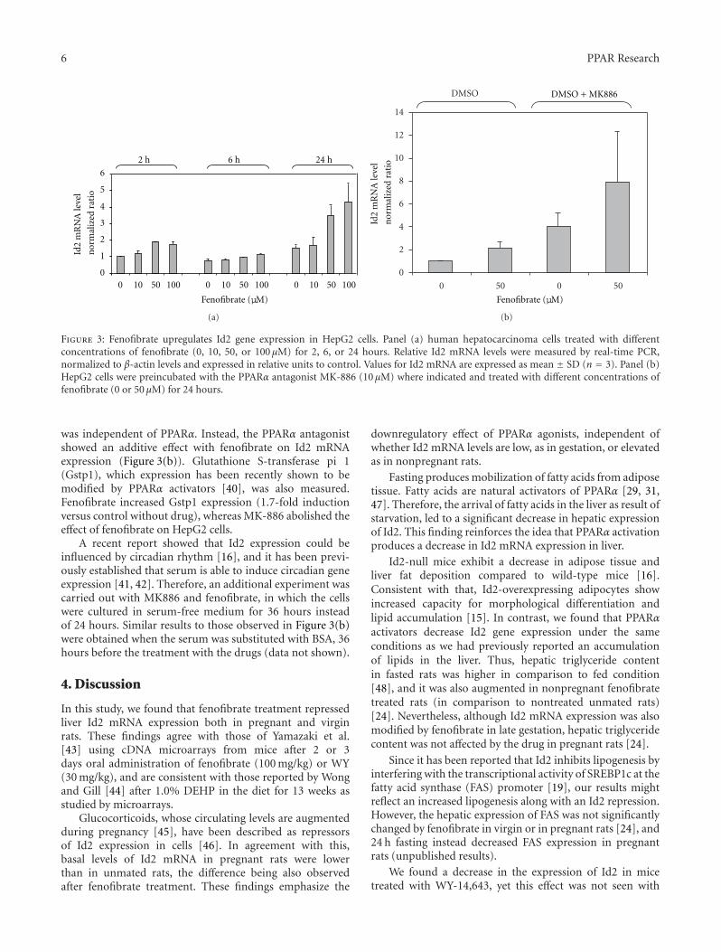
6 PPAR Research
0
1
2
3
4
5
6
0 10 50 100 0 10 50 100 0 10 50 100
2 h 6 h 24 h
Id2
mR
NA
leve
l
nor
mal
ized
rat
io
Fenofibrate (μM)
(a)
0
2
4
6
8
10
12
14
0 50 0 50
DMSO
Id2
mR
NA
leve
l
nor
mal
ized
rat
io
Fenofibrate (μM)
DMSO + MK886
(b)
Figure 3: Fenofibrate upregulates Id2 gene expression in HepG2 cells. Panel (a) human hepatocarcinoma cells treated with differentconcentrations of fenofibrate (0, 10, 50, or 100 μM) for 2, 6, or 24 hours. Relative Id2 mRNA levels were measured by real-time PCR,normalized to β-actin levels and expressed in relative units to control. Values for Id2 mRNA are expressed as mean ± SD (n = 3). Panel (b)HepG2 cells were preincubated with the PPARα antagonist MK-886 (10 μM) where indicated and treated with different concentrations offenofibrate (0 or 50 μM) for 24 hours.
was independent of PPARα. Instead, the PPARα antagonistshowed an additive effect with fenofibrate on Id2 mRNAexpression (Figure 3(b)). Glutathione S-transferase pi 1(Gstp1), which expression has been recently shown to bemodified by PPARα activators [40], was also measured.Fenofibrate increased Gstp1 expression (1.7-fold inductionversus control without drug), whereas MK-886 abolished theeffect of fenofibrate on HepG2 cells.
A recent report showed that Id2 expression could beinfluenced by circadian rhythm [16], and it has been previ-ously established that serum is able to induce circadian geneexpression [41, 42]. Therefore, an additional experiment wascarried out with MK886 and fenofibrate, in which the cellswere cultured in serum-free medium for 36 hours insteadof 24 hours. Similar results to those observed in Figure 3(b)were obtained when the serum was substituted with BSA, 36hours before the treatment with the drugs (data not shown).
4. Discussion
In this study, we found that fenofibrate treatment repressedliver Id2 mRNA expression both in pregnant and virginrats. These findings agree with those of Yamazaki et al.[43] using cDNA microarrays from mice after 2 or 3days oral administration of fenofibrate (100 mg/kg) or WY(30 mg/kg), and are consistent with those reported by Wongand Gill [44] after 1.0% DEHP in the diet for 13 weeks asstudied by microarrays.
Glucocorticoids, whose circulating levels are augmentedduring pregnancy [45], have been described as repressorsof Id2 expression in cells [46]. In agreement with this,basal levels of Id2 mRNA in pregnant rats were lowerthan in unmated rats, the difference being also observedafter fenofibrate treatment. These findings emphasize the
downregulatory effect of PPARα agonists, independent ofwhether Id2 mRNA levels are low, as in gestation, or elevatedas in nonpregnant rats.
Fasting produces mobilization of fatty acids from adiposetissue. Fatty acids are natural activators of PPARα [29, 31,47]. Therefore, the arrival of fatty acids in the liver as result ofstarvation, led to a significant decrease in hepatic expressionof Id2. This finding reinforces the idea that PPARα activationproduces a decrease in Id2 mRNA expression in liver.
Id2-null mice exhibit a decrease in adipose tissue andliver fat deposition compared to wild-type mice [16].Consistent with that, Id2-overexpressing adipocytes showincreased capacity for morphological differentiation andlipid accumulation [15]. In contrast, we found that PPARαactivators decrease Id2 gene expression under the sameconditions as we had previously reported an accumulationof lipids in the liver. Thus, hepatic triglyceride contentin fasted rats was higher in comparison to fed condition[48], and it was also augmented in nonpregnant fenofibratetreated rats (in comparison to nontreated unmated rats)[24]. Nevertheless, although Id2 mRNA expression was alsomodified by fenofibrate in late gestation, hepatic triglyceridecontent was not affected by the drug in pregnant rats [24].
Since it has been reported that Id2 inhibits lipogenesis byinterfering with the transcriptional activity of SREBP1c at thefatty acid synthase (FAS) promoter [19], our results mightreflect an increased lipogenesis along with an Id2 repression.However, the hepatic expression of FAS was not significantlychanged by fenofibrate in virgin or in pregnant rats [24], and24 h fasting instead decreased FAS expression in pregnantrats (unpublished results).
We found a decrease in the expression of Id2 in micetreated with WY-14,643, yet this effect was not seen with
PPAR Research 7
DEHP, indicating that the effect depends on PPARα activatorpotency. In the case of mice that lacked a functional formof PPARα, differences were not observed after the differenttreatments, suggesting that it was an effect mediated byPPARα. This finding is in agreement with that one recentlydescribed by el Azzouzi et al. [49] in murine cardiomyocytecells using cDNA microarrays. One of gene listed by theseauthors to be specifically downregulated by WY was Id2.However, neither GW-5015160 (a PPARβ/δ agonist) norrosiglitazone (a PPARγ agonist) produced any change. Thus,it can be assumed that fibrates affect Id2 gene expressionthrough PPARα.
The decrease found in the expression of Id2 in mice bythe potent PPARα activator WY agrees with the reductionobserved in rats treated with fenofibrate. Since, it appearsthat both STAT3 and C/EBPβ are involved in regulation ofId2 [50, 51], and it is known that PPARα activation interfereswith signalling pathways dependent on C/EBP and STAT[52], we hypothesize that PPARα may negatively regulate Id2through inhibition of STAT3 or C/EBP.
In contrast to rodents, the negative effect of fibrateson Id2 gene expression was not observed in human cells.Fenofibrate enhanced Id2 mRNA levels in these cells aftera 24 h incubation. Moreover, since Grønning et al. [13]have shown in murine macrophages that glucose inducesincreases in protein levels of this transcriptional repressor,we studied the effect of fibrates both at low glucose and highglucose and found a similar fenofibrate-inducing effect onId2 mRNA expression. The effect of fenofibrate was observedafter 24-h incubation, suggesting that regulation of Id2expression by PPAR might occur by an indirect mechanism[53]. Nevertheless, several PPARα target genes [54] werealso induced after 24-hour administration of fibrates butnot earlier. Therefore, we studied the effect of MK-886, anantagonist of PPARα and found that fenofibrate-inducedincreases in Id2 mRNA expression were not abolished bypreincubation with MK-886, confirming that the effectwas not mediated by PPARα. In accordance with thisfinding, TZD, which also modulate Id2 mRNA levels incultured human cells, use a PPARγ-independent mechanism[10]. Fenofibrate and MK-886 functioned synergistically tostimulate Id2 expression. The MK-886-induced increase inId2 mRNA expression could be caused by two mechanisms:(i) specific inactivation of PPARα; (ii) other pathways, suchas inhibition of leukotriene biosynthesis [39]. Therefore, it isassumed that fenofibrate-induced increases in Id2 expressionoccur by a PPARα-independent mechanism. How fenofibrateincreases Id2 expression in HepG2 cells remains elusive.
Finally, Id proteins are HLH transcription factors thatparticipate in development, cell cycle control, differentiation,and tumorigenesis [17]. However, the role of Id2 protein inthe mechanism of action of fibrates has not been elucidated.Altogether, these findings confirm that Id2 gene expressionis responsive to PPARα activators (fibrates and possibly fattyacids). However, as reported for other genes (apoA-I, apoA-V) ([2] and references therein), the effects are oppositein rodents versus humans. Since peroxisome proliferatorsfunction as hepatocarcinogenic agents in rodents, but notin humans [55], and considering the role of Id2 protein
in cell proliferation and cancer [17], we speculate that thedifferential response to fibrate exposure might be related tothe differences in liver tumorigenesis between species.
Abbreviations
PPAR: Peroxisome proliferator-activated receptorGAPDH: Glyceraldehyde-3-phosphate dehydrogenaseId2: Inhibitor of DNA binding 2DEHP: Di-(2-ethylhexyl)phthalateWY: WY-14,643RT-PCR: Reverse transcriptase polymerase chain reactionEMEM: Eagle’s minimal essential mediumA.U.: Arbitrary units.
Acknowledgments
The authors would like to thank Plan Nacional de Inves-tigacion Cientıfica, Desarrollo e Innovacion Tecnologica(I+D+i), Instituto de Salud Carlos III-Subdireccion Generalde Evaluacion y Fomento de la Investigacion (PI-09/02192),and European Community FEDER funds, as well as theFundacion Universitaria San Pablo-CEU (USP-PC 17/09) forfinancial support. M. Carmen Gonzalez was a recipient ofa predoctoral fellowship from the Universidad San Pablo-CEU, Madrid, Spain. The authors thank Milagros Morantefor her excellent technical assistance and Drs. Mitch Rosenand Sheau-Fung Thai for a critical review of the paper. Theinformation in this document has been supported by theU.S. Environmental Protection Agency. It has been subjectedto review by the National Health and Environmental EffectsResearch Laboratory and approved for publication. Approvaldoes not signify that the contents reflect the views of theAgency, nor does mention of trade names or commercialproducts constitute endorsement or recommendation foruse.
References
[1] G. F. Watts and S. B. Dimmitt, “Fibrates, dyslipoproteinaemiaand cardiovascular disease,” Current Opinion in Lipidology,vol. 10, no. 6, pp. 561–574, 1999.
[2] M. I. Panadero, M. C. Gonzalez, E. Herrera, and C. Bocos,“Factors modulating fibrates response: therapeutic implica-tions and alternative strategies,” Endocrine, Metabolic andImmune Disorders, vol. 9, no. 3, pp. 219–236, 2009.
[3] A. M. Lefebvre, J. Peinado-Onsurbe, I. Leitersdorf et al.,“Regulation of lipoprotein metabolism by thiazolidinedionesoccurs through a distinct but complementary mechanismrelative to fibrates,” Arteriosclerosis, Thrombosis, and VascularBiology, vol. 17, no. 9, pp. 1756–1764, 1997.
[4] J. C. Corton, C. Bocos, E. S. Moreno et al., “Rat 17β-hydroxysteroid dehydrogenase type IV is a novel peroxisomeproliferator-inducible gene,” Molecular Pharmacology, vol. 50,no. 5, pp. 1157–1166, 1996.
[5] J. C. Corton, L.-Q. Fan, S. Brown et al., “Down-regulationof cytochrome P450 2C family members and positive acute-phase response gene expression by peroxisome proliferatorchemicals,” Molecular Pharmacology, vol. 54, no. 3, pp. 463–473, 1998.

8 PPAR Research
[6] P. Gervois, R. Kleemann, A. Pilon et al., “Global suppressionof IL-6-induced acute phase response gene expression afterchronic in vivo treatment with the peroxisome proliferator-activated receptor-α activator fenofibrate,” Journal of BiologicalChemistry, vol. 279, no. 16, pp. 16154–16160, 2004.
[7] M. C. Gonzalez, J. C. Corton, R. C. Cattley, E. Herrera, and C.Bocos, “Peroxisome proliferator-activated receptorα (PPARα)agonists down-regulate α2-macroglobulin expression by aPPARα-dependent mechanism,” Biochimie, vol. 91, no. 8, pp.1029–1035, 2009.
[8] M. Guerre-Millo, P. Gervois, E. Raspe et al., “Peroxisomeproliferator-activated receptor α activators improve insulinsensitivity and reduce adiposity,” Journal of Biological Chem-istry, vol. 275, no. 22, pp. 16638–16642, 2000.
[9] C. J. Chou, M. Haluzik, C. Gregory et al., “WY14,643, aperoxisome proliferator-activated receptor α (PPARα) agonist,improves hepatic and muscle steatosis and reverses insulinresistance in lipoatrophic A-ZIP/F-1 mice,” Journal of Biologi-cal Chemistry, vol. 277, no. 27, pp. 24484–24489, 2002.
[10] X. Zhu, Y. Lin, J. Zhang, M. Fu, Z. Mao, and Y. E. Chen,“Thiazolidinediones, a class of anti-diabetic drugs, inhibitId2 expression through a PPARγ-independent pathway inhuman aortic smooth muscle cells,” Cellular and MolecularLife Sciences, vol. 60, no. 1, pp. 212–218, 2003.
[11] J. D. Norton, R. W. Deed, G. Craggs, and F. Sablitzky, “Id helix-loop-helix proteins in cell growth and differentiation,” Trendsin Cell Biology, vol. 8, no. 2, pp. 58–65, 1998.
[12] Y. Yokota, S. Mori, O. Narumi, and K. Kitajima, “In vivofunction of a differentiation inhibitor, Id2,” IUBMB Life, vol.51, no. 4, pp. 207–214, 2001.
[13] L. M. Grønning, R. Tingsabadh, K. Hardy et al., “Glucoseinduces increases in levels of the transcriptional repressor Id2via the hexosamine pathway,” American Journal of Physiology,vol. 290, no. 4, pp. E599–E606, 2006.
[14] D. Vicent, M. Piper, S. Gammeltoft, E. Maratos-Flier, and R. C.Kahn, “Alterations in skeletal muscle gene expression of ob/obmice by mRNA differential display,” Diabetes, vol. 47, no. 12,pp. 1451–1458, 1998.
[15] K. W. Park, H. Waki, C. J. Villanueva et al., “Inhibitor ofDNA binding 2 is a small molecule-inducible modulator ofperoxisome proliferator-activated receptor-γ expression andadipocyte differentiation,” Molecular Endocrinology, vol. 22,no. 9, pp. 2038–2048, 2008.
[16] T. Y. Hou, S. M. Ward, J. M. Murad, N. P. Watson, M. A. Israel,and G. E. Duffield, “ID2 (inhibitor of DNA binding 2) is arhythmically expressed transcriptional repressor required forcircadian clock output in mouse liver,” Journal of BiologicalChemistry, vol. 284, no. 46, pp. 31735–31745, 2009.
[17] M. B. Ruzinova and R. Benezra, “Id proteins in development,cell cycle and cancer,” Trends in Cell Biology, vol. 13, no. 8, pp.410–418, 2003.
[18] J. D. Norton, “ID helix-loop-helix proteins in cell growth,differentiation and tumorigenesis,” Journal of Cell Science, vol.113, no. 22, pp. 3897–3905, 2000.
[19] M. Moldes, M. Boizard, X. Le Liepvre, B. Feve, I. Dugail,and J. Pairault, “Functional antagonism between inhibitor ofDNA binding (Id) and adipocyte determination and differ-entiation factor 1/sterol regulatory element-binding protein-1c (ADD1/SREBP-1c) trans-factors for the regulation of fattyacid synthase promoter in adipocytes,” Biochemical Journal,vol. 344, no. 3, pp. 873–880, 1999.
[20] C. Rustemeijer, J. A. Schouten, H. J. Voerman, H. E. S. J.Hensgens, A. J. M. Donker, and R. J. Heine, “Pravastatincompared to bezafibrate in the treatment of dyslipidemia
in insulintreated patients with Type 2 diabetes mellitus,”Diabetes/Metabolism Research and Reviews, vol. 16, pp. 82–87,2000.
[21] F. Villarroya, C. Bocos, M. Giralt et al., “Pharmacological andGene modification-based models for studying the impact ofperinatal metabolic disturbances in adult life,” Advances inExperimental Medicine and Biology, vol. 646, pp. 141–148,2009.
[22] J. Sevillano, J. de Castro, C. Bocos, E. Herrera, and M.P. Ramos, “Role of insulin receptor substrate-1 serine 307phosphorylation and adiponectin in adipose tissue insulinresistance in late pregnancy,” Endocrinology, vol. 148, no. 12,pp. 5933–5942, 2007.
[23] A. Soria, C. Bocos, and E. Herrera, “Opposite metabolicresponse to fenofibrate treatment in pregnant and virgin rats,”Journal of Lipid Research, vol. 43, no. 1, pp. 74–81, 2002.
[24] A. Soria, M. del Carmen Gonzalez, H. Vidal, E. Her-rera, and C. Bocos, “Triglyceridemia and peroxisomeproliferator-activated receptor-α expression are not connectedin fenofibrate-treated pregnant rats,” Molecular and CellularBiochemistry, vol. 273, no. 1-2, pp. 97–107, 2005.
[25] M. C. Sugden, G. K. Greenwood, N. D. Smith, and M. J.Holness, “Peroxisome proliferator-activated receptor-α activa-tion during pregnancy attenuates glucose-stimulated insulinhypersecretion in vivo by increasing insulin sensitivity, with-out impairing pregnancy-induced increases in β-cell glucosesensing and responsiveness,” Endocrinology, vol. 144, no. 1, pp.146–153, 2003.
[26] M. J. Holness, G. K. Greenwood, N. D. Smith, and M. C.Sugden, “Peroxisome proliferator-activated receptor-α andglucocorticoids interactively regulate insulin secretion duringpregnancy,” Diabetes, vol. 55, no. 12, pp. 3501–3508, 2006.
[27] N. Martınez, V. White, M. Kurtz, R. Higa, E. Capobianco,and A. Jawerbaum, “Activation of the nuclear receptor PPARαregulates lipid metabolism in foetal liver from diabetic rats:implications in diabetes-induced foetal overgrowth,” Dia-betes/Metabolism Research and Reviews, vol. 27, no. 1, pp. 35–46, 2011.
[28] E. Capobianco, N. Martınez, R. Higa, V. White, and A.Jawerbaum, “The effects of maternal dietary treatments withnatural PPAR ligands on lipid metabolism in fetuses fromcontrol and diabetic rats,” Prostaglandins Leukotrienes andEssential Fatty Acids, vol. 79, no. 6, pp. 191–199, 2008.
[29] M. Gottlicher, E. Widmark, Q. Li, and J. A. Gustafsson, “Fattyacids activate a chimera of the clofibric acid-activated receptorand the glucocorticoid receptor,” Proceedings of the NationalAcademy of Sciences of the United States of America, vol. 89, no.10, pp. 4653–4657, 1992.
[30] C. Bocos, M. Gottlicher, K. Gearing et al., “Fatty acid acti-vation of peroxisome proliferator-activated receptor (PPAR),”Journal of Steroid Biochemistry and Molecular Biology, vol. 53,no. 1–6, pp. 467–473, 1995.
[31] M. I. Panadero, E. Herrera, and C. Bocos, “Different sensitivityof PPARα gene expression to nutritional changes in liver ofsuckling and adult rats,” Life Sciences, vol. 76, no. 9, pp. 1061–1072, 2005.
[32] S. S. T. Lee, T. Pineau, J. Drago et al., “Targeted disruptionof the α isoform of the peroxisome proliferator- activatedreceptor gene in mice results in abolishment of the pleiotropiceffects of peroxisome proliferators,” Molecular and CellularBiology, vol. 15, no. 6, pp. 3012–3022, 1995.
[33] H. N. Suh, H. T. Huong, C. H. Song, J. H. Lee, and H. J.Han, “Linoleic acid stimulates gluconeogenesis via Ca2+/PLC,cPLA2, and PPAR pathways through GPR40 in primary

PPAR Research 9
cultured chicken hepatocytes,” American Journal of Physiology,vol. 295, no. 6, pp. C1518–C1527, 2008.
[34] S. L. Lay, S. Krief, C. Farnier et al., “Cholesterol, a cell size-dependent signal that regulates glucose metabolism and geneexpression in adipocytes,” Journal of Biological Chemistry, vol.276, no. 20, pp. 16904–16910, 2001.
[35] I. Inoue, S. Noji, M. Z. Shen, K. Takahashi, and S. Katayama,“The peroxisome proliferator-activated receptor α (PPARα)regulates the plasma thiobarbituric acid-reactive substance(TBARS) level,” Biochemical and Biophysical Research Commu-nications, vol. 237, no. 3, pp. 606–610, 1997.
[36] Y. Kurihara, K. Egawa, S. Kunimoto, T. Takeuchi, and K.Nose, “Induction of p16/INK4a gene expression and cellularsenescence by toyocamycin,” Biological and PharmaceuticalBulletin, vol. 25, no. 10, pp. 1272–1276, 2002.
[37] T. Aoyama, J. M. Peters, N. Iritani et al., “Altered constitutiveexpression of fatty acid-metabolizing enzymes in mice lackingthe peroxisome proliferator-activated receptor α (PPARα),”Journal of Biological Chemistry, vol. 273, no. 10, pp. 5678–5684, 1998.
[38] J. D. Tugwood, I. Issemann, R. G. Anderson, K. R. Bundell,W. L. McPheat, and S. Green, “The mouse peroxisomeproliferator activated receptor recognizes a response elementin the 5’ flanking sequence of the rat acyl CoA oxidase gene,”The EMBO Journal, vol. 11, no. 2, pp. 433–439, 1992.
[39] J. P. Kehrer, S. S. Biswal, E. La et al., “Inhibition of peroxisome-proliferator-activated receptor (PPAR)α by MK886,” Biochem-ical Journal, vol. 356, no. 3, pp. 899–906, 2001.
[40] T. R. Knight, S. Choudhuri, and C. D. Klaassen, “Induction ofhepatic glutathione S-transferases in male mice by prototypesof various classes of microsomal enzyme inducers,” Toxicolog-ical Sciences, vol. 106, no. 2, pp. 329–338, 2008.
[41] A. Balsalobre, F. Damiola, and U. Schibler, “A serum shockinduces circadian gene expression in mammalian tissue cul-ture cells,” Cell, vol. 93, no. 6, pp. 929–937, 1998.
[42] L. Canaple, J. Rambaud, O. Dkhissi-Benyahya et al., “Recip-rocal regulation of brain and muscle Arnt-like protein 1 andperoxisome proliferator-activated receptor α defines a novelpositive feedback loop in the rodent liver circadian clock,”Molecular Endocrinology, vol. 20, no. 8, pp. 1715–1727, 2006.
[43] K. Yamazaki, J. Kuromitsu, and I. Tanaka, “Microarray analysisof gene expression changes in mouse liver induced by perox-isome proliferator-activated receptor α agonists,” Biochemicaland Biophysical Research Communications, vol. 290, no. 3, pp.1114–1122, 2002.
[44] J. S. Wong and S. S. Gill, “Gene expression changes inducedin mouse liver by di(2-ethylhexyl) phthalate,” Toxicology andApplied Pharmacology, vol. 185, no. 3, pp. 180–196, 2002.
[45] A. L. Fowden, J. Li, and A. J. Forhead, “Glucocorticoidsand the preparation for life after birth: are there long-term consequences of the life insurance?” Proceedings of theNutrition Society, vol. 57, no. 1, pp. 113–122, 1998.
[46] V. Zilberfarb, K. Siquier, A. D. Strosberg, and T. Issad, “Effectof dexamethasone on adipocyte differentiation markers andtumour necrosis factor-α expression in human PAZ6 cells,”Diabetologia, vol. 44, no. 3, pp. 377–386, 2001.
[47] S. Kersten, J. Seydoux, J. M. Peters, F. J. Gonzalez, B.Desvergne, and W. Wahli, “Peroxisome proliferator-activatedreceptor α mediates the adaptive response to fasting,” Journalof Clinical Investigation, vol. 103, no. 11, pp. 1489–1498, 1999.
[48] P. Lopez-Luna, J. Olea, and E. Herrera, “Effect of starvation onlipoprotein lipase activity in different tissues during gestationin the rat,” Biochimica et Biophysica Acta, vol. 1215, no. 3, pp.275–279, 1994.
[49] H. el Azzouzi, S. Leptidis, M. Bourajjaj et al., “Peroxisomeproliferator-activated receptor (PPAR) gene profiling uncoversinsulin-like growth factor-1 as a PPARα target gene incardioprotection,” Journal of Biological Chemistry, vol. 286, no.16, pp. 14598–14607, 2011.
[50] K. Karaya, S. Mori, H. Kimoto et al., “Regulation of Id2expression by CCAAT/enhancer binding protein β,” NucleicAcids Research, vol. 33, no. 6, pp. 1924–1934, 2005.
[51] M. Prisco, F. Peruzzi, B. Belletti, and R. Baserga, “Regulation ofId gene expression by type 1 insulin-like growth factor: roles ofSTAT3 and the tyrosine 950 residue of the receptor,” Molecularand Cellular Biology, vol. 21, no. 16, pp. 5447–5458, 2001.
[52] A. Zambon, P. Gervois, P. Pauletto, J.-C. Fruchart, and B.Staels, “Modulation of hepatic inflammatory risk markersof cardiovascular diseases by PPAR-α activators: clinicaland experimental evidence,” Arteriosclerosis, Thrombosis, andVascular Biology, vol. 26, no. 5, pp. 977–986, 2006.
[53] A. Hermanowski-Vosatka, D. Gerhold, S. S. Mundt et al.,“PPARα agonists reduce 11β-hydroxysteroid dehydrogenasetype 1 in the liver,” Biochemical and Biophysical ResearchCommunications, vol. 279, no. 2, pp. 330–336, 2000.
[54] J. C. Corton, E. S. Moreno, A. Merritt, C. Bocos, and R. C.Cattley, “Cloning genes responsive to a hepatocarcinogenicperoxisome proliferator chemical reveals novel targets ofregulation,” Cancer Letters, vol. 134, no. 1, pp. 61–71, 1998.
[55] P. R. Holden and J. D. Tugwood, “Peroxisome proliferator-activated receptor alpha: role in rodent liver cancer and speciesdifferences,” Journal of Molecular Endocrinology, vol. 22, no. 1,pp. 1–8, 1999.
Hindawi Publishing CorporationPPAR ResearchVolume 2012, Article ID 483656, 8 pagesdoi:10.1155/2012/483656
Review Article
Effects of PPARγ Ligands on Leukemia
Yoko Tabe,1 Marina Konopleva,2 Michael Andreeff,2 and Akimichi Ohsaka3
1 Department of Clinical Laboratory Medicine, Juntendo University School of Medicine, Hongo 2-1-1, Bunkyo-ku,Tokyo 113-8421, Japan
2 Department of Leukemia, The University of Texas M.D. Anderson Cancer Center, 1515 Holcombe Boulevard,Houston, TX 77030, USA
3 Department of Transfusion Medicine and Stem Cell Regulation, Juntendo University School of Medicine,Hongo 2-1-1, Bunkyo-ku, Tokyo 113-8421, Japan
Correspondence should be addressed to Yoko Tabe, [email protected]
Received 1 January 2012; Accepted 21 March 2012
Academic Editor: Stefan Alexson
Copyright © 2012 Yoko Tabe et al. This is an open access article distributed under the Creative Commons Attribution License,which permits unrestricted use, distribution, and reproduction in any medium, provided the original work is properly cited.
Peroxisome proliferator-activated receptors (PPARs) and retinoic acid receptors (RARs), members of the nuclear receptorsuperfamily, are transcription factors that regulate a variety of important cellular functions. PPARs form heterodimers retinoidX receptor (RXR), an obligate heterodimeric partner for other nuclear receptors. Several novel links between retinoid metabolismand PPAR responses have been identified, and activation of PPAR/RXR expression has been shown to increase response toretinoids. PPARγ has emerged as a key regulator of cell growth and survival, whose activity is modulated by a number ofsynthetic and natural ligands. While clinical trials in cancer patients with thiazolidinediones (TZD) have been disappointing,novel structurally different PPARγ ligands, including triterpenoids, have entered clinical arena as therapeutic agents for epithelialand hematopoietic malignancies. Here we shall review the antitumor advances of PPARγ, alone and in combination with RARαligands in control of cell proliferation, differentiation, and apoptosis and their potential therapeutic applications in hematologicalmalignancies.
1. Introduction
Acute myelogenous leukemia (AML) remains incurable inmost patients because of the likelihood of relapse and thedevelopment of resistant disease [1]. Many novel agents donot improve survival of patients once relapse occurs, whichenforces the need for more effective treatment strategies forAML exploiting apoptosis and/or differentiation induction.
Ligands of nuclear hormone receptors (NHRs) havebeen shown to induce apoptosis and/or inhibiting prolifer-ation in a variety of preclinical models. The most strikingimprovement in AML therapy was achieved by the treatmentof acute promyelocytic leukemia (APL) using the retinoicacid (RA) receptor- (RAR-) specific ligand, all-trans RA(ATRA) [2, 3]. ATRA, combined with chemotherapy, resultsin complete remission (CR) rates ranging from 72% to 90%in APL patients with the oncogenic transcriptional repressorPML-RARα [4–8]. However, approximately 10% to 30%of patients relapse [8] and frequently develop resistance to
ATRA [9, 10]. Acquisition of specific mutations in the ligandbinding site, which leads to altered interactions with tran-scriptional coregulators, is a well-documented mechanismof acquired ATRA resistance [11, 12]. In addition, severalalternative mechanisms such as DNA methylation [13] orimpaired telomerase regulation [14] have been proposed tocause ATRA-resistant disease.
Considering the potential of using PPARγ ligands in APL“transcriptional” therapy, this paper summarizes the effectsof endogenous and synthetic PPARγ ligands in AML andfocuses on elucidating the mechanisms underlying the anti-tumor effects of novel synthetic PPARγ ligand 2-cyano-3,12-dioxooleana-1,9-dien-28-oic acid (CDDO) in APL.
2. PPARγ and PPARγ Ligands
PPARs belong to the NHR superfamily of ligand-dependenttranscription factors, which includes RAR and RXR amongothers. Three PPAR isotypes have been identified: PPARγ,

2 PPAR Research
O
COOH
O
NC
Figure 1: Molecular structure of CDDO 2-cyano-3,12-dioxooleana-1,9-dien-28-oic acid (CDDO).
PPARα, and PPARβ/δ. PPARγ exists as a heterodimer withRXR, and upon activation by endogenous or syntheticligands, PPARγ/RXR binds to the specific response elementsPPRE in the promoter regions of target genes, respectively,which in turn functions as a transcription factor [15–17].
PPARγ modulates gene networks involved in control-ling growth, cellular differentiation, and apoptosis [18].PPARγ receptor can be activated by endogenous ligands(e.g., prostaglandin D2 (PGD2), 15-deoxy prostaglandin J2(15dPGJ2), or 15-hydroxyeicosatetraenoic acid (15-HETE))[19, 20], and synthetic ligands that include insulin sensitizingantidiabetic thiazolidinediones (TZD); troglitazone (TGZ),rosiglitazone (RGZ), ciglitazone (CGZ), or pioglitazone(PGZ) [21–23]; nonsteroidal anti-inflammatory compoundsindomethacin, ibuprofen, flufenamic acid, or fenoprofen[24]; triterpenoids 2-cyano-3,12-dioxooleana-1,9-dien-28-oic acid (CDDO) [25] are a semisynthetic triterpenoidderived from oleanolic acid, whose structure contains twoα, β-unsaturated carbonyl moieties. CDDO was shown torelease nuclear receptor corepressor (NCoR) and recruitCCAAT/enhancer-binding protein (CBP/p300) to PPARγ[25] (Figure 1).
PPARγ ligands induce differentiation and inhibit prolif-eration in several tumor models [26–34]. The regulation ofgene transcription by ligand-bound PPARγ involves cofactorproteins, which bridge transcription factors to the basaltranscriptional machinery or modify chromatin structure.These include release of small accessory molecules known ascorepressors (e.g., NCoR or silencing mediator for retinoidreceptor and thyroid hormone receptors (SMRT)) andrecruitment of coactivators (e.g., CBP/p300, cyclic adenosinemonophosphate response-element binding protein (CREB),steroid receptor coactivator-1 (SRC-1), receptor interact-ing protein 140(RIP140), or PPARγ interacting protein(PRIP/RAP250) [35–40]. The multiprotein complex inducestranscription by chromatin remodeling and interaction withthe basal transcriptional machinery [41, 42], and the relativelevels of cofactor expression (e.g., availability of cofactorsCBP/p300 versus SRC-1) also control the specificity of thephysiological response to target gene transcription [43].
3. Antitumor Effects of PPARγ in AML
High PPARγ expression was observed in normal bonemarrow and peripheral blood CD34+ progenitor cells [44].Furthermore, significantly higher PPARγ mRNA expressionwas observed in primary AML cases compared to normalperipheral blood or bone marrow mononuclear cells [45,46].
The mechanisms of cell differentiation and cell cyclearrest by activated PPARγ depend heavily on the specificity ofPPARγ ligands. The induction of differentiation by activationof PPARγ may represent a promising novel therapeuticapproach for cancer as already demonstrated for liposarcoma[27] and in xenograft models of prostate [47] and coloncancer [30]. Differentiation therapy may well play a rolein acute myeloid leukemias, analogous to ATRA-induceddifferentiation in APL. PPARγ is known to be induced and/orexpressed in cells of the myeloid/monocytic lineage [48, 49].
In PPARγ expressing AML cell lines, PPARγ ligand TGZsuppressed their clonal growth with G1 cell cycle phasearrest, induced differentiation into monocytes, and increasedapoptosis at higher concentrations [50, 51]. Troglitazone-induced G0/G1 cell cycle arrest with upregulation of p21mRNA in myeloid leukemia cell lines [52]. In concert withthese findings, PPARγ ligand PGZ and 15dPGJ2 suppressedproliferation, and the combined treatment with ATRAsynergistically induced myeloid differentiation in promyelo-cytic leukemia NB4 cells [53]. Furthermore, simultaneoustreatment with TGZ and RXR or RAR ligands resulted inadditive suppression of growth indicating that PPARγ ligandcombined with a retinoid is a potent inhibitor of clonogenicgrowth of AML [50]. CDDO has been reported to inducemonocytic differentiation of human myeloid leukemia cellsand adipogenic differentiation of mouse fibroblasts [54].
CDDO-Me also induced granulo-monocytic differenti-ation in primary AML cells and cell lines. Combinationswith ATRA or the RXR-specific ligand LG100268 enhancedthe effects of CDDO-Me on cell viability and/or terminaldifferentiation of myeloid leukemic cell lines [54]. CDDO-Me-induced enhanced apoptosis when combined with ara-C and retinoids indicating potential activity in the futuretherapy for AML [55].
With respect to the mechanisms of PPARγ-ligand-induced differentiation, CCAAT enhancer-binding proteinalpha (CEBPA) translational upregulation has been reportedto be required for CDDO-induced granulocytic differ-entiation of AML patients samples and cell lines [56].CDDO increases the ratio of transcriptionally active p42and the inactive p30 CEBPA isoform, which in turn leadsto transcriptional activation of CEBPA-regulated genes andassociates with dephosphorylation of eIF2alpha and phos-phorylation of eIF4E [56].
PPARγ ligands are additionally known to induce apop-tosis. The mechanisms of apoptosis induction by activatedPPARγ depend heavily on the specificity of PPARγ ligands.PPARγ activation by natural ligand 15dPGJ2 and syntheticligand TGZ induce apoptosis accompanied by caspase-3 acti-vation and downregulated c-myc gene expression in myeloidleukemic cells [57]. 15dPGJ2 and TGZ have been also

PPAR Research 3
reported to induce upregulation of bax and downregulationof antiapoptotic proteins survivin and bcl-2 in AML andCML [58]. Furthermore, downregulation of cyclooxygenase-2 expression, disruption of mitochondrial membrane poten-tial, activation of caspase-3, downregulation of Bcl-2, Bcl-Xl, and Mcl-1, and upregulation of Bax by these PPARγagonists 15dPGJ2 and TGZ has been reported in humanmonocytic leukemia cells [59]. Semisynthetic oleananetriterpenoid CDDO has potent differentiating, antiprolifer-ative, anti-inflammatory, and apoptosis-inducing properties[54]. CDDO has been reported to activate caspase-8 and-3 and to induce mitochondrial cytochrome c release inleukemic cells and in osteosarcoma cells [60–62]. CDDOhas been further shown to activate the intrinsic pathway ofapoptosis that involves the release of cytochrome c and AIFand initiates caspase-dependent and independent cell deathin AML [63]. The C-28 methyl ester of CDDO, CDDO-Me [55], and C-28 imidazolide imide of CDDO (CDDO-Im) [64] has been shown to be more potent than CDDOin inducing apoptosis and differentiation of acute myeloidleukemia (AML) cells. CDDO-Me is 3- to 5-fold more activethan CDDO in inhibiting the viability of AML cells in anMDR-1- and p53-independent manner, inducing apoptosisthrough a loss of mitochondrial membrane potential, andincreasing caspase-3 cleavage and proapoptotic Bax protein.It has significantly less cytotoxicity against normal CD34+
progenitor cells, assuring therapeutic window [55].In addition, CDDO was shown to inhibit NF-
κB-mediated gene expression in leukemic cells [62].CDDO/tumor-necrosis-factor- (TNF-) induced apoptosisoccurs through selective inhibition of NF-κB-dependentantiapoptotic proteins, bypassing potential mitochondrialresistance mechanisms [62]. CDDO-Me also inhibits bothconstitutive and inducible NF-κB through inhibition of IκBα kinase, leading to the suppression of expression of NF-κB-regulated gene products and enhancement of apoptosisinduced by TNFα [65].
Notably, certain PPARγ ligands execute anti-tumor activ-ities without requiring interaction with the PPAR ligandbinding domain [66]. For example, CDDO, CDDO-Me, andCDDO-Im activate PPARγ-dependent and -independentpathways that inhibit cancer-cell growth [67]. They acti-vate PPARγ in transactivation assays, and CDDO-inducedapoptosis was diminished by dominant-negative PPARγ inmyeloid HL-60 cells and by T007 in myeloid U937 cells [68],but CDDO-Im-induced differentiation in leukemia cells wasnot inhibited by the PPARγ antagonist GW9662 [61], andT007 did not affect inhibition of SKOV3 ovarian cancercell growth by CDDO [69]. In these scenarios, interactionwith the PPARγ receptor is irrelevant to the anti-cancereffects, which may depend on cell type, presence/activityof the receptor(s), and cellular abundance of coactiva-tors/corepressors. PPAR-independent effects of PPARγ lig-ands are due in part to their electrophilic nature, proteasomaldegradation of cell cycle-, and apoptosis-regulatory proteins,transcriptional repression, and other mechanisms [70–72].Both, PPARγ-dependent and -independent pathways thatcontribute to inhibition of cancer cell growth may bebeneficial for cancer chemotherapy [67].
4. Antitumor Effects of PPARγ-ActiveTriterpenoid CDDO on APL
RARs bind with high affinity to the RA-responsive element(RARE) as a heterodimer with RXR, which also heterodimer-izes with other nuclear receptors, such as PPARγ.
In APL cells, the oncogenic transcription factor PML-RARα, a dominant negative transcriptional repressor, targetsconsist of two copies of an AGGTCA, a highly conservedconsensus for RARα. PML-induced dimerization allows thetwo RARα moieties of PML-RARα to bind very distantmonomeric DNA sites. The spectrum of response elementsfor PML-RARα and PML-RARα-RXR (DR1-DR16 responseelements) is much broader than one for the wild-type RAR-RXR (DR1, DR2, and DR5), and PML-RARα-RXR oligomerssilence a wide range of nuclear receptor target genes [73].
X-RARα fusion proteins in APL have been demonstratedto negatively affect transactivation of PPARγ [74], indicatingthat inhibition of PPARγ activity may contribute to thepathophysiology of the differentiation block in APL, and thatPPARγ ligands could sensitize APL cells to the differentiatingeffects of ATRA including ATRA-resistant cells [45].
PML-RARα recruits the nuclear corepressors and histonedeacetylase (HDAC), which leads to histone condensationand transcriptional repression [75–77]. ATRA acts by caus-ing the PML-RARα/HDAC complex to dissociate, therebyconverting PML-RARα into a transcriptional activator [76].Reactivation of ATRA target genes by inducing an appro-priate level of histone acetylation in their promoters is apotential strategy for restoring anticancer effects of ATRA inrefractory APL [77]. Differentiating agents including ATRA,arsenic, cAMP, HDAC inhibitors, and rexinoids relieve thisrepression through various molecular mechanisms, allowingspontaneous differentiation of leukemic blasts [73].
In fact, it has been demonstrated that HDAC inhibitors(HDACI) such as trichostatin A (TSA), sodium phenylbu-tyrate (PB), and suberoylanilide hydroxamic acid (SAHA)can augment the cell growth inhibition induced by ATRA,and that ATRA combined with SAHA increased survival andinduced remissions in APL transgenic mice harboring thePLZF-RARα translocation [78]. In addition, the PML-RARαfusion protein was observed to induce hypermethylation onRAR promoter, and the DNA methyltransferase inhibitor 5-asa-2′-deoxycytidine (5-Aza-dC) enhanced ATRA-inducedRAR promoter transactivation in APL cells [13].
Induction of APL cell differentiation by ATRA is asso-ciated with modulation of several critical genes, includingRARβ2 [78], C/EBPβ [79], p21 [80], PU.1 [81], or a domi-nant repressor of RAR signaling PRAME [82]. Notably, PML-RARα has a significant affinity for DR1 [83], a binding sitefor RXR/PPARγ heterodimers, and negatively contributes totransactivation by ligand-activated PPRE.
The RA-target gene RARβ plays a crucial role in medi-ating the growth-inhibitory and tumor suppressive effectsof retinoids in various cancer cells [84–87], and RARβis silenced in many tumors [84, 87, 88] and myeloidleukemias [89, 90] including APL [13]. Its upregulationhas been proposed as a general mechanism of retinoid-induced growth inhibition and differentiation induction
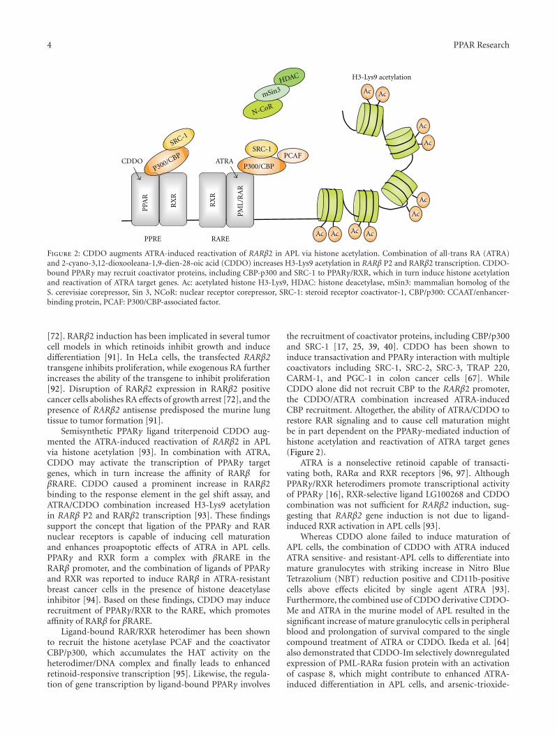
4 PPAR Research
RX
R
RX
R
RAREPPRE
PCAF
P300/CBP
SRC-1
Ac
Ac
Ac
Ac
Ac Ac Ac
Ac
H3-Lys9 acetylation
Ac
Ac
PPA
R
PM
L/R
AR
CDDO ATRA
SRC-1
P300/CBP
N-CoR
mSin3HDAC
Figure 2: CDDO augments ATRA-induced reactivation of RARβ2 in APL via histone acetylation. Combination of all-trans RA (ATRA)and 2-cyano-3,12-dioxooleana-1,9-dien-28-oic acid (CDDO) increases H3-Lys9 acetylation in RARβ P2 and RARβ2 transcription. CDDO-bound PPARγ may recruit coactivator proteins, including CBP-p300 and SRC-1 to PPARγ/RXR, which in turn induce histone acetylationand reactivation of ATRA target genes. Ac: acetylated histone H3-Lys9, HDAC: histone deacetylase, mSin3: mammalian homolog of theS. cerevisiae corepressor, Sin 3, NCoR: nuclear receptor corepressor, SRC-1: steroid receptor coactivator-1, CBP/p300: CCAAT/enhancer-binding protein, PCAF: P300/CBP-associated factor.
[72]. RARβ2 induction has been implicated in several tumorcell models in which retinoids inhibit growth and inducedifferentiation [91]. In HeLa cells, the transfected RARβ2transgene inhibits proliferation, while exogenous RA furtherincreases the ability of the transgene to inhibit proliferation[92]. Disruption of RARβ2 expression in RARβ2 positivecancer cells abolishes RA effects of growth arrest [72], and thepresence of RARβ2 antisense predisposed the murine lungtissue to tumor formation [91].
Semisynthetic PPARγ ligand triterpenoid CDDO aug-mented the ATRA-induced reactivation of RARβ2 in APLvia histone acetylation [93]. In combination with ATRA,CDDO may activate the transcription of PPARγ targetgenes, which in turn increase the affinity of RARβ forβRARE. CDDO caused a prominent increase in RARβ2binding to the response element in the gel shift assay, andATRA/CDDO combination increased H3-Lys9 acetylationin RARβ P2 and RARβ2 transcription [93]. These findingssupport the concept that ligation of the PPARγ and RARnuclear receptors is capable of inducing cell maturationand enhances proapoptotic effects of ATRA in APL cells.PPARγ and RXR form a complex with βRARE in theRARβ promoter, and the combination of ligands of PPARγand RXR was reported to induce RARβ in ATRA-resistantbreast cancer cells in the presence of histone deacetylaseinhibitor [94]. Based on these findings, CDDO may inducerecruitment of PPARγ/RXR to the RARE, which promotesaffinity of RARβ for βRARE.
Ligand-bound RAR/RXR heterodimer has been shownto recruit the histone acetylase PCAF and the coactivatorCBP/p300, which accumulates the HAT activity on theheterodimer/DNA complex and finally leads to enhancedretinoid-responsive transcription [95]. Likewise, the regula-tion of gene transcription by ligand-bound PPARγ involves
the recruitment of coactivator proteins, including CBP/p300and SRC-1 [17, 25, 39, 40]. CDDO has been shown toinduce transactivation and PPARγ interaction with multiplecoactivators including SRC-1, SRC-2, SRC-3, TRAP 220,CARM-1, and PGC-1 in colon cancer cells [67]. WhileCDDO alone did not recruit CBP to the RARβ2 promoter,the CDDO/ATRA combination increased ATRA-inducedCBP recruitment. Altogether, the ability of ATRA/CDDO torestore RAR signaling and to cause cell maturation mightbe in part dependent on the PPARγ-mediated induction ofhistone acetylation and reactivation of ATRA target genes(Figure 2).
ATRA is a nonselective retinoid capable of transacti-vating both, RARα and RXR receptors [96, 97]. AlthoughPPARγ/RXR heterodimers promote transcriptional activityof PPARγ [16], RXR-selective ligand LG100268 and CDDOcombination was not sufficient for RARβ2 induction, sug-gesting that RARβ2 gene induction is not due to ligand-induced RXR activation in APL cells [93].
Whereas CDDO alone failed to induce maturation ofAPL cells, the combination of CDDO with ATRA inducedATRA sensitive- and resistant-APL cells to differentiate intomature granulocytes with striking increase in Nitro BlueTetrazolium (NBT) reduction positive and CD11b-positivecells above effects elicited by single agent ATRA [93].Furthermore, the combined use of CDDO derivative CDDO-Me and ATRA in the murine model of APL resulted in thesignificant increase of mature granulocytic cells in peripheralblood and prolongation of survival compared to the singlecompound treatment of ATRA or CDDO. Ikeda et al. [64]also demonstrated that CDDO-Im selectively downregulatedexpression of PML-RARα fusion protein with an activationof caspase 8, which might contribute to enhanced ATRA-induced differentiation in APL cells, and arsenic-trioxide-

PPAR Research 5
(ATO-) induced apoptosis in both ATRA-sensitive NB4 andresistant R2 cell lines and primary APL cells.
RA signaling is a common mechanism in AML other thanAPL, and HDAC inhibitors have been shown to restore RA-dependent transcriptional activation and trigger terminaldifferentiation of primary blasts from AML patients [89].Recent reports of in vivo differentiation of the leukemic clonefollowing HDAC inhibitor valproic acid/ATRA treatment inAML patients [98] further suggest the possibility that theATRA/CDDO or its more potent derivatives combinationmay be useful transcriptional/differentiation therapy in non-APL AML. Randomized trial AML HD98B showed thatadministration of ATRA in addition to intensive chemother-apy improved the outcomes of the patients with genotype of“mutant (mt-) NPM1 without FLT3-ITD” [99]. NPM1 hasbeen reported to be a possible transcriptional corepressor[100]. Inhibition of NPM1 oligomerization or knockdownof NPM1-induced apoptosis and sensitized to ATRA in mt-NPM1-bearing AML cells [101]. These findings suggest newavenues of exploration for ATRA and CDDO derivativescombination therapy targeting “mt-NPM1 wt-FLT3” geno-type AML.
Acknowledgment
The authors thank an anonymous for the paper review. Thereis no conflict of interests between the paper’s authors and thecompanies involved in the paper.
References
[1] E. H. Estey, “Treatment of relapsed and refractory acutemyelogenous leukemia,” Leukemia, vol. 14, no. 3, pp. 476–479, 2000.
[2] L. Degos, C. Chomienne, M. T. Daniel et al., “Treatment offirst relapse in acute promyelocytic leukaemia with all-transretinoic acid,” The Lancet, vol. 336, no. 8728, pp. 1440–1441,1990.
[3] Z. X. Chen, Y. Q. Xue, R. Zhang et al., “A clinical andexperimental study on all-trans retinoic acid-treated acutepromyelocytic leukemia patients,” Blood, vol. 78, no. 6, pp.1413–1419, 1991.
[4] H. Meng-er, Y. Yu-chen, C. Shu-rong et al., “Use of all-trans retinoic acid in the treatment of acute promyelocyticleukemia,” Blood, vol. 72, no. 2, pp. 567–572, 1988.
[5] R. P. Warrell, S. R. Frankel, W. H. Miller et al., “Differentia-tion therapy of acute promyelocytic leukemia with tretinoin(all-trans-retinoic acid),” New England Journal of Medicine,vol. 324, no. 20, pp. 1385–1393, 1991.
[6] F. Mandelli, D. Diverio, G. Avvisati et al., “Molecular remis-sion in PML/RARα-positive acute promyelocytic leukemiaby combined all-trans retinoic acid and Idarubicin (AIDA)therapy,” Blood, vol. 90, no. 3, pp. 1014–1021, 1997.
[7] S. Castaigne, C. Chomienne, M. T. Daniel et al., “All-trans retinoic acid as a differentiation therapy for acutepromyelocytic leukemia. I. Clinical results,” Blood, vol. 76,no. 9, pp. 1704–1709, 1990.
[8] M. S. Tallman, “Therapy of acute promyelocytic leukemia:all-trans retinoic acid and beyond,” Leukemia, vol. 12, no. 1,pp. S37–S40, 1998.
[9] R. P. Warrell, “Retinoid resistance in acute promyelocyticleukemia: new mechanisms, strategies, and implications,”Blood, vol. 82, no. 7, pp. 1949–1953, 1993.
[10] M. Cornic and C. Chomienne, “Induction of retinoidresistance by all-trans retinoic acid in acute promyelocyticleukemia after remission,” Leukemia and Lymphoma, vol. 18,no. 3-4, pp. 249–257, 1995.
[11] W. Shao, L. Benedetti, W. W. Lamph, C. Nervi, and W. H.Miller, “A retinoid-resistant acute promyelocytic leukemiasubclone expresses a dominant negative PML-RARα muta-tion,” Blood, vol. 89, no. 12, pp. 4282–4289, 1997.
[12] S. Cote, D. Zhou, A. Bianchini, C. Nervi, R. E. Gallagher,and W. H. Miller, “Altered ligand binding and transcriptionalregulation by mutations in the PML/RARα ligand-bindingdomain arising in retinoic acid-resistant patients with acutepromyelocytic leukemia,” Blood, vol. 96, no. 9, pp. 3200–3208, 2000.
[13] L. Di Croce, V. A. Raker, M. Corsaro et al., “Methyltransferaserecruitment and DNA hypermethylation of target promotersby an oncogenic transcription factor,” Science, vol. 295, no.5557, pp. 1079–1082, 2002.
[14] F. Pendino, T. Sahraoui, M. Lanotte, and E. Segal-Bendirdijian, “A novel mechanism of retinoic acid resistancein acute promyelocytic leukemia cells through a defectivepathway in telomerase regulation,” Leukemia, vol. 16, no. 5,pp. 826–832, 2002.
[15] D. J. Mangelsdorf, C. Thummel, M. Beato et al., “The nuclearreceptor super-family: the second decade,” Cell, vol. 83, no. 6,pp. 835–839, 1995.
[16] I. G. Schulman, G. Shao, and R. A. Heyman, “Transactivationby retinoid X receptor-peroxisome proliferator-activatedreceptor γ (PPARγ) heterodimers: intermolecular synergyrequires only the PPRAγ hormone-dependent activationfunction,” Molecular and Cellular Biology, vol. 18, no. 6, pp.3483–3494, 1998.
[17] E. D. Rosen and B. M. Spiegelman, “PPARγ: a nuclearregulator of metabolism, differentiation, and cell growth,”Journal of Biological Chemistry, vol. 276, no. 41, pp. 37731–37734, 2001.
[18] M. Pfahl, R. Apfel, I. Bendik et al., “Nuclear retinoidreceptors and their mechanism of action,” Vitamins andHormones, vol. 49, no. C, pp. 327–382, 1994.
[19] J. T. Huang, J. S. Welch, M. Ricote et al., “Interleukin-4-dependent production of PPAR-γ ligands in macrophages by12/15-lipoxygenase,” Nature, vol. 400, no. 6742, pp. 378–382,1999.
[20] L. Nagy, P. Tontonoz, J. G. A. Alvarez, H. Chen, and R. M.Evans, “Oxidized LDL regulates macrophage gene expressionthrough ligand activation of PPARγ,” Cell, vol. 93, no. 2, pp.229–240, 1998.
[21] J. Berger, P. Bailey, C. Biswas et al., “Thiazolidinediones pro-duce a conformational change in peroxisomal proliferator-activated receptor-γ: binding and activation correlate withantidiabetic actions in db/db mice,” Endocrinology, vol. 137,no. 10, pp. 4189–4195, 1996.
[22] J. M. Lehmann, L. B. Moore, T. A. Smith-Oliver, W. O.Wilkison, T. M. Willson, and S. A. Kliewer, “An antidiabeticthiazolidinedione is a high affinity ligand for peroxisomeproliferator-activated receptor γ (PPARγ),” Journal of Biolog-ical Chemistry, vol. 270, no. 22, pp. 12953–12956, 1995.
[23] K. G. Lambe and J. D. Tugwood, “A human peroxisome-proliferator-activated receptor-γ is activated by inducers ofadipogenesis, including thiazalidinedione drugs,” EuropeanJournal of Biochemistry, vol. 239, no. 1, pp. 1–7, 1996.

6 PPAR Research
[24] J. M. Lehmann, J. M. Lenhard, B. B. Oliver, G. M. Ringold,and S. A. Kliewer, “Peroxisome proliferator-activated recep-tors α and γ are activated by indomethacin and othernon-steroidal anti-inflammatory drugs,” Journal of BiologicalChemistry, vol. 272, no. 6, pp. 3406–3410, 1997.
[25] Y. Wang, W. W. Porter, N. Suh et al., “A synthetic triterpenoid,2-cyano-3,12-dioxooleana-1,9-dien-28-oic acid (CDDO), isa ligand for the peroxisome proliferator-activated receptorγ,” Molecular Endocrinology, vol. 14, no. 10, pp. 1550–1556,2000.
[26] P. Tontonoz, S. Singer, B. M. Forman et al., “Terminal differ-entiation of human liposarcoma cells induced by ligands forperoxisome proliferator-activated receptor γ and the retinoidX receptor,” Proceedings of the National Academy of Sciences ofthe United States of America, vol. 94, no. 1, pp. 237–241, 1997.
[27] G. D. Demetri, C. D. M. Fletcher, E. Mueller et al., “Inductionof solid tumor differentiation by the peroxisome proliferator-activated receptor-γ ligand troglitazone in patients withliposarcoma,” Proceedings of the National Academy of Sciencesof the United States of America, vol. 96, no. 7, pp. 3951–3956,1999.
[28] E. Elstner, C. Muller, K. Koshizuka et al., “Ligands forperoxisome proliferator-activated receptory and retinoic acidreceptor inhibit growth and induce apoptosis of humanbreast cancer cells in vitro and in BNX mice,” Proceedingsof the National Academy of Sciences of the United States ofAmerica, vol. 95, no. 15, pp. 8806–8811, 1998.
[29] E. Mueller, P. Sarraf, P. Tontonoz et al., “Terminal differentia-tion of human breast cancer through PPARγ,” Molecular Cell,vol. 1, no. 3, pp. 465–470, 1998.
[30] P. Sarraf, E. Mueller, D. Jones et al., “Differentiation andreversal of malignant changes in colon cancer throughPPARγ,” Nature Medicine, vol. 4, no. 9, pp. 1046–1052, 1998.
[31] P. Sarraf, E. Mueller, W. M. Smith et al., “Loss-of-functionmutations in PPARγ associated with human colon cancer,”Molecular Cell, vol. 3, no. 6, pp. 799–804, 1999.
[32] E. Mueller, M. Smith, P. Sarraf et al., “Effects of ligandactivation of peroxisome proliferator-activated receptor γ inhuman prostate cancer,” Proceedings of the National Academyof Sciences of the United States of America, vol. 97, no. 20, pp.10990–10995, 2000.
[33] T. H. Chang and E. Szabo, “Induction of differentiationand apoptosis by ligands of peroxisome proliferator-activatedreceptor γ in non-small cell lung cancer,” Cancer Research,vol. 60, no. 4, pp. 1129–1138, 2000.
[34] F. Marra, E. Efsen, R. G. Romanelli et al., “Ligands ofperoxisome proliferator-activated receptor γ modulate profi-brogenic and proinflammatory actions in hepatic stellatecells,” Gastroenterology, vol. 119, no. 2, pp. 466–478, 2000.
[35] S. Westin, R. Kurokawa, R. T. Nolte et al., “Interactionscontrolling the assembly of nuclear-receptor heterodimersand co-activators,” Nature, vol. 395, no. 6698, pp. 199–202,1998.
[36] P. Puigserver, G. Adelmant, Z. Wu et al., “Activation of PPARγcoactivator-1 through transcription factor docking,” Science,vol. 286, no. 5443, pp. 1368–1371, 1999.
[37] P. Tontonoz, E. Hu, and B. M. Spiegelman, “Stimulationof adipogenesis in fibroblasts by PPARγ2, a lipid-activatedtranscription factor,” Cell, vol. 79, no. 7, pp. 1147–1156, 1994.
[38] R. T. Nolte, G. B. Wisely, S. Westin et al., “Ligand bindingand co-activator assembly of the peroxisome proliferator-activated receptor-γ,” Nature, vol. 395, no. 6698, pp. 137–143,1998.
[39] W. Yang, C. Rachez, and L. P. Freedman, “Discrete rolesfor peroxisome proliferator-activated receptor γ and retinoidX receptor in recruiting nuclear receptor coactivators,”Molecular and Cellular Biology, vol. 20, no. 21, pp. 8008–8017,2000.
[40] Y. Kodera, K. I. Takeyama, A. Murayama, M. Suzawa, Y.Masuhiro, and S. Kato, “Ligand type-specific interactions ofperoxisome proliferator-activated receptor γ with transcrip-tional coactivators,” Journal of Biological Chemistry, vol. 275,no. 43, pp. 33201–33204, 2000.
[41] J. Chen, H. K. Kinyamu, and T. K. Archer, “Changes inattitude, changes in latitude: nuclear receptors remodelingchromatin to regulate transcription,” Molecular Endocrinol-ogy, vol. 20, no. 1, pp. 1–13, 2006.
[42] M. G. Rosenfeld, V. V. Lunyak, and C. K. Glass, “Sensorsand signals: a coactivator/corepressor/epigenetic code forintegrating signal-dependent programs of transcriptionalresponse,” Genes and Development, vol. 20, no. 11, pp. 1405–1428, 2006.
[43] J. N. Feige, L. Gelman, L. Michalik, B. Desvergne, andW. Wahli, “From molecular action to physiological out-puts: peroxisome proliferator-activated receptors are nuclearreceptors at the crossroads of key cellular functions,” Progressin Lipid Research, vol. 45, no. 2, pp. 120–159, 2006.
[44] T. Ikezoe, C. W. Miller, S. Kawano et al., “Mutational analysisof the peroxisome proliferator-activated receptor γ in humanmalignancies,” Cancer Research, vol. 61, no. 13, pp. 5307–5310, 2001.
[45] M. Konopleva and M. Andreeff, “Role of peroxisomeproliferator-activated receptor-γ in hematologic malignan-cies,” Current Opinion in Hematology, vol. 9, no. 4, pp. 294–302, 2002.
[46] T. Tsao, S. Kornblau, S. Safe et al., “Role of peroxisomeproliferator-activated receptor-γ and its coactivator DRIP205in cellular responses to CDDO (RTA-401) in acute myeloge-nous leukemia,” Cancer Research, vol. 70, no. 12, pp. 4949–4960, 2010.
[47] T. Kubota, K. Koshizuka, E. A. Williamson et al., “Ligand forperoxisome proliferator-activated receptor γ (Troglitazone)has potent antitumor effect against human prostate cancerboth in vitro and in vivo,” Cancer Research, vol. 58, no. 15,pp. 3344–3352, 1998.
[48] P. Tontonoz, L. Nagy, J. G. A. Alvarez, V. A. Thomazy,and R. M. Evans, “PPARγ promotes monocyte/macrophagedifferentiation and uptake of oxidized LDL,” Cell, vol. 93, no.2, pp. 241–252, 1998.
[49] K. J. Moore, E. D. Rosen, M. L. Fitzgerald et al., “The roleof PPAR-γ in macrophage differentiation and cholesteroluptake,” Nature Medicine, vol. 7, no. 1, pp. 41–47, 2001.
[50] H. Asou, W. Verbeek, E. Williamson et al., “Growth inhi-bition of myeloid leukemia cells by troglitazone, a ligandfor peroxisome proliferator activated receptor gamma, andretinoids,” International Journal of Oncology, vol. 15, no. 5,pp. 1027–1031, 1999.
[51] S. Fujimura, J. Suzumiya, K. Nakamura, and J. Ono,“Effects of troglitazone on the growth and differentiation ofhematopoietic cell lines,” International Journal of Oncology,vol. 13, no. 6, pp. 1263–1267, 1998.
[52] A. Sugimura, Y. Kiriyama, H. Nochi et al., “Troglitazone sup-presses cell growth of myeloid leukemia cell lines by induc-tion of p21WAF1/CIP1 cyclin-dependent kinase inhibitor,”Biochemical and Biophysical Research Communications, vol.261, no. 3, pp. 833–837, 1999.
PPAR Research 7
[53] E. Yasugi, A. Horiuchi, I. Uemura et al., “Peroxisomeproliferator-activated receptor γ ligands stimulate myeloiddifferentiation and lipogenensis in human leukemia NB4cells,” Development Growth and Differentiation, vol. 48, no.3, pp. 177–188, 2006.
[54] N. Suh, Y. Wang, T. Honda et al., “A novel syntheticoleanane triterpenoid, 2-cyano-3,12-dioxoolean-1,9- dien-28-oic acid, with potent differentiating, antiproliferative, andanti- inflammatory activity,” Cancer Research, vol. 59, no. 2,pp. 336–341, 1999.
[55] M. Konopleva, T. Tsao, P. Ruvolo et al., “Novel triterpenoidCDDO-Me is a potent inducer of apoptosis and differentia-tion in acute myelogenous leukemia,” Blood, vol. 99, no. 1,pp. 326–335, 2002.
[56] S. Koschmieder, F. D’Alo, H. Radomska et al., “CDDOinduces granulocytic differentiation of myeloid leukemicblasts through translational up-regulation of p42 CCAATenhancer-binding protein alpha,” Blood, vol. 110, no. 10, pp.3695–3705, 2007.
[57] N. Yamakawa-Karakida, K. Sugita, T. Inukai et al., “Ligandactivation of peroxisome proliferator-activated receptor γinduces apoptosis of leukemia cells by down-regulating thec-myc gene expression via blockade of the Tcf-4 activity,” CellDeath and Differentiation, vol. 9, no. 5, pp. 513–526, 2002.
[58] J. J. Liu, R. W. Huang, D. J. Lin et al., “Expression of survivinand bax/bcl-2 in peroxisome proliferator activated receptor-γligands induces apoptosis on human myeloid leukemia cellsin vitro,” Annals of Oncology, vol. 16, no. 3, pp. 455–459,2005.
[59] J. J. Liu, P. Q. Liu, D. J. Lin et al., “Downregulationof cyclooxygenase-2 expression and activation of caspase-3are involved in peroxisome proliferator-activated receptor-γ agonists induced apoptosis in human monocyte leukemiacells in vitro,” Annals of Hematology, vol. 86, no. 3, pp. 173–183, 2007.
[60] Y. Ito, P. Pandey, A. Place et al., “The novel triterpenoid 2-cyano-3,12-dioxoolean-1,9-dien-28-oic acid induces apop-tosis of human myeloid leukemia cells by a caspase-8-dependent mechanism,” Cell Growth and Differentiation, vol.11, no. 5, pp. 261–267, 2000.
[61] Y. Ito, P. Pandey, M. B. Sporn, R. Datta, S. Kharbanda, and D.Kufe, “The novel triterpenoid CDDO induces apoptosis anddifferentiation of human osteosarcoma cells by a caspase-8dependent mechanism,” Molecular Pharmacology, vol. 59, no.5, pp. 1094–1099, 2001.
[62] T. A. Stadheim, N. Suh, N. Ganju, M. B. Sporn, and A. East-man, “The novel triterpenoid 2-cyano-3,12-dioxooleana-1,9-dien-28-oic acid (CDDO) potently enhances apoptosisinduced by tumor necrosis factor in human leukemia cells,”Journal of Biological Chemistry, vol. 277, no. 19, pp. 16448–16455, 2002.
[63] M. Konopleva, T. Tsao, Z. Estrov et al., “The synthetictriterpenoid 2-cyano-3,12-dioxooleana-1,9-dien-28-oic acidinduces caspase-dependent and -independent apoptosis inacute myelogenous leukemia,” Cancer Research, vol. 64, no.21, pp. 7927–7935, 2004.
[64] T. Ikeda, F. Kimura, Y. Nakata et al., “Triterpenoid CDDO-Im downregulates PML/RARα expression in acute promyelo-cytic leukemia cells,” Cell Death and Differentiation, vol. 12,no. 5, pp. 523–531, 2005.
[65] S. Shishodia, G. Sethi, M. Konopleva, M. Andreeff, and B.B. Aggarwal, “A synthetic triterpenoid, CDDO-Me, inhibitsIκBα kinase and enhances apoptosis induced by TNF
and chemotherapeutic agents through down-regulation ofexpression of nuclear factor κB-regulated gene products inhuman leukemic cells,” Clinical Cancer Research, vol. 12, no.6, pp. 1828–1838, 2006.
[66] P. J. Simpson-Haidaris, S. J. Pollock, S. Ramon et al.,“Anticancer role of PPARgamma agonists in hematologicalmalignancies found in the vasculature, marrow, and eyes,”PPAR Research, vol. 2010, article 814609, 2010.
[67] S. Chintharlapalli, S. Papineni, M. Konopleva, M. Andreef,I. Samudio, and S. Safe, “2-Cyano-3,12-dioxoolean-1,9-dien-28-oic acid and related compounds inhibit growth of coloncancer cells through peroxisome proliferator-activated recep-tor γ-dependent and -independent pathways,” MolecularPharmacology, vol. 68, no. 1, pp. 119–128, 2005.
[68] M. Konopleva, E. Elstner, T. J. McQueen et al., “Peroxisomeproliferator-activated receptor and retinoid X receptor lig-ands are potent inducers of differentiation and apoptosis inleukemias,” Molecular Cancer Therapeutics, vol. 3, no. 10, pp.1249–1262, 2004.
[69] B. Melichar, M. Konopleva, W. Hu, K. Melicharova, M.Andreeff, and R. S. Freedman, “Growth-inhibitory effectof a novel synthetic triterpenoid, 2-cyano-3,12-dioxoolean-1,9-dien-28-oic acid, on ovarian carcinoma cell lines notdependent on peroxisome proliferator-activated receptor-γexpression,” Gynecologic Oncology, vol. 93, no. 1, pp. 149–154, 2004.
[70] D. M. Ray, K. M. Morse, S. P. Hilchey et al., “The noveltriterpenoid 2-cyano-3,12-dioxooleana-1,9-dien-28-oic acid(CDDO) induces apoptosis of human diffuse large B-cell lymphoma cells through a peroxisome proliferator-activated receptor gamma-independent pathway,” Experi-mental Hematology, vol. 34, no. 9, pp. 1202–1211, 2006.
[71] D. M. Ray, F. Akbiyik, and R. P. Phipps, “The peroxisomeproliferator-activated receptor γ (PPARγ) ligands 15-deoxy-Δ12,14-prostaglandin J2 and ciglitazone induce human Blymphocyte and B cell lymphoma apoptosis by PPARγ-independent mechanisms,” Journal of Immunology, vol. 177,no. 8, pp. 5068–5076, 2006.
[72] S. Wei, J. Yang, S. L. Lee, S. K. Kulp, and C. S. Chen, “PPARγ-independent antitumor effects of thiazolidinediones,” CancerLetters, vol. 276, no. 2, pp. 119–124, 2009.
[73] D. Kamashev, D. Vitoux, and H. De The, “PML-RARA-RXRoligomers mediate retinoid and rexinoid/cAMP cross-talk inacute promyelocytic leukemia cell differentiation,” Journal ofExperimental Medicine, vol. 199, no. 8, pp. 1163–1174, 2004.
[74] S. A. Hamadani, T. Zhang, C. Dorrell et al., “X-retinoicacid receptor a fusion genes in acute promyelocytic leukemiainterfere with retinoid and peroxisome-proliferator signalingpathways,” Blood, vol. 98, p. 88a, 2001.
[75] L. Z. He, F. Guidez, C. Tribioli et al., “Distinct interactions ofPML-RARα and PLZF-RARα with co-repressors determinedifferential responses to RA in APL,” Nature Genetics, vol. 18,no. 2, pp. 126–135, 1998.
[76] F. Grignani, S. De Matteis, C. Nervi et al., “Fusion proteinsof the retinoic acid receptor-α recruit histone deacetylase inpromyelocytic leukaemia,” Nature, vol. 391, no. 6669, pp.815–818, 1998.
[77] L. Z. He, T. Tolentino, P. Grayson et al., “Histone deacetylaseinhibitors induce remission in transgenic models of therapy-resistant acute promyelocytic leukemia,” Journal of ClinicalInvestigation, vol. 108, no. 9, pp. 1321–1330, 2001.
[78] T. N. Faria, C. Mendelsohn, P. Chambon, and L. J. Gudas,“The targeted disruption of both alleles of RARβ2 in F9 cells
8 PPAR Research
results in the loss of retinoic acid-associated growth arrest,”Journal of Biological Chemistry, vol. 274, no. 38, pp. 26783–26788, 1999.
[79] E. Duprez, K. Wagner, H. Koch, and D. G. Tenen, “C/EBPβ: amajor PML-RARA-responsive gene in retinoic acid-induceddifferentiation of APL cells,” EMBO Journal, vol. 22, no. 21,pp. 5806–5816, 2003.
[80] M. Liu, A. Iavarone, and L. P. Freedman, “Transcriptionalactivation of the human p21(WAF1/CIP1) gene by retinoicacid receptor. Correlation with retinoid induction of U937cell differentiation,” Journal of Biological Chemistry, vol. 271,no. 49, pp. 31723–31728, 1996.
[81] B. U. Mueller, T. Pabst, J. Fos et al., “ATRA resolves thedifferentiation block in t(15;17) acute myeloid leukemia byrestoring PU.1 expression,” Blood, vol. 107, no. 8, pp. 3330–3338, 2006.
[82] M. T. Epping, L. Wang, M. J. Edel, L. Carlee, M. Hernandez,and R. Bernards, “The human tumor antigen PRAME is adominant repressor of retinoic acid receptor signaling,” Cell,vol. 122, no. 6, pp. 835–847, 2005.
[83] A. Perez, P. Kastner, S. Sethi, Y. Lutz, C. Reibel, and P.Chambon, “PMLRAR homodimers: distinct DNA bindingproperties and heteromeric interactions with RXR,” EMBOJournal, vol. 12, no. 8, pp. 3171–3182, 1993.
[84] Y. Li, M. I. Dawson, A. Agadir et al., “Regulation of RARβexpression by RAR- and RXR-selective retinoids in humanlung cancer cell lines: effect on growth inhibition andapoptosis induction,” International Journal of Cancer, vol. 75,pp. 88–95, 1998.
[85] B. Houle, C. Rochette-Egly, and W. E. C. Bradley, “Tumor-suppressive effect of the retinoic acid receptor β in humanepidermoid lung cancer cells,” Proceedings of the NationalAcademy of Sciences of the United States of America, vol. 90,no. 3, pp. 985–989, 1993.
[86] Y. Liu, M. O. Lee, H. G. Wang et al., “Retinoic acid receptorβ mediates the growth-inhibitory effect of retinoic acid bypromoting apoptosis in human breast cancer cells,” Molecularand Cellular Biology, vol. 16, no. 3, pp. 1138–1149, 1996.
[87] C. Li and Y. J. Y. Wan, “Differentiation and antiproliferationeffects of retinoic acid receptor β in hepatoma cells,” CancerLetters, vol. 124, no. 2, pp. 205–211, 1998.
[88] B. R. Haugen, L. L. Larson, U. Pugazhenthi et al., “Retinoicacid and retinoid X receptors are differentially expressed inthyroid cancer and thyroid carcinoma cell lines and predictresponse to treatment with retinoids,” Journal of ClinicalEndocrinology and Metabolism, vol. 89, no. 1, pp. 272–280,2004.
[89] F. F. Ferrara, F. Fazi, A. Bianchini et al., “Histone deacetylase-targeted treatment restores retinoic acid signaling and differ-entiation in acute myeloid leukemia,” Cancer Research, vol.61, no. 1, pp. 2–7, 2001.
[90] S. Lehmann, C. Paul, and H. Torma, “The expression ofcellular retinoid binding proteins in non-APL leukemic cellsand its association with retinoid sensitivity,” Leukemia andLymphoma, vol. 43, no. 4, pp. 851–858, 2002.
[91] P. Chambon, “A decade of molecular biology of retinoic acidreceptors,” FASEB Journal, vol. 10, no. 9, pp. 940–954, 1996.
[92] S. P. Si, X. Lee, H. C. Tsou et al., “ARγ2-mediated growthinhibition in HeLa cells,” Experimental Cell Research, vol. 223,pp. 102–111, 1996.
[93] Y. Tabe, M. Konopleva, Y. Kondo et al., “PPARγ-activetriterpenoid CDDO enhances ATRA-induced differentiation
in APL,” Cancer Biology and Therapy, vol. 6, no. 12, pp. 1967–1977, 2007.
[94] S. Y. James, F. Lin, S. K. Kolluri, M. I. Dawson, and X. K.Zhang, “Regulation of retinoic acid receptor β expressionby peroxisome proliferator-activated receptor γ ligands incancer cells,” Cancer Research, vol. 63, no. 13, pp. 3531–3538,2003.
[95] J. C. G. Blanco, S. Minucci, J. Lu et al., “The histoneacetylase PCAF is a nuclear receptor coactivator,” Genes andDevelopment, vol. 12, no. 11, pp. 1638–1651, 1998.
[96] D. J. Mangelsdorf, E. S. Ong, J. A. Dyck, and R. M.Evans, “Nuclear receptor that identifies a novel retinoic acidresponse pathway,” Nature, vol. 345, no. 6272, pp. 224–229,1990.
[97] R. A. Heyman, D. J. Mangelsdorf, J. A. Dyck et al., “9-Cis retinoic acid is a high affinity ligand for the retinoid Xreceptor,” Cell, vol. 68, no. 2, pp. 397–406, 1992.
[98] G. Cimino, F. Lo-Coco, S. Fenu et al., “Sequential valproicacid/all-trans retinoic acid treatment reprograms differenti-ation in refractory and high-risk acute myeloid leukemia,”Cancer Research, vol. 66, no. 17, pp. 8903–8911, 2006.
[99] R. F. Schlenk, K. Dohner, M. Kneba et al., “German-AustrianAML Study Group (AMLSG). Gene mutations and responseto treatment with all-trans retinoic acid in elderly patientswith acute myeloid leukemia: results from the AMLSG TrialAML HD98B,” Haematologica, vol. 94, pp. 54–60, 2009.
[100] H. Liu, B. C. M. Tan, K. H. Tseng et al., “Nucleophosmin actsas a novel AP2α-binding transcriptional corepressor duringcell differentiation,” EMBO Reports, vol. 8, no. 4, pp. 394–400, 2007.
[101] R. Balusu, W. Fiskus, R. Rao et al., “Targeting levels oroligomerization of nucleophosmin 1 induces differentiationand loss of survival of human AML cells with mutant NPM1,”Blood, vol. 118, pp. 3096–3106, 2011.
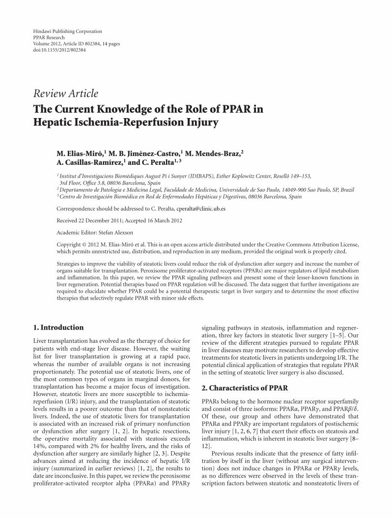
Hindawi Publishing CorporationPPAR ResearchVolume 2012, Article ID 802384, 14 pagesdoi:10.1155/2012/802384
Review Article
The Current Knowledge of the Role of PPAR inHepatic Ischemia-Reperfusion Injury
M. Elias-Miro,1 M. B. Jimenez-Castro,1 M. Mendes-Braz,2
A. Casillas-Ramırez,1 and C. Peralta1, 3
1 Institut d’Investigacions Biomediques August Pi i Sunyer (IDIBAPS), Esther Koplowitz Center, Rosello 149–153,3rd Floor, Office 3.8, 08036 Barcelona, Spain
2 Departamento de Patologia e Medicina Legal, Faculdade de Medicina, Universidade de Sao Paulo, 14049-900 Sao Paulo, SP, Brazil3 Centro de Investigacion Biomedica en Red de Enfermedades Hepaticas y Digestivas, 08036 Barcelona, Spain
Correspondence should be addressed to C. Peralta, [email protected]
Received 22 December 2011; Accepted 16 March 2012
Academic Editor: Stefan Alexson
Copyright © 2012 M. Elias-Miro et al. This is an open access article distributed under the Creative Commons Attribution License,which permits unrestricted use, distribution, and reproduction in any medium, provided the original work is properly cited.
Strategies to improve the viability of steatotic livers could reduce the risk of dysfunction after surgery and increase the number oforgans suitable for transplantation. Peroxisome proliferator-activated receptors (PPARs) are major regulators of lipid metabolismand inflammation. In this paper, we review the PPAR signaling pathways and present some of their lesser-known functions inliver regeneration. Potential therapies based on PPAR regulation will be discussed. The data suggest that further investigations arerequired to elucidate whether PPAR could be a potential therapeutic target in liver surgery and to determine the most effectivetherapies that selectively regulate PPAR with minor side effects.
1. Introduction
Liver transplantation has evolved as the therapy of choice forpatients with end-stage liver disease. However, the waitinglist for liver transplantation is growing at a rapid pace,whereas the number of available organs is not increasingproportionately. The potential use of steatotic livers, one ofthe most common types of organs in marginal donors, fortransplantation has become a major focus of investigation.However, steatotic livers are more susceptible to ischemia-reperfusion (I/R) injury, and the transplantation of steatoticlevels results in a poorer outcome than that of nonsteatoticlivers. Indeed, the use of steatotic livers for transplantationis associated with an increased risk of primary nonfunctionor dysfunction after surgery [1, 2]. In hepatic resections,the operative mortality associated with steatosis exceeds14%, compared with 2% for healthy livers, and the risks ofdysfunction after surgery are similarly higher [2, 3]. Despiteadvances aimed at reducing the incidence of hepatic I/Rinjury (summarized in earlier reviews) [1, 2], the results todate are inconclusive. In this paper, we review the peroxisomeproliferator-activated receptor alpha (PPARα) and PPARγ
signaling pathways in steatosis, inflammation and regener-ation, three key factors in steatotic liver surgery [1–5]. Ourreview of the different strategies pursued to regulate PPARin liver diseases may motivate researchers to develop effectivetreatments for steatotic livers in patients undergoing I/R. Thepotential clinical application of strategies that regulate PPARin the setting of steatotic liver surgery is also discussed.
2. Characteristics of PPAR
PPARs belong to the hormone nuclear receptor superfamilyand consist of three isoforms: PPARα, PPARγ, and PPARβ/δ.Of these, our group and others have demonstrated thatPPARα and PPARγ are important regulators of postischemicliver injury [1, 2, 6, 7] that exert their effects on steatosis andinflammation, which is inherent in steatotic liver surgery [8–12].
Previous results indicate that the presence of fatty infil-tration by itself in the liver (without any surgical interven-tion) does not induce changes in PPARα or PPARγ levels,as no differences were observed in the levels of these tran-scription factors between steatotic and nonsteatotic livers of
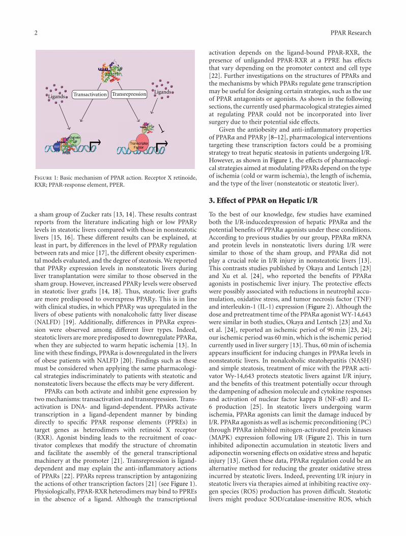
2 PPAR Research
Transactivation Transrepression LigandsLigands
Figure 1: Basic mechanism of PPAR action. Receptor X retinoide,RXR; PPAR-response element, PPER.
a sham group of Zucker rats [13, 14]. These results contrastreports from the literature indicating high or low PPARγlevels in steatotic livers compared with those in nonsteatoticlivers [15, 16]. These different results can be explained, atleast in part, by differences in the level of PPARγ regulationbetween rats and mice [17], the different obesity experimen-tal models evaluated, and the degree of steatosis. We reportedthat PPARγ expression levels in nonsteatotic livers duringliver transplantation were similar to those observed in thesham group. However, increased PPARγ levels were observedin steatotic liver grafts [14, 18]. Thus, steatotic liver graftsare more predisposed to overexpress PPARγ. This is in linewith clinical studies, in which PPARγ was upregulated in thelivers of obese patients with nonalcoholic fatty liver disease(NALFD) [19]. Additionally, differences in PPARα expres-sion were observed among different liver types. Indeed,steatotic livers are more predisposed to downregulate PPARα,when they are subjected to warm hepatic ischemia [13]. Inline with these findings, PPARα is downregulated in the liversof obese patients with NALFD [20]. Findings such as thesemust be considered when applying the same pharmacologi-cal strategies indiscriminately to patients with steatotic andnonsteatotic livers because the effects may be very different.
PPARs can both activate and inhibit gene expression bytwo mechanisms: transactivation and transrepression. Trans-activation is DNA- and ligand-dependent. PPARs activatetranscription in a ligand-dependent manner by bindingdirectly to specific PPAR response elements (PPREs) intarget genes as heterodimers with retinoid X receptor(RXR). Agonist binding leads to the recruitment of coac-tivator complexes that modify the structure of chromatinand facilitate the assembly of the general transcriptionalmachinery at the promoter [21]. Transrepression is ligand-dependent and may explain the anti-inflammatory actionsof PPARs [22]. PPARs repress transcription by antagonizingthe actions of other transcription factors [21] (see Figure 1).Physiologically, PPAR-RXR heterodimers may bind to PPREsin the absence of a ligand. Although the transcriptional
activation depends on the ligand-bound PPAR-RXR, thepresence of unliganded PPAR-RXR at a PPRE has effectsthat vary depending on the promoter context and cell type[22]. Further investigations on the structures of PPARs andthe mechanisms by which PPARs regulate gene transcriptionmay be useful for designing certain strategies, such as the useof PPAR antagonists or agonists. As shown in the followingsections, the currently used pharmacological strategies aimedat regulating PPAR could not be incorporated into liversurgery due to their potential side effects.
Given the antiobesity and anti-inflammatory propertiesof PPARα and PPARγ [8–12], pharmacological interventionstargeting these transcription factors could be a promisingstrategy to treat hepatic steatosis in patients undergoing I/R.However, as shown in Figure 1, the effects of pharmacologi-cal strategies aimed at modulating PPARs depend on the typeof ischemia (cold or warm ischemia), the length of ischemia,and the type of the liver (nonsteatotic or steatotic liver).
3. Effect of PPAR on Hepatic I/R
To the best of our knowledge, few studies have examinedboth the I/R-inducedexpression of hepatic PPARα and thepotential benefits of PPARα agonists under these conditions.According to previous studies by our group, PPARα mRNAand protein levels in nonsteatotic livers during I/R weresimilar to those of the sham group, and PPARα did notplay a crucial role in I/R injury in nonsteatotic livers [13].This contrasts studies published by Okaya and Lentsch [23]and Xu et al. [24], who reported the benefits of PPARαagonists in postischemic liver injury. The protective effectswere possibly associated with reductions in neutrophil accu-mulation, oxidative stress, and tumor necrosis factor (TNF)and interleukin-1 (IL-1) expression (Figure 2). Although thedose and pretreatment time of the PPARα agonist WY-14,643were similar in both studies, Okaya and Lentsch [23] and Xuet al. [24], reported an ischemic period of 90 min [23, 24];our ischemic period was 60 min, which is the ischemic periodcurrently used in liver surgery [13]. Thus, 60 min of ischemiaappears insufficient for inducing changes in PPARα levels innonsteatotic livers. In nonalcoholic steatohepatitis (NASH)and simple steatosis, treatment of mice with the PPAR acti-vator Wy-14,643 protects steatotic livers against I/R injury,and the benefits of this treatment potentially occur throughthe dampening of adhesion molecule and cytokine responsesand activation of nuclear factor kappa B (NF-κB) and IL-6 production [25]. In steatotic livers undergoing warmischemia, PPARα agonists can limit the damage induced byI/R. PPARα agonists as well as ischemic preconditioning (PC)through PPARα inhibited mitogen-activated protein kinases(MAPK) expression following I/R (Figure 2). This in turninhibited adiponectin accumulation in steatotic livers andadiponectin worsening effects on oxidative stress and hepaticinjury [13]. Given these data, PPARα regulation could be analternative method for reducing the greater oxidative stressincurred by steatotic livers. Indeed, preventing I/R injury insteatotic livers via therapies aimed at inhibiting reactive oxy-gen species (ROS) production has proven difficult. Steatoticlivers might produce SOD/catalase-insensitive ROS, which

PPAR Research 3
PC
PCPioglitazoneRBP4
Ang IIblockersEGF/IGFWY-14643
↑PPAR- ↑PPAR- ↑PPAR- ↑PPAR- ↑PPAR-↓PPAR-
NF BIL-6
↓MAPKs
↓Adiponectin
Oxidativestress
IL-1TNFROS
Mechanisms?
↓ Liver injury
Inflammatorycytokines
Neutrophilaccumulation
↑
↓↓
Figure 2: PPAR and hepatic I/R injury. Angiotensin II, Ang II; epidermal growth factor, EGF; insulin-like growth factor, IGF; interleukin-6, IL-6; mitogen-activated protein kinases, MAPKs; nuclear factor kappa B, NFκB; PPARα agonist; pioglitazone, peroxisome proliferator-activated receptors, PPAR; ischemic preconditioning, PC; retinol binding protein, RBP4, PPARα agonist; Wy-14,643.
may be involved in the mechanism of failure of steatotic liversafter transplantation [26]. Moreover, gene therapy based onantioxidant overexpression is limited by the toxicity of thevectors [2, 27]. In a recent study of nonsteatotic livers under-going warm hepatic ischemia, the dietary supplementationwith n-3 polyunsaturated fatty acids (PUFAs) increasedhepatic n-3 PUFA content and reduced hepatic n-6/n-3PUFA content. This was associated with PPARα upregula-tion, which in turn reduced NF-κB signaling and oxidativestress, leading to a reduced inflammatory response [28].
The function of PPARγ in hepatic I/R injury is unclear.Previous results in liver transplantation studies indicated thatI/R did not induce changes in PPARγ expression in non-steatotic livers, and consequently, strategies based on PPARγregulation had no effect on hepatic injury [14]. These resultswere different from those observed in nonsteatotic liversunder warm ischemia conditions [6]. In that study, treatmentwith pioglitazone, a PPARγ agonist, significantly inhibitedhepatic I/R injury (Figure 2). The protective effect was asso-ciated with the downregulation of several proinflammatory
cytokines and chemokines and neutrophil accumulation [7].This is in line with other results indicating that PPARγ-deficient mice displayed more severe injuries than untreatedmice under warm ischemia conditions [6]. Furthermore,pioglitazone treatment inhibited apoptosis and significantlyimproved the survival of mice in a lethal model of hepatic I/Rinjury [7]. Previous studies indicated that PPARγ activationinhibits the release of TNFα, IL-1, and IL-6 by macrophages[29, 30], which could be of interest in steatotic livers. Indeed,under warm hepatic ischemia, higher IL-1 and lower IL-10 levels were detected in steatotic livers after reperfusionthan in nonsteatotic livers [31]. This imbalance between pro-and anti-inflammatory ILs increased oxidative stress anddecreased the tolerance of steatotic livers to I/R. In addition,different studies have reported proinflammatory and anti-inflammatory roles of TNF-α and IL-6, respectively, in thevulnerability of steatotic livers undergoing I/R [2, 32].
Previous results indicated that PPARγ activation inhepatocytes by rosiglitazone treatment increases autophagyand protects against hepatic I/R injury. Autophagy is

4 PPAR Research
an evolutionarily conserved cellular process for recycling ofold proteins and organelles via the lysosomal degradation[33]. Thus, these results suggest that PPARγ has anti-inflam-matory properties and therefore may be relevant during hep-atic I/R injury. In line with these data, PPARγ upregulationis a key mechanism of the benefits of different pharmaco-logical or surgical strategies for steatotic livers undergoingI/R. Thus, some results based on isolated perfused liversindicated that the addition of growth factors (epidermalgrowth factor (EGF) and insulin-like growth factor-1 (IGF-I)) to University of Wisconsin (UW) preservation solutionprotected steatotic livers due to PPARγ overexpression [34].Similarly, EGF pretreatment mediated by PPARγ overexpres-sion protected steatotic livers undergoing warm ischemia[35] (Figure 2). Moreover, in warm hepatic ischemia, PPARγupregulation was a key mechanism of the benefits of pharma-cological blockers of angiotensin II (angiotensin-converting-enzyme (ACE) inhibitors and Ang II receptor antagonists) onsteatotic livers [36]. However, the role of PPARγ in hepaticI/R injury could depend on the surgical conditions, as arecent study of liver transplantation indicated that treatmentwith a PPARγ antagonist was effective in steatotic livers,suggesting a detrimental role of PPARγ under these condi-tions [14]. In line with this finding, PPARγ inhibition wasa key mechanism of the benefits of RBP4 treatment andPC on steatotic liver grafts [14]. Considering these results,drugs targeting PPARγ regulation can potentially increase thenumber of organs suitable for transplantation, as these drugscan improve the outcome for marginal grafts that wouldnot otherwise have been transplanted. However, the dataon PPARγ reported in steatotic liver transplantation modelswith standard liver graft sizes should not be extrapolatedto small-size steatotic liver grafts. In the case of small livertransplants, the liver regeneration inherent in this surgicalprocedure and the mechanism of hepatic damage derivedfrom the removal of hepatic mass should be considered[1, 31, 36]. In small liver grafts the periods of ischemiaranged 40–60 min, whereas the periods of ischemia ranged6–8 hours for cadaveric donor liver transplantation.
4. Effect of PPAR on Hepatic Steatosis
Numerous studies suggest that the actions of PPARα canprevent steatosis. Mice deficient in PPARα develop hepaticsteatosis when fasted or fed a high-fat diet [37, 46, 57].Treatment with a PPARα agonist decreased hepatic steatosisin mice on a methionine- and choline-deficient (MCD) diet[37]. Activation of PPARα by the agonist Wy-14,643 amelio-rated alcoholic fatty liver- and MCD-induced steatohepatitis[37, 38]. The critical role of PPARα in ameliorating steatosisis mediated through the regulation of a wide variety of genesinvolved in peroxisomal, mitochondrial, and microsomalFA β-oxidation systems in the liver [58]. When steatoticlivers are submitted to certain stresses much as partialhepatectomy, the activation of PPARα by bezafibrate reducesthe availability of FAs from circulation, reducing thus thehepatic sphingolipid synthesis [40] (see Table 1).
It is well known that n-3 PUFAs and their derivative FAsactivate PPARα [59–61], which then heterodimerizes with
RXR and liver X receptor, leading to the transcription of alarge number of genes involved in lipid metabolism. It hasbeen reported that n-3 PUFAs are more potent than then-6 PUFAs as in vivo activators of PPARα [59]. In addition,PUFA metabolites such as eicosanoids or oxidized FAs haveone to two orders of magnitude greater affinity for PPARαand are consequently far more potent transcriptional activa-tors of PPARα-dependent genes [59].
The interaction of PPARα with its DNA recognition siteis markedly enhanced by ligands such as hypotriglyceridemicfibrate drugs, conjugated linoleic acid, and PUFAs [59]. Thediscovery of PPARα led quickly to the idea that PPARα wasa “master switch” transcription factor that was targeted byPUFA to coordinately suppress genes encoding lipid synthe-sis proteins and to induce genes encoding lipid oxidationproteins [59]. In line with this idea, recent studies suggestedthat n-3 FAs serve as important mediators of gene expression,working via the PPARs to control the expression of the genesinvolved in lipid and glucose metabolism and adipogenesis[61]. Neschen et al. [62] demostrated that the administrationof dietary fish oil (n-3) to rats increases the FA capacityof their livers through its ability to function as a ligandactivator of PPARα and thereby induces the transcription ofseveral gene-encoding proteins affiliated with FA oxidation.Of interest, other studies examining the effects of fish oilfeeding on the expression of several genes of PPAR knockoutmice clearly indicated that hepatic gene regulation by fishoil feeding involves at least two different pathways: PPARα-dependent and PPARα-independent pathways. Enzymes forperoxisomal (CYP4A2) and microsomal (AOX) oxidationare PPARα-dependent and upregulated by fish oil feeding,whereas those for lipid synthesis (FAS; S14) are PPARα-independent and downregulated. This indicates that the FAregulation of de novo hepatic lipogenesis and FA oxidationare not mediated through a common factor (e.g., PPARα)[61].
Given all these data into in account, the regulation ofPPARα by PUFA, particularly n-3 PUFA and possibly conju-gated linoleic acid, may offer an explanation for the reportedbenefits of these FAs in different pathologies.
In obese NAFLD patients, the increased production ofROS leads to the depletion of n-3 PUFAs due to enhancedlipid peroxidation. As PPARα is activated through directbinding to n-3 PUFA, liver PPARα function is compromisedin obesity. This prevented the upregulation of genes involvedin lipid transport, FA β-oxidation and thermogenesis, favor-ing FA and triacylglycerol synthesis over FA β-oxidation andthus promoting hepatic steatosis [20]. Thus, PPARα acti-vation by n-3 PUFA supplementation ameliorated hepaticsteatosis in obese NAFLD patients [20]. In line with this,NASH patients have low levels of circulating n-3 PUFA, witha consequent increase of the n-6/n-3 FA ratio and impairedPPARα activity in the liver [42, 43]. NASH patients treatedwith eicosapentaenoic acid (EPA) or n-3 PUFAs, a mixtureof EPA and docosahexaenoic acid, exhibited improvementsin hepatic steatosis and necroinflammation in humans andrats with NASH, probably due to the reduction of hepaticTNFα expression and improvement of insulin sensitivity[41–43]. Moreover, PUFAs activate PPARα, leading to

PPAR Research 5
Table 1: Effect of strategies that regulate PPAR on hepatic injury, steatosis, and regeneration in experimental models and patients. Angiot-ensin II: Ang II; choline deficient: CD; epidermal growth factor: EGF; high-fat diet: HFD; insulin-like growth factor 1: IGF-1; methioninecholine deficient: MCD; nonalcoholic Steatohepatitis: NASH; peroxisome proliferator-activated receptors: PPARs; polyunsaturated fattyacids: PUFAs; ischemic preconditioning: PC; retinol binding protein-4: RBP4.
PPARα
PPARα activators
Strategies Time EffectExperimentalmodel and patients
Steatosis and hepatic injury Regeneration
WY-14,643 (30 μmol/kg/d) [17] 3 weeks ↑ PPARα Obese Zucker rats ↑ β-oxidation of fatty acids Not evaluated
WY-14,643 (180 μmol/kg/d) [17] 1 week ↑ PPARα Ob/ob mice↑ β-oxidation of fatty acids;↓ triglycerides
Not evaluated
WY-14,643 (10 mg/kg) [23, 24]1 h beforeischemia
↑ PPARαMice or Rats;warm ischemia(90 min)
↓ hepatic injury Not evaluated
WY-14,643 (10 mg/kg) [13]1 h beforeischemia
↑ PPARαZucker obese rats;warm ischemia(60 min)
↓ hepatic injury Not evaluated
WY-14,643 (10 mg/kg) [25]10 days beforesurgery
↑ PPARα
Foz/foz mice;steatotic livers;warm ischemia(90 min)
↓ hepatic injury↑ cell cycleentry
Wy-14,643 (0.1%) [37] 5 weeks ↑ PPARα Mice fed MCD diet ↓ steatohepatitis Not evaluated
Wy-14,643 (0.1%) [38] 12 days ↑ PPARα Mice fed MCD diet↓ steatohepatitis;↑ hepatic fatty acidoxidation
Not evaluated
Bezafibrate [39] 5 weeks ↑ PPARα Mice fed MCD↓ hepatic triglycerides;↑ hepatic fatty acidoxidation
Not evaluated
Benzafibrate (75 mg/kg) [40] 7 days ↑ PPARαRats;partialhepatectomy
↓ availability of fatty acids;sphingolipid synthesis
↓ liverregeneration
PC (5 min/10 min) [13]Immediatelybefore ischemia
↑ PPARαObese Zucker rats;warm ischemia(60 min)
↓ hepatic injury Not evaluated
n-3 PUFA (EPA (270 mg/kg) andDHA (180 mg/kg)) [28]
7 days ↑ PPARαSprague-Dawleyrats;warm ischemia
↓ hepatic injury,inflammation, andoxidative stress
Not evaluated
EPA (2700 mg/d) [41] 1 year ↑ PPARα NAFLD patients↓ steatosis, hepatic injury,necroinflammation, andoxidative stress
Not evaluated
n-3 PUFA (1 g/day) [42] 1 year ↑ PPARα NAFLD patients↓ steatosis, hepatic injury,and necroinflammation
Not evaluated
n-3 PUFA (2 g/day) [43] 6 months ↑ PPARα NAFLD patients↓ steatosis, hepatic injury,necroinflammation, andhepatic injury
Not evaluated
n-3 PUFA (2 g, 3 times daily)[44]
24 weeks ↑ PPARαNAFLD patientswithhyperlipidemia
↓ steatosis and hepaticinjury
Not evaluated
Ω-3 FA (5 mL, thrice daily) [45] 24 weeks ↑ PPARαNAFLD patientswith dyslipidemia
↓ steatosis and hepaticinjury
Not evaluated
Atorvastatin (20 mg/daily) [45] 24 weeks ↑ PPARαNAFLD patientswith dyslipidemia
↓ steatosis and hepaticinjury
Not evaluated
Orlistat (120 mg, thrice daily)[45]
24 weeks ↑ PPARαNAFLD patientswith dyslipidemia
↓ steatosis and hepaticinjury
Not evaluated

6 PPAR Research
Table 1: Continued.
PPARα knockout
Strategies Time Effect Experimental modelSteatosis and hepaticinjury
Regeneration
PPARα-knockout [23] — ↓ PPARαPPARα-null miceWarm ischemia(90 min)
↑ hepatic injury Not evaluated
PPARα-knockout [46] — ↓ PPARαPPARα-null mice fedHF diet
↑ hepatic β-oxidation Not evaluated
PPARα-knockout [47] — ↓ PPARαPPARα-null micePartial hepatectomy
Not evaluated↓ liverregeneration
PPARγ
PPARγ activator
Strategies Time Effect Experimental modelSteatosis and hepaticinjury
Regeneration
Rosiglitazone (10 mg/kg) [6]30 min beforeischemia
↑ PPARγ PPARγ± mice ↓ hepatic injury Not evaluated
Rosiglitazone (2.5 μmol/kg/d)[17]
1 week ↑ PPARγ Ob/ob mice ↓ triglycerides Not evaluated
Rosiglitazone (3 mg/kg/day) [48] 5 weeks ↑ PPARγPPARγfl/fl mice fedHFD diet
↑ steatosis Not evaluated
Rosiglitazone (1 mg/kg/day) [49] 12 weeks ↑ PPARγ Obese C57BL/6J mice ↑ steatosis Not evaluated
Rosiglitazone (10 mg/kg) [50]2 days beforesurgery
↑ PPARγMice partialhepatectomy
Not evaluated↓ hepaticregeneration
Troglitazone (0.1%) + adPPARγ[51]
adPPARγ (5th day)troglitazone (5 days)
↑ PPARγPPARα-null mice fedCD diet
↑ steatosis Not evaluated
Pioglitazone (500 μg/Kg) [52] 8 weeks ↑ PPARγRat fed liquid diet +alcohol
↓ liver injury Not evaluated
Pioglitazone (30 mg) [53] 96 weeks ↑ PPARγ Patients with NASH ↓ steatosis Not evaluated
Pioglitazone (25 mg/kg/day) [54]5 days beforesurgery
↑ PPARγKK-AY, micepartial hepatectomy
Not evaluated↑ hepaticregeneration
Pioglitazone (20 mg/kg) [7]1.5 h beforeischemia
↑ PPARγMiceWarm ischemia(60 min)
↓ hepatic injury Not evaluated
Ang II blockersCaptopril (100 mg/kg) orPD123319 (30 mg/kg) [36]
Immediately beforeischemia
↑ PPARγObese Zucker rats;warm ischemia(60 min)
↓ hepatic injury Not evaluated
EGF and IGF-1 (10 μg/L) [34] 24 h in UW solution ↑ PPARγObese Zucker rats;isolated liver perfused(24 h cold ischemia)
↓ hepatic injury Not evaluated
EGF (100 μg/Kg) [35]3 doses (every 8 h)starting beforesurgery
↑ PPARγObese Zucker rats;warm ischemia(60 min)
↓ hepatic injury Not evaluated
IGF-I (400 μg/Kg) [35]2 doses (every 12 h)starting beforesurgery
↑ PPARγObese Zucker rats;warm ischemia(60 min)
↓ hepatic injury Not evaluated
Adenovirus PPARγ +rosiglitazone (50 mg/kg/day)[55]
8 weeks ↑ PPARγC57BL/6J mice fedMCD diet
↓ steatohepatitis andfibrosis
Not evaluated
PC (5 min/10 min) [36]Immediately beforeischemia
↑ PPARγObese Zucker rats;warm ischemia(60 min)
↓ hepatic injury Not evaluated

PPAR Research 7
Table 1: Continued.
PPARγ inhibitor
Strategy Time Effect Experimental modelSteatosis and hepaticinjury
Regeneration
GW9662 (1 mg/kg) [14] 1 h before surgery ↓ PPARγLiver transplantation(6 h cold ischemia)
Does not change inhepatic injury
Not evaluated
GW9662 (1 mg/kg) [14] 1 h before surgery ↓ PPARγSteatotic livertransplantation (6 h coldischemia)
↓ hepatic injury Not evaluated
GW9662 (1 mg/kg, 3times/week) [55]
8 weeks ↓ PPARγC57BL/6J mice fed MCDdiet
↑ steatohepatitis,fibrosis and hepaticinjury
Not evaluated
RBP4 (150 μg/kg) [14] 30 min before surgery ↓ PPARγSteatotic livertransplantation (6 h coldischemia)
↓ hepatic injury Not evaluated
PC (5 min/10 min) [14]Immediately beforeischemia
↓ PPARγSteatotic livertransplantation (6 h ofcold ischemia)
↓ hepatic injury Not evaluated
PPARγ inhibitor
Strategies Time Effect Experimental modelSteatosis and hepaticinjury
Regeneration
PPARγ-knockout [56] — ↓ PPARγLiver-specificPPARγ-null mice
↓ steatosis Not evaluated
increased FA β-oxidation; hence, they can shift the energybalance from storage to consumption [41, 43]. n-3 PUFAshave also been proved as safe and efficacious for patientswith NAFLD associated with hyperlipidemia, as indicatedby reduced hepatic damage and serum lipid levels [44]. Inanother study, the efficacy and safety of three hypolipidemic,agents in patients with NAFLD with dyslipidemia were eval-uated. In this context, predominantly hypertriglyceridemic,hypercholesterolemic, and overweight patients were treatedwith n-3 FAs, atorvastatin, and orlistat, respectively. Thethree different groups of patients exhibited reduced hepaticdamage, normalized of hepatic steatosis, and reduced serumlipids [45].
Considering that steatosis is a risk factor in liver surgery,strategies aimed to reduce steatosis could increase the toler-ance of steatotic livers to I/R. There is considerable evidencethat liver regeneration is impaired in certain genetic modelsin which the liver contains excess fat. For example, steatoticlivers from Ob mice exhibit defective liver regeneration andhigh mortality following partial hepatectomy [63]. Similarly,impaired liver regeneration was observed in steatotic liversundergoing partial hepatectomy under vascular occlusioncompared with that in nonsteatotic livers [31]. On thecontrary, drugs that reduce hepatic steatosis, such as PPARαregulators, should be considered with caution in clinical liversurgery, as other studies indicate that genetic or pharmaco-logic approaches that reduce lipid accumulation may alsohinder liver regeneration [63–66]. Thus, a question is to whatdegree should we reduce steatosis in steatotic livers to protectthis type of liver. Another question is whether we shouldreduce steatosis before the surgical procedure and thereforeavoid the vulnerability of steatotic livers to I/R, or in contrast,should we use drugs aimed at reducing hepatic triglycerides
during surgery and thus conserve the energy required forliver regeneration. Moreover, research evaluating whetherthe short-term administration of PPARα agonists mightalleviate hepatic steatosis in steatotic livers before I/R wouldbe of interest for clinical practice because there are obviousdifficulties concerning the feasibility of long-term PPARαagonist administration in some I/R processes, in particularliver transplantation from cadaveric donors, because this isan emergency procedure in which there is very little time topretreat the donor with PPARα agonists.
Several studies attribute a causal role to PPARγ in thedevelopment of steatosis by mechanisms involving the acti-vation of lipogenic genes and de novo lipogenesis [48, 51].In accordance, targeted deletion of PPARγ in hepatocytesprotects mice against diet-induced hepatic steatosis [67],suggesting a prosteatotic role of PPARγ. Similarly, micewith liver-specific PPARγ silencing are protected againsthepatic steatosis [56]. Additionally, treatment of ob/ob micewith rosiglitazone increased liver steatosis [49]. By contrast,different results have been reported regarding the effectof PPARγ on hepatic steatosis. Indeed, PPARγ-deficientmice develop more severe MCD-induced NAFLD, whereasadenovirus-mediated PPARγ overexpression attenuated theprogression of NASH [55]. In line with this finding,rosiglitazone treatment prevented the development of NASHin a model of MCD-treated mice [55], and similar resultswere obtained using the PPARγ agonist pioglitazone [52,53]. These different results can be partially explained bydifferences in the studies such as the species, type of PPARagonist, method to induce hepatic steatosis, the type ofgenetic strategy used to induce PPARγ overexpression ordeficiency in PPARγ expression as well as differences in thepretreatment times of the drugs used (see Table 1).

8 PPAR Research
5. Effect of PPAR on Hepatic Regeneration
Recent studies demostrated that liver regeneration is im-paired in a number of animal models of fatty liver disease[68–73]. PPARα-null mice subjected to partial hepatectomy(PH) have an impaired ability to regenerate hepatic mass.Emerging evidence suggests that PPARα is a critical mod-ulator of the energy flux important for the repair of liverdamage. For example, hepatocytes in the periportal regions,which divide and replicate after PH, require mitochondrialoxidation of FAs to generate energy [74]. PPARα controls theconstitutive expression of genes involved in mitochondrialFA oxidation, including carnitine palmitoyltransferase-1 [46,75]. In mice deficient in PPARα, the impaired hepatic regen-eration is also associated with the altered expression of genesinvolved in cell cycle control and cytokine signaling. Studieswith PPARα agonists indicate that PPARα upregulates genesinvolved in the cycle cell (Ccnd1 and cMyc) as well as IL1r1and IL-6r [76] (Figure 3).
It is well known that PPARα affects the transcription of anumber of genes involved in lipid turnover and peroxisomaland mitochondrial β-oxidation, resulting in the generationof ATP, which is required to “fuel” liver repair and regener-ation [76]. By contrast, in conditions in which PPARα func-tion and/or expression is altered such as hepatic steatosis, andsmall-size liver grafts, FA metabolism is deviated toward theaccumulation of inadequately metabolized fat, favoring ROSgeneration. Consequently, ATP production is decreased, andthe demise of hepatocytes via necrotic cell death is increased,halting liver repair [77] (Figure 3). Accordingly, mice withtargeted PPARα disruption exhibit increased inflammationand necrosis and delayed liver regeneration following partialhepatectomy [47].
Previous results indicate that the impaired liver regener-ation of steatotic rats was partially due to PPARα downreg-ulation through the AdipoR2 axis. The inhibition of PPARαsignaling, increased triglyceride (TG) accumulation in hepa-tocytes and inhibited the expression of hepatic enzymes thatcontribute to FA oxidation (Figure 3). This was associatedwith increased lipid peroxidation and decreased antioxidantlevels [78].
In contrast with the aforementioned data indicating thebeneficial effects of PPARα on hepatic regeneration, a recentreport indicated that PPARα activation by bezafibrate hadnegative effects on liver regeneration, which can be attributedto the inhibition of de novo sphingolipid synthesis [40]. Pre-sumably, bezafibrate affects de novo sphingolipid synthesisby decreasing FA availability (Figure 3). The activation ofPPARα by bezafibrate virtually obliterated the postoperativeincrease in plasma nonesterified FAs induced by PH. Thiscan be explained by the inhibition of hormone-sensitivelipase activity in adipose tissue by PPARα ligands and theiranti-inflammatory properties, which decrease the releaseof cytokines such as TNF and IL-6. Both events inhibitedlipolysis in isolated white adipocytes, resulting in reduced FArelease from extrahepatic sources after PH [40].
PPARγ activity is likely to be regulated during normalliver regeneration, and the disruption of this regulation couldimpair the regenerative response. Pioglitazone improved
hepatic regeneration failure in obese mice. This effect wasassociated with reduced TNFα and IL-6 levels. Additionally,pioglitazone prevented the increased mRNA expression ofsignal transducer and activators of transcription-3 phospho-rylation and suppressor of cytokine signaling-3 mRNA in thelivers of obese mice [54]. However, inconsistent results havebeen obtained regarding the effect of PPARγ of liver regen-eration. Indeed, rosiglitazone inhibited hepatocyte prolifer-ation in mice undergoing partial hepatectomy by reducingp38 and cyclin expression [50] (see Figure 3).
On the basis of the inconsistent results reported to dateon the role of PPAR in hepatic regeneration, it is difficultto discern whether we should attempt to inhibit PPAR oradminister PPAR activators to promote liver regeneration insurgery.
6. Modulators of PPAR in Clinical Practice
Based on the data reported in experimental models (asreviewed above), different strategies (which have been sum-marized in Table 1) could exert effects on steatosis, inflam-mation, or regeneration by regulating PPAR. Whether thesepharmacological approaches can be translated into treat-ments for clinical liver surgery remains unknown. Forexample, thiazolidinediones (TZDs) should not be applied inclinical liver surgery due to their potential side effects. TZDs(pioglitazone, troglitazone, and rosiglitazone) are syntheticPPARγ agonists that are widely used as antidiabetic agents[79–81]. However, prolonged treatment of obese and dia-betic mice with TZDs resulted in the development of severesteatosis, which can lead to steatohepatitis and/or fibrosis.Troglitazone administration was associated with the devel-opment of idiosyncratic acute liver failure and was thereforewithdrawn from clinical use [82, 83]. Hepatotoxicity hassubsequently been reported in patients taking pioglitazoneand rosiglitazone [83, 84]. These data provide support forcurrent clinical practices in which these drugs are avoided orused judiciously in patients with known or suspected liverdisease. Further experiments should be initiated to devise apharmaceutical form appropriate for clinical use.
PPARα agonists are clinically and functionally relevantas fibrate therapeutics against hyperlipidemia and agents forreducing the complications of peripheral vascular diseasein diabetic patients [85]. Despite their potentially beneficialroles, PPARα agonists should be used judiciously. Short-termadministration in humans (1–10 days) would be unlikelyto produce permanent genotoxic effects. However, long-term exposure to these drugs, which would be required toreduce hepatic steatosis, can result in oxidative DNA damage,among other effects [86–90] (Figure 4).
Further studies will also be required to elucidate whethergrowth factors, Ang II blockers, or RBP4 may be safer protec-tive pharmacologic strategies for regulating PPAR in hepaticI/R injury in clinical practice (Figure 4). Nevertheless, noneof the aforementioned strategies is specific for PPAR.
To avoid the potential side effects of PPAR agonists,strategies that regulate PPARα, such as the induction of PCcould be of clinical interest. PC is an adaptive mechanismthat consists of a brief period of I/R, resulting in marked

PPAR Research 9
↑Genes involved in cell cycle
(Cnd1; cMyc)
↑IL-1R1; IL-6R ↑Triglycerides
↓ -oxidation
↑Oxidative stress
↑ROS
↓ATP
↑Hepatic injury
↓Hepatic regeneration
↑FAS
↓IL-6
Adipose tissue
↓Lipase
↓FA
↓Sphingolipids
↓Hepatic regeneration
PPARalteration
alteration
↓p38
↓Cyclin
↑Regeneration↓TNF ; IL-6↑STAT3; SOC3
PPAR
expression
activation
PPARactivation
↓FA
↓TNF
↓Lipolysis
PPARinhibition
PPA
Rac
tiva
tion
PPA
Rac
tiva
tion
PPA
Rac
tiva
tion
PPAR
Figure 3: PPAR and hepatic regeneration. Adenosine triphosphate: ATP; fatty acid: FA; interleukin-1 receptor: IL-1R; interleukin-6: IL-6;interleukin-6 receptor: IL-6R; tumor necrosis factor-alpha: TNF-α; signal transducer and activator of transcription 3: STAT3; suppressor ofcytokine signalling 3: SOC3; reactive oxygen species: ROS.
resistance in the liver, prior to a subsequent prolongedischemic stress. Our successes regarding the efficacy of PC innonsteatotic and steatotic livers undergoing warm ischemia(associated with PH) and liver transplantation [1, 2, 14, 91–93] have resulted in the clinical application of PC.
Several studies have demonstrated the effectiveness of PCin the resection of steatotic and nonsteatotic livers in clinicalpractice [94–96]. In such studies, the authors primarilyperformed liver resection via a continuous Pringle maneuver.However, other data indicate that PC does not improvepostoperative liver function and does not affect morbidityor mortality after hepatectomy under vascular exclusion ofthe liver with the preservation of caval flow [97, 98]. Thediscrepancy between these differential effects of PC duringhepatic resection might have arisen from the absence ofback flow perfusion of the liver during vascular exclusioncompared with that during the Pringle maneuver, whichinvolves interruptions only to the inflow to the liver. Inaddition, the ischemic period used by Azoulay et al. [97] waslonger (10 min on average) that that used by Clavien et al.[94]. All of these could explain, at least partially, the differenteffectiveness of PC in the clinical practice of liver surgery.
In the past decade, serious efforts have commenced totranslate some of the robust benefits of PC against ischemiareperfusion to liver transplantation in clinical practice. Itis fair to conclude that the overall clinical results havebeen less impressive than the observations in experimentalanimals. There are different data on the effectiveness of PCin I/R injury associated with liver transplantation [99–102].However, these differential effects cannot be explained by theuse of PC periods that have proved experimentally ineffectiveor by the clinical use of different cold ischemic timesfrom those evaluated experimentally. However, the reducedproportion of subjects with steatosis enrolled in PC trials andthe presence of brain death in clinical liver transplantation,which has thus far been evaluated in experimental studies ofliver transplantation, should be considered.
As previously mentioned, the proportion of subjects withsteatosis who have been enrolled in PC trials to date hasbeen small (10%). Thus, in the future, clinical trials mustmake serious efforts to include a larger proportion of donorwith steatotic livers to clarify the effectiveness of PC inliver transplantation in clinical practice. The benefits of PCare more likely to become clinically meaningful in patient
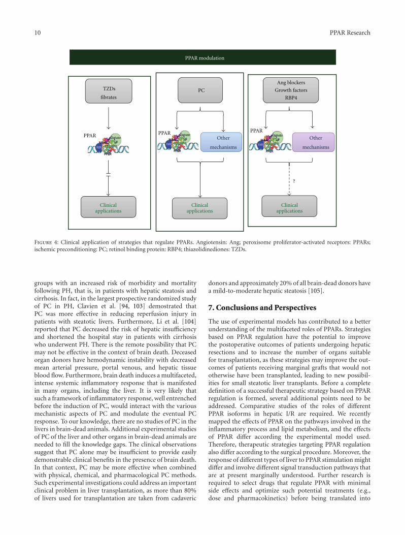
10 PPAR Research
TZDs
fibratesPC
Other
mechanisms
Other
mechanisms
?
PPAR PPARPPAR
Ang blockers
Growth factors
RBP4
Clinicalapplications
Clinicalapplications
Clinicalapplications
PPAR modulation
Figure 4: Clinical application of strategies that regulate PPARs. Angiotensin: Ang; peroxisome proliferator-activated receptors: PPARs;ischemic preconditioning: PC; retinol binding protein: RBP4; thiazolidinediones: TZDs.
groups with an increased risk of morbidity and mortalityfollowing PH, that is, in patients with hepatic steatosis andcirrhosis. In fact, in the largest prospective randomized studyof PC in PH, Clavien et al. [94, 103] demostrated thatPC was more effective in reducing reperfusion injury inpatients with steatotic livers. Furthermore, Li et al. [104]reported that PC decreased the risk of hepatic insufficiencyand shortened the hospital stay in patients with cirrhosiswho underwent PH. There is the remote possibility that PCmay not be effective in the context of brain death. Deceasedorgan donors have hemodynamic instability with decreasedmean arterial pressure, portal venous, and hepatic tissueblood flow. Furthermore, brain death induces a multifaceted,intense systemic inflammatory response that is manifestedin many organs, including the liver. It is very likely thatsuch a framework of inflammatory response, well entrenchedbefore the induction of PC, would interact with the variousmechanistic aspects of PC and modulate the eventual PCresponse. To our knowledge, there are no studies of PC in thelivers in brain-dead animals. Additional experimental studiesof PC of the liver and other organs in brain-dead animals areneeded to fill the knowledge gaps. The clinical observationssuggest that PC alone may be insufficient to provide easilydemonstrable clinical benefits in the presence of brain death.In that context, PC may be more effective when combinedwith physical, chemical, and pharmacological PC methods.Such experimental investigations could address an importantclinical problem in liver transplantation, as more than 80%of livers used for transplantation are taken from cadaveric
donors and approximately 20% of all brain-dead donors havea mild-to-moderate hepatic steatosis [105].
7. Conclusions and Perspectives
The use of experimental models has contributed to a betterunderstanding of the multifaceted roles of PPARs. Strategiesbased on PPAR regulation have the potential to improvethe postoperative outcomes of patients undergoing hepaticresections and to increase the number of organs suitablefor transplantation, as these strategies may improve the out-comes of patients receiving marginal grafts that would nototherwise have been transplanted, leading to new possibil-ities for small steatotic liver transplants. Before a completedefinition of a successful therapeutic strategy based on PPARregulation is formed, several additional points need to beaddressed. Comparative studies of the roles of differentPPAR isoforms in hepatic I/R are required. We recentlymapped the effects of PPAR on the pathways involved in theinflammatory process and lipid metabolism, and the effectsof PPAR differ according the experimental model used.Therefore, therapeutic strategies targeting PPAR regulationalso differ according to the surgical procedure. Moreover, theresponse of different types of liver to PPAR stimulation mightdiffer and involve different signal transduction pathways thatare at present marginally understood. Further research isrequired to select drugs that regulate PPAR with minimalside effects and optimize such potential treatments (e.g.,dose and pharmacokinetics) before being translated into
PPAR Research 11
treatments for human disease. Pharmacological strategiesthat specifically regulate PPAR including fibrates and TZDsmight be inappropriate for clinical liver surgery due to theirpotential side effects. Conversely, surgical strategies such asPC have been applied in clinical surgery; however, thesestrategies do not exert their effects exclusively on PPAR, asthey affect multiple aspects of I/R injury. Only a full appraisalof the role of PPAR in hepatic I/R and studies on the structureof this transcription factor will permit the design of newprotective strategies for clinical liver surgery based on thespecific regulation of PPAR without adverse effects.
Financial Support
This research was supported by the Ministerio de Ciencia eInnovacion (Project Grant BFU2009-07410) Madrid, Spain;the ACC1O (Project Grant VALTEC08-2-0033) Barcelona,Spain.
Acknowledgments
The authors thank Bioscience Writers for revising theEnglish text. M. Mendes-Braz is in receipt of a fellowshipfrom CAPES Foundation, Ministry of Education of Brazilia,Brasılia. M. B. Jimenez-Castro is in receipt of a fellowshipfrom SETH Foundation (Sociedad Espanola de TransplanteHepatico), Spain.
References
[1] M. Massip-salcedo, J. Rosello-Catafau, J. Prieto, M. A. Avıla,and C. Peralta, “The response of the hepatocyte to ischemia,”Liver International, vol. 27, no. 1, pp. 6–16, 2007.
[2] A. Casillas-Ramırez, I. B. Mosbah, F. Ramalho, J. Rosello-Catafau, and C. Peralta, “Past and future approaches toischemia-reperfusion lesion associated with liver transplan-tation,” Life Sciences, vol. 79, no. 20, pp. 1881–1894, 2006.
[3] M. Selzner and P. A. Clavien, “Fatty liver in liver transplan-tation and surgery,” Seminars in Liver Disease, vol. 21, no. 1,pp. 105–113, 2001.
[4] C. Peralta and J. Rosello-Catafau, “The future of fatty livers,”Journal of Hepatology, vol. 41, no. 1, pp. 149–151, 2004.
[5] R. W. Busuttil and K. Tanaka, “The utility of marginal donorsin liver transplantation,” Liver Transplantation, vol. 9, no. 7,pp. 651–663, 2003.
[6] S. Kuboki, T. Shin, N. Huber et al., “Peroxisome proliferator-activated receptor-γ protects against hepatic ischemia/reper-fusion injury in mice,” Hepatology, vol. 47, no. 1, pp. 215–224, 2008.
[7] T. Akahori, M. Sho, K. Hamada et al., “Importance of perox-isome proliferator-activated receptor-γ in hepatic ischemia/reperfusion injury in mice,” Journal of Hepatology, vol. 47,no. 6, pp. 784–792, 2007.
[8] A. Tailleux, K. Wouters, and B. Staels, “Roles of PPARs inNAFLD: potential therapeutic targets,” Biochimia et Biophys-ica Acta, vol. 1821, no. 5, pp. 809–818, 2012.
[9] U. A. Boelsterli and M. Bedoucha, “Toxicological conse-quences of altered peroxisome proliferator-activated receptorγ (PPARγ) expression in the liver: Insights from models ofobesity and type 2 diabetes,” Biochemical Pharmacology, vol.63, no. 1, pp. 1–10, 2002.
[10] C. N. Lumeng and A. R. Saltiel, “Inflammatory links betweenobesity and metabolic disease,” Journal of Clinical Investiga-tion, vol. 121, no. 6, pp. 2111–2117, 2011.
[11] A. Yessoufou and W. Wahli, “Multifaceted roles of peroxi-some proliferator-activated receptors (PPARs) at the cellularand whole organism levels,” Swiss Medical Weekly, vol. 140,pp. w13071–w13080, 2010.
[12] D. S. Straus and C. K. Glass, “Anti-inflammatory actions ofPPAR ligands: new insights on cellular and molecular mech-anisms,” Trends in Immunology, vol. 28, no. 12, pp. 551–558,2007.
[13] M. Massip-Salcedo, M. A. Zaouali, S. Padrissa-Altes et al.,“Activation of peroxisome proliferator-activated receptor-αinhibits the injurious effects of adiponectin in rat steatoticliver undergoing ischemia-reperfusion,” Hepatology, vol. 47,no. 2, pp. 461–472, 2008.
[14] A. Casillas-Ramırez, I. Alfany-Fernandez, M. Massip-Salcedoet al., “Retinol-binding protein 4 and peroxisome prolifera-tor-activated receptor-γ in steatotic liver transplantation,”Journal of Pharmacology and Experimental Therapeutics, vol.338, no. 1, pp. 143–153, 2011.
[15] C. Y. Zhao, L. L. Jiang, L. Li, Z. J. Deng, B. L. Liang, andJ. M. Li, “Peroxisome proliferator activated receptor-γ inpathogenesis of experimental fatty liver disease,” World Jour-nal of Gastroenterology, vol. 10, no. 9, pp. 1329–1332, 2004.
[16] M. Inoue, T. Ohtake, W. Motomura et al., “Increased expres-sion of PPARγ in high fat diet-induced liver steatosis in mice,”Biochemical and Biophysical Research Communications, vol.336, no. 1, pp. 215–222, 2005.
[17] B. Lanne, B. Dahllof, C. Lindahl et al., “PPARα and PPARγregulation of liver and adipose proteins in obese and dyslipi-demic rodents,” Journal of Proteome Research, vol. 5, no. 8,pp. 1850–1859, 2006.
[18] C. Duval, U. Thissen, S. Keshtkar et al., “Adipose tissue dys-function signals progression of hepatic steatosis towardsnonalcoholic steatohepatitis in C57Bl/6 mice,” Diabetes, vol.59, no. 12, pp. 3181–3191, 2010.
[19] P. Pettinelli and L. A. Videla, “Up-regulation of PPAR-γmRNA expression in the liver of obese patients: an additionalreinforcing lipogenic mechanism to SREBP-1c induction,”Journal of Clinical Endocrinology and Metabolism, vol. 96, no.5, pp. 1424–1430, 2011.
[20] P. Pettinelli, T. del Pozo, J. Araya et al., “Enhancementin liver SREBP-1c/PPAR-α ratio and steatosis in obesepatients: correlations with insulin resistance and n-3 long-chain polyunsaturated fatty acid depletion,” Biochimica etBiophysica Acta, vol. 1792, no. 11, pp. 1080–1086, 2009.
[21] M. Ricote and C. K. Glass, “PPARs and molecular mecha-nisms of transrepression,” Biochimica et Biophysica Acta, vol.1771, no. 8, pp. 926–935, 2007.
[22] T. M. Willson, M. H. Lambert, and S. A. Kliewer, “Per-oxisome proliferator-activated receptor γ and metabolicdisease,” Annual Review of Biochemistry, vol. 70, pp. 341–367,2001.
[23] T. Okaya and A. B. Lentsch, “Peroxisome proliferator-activated receptor-α regulates postischemic liver injury,”American Journal of Physiology, vol. 286, no. 4, pp. G606–G612, 2004.
[24] S. Q. Xu, Y. H. Li, S. H. Hu, K. Chen, and L. Y. Dong, “Effectsof Wy14643 on hepatic ischemia reperfusion injury in rats,”World Journal of Gastroenterology, vol. 14, no. 45, pp. 6936–6942, 2008.
[25] N. C. Teoh, J. Williams, J. Hartley, J. Yu, R. S. McCuskey,and G. C. Farrell, “Short-term therapy with peroxisome

12 PPAR Research
proliferation-activator receptor-alpha agonist Wy-14,643protects murine fatty liver against ischemia-reperfusioninjury,” Hepatology, vol. 51, no. 3, pp. 996–1006, 2010.
[26] W. Gao, H. D. Connor, J. J. Lemasters, R. P. Mason, and R. G.Thurman, “Primary nonfunction of fatty livers produced byalcohol is associated with a new, antioxidant-insensitive freeradical species,” Transplantation, vol. 59, no. 5, pp. 674–679,1995.
[27] M. D. Wheeler, M. Katuna, O. M. Smutney et al., “Com-parison of the effect of adenoviral delivery of three super-oxide dismutase genes against hepatic ischemia-reperfusioninjury,” Human Gene Therapy, vol. 12, no. 18, pp. 2167–2177,2001.
[28] J. Zuniga, M. Cancino, F. Medina et al., “N-3 PUFA supple-mentation triggers PPAR-α activation and PPAR-α/NF-κBinteraction: anti-inflammatory implications in liver ische-mia-reperfusion injury,” PLoS One, vol. 16, no. 12, Article IDe28502, 2011.
[29] C. Jiang, A. T. Ting, and B. Seed, “PPAR-γ agonists inhibitproduction of monocyte inflammatory cytokines,” Nature,vol. 391, no. 6662, pp. 82–86, 1998.
[30] A. von Knethen and B. Brune, “PPARγ: an important regu-lator of monocyte/macrophage function,” Archivum Immu-nologiae et Therapiae Experimentalis, vol. 51, no. 4, pp. 219–226, 2003.
[31] F. S. Ramalho, I. Alfany-Fernandez, A. Casillas-Ramırez etal., “Are angiotensin II receptor antagonists useful strategiesin steatotic and nonsteatotic livers in conditions of partialhepatectomy under ischemia-reperfusion?” Journal of Phar-macology and Experimental Therapeutics, vol. 329, no. 1, pp.130–140, 2009.
[32] R. F. Saidi, J. Chang, and S. Verb, “The effect of methylpred-nisolone on warm ischemia-reperfusion injury in the liver,”American Journal of Surgery, vol. 193, no. 3, pp. 345–347,2007.
[33] T. Yorimitsu and D. J. Klionsky, “Autophagy: molecularmachinery for self-eating,” Cell Death and Differentiation,vol. 12, no. 2, pp. 1542–1552, 2005.
[34] M. A. Zaouali, S. Padrissa-Altes, I. B. Mosbah et al., “Im-proved rat steatotic and nonsteatotic liver preservation bythe addition of epidermal growth factor and insulin-likegrowth factor-I to University of Wisconsin solution,” LiverTransplantation, vol. 16, no. 9, pp. 1098–1111, 2010.
[35] A. Casillas-Ramırez, A. Zaouali, S. Padrissa-Altes et al.,“Insulin-like growth factor and epidermal growth factortreatment: New approaches to protecting steatotic liversagainst ischemia-reperfusion injury,” Endocrinology, vol. 150,no. 7, pp. 3153–3161, 2009.
[36] A. Casillas-Ramırez, M. Amine-Zaouali, M. Massip-Salcedoet al., “Inhibition of angiotensin II action protects ratsteatotic livers against ischemia-reperfusion injury,” CriticalCare Medicine, vol. 36, no. 4, pp. 1256–1266, 2008.
[37] E. Ip, G. C. Farrell, G. Robertson, P. Hall, R. Kirsch, andI. Leclercq, “Central role of PPARα-dependent hepatic lipidturnover in dietary steatohepatitis in mice,” Hepatology, vol.38, no. 1, pp. 123–132, 2003.
[38] E. Ip, G. Farrell, P. Hall, G. Robertson, and I. Leclercq,“Administration of the potent PPARα agonist, Wy-14,643,reverses nutritional fibrosis and steatohepatitis in mice,”Hepatology, vol. 39, no. 5, pp. 1286–1296, 2004.
[39] T. Nagasawa, Y. Inada, S. Nakano et al., “Effects of bezafibrate,PPAR pan-agonist, and GW501516, PPARδ agonist, ondevelopment of steatohepatitis in mice fed a methionine- and
choline-deficient diet,” European Journal of Pharmacology,vol. 536, no. 1-2, pp. 182–191, 2006.
[40] P. Zabielski, A. Blachnio-Zabielska, M. Baranowski, M.Zendzian-Piotrowska, and J. Gorski, “Activation of PPARαby bezafibrate negatively affects de novo synthesis of sphin-golipids in regenerating rat liver,” Prostaglandins and OtherLipid Mediators, vol. 93, no. 3-4, pp. 120–125, 2010.
[41] N. Tanaka, K. Sano, A. Horiuchi, E. Tanaka, K. Kiyosawa, andT. Aoyama, “Highly purified eicosapentaenoic acid treatmentimproves nonalcoholic steatohepatitis,” Journal of ClinicalGastroenterology, vol. 42, no. 4, pp. 413–418, 2008.
[42] M. Capanni, F. Calella, M. R. Biagini et al., “Prolonged n-3polyunsaturated fatty acid supplementation ameliorates hep-atic steatosis in patients with non-alcoholic fatty liver disease:a pilot study,” Alimentary Pharmacology and Therapeutics,vol. 23, no. 8, pp. 1143–1151, 2006.
[43] L. Spadaro, O. Magliocco, D. Spampinato et al., “Effects ofn-3 polyunsaturated fatty acids in subjects with nonalcoholicfatty liver disease,” Digestive and Liver Disease, vol. 40, no. 3,pp. 194–199, 2008.
[44] F. S. Zhu, S. Liu, X. M. Chen, Z. G. Huang, and D. W. Zhang,“Effects of n-3 polyunsaturated fatty acids from seal oilson nonalcoholic fatty liver disease associated with hyperlipi-demia,” World Journal of Gastroenterology, vol. 14, no. 41, pp.6395–6400, 2008.
[45] A. Hatzitolios, C. Savopoulos, G. Lazaraki et al., “Efficacyof omega-3 fatty acids, atorvastatin and orlistat in non-alcoholic fatty liver disease with dyslipidemia,” Indian Journalof Gastroenterology, vol. 23, no. 4, pp. 131–134, 2004.
[46] S. Kersten, J. Seydoux, J. M. Peters, F. J. Gonzalez, B.Desvergne, and W. Wahli, “Peroxisome proliferator-activatedreceptor α mediates the adaptive response to fasting,” Journalof Clinical Investigation, vol. 103, no. 11, pp. 1489–1498, 1999.
[47] S. P. Anderson, L. Yoon, E. B. Richard, C. S. Dunn, R. C.Cattley, and J. C. Corton, “Delayed liver regeneration inperoxisome proliferator-activated receptor-α-null mice,”Hepatology, vol. 36, no. 3, pp. 544–554, 2002.
[48] O. Gavrilova, M. Haluzik, K. Matsusue et al., “Liver perox-isome proliferator-activated receptor gamma contributes tohepatic steatosis, triglyceride clearance, and regulation ofbody fat mass,” The Journal of Biological Chemistry, vol. 278,no. 36, pp. 34268–34276, 2003.
[49] I. Garcıa-Ruiz, C. Rodrıguez-Juan, T. Dıaz-Sanjuan, M. A.Martınez, T. Munoz-Yague, and J. A. Solıs-Herruzo, “Effectsof rosiglitazone on the liver histology and mitochondrialfunction in ob/ob mice,” Hepatology, vol. 46, no. 2, pp. 414–423, 2007.
[50] Y. P. Turmelle, O. Shikapwashya, S. Tu, P. W. Hruz, Q. Yan,and D. A. Rudnick, “Rosiglitazone inhibits mouse liver regen-eration,” The FASEB Journal, vol. 20, no. 14, pp. 2609–2611,2006.
[51] S. Yu, K. Matsusue, P. Kashireddy et al., “Adipocyte-specific gene expression and adipogenicsteatosis in the mouseliver due to peroxisome proliferator-activated receptor γ1(PPARγ1) overexpression,” The Journal of Biological Chem-istry, vol. 278, no. 1, pp. 498–505, 2003.
[52] N. Enomoto, Y. Takei, M. Hirose et al., “Prevention ofethanol-induced liver injury in rats by an agonist of peroxi-some proliferator-activated receptor-γ, pioglitazone,” Journalof Pharmacology and Experimental Therapeutics, vol. 306, no.3, pp. 846–854, 2003.
[53] A. J. Sanyal, N. Chalasani, K. V. Kowdley et al., “Pioglitazone,vitamin E, or placebo for nonalcoholic steatohepatitis,”

PPAR Research 13
The New England Journal of Medicine, vol. 362, no. 18, pp.1675–1685, 2010.
[54] T. Aoyama, K. Ikejima, K. Kon, K. Okumura, K. Arai, andS. Watanabe, “Pioglitazone promotes survival and preventshepatic regeneration failure after partial hepatectomy inobese and diabetic KK-Ay mice,” Hepatology, vol. 49, no. 5,pp. 1636–1644, 2009.
[55] Y. M. Nan, F. Han, L. B. Kong et al., “Adenovirus-mediatedperoxisome proliferator activated receptor gamma overex-pression prevents nutritional fibrotic steatohepatitis in mice,”Scandinavian Journal of Gastroenterology, vol. 46, no. 3, pp.358–369, 2011.
[56] K. Matsusue, M. Haluzik, G. Lambert et al., “Liver-specificdisruption of PPARγ in leptin-deficient mice improves fattyliver but aggravates diabetic phenotypes,” Journal of ClinicalInvestigation, vol. 111, no. 5, pp. 737–747, 2003.
[57] S. S. T. Lee, T. Pineau, J. Drago et al., “Targeted disruption ofthe α isoform of the peroxisome proliferator- activated recep-tor gene in mice results in abolishment of the pleiotropiceffects of peroxisome proliferators,” Molecular and CellularBiology, vol. 15, no. 6, pp. 3012–3022, 1995.
[58] J. K. Reddy, “Nonalcoholic steatosis and steatohepatitisIII. Peroxisomal β-oxidation, PPARα, and steatohepatitis,”American Journal of Physiology, vol. 281, no. 6, pp. G1333–G1339, 2001.
[59] S. D. Clarke, “Polyunsaturated fatty acid regulation ofgene transcription: a molecular mechanism to improve themetabolic syndrome,” Journal of Nutrition, vol. 131, no. 4, pp.1129–1132, 2001.
[60] J. Delarue, C. LeFoll, C. Corporeau, and D. Lucas, “N-3long chain polyunsaturated fatty acids: a nutritional tool toprevent insulin resistance associated to type 2 diabetes andobesity?” Reproduction Nutrition Development, vol. 44, no. 3,pp. 289–299, 2004.
[61] Y. B. Lombardo and A. G. Chicco, “Effects of dietarypolyunsaturated n-3 fatty acids on dyslipidemia and insulinresistance in rodents and humans. A review,” Journal ofNutritional Biochemistry, vol. 17, no. 1, pp. 1–13, 2006.
[62] S. Neschen, I. Moore, W. Regittnig et al., “Contrasting effectsof fish oil and safflower oil on hepatic peroxisomal and tissuelipid content,” American Journal of Physiology, vol. 282, no. 2,pp. E395–E401, 2002.
[63] E. Shteyer, Y. Liao, L. J. Muglia, P. W. Hruz, and D. A.Rudnick, “Disruption of hepatic adipogenesis is associatedwith impaired liver regeneration in mice,” Hepatology, vol.40, no. 6, pp. 1322–1332, 2004.
[64] H. Ezaki, Y. Yoshida, Y. Saji et al., “Delayed liver regenerationafter partial hepatectomy in adiponectin knockout mice,”Biochemical and Biophysical Research Communications, vol.378, no. 1, pp. 68–72, 2009.
[65] M. A. Fernandez, C. Albor, M. Ingelmo-Torres et al.,“Caveolin-1 is essential for liver regeneration,” Science, vol.313, no. 5793, pp. 1628–1632, 2006.
[66] J. Walldorf, C. Hillebrand, H. Aurich et al., “Propra-nolol impairs liver regeneration after partial hepatectomyin C57Bl/6-mice by transient attenuation of hepatic lipidaccumulation and increased apoptosis,” Scandinavian Journalof Gastroenterology, vol. 45, no. 4, pp. 468–476, 2010.
[67] E. Moran-Salvador, M. Lopez-Parra, V. Garcia-Alonso etal., “Role for PPARγ in obesity-induced hepatic steatosis asdetermined by hepatocyte- and macrophage-specific condi-tional knockouts,” The FASEB Journal, vol. 25, no. 8, pp.2538–2550, 2011.
[68] M. Selzner and P. A. Clavien, “Failure of regeneration of thesteatotic rat liver: disruption at two different levels in theregeneration pathway,” Hepatology, vol. 31, no. 1, pp. 35–42,2000.
[69] S. Q. Yang, A. K. Mandal, J. Huang, and A. M. Diehl, “Dis-rupted signaling and inhibited regeneration in obese micewith fatty livers: implications for nonalcoholic fatty liverdisease pathophysiology,” Hepatology, vol. 34, no. 4 I, pp.694–706, 2001.
[70] I. A. Leclercq, J. Field, and G. C. Farrell, “Leptin-specificmechanisms for impaired liver regeneration in ob/ob miceafter toxic injury,” Gastroenterology, vol. 124, no. 5, pp. 1451–1464, 2003.
[71] H. Yamauchi, K. Uetsuka, T. Okada, H. Nakayama, and K.Doi, “Impaired liver regeneration after partial hepatectomyin db/db mice,” Experimental and Toxicologic Pathology, vol.54, no. 4, pp. 281–286, 2003.
[72] R. A. DeAngelis, M. M. Markiewski, R. Taub, and J. D. Lam-bris, “A high-fat diet impairs liver regeneration in C57BL/6mice through overexpression of the NF-κB inhibitor, IκBα,”Hepatology, vol. 42, no. 5, pp. 1148–1157, 2005.
[73] D. A. Rudnick, “Trimming the fat from liver regeneration,”Hepatology, vol. 42, no. 5, pp. 1001–1003, 2005.
[74] H. S. Lai and W. J. Chen, “Alterations of remnant livercarnitine palmitoyltransferase I activity and serum carnitineconcentration after partial hepatectomy in rats,” Journal ofSurgical Research, vol. 59, no. 6, pp. 754–758, 1995.
[75] T. Aoyama, J. M. Peters, N. Iritani et al., “Altered con-stitutive expression of fatty acid-metabolizing enzymes inmice lacking the peroxisome proliferator-activated receptorα (PPARα),” The Journal of Biological Chemistry, vol. 273, no.10, pp. 5678–5684, 1998.
[76] G. C. Farrell, “Probing Prometheus: fat fueling the fire?”Hepatology, vol. 40, no. 6, pp. 1252–1255, 2004.
[77] G. Chinetti, J. C. Fruchart, and B. Staels, “Peroxisomeproliferator-activated receptors (PPARs): nuclear receptors atthe crossroads between lipid metabolism and inflammation,”Inflammation Research, vol. 49, no. 10, pp. 497–505, 2000.
[78] C. Y. Tsai, Y. S. Lin, T. S. Yeh et al., “Disrupted hepaticadiponectin signaling impairs liver regeneration of steatoticrats,” Chang Gung Medical Journal, vol. 34, no. 3, pp. 248–259, 2011.
[79] K. Schoonjans and J. Auwerx, “Thiazolidinediones: anupdate,” The Lancet, vol. 355, no. 9208, pp. 1008–1010, 2000.
[80] H. Yki-Jarvinen, “Thiazolidinediones,” The New EnglandJournal of Medicine, vol. 351, no. 11, pp. 1106–1118, 2004.
[81] J. Plutzky, “The potential role of peroxisome proliferator-activated receptors on inflammation in type 2 diabetes melli-tus and atherosclerosis.,” The American Journal of Cardiology,vol. 92, no. 4, pp. 34J–41J, 2003.
[82] J. Kohlroser, J. Mathai, J. Reichheld, B. F. Banner, and H.L. Bonkovsky, “Hepatotoxicity due to troglitazone: report oftwo cases and review of adverse events reported to the UnitedStates food and drug administration,” American Journal ofGastroenterology, vol. 95, no. 1, pp. 272–276, 2000.
[83] K. G. Tolman, “Thiazolidinedione hepatotoxicity: a classeffect?” International Journal of Clinical Practice, no. 113, pp.29–34, 2000.
[84] T. R. Marcy, M. L. Britton, and S. M. Blevins, “Second-generation thiazolidinediones and hepatotoxicity,” Annals ofPharmacotherapy, vol. 38, no. 9, pp. 1419–1423, 2004.
[85] A. A. Bulhak, C. Jung, C. G. Ostenson, J. O. Lundberg, P.O. Sjoquist, and J. Pernow, “PPAR-α activation protects thetype 2 diabetic myocardium against ischemia-reperfusion

14 PPAR Research
injury: involvement of the PI3-kinase/Akt and NO pathway,”American Journal of Physiology, vol. 296, no. 3, pp. H719–H727, 2009.
[86] C. G. Woods, A. M. Burns, B. U. Bradford et al., “WY-14,643-induced cell proliferation and oxidative stress in mouse liverare independent of NADPH oxidase,” Toxicological Sciences,vol. 98, no. 2, pp. 366–374, 2007.
[87] A. E. Lovett-Racke, R. Z. Hussain, S. Northrop et al., “Per-oxisome proliferator-activated receptor α agonists as therapyfor autoimmune disease,” Journal of Immunology, vol. 172,no. 9, pp. 5790–5798, 2004.
[88] I. Rusyn, S. Asakura, B. Pachkowski et al., “Expression of baseexcisionDNArepair genes is a sensitive biomarker for in vivodetection of chemical-induced chronic oxidative stress: iden-tification of the molecular marker source of radicals respon-sible for DNA damage by peroxisome proliferators,” CancerResearch, vol. 64, no. 3, pp. 1050–1057, 2004.
[89] Y. Li, L. K. Leung, H. P. Glauert, and B. T. Spear, “Treatmentof rats with the peroxisome proliferator ciprofibrate resultsin increased liver NF-κB activity,” Carcinogenesis, vol. 17, no.11, pp. 2305–2309, 1996.
[90] I. Rusyn, S. Yamashina, B. H. Segal et al., “Oxidants fromnicotinamide adenine dinucleotide phosphate oxidase areinvolved in triggering cell proliferation in the liver due toperoxisome proliferators,” Cancer Research, vol. 60, no. 17,pp. 4798–4803, 2000.
[91] A. Serafın, J. Rosello-Catafau, N. Prats, E. Gelpı, J. Rodes,and C. Peralta, “Ischemic preconditioning affects interleukinrelease in fatty livers of rats undergoing ischemia/reperfu-sion,” Hepatology, vol. 39, no. 3, pp. 688–698, 2004.
[92] M. Elias-Miro, M. Massip-Salcedo, M. Jimenez-Castro, andC. Peralta, “Does adiponectin benefit steatotic liver trans-plantation?” Liver Transplantation, vol. 17, no. 9, pp. 993–1004, 2011.
[93] M. B. Jimenez-Castro, A. Casillas-Ramırez, M. Massip-Salcedo et al., “Cyclic adenosine 3′,5′-monophosphate in ratsteatotic liver transplantation,” Liver Transplantation, vol. 17,no. 9, pp. 1099–1110, 2011.
[94] P. A. Clavien, M. Selzner, H. A. Rudiger et al., “A prospectiverandomized study in 100 consecutive patients undergoingmajor liver resection with versus without ischemic precon-ditioning,” Annals of Surgery, vol. 238, no. 6, pp. 843–852,2003.
[95] A. Chouker, A. Martignoni, R. Schauer et al., “Beneficialeffects of ischemic preconditioning in patients undergoinghepatectomy: the role of neutrophils,” Archives of Surgery, vol.140, no. 2, pp. 129–136, 2005.
[96] G. Nuzzo, F. Giuliante, M. Vellone et al., “Pedicle clampingwith ischemic preconditioning in liver resection,” LiverTransplantation, vol. 10, supplement 1, no. 2, pp. S53–S57,2004.
[97] D. Azoulay, V. Lucidi, P. Andreani et al., “Ischemic precondi-tioning for major liver resection under vascular exclusion ofthe liver preserving the caval flow: a randomized prospectivestudy,” Journal of the American College of Surgeons, vol. 202,no. 2, pp. 203–211, 2006.
[98] N. N. Rahbari, M. N. Wente, P. Schemmer et al., “Systematicreview and meta-analysis of the effect of portal triadclamping on outcome after hepatic resection,” British Journalof Surgery, vol. 95, no. 4, pp. 424–432, 2008.
[99] D. Azoulay, M. Del Gaudio, P. Andreani et al., “Effects of 10minutes of ischemic preconditioning of the cadaveric liver onthe graft’s preservation and function,” Annals of Surgery, vol.242, no. 1, pp. 133–139, 2005.
[100] M. Cescon, G. L. Grazi, A. Grassi et al., “Effect of ischemicpreconditioning in whole liver transplantation from deceaseddonors. A pilot study,” Liver Transplantation, vol. 12, no. 4,pp. 628–635, 2006.
[101] A. Amador, L. Grande, J. Martı et al., “Ischemic pre-conditioning in deceased donor liver transplantation: aprospective randomized clinical trial,” American Journal ofTransplantation, vol. 7, no. 9, pp. 2180–2189, 2007.
[102] B. Koneru, A. Shareef, G. Dikdan et al., “The ischemicpreconditioning paradox in deceased donor liver trans-plantation—evidence from a prospective randomized singleblind clinical trial,” American Journal of Transplantation, vol.7, no. 12, pp. 2788–2796, 2007.
[103] P. A. Clavien, S. Yadav, D. Sindram, and R. C. Bentley, “Pro-tective effects of ischemic preconditioning for liver resectionperformed under inflow occlusion in humans,” Annals ofSurgery, vol. 232, no. 2, pp. 155–162, 2000.
[104] S. Q. Li, L. J. Liang, J. F. Huang, and Z. Li, “Ischemic pre-conditioning protects liver from hepatectomy under hepaticinflow occlusion for hepatocellular carcinoma patients withcirrhosis,” World Journal of Gastroenterology, vol. 10, no. 17,pp. 2580–2584, 2004.
[105] K. Yamagami, J. Hutter, Y. Yamamoto et al., “Synergisticeffects of brain death and liver steatosis on the hepatic micro-circulation,” Transplantation, vol. 80, no. 4, pp. 500–505,2005.

Hindawi Publishing CorporationPPAR ResearchVolume 2012, Article ID 976089, 11 pagesdoi:10.1155/2012/976089
Research Article
PPARα Activation Protects against Anti-Thy1 Nephritis bySuppressing Glomerular NF-κB Signaling
Koji Hashimoto,1, 2 Yuji Kamijo,1, 2 Takero Nakajima,1 Makoto Harada,1, 2 Makoto Higuchi,2
Takashi Ehara,3 Hidekazu Shigematsu,3 and Toshifumi Aoyama1
1 Department of Metabolic Regulation, Institute on Aging and Adaptation, Shinshu University Graduate School of Medicine,3-1-1 Asahi, Matsumoto 390-8621, Japan
2 Department of Nephrology, Shinshu University School of Medicine, 3-1-1 Asahi, Matsumoto 390-8621, Japan3 Department of Pathology, Shinshu University School of Medicine, 3-1-1 Asahi, Matsumoto 390-8621, Japan
Correspondence should be addressed to Yuji Kamijo, [email protected]
Received 4 January 2012; Accepted 21 February 2012
Academic Editor: Christopher J. Nicol
Copyright © 2012 Koji Hashimoto et al. This is an open access article distributed under the Creative Commons AttributionLicense, which permits unrestricted use, distribution, and reproduction in any medium, provided the original work is properlycited.
The vast increase of chronic kidney disease (CKD) has attracted considerable attention worldwide, and the development of anovel therapeutic option against a representative kidney disease that leads to CKD, mesangial proliferative glomerulonephritis(MsPGN) would be significant. Peroxisome proliferator-activated receptor α (PPARα), a member of the steroid/nuclear receptorsuperfamily, is known to perform various physiological functions. Recently, we reported that PPARα in activated mesangial cellsexerted anti-inflammatory effects and that the deficiency of PPARα resulted in high susceptibility to glomerulonephritis. Toinvestigate whether PPARα activation improves the disease activity of MsPGN, we examined the protective effects of a PPARαagonist, clofibrate, in a well-established model of human MsPGN, anti-Thy1 nephritis, for the first time. This study demonstratedthat pretreatment with clofibrate (via a 0.02% or 0.1% clofibrate-containing diet) continuously activated the glomerular PPARα,which outweighed the PPARα deterioration associated with the nephritic process. The PPARα activation appeared to suppress theNF-κB signaling pathway in glomeruli by the induction of IκBα, resulting in the reduction of proteinuria and the ameliorationof the active inflammatory pathologic glomerular changes. These findings suggest the antinephritic potential of PPARα-relatedmedicines against MsPGN. PPARα-related medicines might be useful as a treatment option for CKD.
1. Introduction
The vast increase in chronic kidney disease (CKD) hasattracted considerable attention worldwide, since CKD is oneof the most important risk factors for cardiovascular events,the induction of kidney replacement therapies, and death[1]. Among many types of primary kidney disease, mesangialproliferative glomerulonephritis (MsPGN) including IgAnephropathy is a representative proteinuric kidney diseasethat leads to CKD [2, 3]. Various medications such asangiotensin-converting enzyme inhibitors, angiotensin IIreceptor blockers, fish oil, statins, hydroxymethylglutaryl-CoA reductase inhibitors, immunosuppressive therapy, anti-platelets, and anticoagulants have been proposed; however, itremains difficult to control the nephritic activity associated
with severe inflammatory pathologic glomerular changes[4]. It is known that the marked activation of nuclearfactor kappa B (NF-κB) was detected in various kidney cellsfrom MsPGN patients, including mesangial cells, glomerularendothelial and epithelial cells, tubular epithelial cells, andinfiltrating cells and that the NF-κB transcriptional activa-tion is significantly involved in the progression of kidneytissue injury [5]. Therefore, the development of a novel ther-apeutic option against NF-κB activation in active MsPGNwould be significant.
Peroxisome proliferator-activated receptor α (PPARα),a member of the steroid/nuclear receptor superfamily ofligand-dependent transcription factors, is known to performvarious physiological functions, including the maintenanceof lipid and glucose homeostasis, the regulation of cell

2 PPAR Research
proliferation, and anti-inflammatory effects via suppressionof the NF-κB pathway [6–13]. Recently, we reported that theactivated mesangial cells expressed a significant amount ofPPARα and that the representative PPARα agonists, fibrates,exert anti-inflammatory effects in an in vitro study usingmurine mesangial cells stimulated by lipopolysaccharide[14]. Moreover, we also showed that a deficiency of PPARαresulted in high susceptibility to glomerulonephritis in an invivo murine study [15]. These findings suggest that glomeru-lar PPARα activation might contribute to the treatment ofMsPGN.
To obtain basic evidence concerning the beneficial poten-tial of PPARα ligand against MsPGN, we examined theglomerular protective effects of a PPARα agonist, clofibrate,in a well-established rat model of human MsPGN, anti-Thy1 nephritis. Anti-Thy1 nephritis, induced by anti-Thy1antibody binding to the corresponding antigen on the mem-brane of mesangial cells, is marked by obvious transientinflammatory glomerular lesions, such as mesangial cellproliferation, mesangiolysis, glomerular capillary aneurysmformation, and extracapillary proliferation [16]. Severalearlier studies demonstrated that upregulation of the NF-κBgene was greatly involved in the developmental process ofanti-Thy1 nephritis [17, 18]. The current study reveals forthe first time that PPARα activation via clofibrate treatmentwould attenuate the disease activity of anti-Thy1 nephritis bysuppressing glomerular NF-κB signaling.
2. Materials and Methods
2.1. Animals and Experimental Design. Male Wistar rats wereused in this study (age, 8 weeks; purchased from Nihon SLC,Hamamatsu, Japan). All rats were maintained in a facility freeof specific pathogens, housed in a temperature- and light-controlled environment (25◦C; 12-h light/dark cycle), andgiven tap water ad libitum. All procedures were performedin accordance with the guidelines of the Shinshu University,the National Institutes of Health, and the Association forAssessment and Accreditation of Laboratory Animal Care.The rats were divided into three groups: a regular diet group(Fib(−); n = 24), a low-dose clofibrate-containing-dietgroup (0.02% Fib; n = 12), and a high-dose clofibrate-containing-diet group (0.1% Fib; n = 24). The rats inthe Fib(−) group were fed a regular diet throughout theexperimental period. The clofibrate-treated rats were fed a0.02 or 0.1% clofibrate-containing diet (drug weight/foodweight) beginning 5 days before the injection of anti-Thy1 antibody, respectively. We measured the animals’ bodyweight and daily food consumption every day. The meanbody weight and food consumption values in each group didnot change significantly throughout the experimental period,and did not differ among groups (Table 1). Using these data,the mean ± SD clofibrate dosage was calculated (Table 1).Clofibrate was obtained from Wako (Tokyo, Japan). Anti-Thy1 MsPGN was induced by a single intravenous injectionof a mouse anti-Thy1 monoclonal antibody-containingsolution. Concentrated anti-Thy1 antibody solution wasobtained from Cedarlane Laboratories (Ontario, Canada,
catalog no. CL005A). One vial of the commercial antibodysolution was diluted with 300 μL of sterile saline, and itwas injected into each rat at a dose of 25 μL/100 g bodyweight. No rat in any group died except those sacrificedaccording to the study protocol throughout the experimentalperiod. Some rats were sacrificed for analysis according to theprotocol at days 0, 4, 7, and 14. The numbers of rats subjectedto analyses at days 0, 4, 7, and 14 were as follows: Fib(−)group, n = 6; 0.02% group, n = 3; 0.1% group, n = 6, ateach day, respectively. The possibility of the induction failureof nephritis was checked by means of the measurement ofurine protein excretion in the early phase, as described below.In the current study, all rats, which were injected to anti-Thy1 antibody solution, developed significant increases ofproteinuria at day 2, indicating perfect induction of anti-Thy1 nephritis.
2.2. Pathological Analyses. Tissues from the kidneys of ratsin each group were fixed in 4% paraformaldehyde. Deparaf-finized sections were stained with hematoxylin & eosin,periodic acid Schiff, or periodic acid-methenamine-silver.Since anti-Thy1 nephritis markedly caused various acuteinflammatory glomerular changes including mesangial cellproliferation, mesangiolysis, glomerular capillary aneurysmformation, and extracapillary proliferation, we evaluatedthese inflammatory glomerular changes using semiquanti-tative pathologic analyses. For the analyses, 50 randomlyselected glomeruli from each kidney section were studied.The degree of mesangial cell proliferation was estimatedusing a scale that ranged from 0 to 3 (0, normal; 1, mild; 2,moderate; 3, severe). Indices were calculated using thefollowing formula: Index = (n0 × 0) + (n1 × 1) + (n2 × 2) +(n3 × 3)/50 (
∑n = 50). The levels of severity of the me-
sangiolysis, glomerular capillary aneurysm formation, andcrescent formation were assessed by the appearance rate ofeach finding (% of the damaged glomeruli). These pathologicanalyses were performed in a blinded manner by twoobservers who were unaware of the study protocol.
2.3. Intranuclear Transcription Factor Assay. The specifictranscription factor DNA-binding activities of PPARα or NF-κB in nuclear extracts were analyzed using enzyme-linkedimmunosorbent assay (ELISA) kits (Cayman Chemical, CA,USA). The specific double-stranded DNA sequence contain-ing the PPAR or NF-κB response element was immobilizedonto the bottoms of the wells of a 96-well plate. PPARαor NF-κB, contained in a nuclear extract, bound to eachspecific response element and was detected by the additionof a specific primary antibody. After secondary antibodybinding, the DNA-binding activity was visualized calori-metrically. These ELISA assays are nonradioactive, sensitiveestablished methods, and recently replaced the radioactiveelectrophoretic mobility shift assay. Nuclear protein wasextracted from isolated glomeruli using the NE-PER Nuclearand Cytoplasmic Extraction Kit (Thermo Scientific, MA,USA). Glomeruli were isolated from the kidney cortex ofeach rat by mechanical sieving techniques as describedpreviously [19]. The nuclear protein samples, as well as

PPAR Research 3
Table 1: Systemic change and estimated dose of clofibrate in anti-Thy1 nephritis rats.
ParameterFibrate (−) group 0.02% fibrate group 0.1% fibrate group
Day 0 Day 4 Day 7 Day 14 Day 0 Day 4 Day 7 Day 14 Day 0 Day 4 Day 7 Day 14BW (g) 248 ± 26 254 ± 33 256 ± 38 267 ± 54 257±16 259 ± 48 264 ± 35 265 ± 47 254 ± 21 258 ± 39 263 ± 30 267 ± 40sBP (mmHg) 152 ± 17 158 ± 20 158 ± 15 150±10 155 ± 10 149 ± 12 152 ± 18 153 ± 6 151 ± 16 157 ± 15 155 ± 20 158 ± 15HR (beat/min) 400 ± 16 407 ± 30 410 ± 20 400 ± 20 393 ± 11 400 ± 10 406 ± 21 404±25 396 ± 26 405 ± 36 403 ± 30 402±15FC (g/day) 20.1 ± 5 19 ± 3 18.5 ± 5 18.3 ± 5 18.2 ± 2 18.7 ± 2 19.6 ± 2 18.6 ± 5 18.9 ± 3 18.7 ± 1 19.5 ± 3 19 ± 6Clo dose (mg/kg) 0 0 0 0 14.2 14.1 14.8 14.1 74.5 72.4 74.1 71.2
BW, body weight; sBP, systolic bood pressure; HR, heart rate; FC, food consumption; Clo, clofibrate.These parameters were not affected by the induction of anti-Thy1 nephritis. There was no significant difference among groups.
Table 2: Primer sequences for quantitative real-time PCR assay.
Gene name Primers GenBank access no.
PPARαForward: 5′-GACAAGGCTCAGGATACCACTATG-3′
Reverse: 5′-TTGCAGCTTCGATCACACTTGTC-3′NM 013196
ACOXForward: 5′-GGGCCTGACAGAAGCCTACAAG-3′
Reverse: 5′-AAGGTCGACAGAGGTTAGGTTCCA-3′NM 017340
IκBαForward: 5′-TGACCATGGAAGTGATTGGTCAG-3′
Reverse: 5′-GATCACAGCCAAGTGGAGTGGA-3′NM 001105720
COX2Forward: 5′-GCGACTGTTCCAAACCAGCA-3′
Reverse: 5′-TGGGTCGAACTTGAGTTTGAAGTG-3′NM 017232
ICAM1Forward: 5′-ACAAGTGCCGTGCCTTTAGCTC-3′
Reverse: 5′-GATCACGAAGCCCGCAATG-3′NM 012967
TNFαForward: 5′-AACTCGAGTGACAAGCCCGTAG-3′
Reverse: 5′-GTACCACCAGTTGGTTGTCTTTGA-3′NM 012675
PPARα, peroxisome proliferator-activated receptor α; ACOX, acyl-CoA oxidase; IκBα, inhibitory factor κBα; COX2, cyclooxygenase-2; ICAM1, intercellularadhesion molecule-1; TNFα, tumor necrosis factor-α.
commercial positive control protein reagents and blanksamples, were subjected to ELISA in triplicate. The meanoptical density (OD) of the blank sample was subtractedfrom the OD of each sample, and the value was normalizedto each nuclear protein amount and subsequently expressedas the change relative to the value of the control rats (Fib(−)group at day 0).
2.4. Analyses of mRNA. Analyses of mRNA were performedusing quantitative real-time PCR as described previously[20–22]. One microgram of total RNA, extracted fromisolated glomeruli obtained from each rat, was reverse-transcribed using oligo(dT) primers and Superscript reversetranscriptase (Invitrogen, CA). The cDNAs were quanti-fied with an ABI PRISM 7700 sequence detection system(Applied Biosystems, CA) using specific primers and SYBRGreen double-stranded DNA binding dye I. The specificprimers were designed as shown in Table 2. For relativequantification of mRNA, glyceraldehyde-3-phosphate dehy-drogenase was used as an internal control, and the relativeexpression of RNA was calculated by the comparativethreshold cycle (Ct) method. The expression was expressedin terms of the change relative to the expression of the control[Fib(−) group of rats at day 0]. PCR reactions were carriedout in triplicate and averaged for analysis.
2.5. Miscellaneous Methods. Throughout the experimentalperiod, urine collections were carried out daily. Urine proteinconcentrations were measured as described previously [7].
Serum urea nitrogen and serum creatinine were measured byenzymatic methods using a clinical analyzer (JCA-BM2250;JEOL, Tokyo, Japan).
2.6. Statistical Analysis. Analysis of significant differenceswith respect to the interactive effects of the two factors(fibrate treatment and anti-Thy1 antibody injection) wasperformed using one-way ANOVA. Throughout the paper,significant differences from the respective day 0 group areindicated with number signs (#P < 0.05, ##P < 0.01, ###P <0.001), while significant differences between regular-diet andclofibrate-diet groups are indicated with asterisks (∗P < 0.05,∗∗P < 0.01, ∗∗∗P < 0.001).
3. Results
3.1. The Antiproteinuric Effect by Clofibrate Treatment in Anti-Thy1 Nephritis. Pretreatment with clofibrate for 5 days andthe inductive procedure of anti-Thy1 nephritis did not causeany systemic changes to body weight, food consumption,urine volume, blood pressure, or heart rate (Table 1).Pretreatment with clofibrate did not affect the urinalysis inany group of rats; however, the anti-Thy1 antibody injectionimmediately and dramatically increased daily urine proteinexcretion in all groups (Figure 1). Especially in the Fib(−)group, massive proteinuria appeared within 2 days and thengradually decreased. The clofibrate treatment attenuated themarked elevation of proteinuria throughout the experimen-tal period in a dose-dependent manner. Serum levels of urea

4 PPAR Research
Anti-Thy1 injection
Clofibrate-containing diet
0.02% fibrate 0.1% fibrate
Tota
l uri
ne
prot
ein
(m
g/da
y)
160
120
80
40
0
(day)
−5 −2 0 2 4 6 8 10 12 14
###
###
######
###
### ###
###
###
######
######
###
###### ###
######
### ###
∗∗∗
∗∗
∗
∗∗∗ ∗∗∗
∗∗∗∗∗∗
∗∗∗
∗∗
∗∗
Fibrate (–)
(a)
0.02% fibrate0.1% fibrate
30
20
10
0
Seru
m u
rea
nit
roge
n (
mg/
dL)
(day)
0 4 7 14
Fibrate (–)
(b)
0.02% fibrate0.1% fibrate
Seru
m c
reat
inin
e (m
g/dL
)
(day)
0 4 7 14
0.3
0.2
0.1
0
Fibrate (–)
(c)
Figure 1: Alteration of kidney function by clofibrate pretreatment in anti-Thy1 nephritis rat. (a) Time course of the daily urinary proteinexcretion in anti-Thy1 nephritis rats. ((b) and (c)) Serum concentrations of urea nitrogen and creatinine, respectively. The clofibrate-pretreatment group was fed a 0.02% or 0.1% clofibrate-containing diet from 5 days before anti-Thy1 antibody injection. The start timeof the anti-Thy1 antibody injection was designated as day 0. Values represent means ± SD (n = 24, 12, and 24 for the Fib(−), 0.02% fibrate,and 0.1% fibrate groups, resp.). Significant differences from the respective day 0 groups are indicated with number signs (###P < 0.001), whilesignificant differences between regular-diet and clofibrate-diet groups are indicated with asterisks (∗P < 0.05, ∗∗P < 0.01, ∗∗∗P < 0.001).
nitrogen and creatinine were prone to increase in all groups;however, there were no significant differences among thethree groups. These findings suggest an antiproteinuric effectof clofibrate treatment against anti-Thy1 nephritis.
3.2. The Amelioration of Glomerular Active Lesions by Clofi-brate Treatment. To evaluate kidney damage, we carried out
pathological analyses. In the Fib(−) group, an acute findingof mesangial damage, mesangiolysis, induced by anti-Thy1antibody appeared within 4 days, followed by varioussevere glomerular inflammatory changes, such as glomeru-lar capillary aneurysm formation, crescent formation, andmesangial cell proliferation (Figure 2). The semiquantitativepathological analyses demonstrated that the levels of severityof these acute glomerular lesions reached peak levels on day 7

PPAR Research 5
Fibr
ate
(–)
Day 0 Day 4 Day 7 Day 14
0.02
% fi
brat
e0.
1% fi
brat
e
(a)
0.02% fibrate0.1% fibrate
Mes
angi
al c
ell p
rolif
erat
ion
inde
x
2
1
0
10
5
0
(day)
0 4 7 14
0 4 7 14
(day)
Mes
angi
olys
is r
ate
(%)
100
50
00 4 7 14
(day)
An
eury
sm a
ppea
ran
ce r
ate
(%)
10
0
0.02% fibrate0.1% fibrate
0 4 7 14
20
(day)
Ext
raca
pilla
ry p
rolif
erat
ion
rat
e (%
)
Fibrate (–)Fibrate (–)
##
∗∗∗
###
#########
######
∗∗∗
∗∗∗
∗∗∗
∗∗∗∗∗∗
∗∗∗
######
######
###
###
∗∗∗∗∗∗
∗∗∗
∗∗∗
∗∗∗
∗∗∗
###
######
###
######
######
###
###
#########
######
###
######
(b)
Figure 2: Light microscopic analyses of kidney injuries. (a) Representative micrographs of the kidney in anti-Thy1 nephritis rats. Kidneysections were stained with periodic acid methenamine silver (PAM). (b) Semiquantification of pathologic changes including mesangialcell proliferation, mesangiolysis, glomerular capillary aneurysm formation, and crescent formation. Values are means ± SD. Significantdifferences from the respective day 0 groups are indicated with number signs (##P < 0.01, ###P < 0.001), while significant differences betweenregular-diet and clofibrate-diet groups are indicated with asterisks (∗∗∗P < 0.001).

6 PPAR Research
in the Fib(−) group. The findings of mesangiolysis, capillaryaneurysm, and crescent improved spontaneously on day 14,while the high level of mesangial cell proliferation continuedin this group. The pretreatment with clofibrate causedno glomerular change at day 0. This treatment markedlymoderated the acute findings induced by anti-Thy1 antibodyin a dose-dependent manner throughout the experimentalperiod. These findings suggest that pretreatment with clofi-brate ameliorated the glomerular active lesions of anti-Thy1nephritis.
3.3. The Activation of Glomerular PPARα by ClofibrateTreatment. To investigate the degree of PPARα activation viaclofibrate treatment, we examined the binding activities ofintranuclear PPARα with PPAR response element (PPRE),using nuclear protein samples from isolated glomeruli ofeach group. In the Fib(−) group, the induction of anti-Thy1nephritis obviously decreased the PPRE binding activityof PPARα at days 7 and 14 in a time-dependent manner(Figure 3). The pretreatment with clofibrate increased theglomerular PPARα activity at day 0 (before the anti-Thy1antibody injection), in a dose-dependent manner. In spite ofthe induction of anti-Thy1 nephritis, the high-dose clofibratetreatment further enhanced the increase of PPARα activity,and the low-dose treatment maintained the activated level asof day 7. Then, the PPARα activities of both clofibrate groupsdecreased at day 14, but the level of activity was still high ascompared to that of the control rats. These findings suggestthat the PPRE binding activity of PPARα deteriorated due tothe development of anti-Thy1 nephritis in the control group;however, the pretreatment with clofibrate outweighed thisdeterioration and continuously activated glomerular PPARα.To verify the enhancement of the transcriptional activity ofglomerular PPARα, we next examined the mRNA expressionlevels of PPARα and of its representative target molecule,acyl-CoA oxidase (ACOX). In the Fib(−) group, the induc-tion of anti-Thy1 nephritis decreased the mRNA expressionsof PPARα and ACOX, a result that was identical to the resultsof the PPRE binding assay and suggesting the deteriorationof glomerular PPARα (Figure 4). The pretreatment withclofibrate increased the mRNA expression of PPARα andACOX at day 0 in a dose-dependent manner. The inductionof anti-Thy1 nephritis decreased these expressions in eachgroup; however, the PPARα and ACOX expressions of bothclofibrate treatment groups remained over the baseline levelof the control rats. These findings support the finding ofcontinuous activation of PPARα via the clofibrate treatment.This activation was resistant to the PPARα deteriorationassociated with the nephritic process.
3.4. The Suppression of the NF-κB Pathway by Clofibrate Treat-ment. Since many earlier studies have demonstrated that theactivated PPARα exerts anti-inflammatory effects throughsuppression of the NF-κB pathway [23, 24], we next exam-ined the binding activities of nuclear NF-κB (p65) with a NF-κB response element. The response element binding activitiesof NF-κB did not differ among the groups at day 0. However,anti-Thy1 antibody injection increased the NF-κB binding
activities in the Fib(−) and low-dose clofibrate groups(Figure 3). The peak phase of NF-κB activation in bothgroups appeared to be around day 7. On the other hand, thehigh-dose clofibrate treatment dramatically suppressed theNF-κB activation throughout the experimental period. Thetime course of NF-κB activation appeared to be consistentwith that of the pathological activities of anti-Thy1 nephritisin each group. It is known that activated PPARα suppressesthe NF-κB pathway via the induction of the inhibitoryfactor κBα (IκBα) [23]. The mRNA analyses demonstratedthat the high-dose clofibrate treatment continuously inducedIκBα expression and decreased the high mRNA levelsof proinflammatory mediators including cyclooxygenase-2(COX2), tumor necrosis factor-α (TNFα), and intercellularadhesion molecule-I (ICAM1), which were NF-κB targetmolecules (Figure 4). These findings suggest that the anti-Thy1 antibody injection induced the continuous activationof the NF-κB signaling pathway in glomeruli followed byan increase of proinflammatory mediators and that thisproinflammatory pathway was suppressed considerably bythe induction of IκBα, which might be mediated by PPARαactivation.
4. Discussion
The current study demonstrated that pretreatment withclofibrate exerted antiproteinuric effects and amelioratedthe active glomerular pathologic inflammatory changes inrat anti-Thy1 nephritis. The pretreatment with clofibratecontinuously activated the glomerular PPARα, which out-weighed the PPARα deterioration associated with the neph-ritic process. This glomerular PPARα activation wouldsuppress the NF-κB signaling pathway via the inductionof IκBα and result in beneficial antinephritic effects. Thesefindings indicate the anti-nephritic potentiality of PPARα-related medicines.
Several metabolic experimental studies, includingmurine studies employing a high-fat-diet-induced glomer-ular injury model or a diabetic nephropathy model, have alsodemonstrated the beneficial properties of the PPARα agonistfibrates in reducing glomerular lesions [25–28]. Thesestudies suggested various beneficial glomerular protectiveeffects of fibrates as follows. First, PPARα activation improvesthe lipid metabolic abnormality in glomeruli. Second,PPARα activation attenuates the glomerular oxidative stress.Third, PPARα activation ameliorates systemic insulinresistance, lipid abnormality, energy homeostasis, hyper-tension, and vascular injuries. These pathogenic abnormal-ities are known to induce secondary activation of the NF-κBsignaling pathway and accumulation of the extracellularmatrix in glomeruli, resulting in glomerular failure [25].Using these metabolic experimental models, it might bedifficult to detect whether PPARα agonists have directanti-inflammatory effects that protect glomeruli. Incontrast to these metabolic models, the mechanism ofglomerular injury of anti-Thy1 nephritis is due to thedirect inflammatory response by complement (C5b-9)-induced activation of the NF-κB signaling pathway [16, 17].
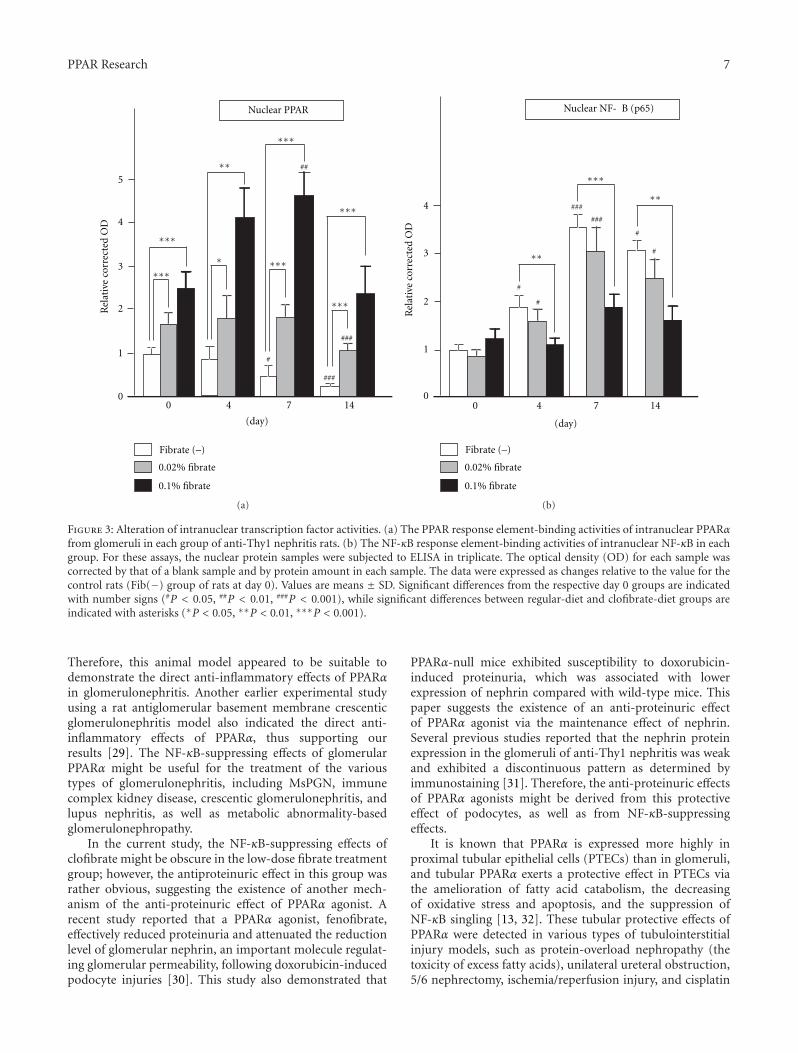
PPAR Research 7
0.02% fibrate
0.1% fibrate
Rel
ativ
e co
rrec
ted
OD
5
4
3
2
1
0
(day)
0 4 7 14
Nuclear PPAR
Fibrate (–)
##
∗
∗∗
∗∗∗
#
###
∗∗∗
∗∗∗
∗∗∗
∗∗∗
###
∗∗∗
(a)
Rel
ativ
e co
rrec
ted
OD
4
3
2
1
0
(day)
0 4 7 14
0.02% fibrate
0.1% fibrate
Nuclear NF- B (p65)
Fibrate (–)
∗∗
∗∗∗
#
###
∗∗ #
#
#
###
(b)
Figure 3: Alteration of intranuclear transcription factor activities. (a) The PPAR response element-binding activities of intranuclear PPARαfrom glomeruli in each group of anti-Thy1 nephritis rats. (b) The NF-κB response element-binding activities of intranuclear NF-κB in eachgroup. For these assays, the nuclear protein samples were subjected to ELISA in triplicate. The optical density (OD) for each sample wascorrected by that of a blank sample and by protein amount in each sample. The data were expressed as changes relative to the value for thecontrol rats (Fib(−) group of rats at day 0). Values are means ± SD. Significant differences from the respective day 0 groups are indicatedwith number signs (#P < 0.05, ##P < 0.01, ###P < 0.001), while significant differences between regular-diet and clofibrate-diet groups areindicated with asterisks (∗P < 0.05, ∗∗P < 0.01, ∗∗∗P < 0.001).
Therefore, this animal model appeared to be suitable todemonstrate the direct anti-inflammatory effects of PPARαin glomerulonephritis. Another earlier experimental studyusing a rat antiglomerular basement membrane crescenticglomerulonephritis model also indicated the direct anti-inflammatory effects of PPARα, thus supporting ourresults [29]. The NF-κB-suppressing effects of glomerularPPARα might be useful for the treatment of the varioustypes of glomerulonephritis, including MsPGN, immunecomplex kidney disease, crescentic glomerulonephritis, andlupus nephritis, as well as metabolic abnormality-basedglomerulonephropathy.
In the current study, the NF-κB-suppressing effects ofclofibrate might be obscure in the low-dose fibrate treatmentgroup; however, the antiproteinuric effect in this group wasrather obvious, suggesting the existence of another mech-anism of the anti-proteinuric effect of PPARα agonist. Arecent study reported that a PPARα agonist, fenofibrate,effectively reduced proteinuria and attenuated the reductionlevel of glomerular nephrin, an important molecule regulat-ing glomerular permeability, following doxorubicin-inducedpodocyte injuries [30]. This study also demonstrated that
PPARα-null mice exhibited susceptibility to doxorubicin-induced proteinuria, which was associated with lowerexpression of nephrin compared with wild-type mice. Thispaper suggests the existence of an anti-proteinuric effectof PPARα agonist via the maintenance effect of nephrin.Several previous studies reported that the nephrin proteinexpression in the glomeruli of anti-Thy1 nephritis was weakand exhibited a discontinuous pattern as determined byimmunostaining [31]. Therefore, the anti-proteinuric effectsof PPARα agonists might be derived from this protectiveeffect of podocytes, as well as from NF-κB-suppressingeffects.
It is known that PPARα is expressed more highly inproximal tubular epithelial cells (PTECs) than in glomeruli,and tubular PPARα exerts a protective effect in PTECs viathe amelioration of fatty acid catabolism, the decreasingof oxidative stress and apoptosis, and the suppression ofNF-κB singling [13, 32]. These tubular protective effects ofPPARα were detected in various types of tubulointerstitialinjury models, such as protein-overload nephropathy (thetoxicity of excess fatty acids), unilateral ureteral obstruction,5/6 nephrectomy, ischemia/reperfusion injury, and cisplatin

8 PPAR Research
mR
NA
rel
ativ
e ex
pres
sion
2
1
00 4 7 14
(day)m
RN
A r
elat
ive
expr
essi
on
2
1
00 4 7 14
(day)
mR
NA
rel
ativ
e ex
pres
sion
2
1
00 4 7 14
(day)
ACOXPPAR I B
∗∗∗
## ###
∗∗∗∗∗∗
∗∗∗
######
#####
##
∗∗
∗∗∗
###
##
####
∗∗∗∗∗∗
∗∗∗
###
######
∗ ∗∗
∗∗∗
###
###∗∗
##
#
#
##
(a)
∗∗∗
∗∗∗
#
##
∗∗∗∗∗
##
##
##
##
∗∗∗
∗∗∗
#
##
#
∗
∗
##
∗∗∗∗
#
###
∗∗∗
##
mR
NA
rel
ativ
e ex
pres
sion
2
1
0
COX2
0 4 7 14
(day)
0.02% fibrate0.1% fibrate
mR
NA
rel
ativ
e ex
pres
sion
2
1
00 4 7 14
(day)
0.02% fibrate0.1% fibrate
mR
NA
rel
ativ
e ex
pres
sion
2
1
00 4 7 14
(day)
0.02% fibrate0.1% fibrate
3
ICAM1TNF
Fibrate (–)Fibrate (–) Fibrate (–)
(b)
Figure 4: Alteration of mRNA expression of target molecules of transcription factors. mRNAs were obtained from glomeruli of each groupof rats. The expression of mRNAs for the target molecules of PPARα (a) and NF-κB (b), including PPARα, ACOX, IκBα, COX2, TNFα, andICAM1, was measured with real-time PCR. For relative quantification of mRNA, glyceraldehyde-3-phosphate dehydrogenase was used asan internal control, and the relative expression of RNA was calculated by the comparative threshold cycle (Ct) method. The expression wasexpressed as the change relative to that of the control rats (Fib(−) group of rats at day 0). PCR reactions were carried out in triplicate andaveraged for analysis. Values represent means ± SD. Significant differences from the respective day 0 groups are indicated with number signs(#P < 0.05, ##P < 0.01, ###P < 0.001), while significant differences between regular-diet and clofibrate-diet groups are indicated with asterisks(∗P < 0.05, ∗∗P < 0.01, ∗∗∗P < 0.001).
injury [13, 33–35]. In the current study, anti-Thy1 nephritisinduced a high level of proteinuria, a representative tubu-lotoxic factor; however, the proteinuria was transient, sotubulointerstitial changes were scarcely detected throughoutthe experimental period. Therefore, we could not detect theprotective effects of PPARα against tubulointerstitial injuriesderived from a high level of proteinuria in this model. Inorder to detect such effects, we would have to performanother experiment using models exhibiting continuousexcretion of proteinuria in the future.
In the current study, we used clofibrate to investigate theantinephritic potential of PPARα-related medicines, since
this molecule was established as a representative beneficialmedicine activating PPARα. However, we recommend thecareful use of fibrates when clinical physicians treat kidneydisease patients because the renal toxicity by excess serumaccumulation of the fibrates was often detected in theanimal models of kidney dysfunction [36]. The mechanismof the renal toxicity of fibrates was not fully understood;however, our earlier studies reported that excess-dose clofi-brate treatment induced considerable oxidative stress dueto a PPARα-dependent mechanism, such as the inductionof PPARα-regulated ROS-generating enzymes (acyl-CoAoxidase, cytochrome P450 4A, and NADPH oxidase) and

PPAR Research 9
the enhancement of mitochondrial fatty acid β-oxidation[13, 37]. Furthermore, our recent study reported that fibratescould also enhance oxidative stress in a PPARα-independentmanner, as well as by a PPARα-dependent mechanism[36]. The unfavorable oxidative effect of fibrates treatmentappeared to surpass the antioxidative effects of PPARαactivation in the situation of excess drug accumulation in theserum; therefore adequate dose management would be essen-tial for patients with kidney dysfunction. The earlier studydemonstrated that the marked elevation of oxidative stress,induced by excess serum accumulation of fibrates, exertedproximal tubular epithelial cell toxicities such as tubulardilatation, tubular atrophy, and tubular cast formation [36].Interestingly, the glomerular toxicity of fibrates was notdetected, suggesting that excess fibrates exert only tubulartoxicity without glomerular toxicity [36]. In the currentstudy, the anti-Thy1 nephritic process resulted in transientglomerular damage without tubular damage; therefore, thislimited situation might contribute to the good resultsof fibrate effects, obvious glomerular protection, and lesstubular toxicity. On the other hand, in the human case ofmany types of chronic progressing glomerulonephritis, aconsiderable level of gradual secondary tubular damage gen-erally appears; therefore, the tubular toxic effects of fibratesmight become obvious, especially after CKD has progressed.With regard to the safe use of fibrates, we must be clearthat the results of this study would not provide long-termsafety verification for CKD patients. Furthermore, in orderto prevent excess drug accumulation and the associatedtoxicities, we employed a pretreatment protocol establishedvia past animal study [36] in which an adequate dose offibrate was started before the appearance of apparent kidneydysfunction. In these specific situations, we succeeded indetecting beneficial anti-nephritic effects of fibrates withoutadverse renal effects in the current study. We believe thatthe results are important when considering the beneficialpotential of PPARα-related medicine in treating glomeru-lonephritis. In humans, two clinical trials have reported thatfibrates suppressed microalbuminuria in patients with earlydiabetic nephropathy; however, kidney dysfunction was notobviously improved [38, 39]. The results of these clinicaltrials might be derived from the delicate balance between thebeneficial effects of PPARα activation and the renal toxicityof fibrates. In the future, the development of a novel PPARαagonist exhibiting stable pharmacokinetics under kidneydysfunction is needed.
5. Conclusions
Taken together, the current results suggest that pretreatmentwith a representative PPARα agonist, clofibrate, exerts aprotective function against anti-Thy1 nephritis via thesuppression of glomerular NF-κB signaling for the first time.The developmental process of anti-Thy1 nephritis decreasedglomerular PPARα expression and weakened its function,while the pretreatment with an appropriate dose of clofibrateappeared to outweigh this deterioration. However, there areseveral limitations to our study. First, the use of pretreatment
before nephritis might not fit the actual clinical situation ofthe treatment for human kidney disease. The investigationof the beneficial effects of a treatment administered after theinitiation of anti-Thy1 nephritis using a novel medicine, ahigh serum concentration of which is not caused by kidneydysfunction or scarcely exerts toxicity, is needed in thefuture. Second, there are known to be species differencesin PPARα activation via fibrate treatment between rodentsand humans [40]. Therefore, we could not directly apply theresults of the current study to human patients. In order toevaluate the anti-nephritic effect of human PPARα function,an investigation using PPARα-humanized mice might beuseful [41]. Third, anti-Thy1 nephritis is a very famous ratmodel resembling human MsPGN; however this nephritiscould be produced only in rats. Therefore, we have to verifythe anti-nephritic effects of PPARα agonists using variousnephritic models in the future. Nevertheless, the potentialanti-nephritic effects of PPARα activation suggested in thecurrent study will be valuable for the development of a usefultherapeutic strategy to treat glomerulonephritis.
Conflict of Interests
The authors declare that there are no conflicts of interest.
Authors’ Contribution
Koji Hashimoto and Yuji Kamijo contributed equally to thiswork.
Acknowledgments
This work was supported by a grant (Grant number22590238) from the Japan Society for the Promotion ofScience (JSPS).
References
[1] M. J. Sarnak, A. S. Levey, A. C. Schoolwerth et al., “Kidney dis-ease as a risk factor for development of cardiovascular disease:a statement from the American heart association councils onkidney in cardiovascular disease, high blood pressure research,clinical cardiology, and epidemiology and prevention,” Circu-lation, vol. 108, no. 17, pp. 2154–2169, 2003.
[2] Y. Tomino, “Immunoglobulin A nephropathy and chronickidney disease,” Nephrology, vol. 15, supplement 2, pp. 23–26,2010.
[3] R. Waikhom, D. Sarka, and K. Patil, “Non-IgA mesangiopro-liferative glomerulonephritis: a benignentity?” NephrologyDialysis Transplantation. In press.
[4] J. L. Rosselli, S. M. Thacker, J. P. Karpinski, and K. A.Petkewicz, “Treatment of IgA nephropathy: an update,” Annalsof Pharmacotherapy, vol. 45, no. 10, pp. 1284–1296, 2011.
[5] M. Ashizawa, M. Miyazaki, K. Abe et al., “Detection of nuclearfactor-κb in IgA nephropathy using South Western histochem-istry,” American Journal of Kidney Diseases, vol. 42, no. 1, pp.76–86, 2003.
[6] T. Aoyama, J. M. Peters, N. Iritani et al., “Altered constitutiveexpression of fatty acid-metabolizing enzymes in mice lackingthe peroxisome proliferator-activated receptor α (pparα),”
10 PPAR Research
Journal of Biological Chemistry, vol. 273, no. 10, pp. 5678–5684, 1998.
[7] Y. Kamijo, K. Hora, N. Tanaka et al., “Identification offunctions of peroxisome proliferator-activated receptor α inproximal tubules,” Journal of the American Society of Nephrol-ogy, vol. 13, no. 7, pp. 1691–1702, 2002.
[8] K. Watanabe, H. Fujii, T. Takahashi et al., “Constitutive reg-ulation of cardiac fatty acid metabolism through peroxi-some proliferator-activated receptor α associated with age-dependent cardiac toxicity,” Journal of Biological Chemistry,vol. 275, no. 29, pp. 22293–22299, 2000.
[9] M. Guerre-Millo, P. Gervois, E. Raspe et al., “Peroxisomeproliferator-activated receptor α activators improve insulinsensitivity and reduce adiposity,” Journal of Biological Chem-istry, vol. 275, no. 22, pp. 16638–16642, 2000.
[10] J. M. Peters, T. Aoyama, R. C. Cattley, U. Nobumitsu, T.Hashimoto, and F. J. Gonzalez, “Role of peroxisome prolif-erator-activated receptor α in altered cell cycle regulation inmouse liver,” Carcinogenesis, vol. 19, no. 11, pp. 1989–1994,1998.
[11] N. Tanaka, K. Moriya, K. Kiyosawa, K. Koike, F. J. Gonzalez,and T. Aoyama, “PPARα activation is essential for HCVcore protein-induced hepatic steatosis and hepatocellularcarcinoma in mice,” Journal of Clinical Investigation, vol. 118,no. 2, pp. 683–694, 2008.
[12] T. Nakajima, Y. Kamijo, N. Tanaka et al., “Peroxisome prolif-erator-activated receptor α protects against alcohol-inducedliver damage,” Hepatology, vol. 40, no. 4, pp. 972–980, 2004.
[13] Y. Kamijo, K. Hora, K. Kono et al., “PPARα protects proximaltubular cells from acute fatty acid toxicity,” Journal of theAmerican Society of Nephrology, vol. 18, no. 12, pp. 3089–3100,2007.
[14] K. Kono, Y. Kamijo, K. Hora et al., “PPARα attenuatesthe proinflammatory response in activated mesangial cells,”American Journal of Physiology-Renal Physiology, vol. 296, no.2, pp. F328–F336, 2009.
[15] Y. Kamijo, K. Hora, T. Nakajima et al., “Peroxisome prolif-erator-activated receptor α protects against glomerulonephri-tis induced by long-term exposure to the plasticizer di-(2-ethylhexyl)phthalate,” Journal of the American Society ofNephrology, vol. 18, no. 1, pp. 176–188, 2007.
[16] J. Brandt, J. Pippin, M. Schulze et al., “Role of the complementmembrane attack complex (C5b-9) in mediating experimentalmesangioproliferative glomerulonephritis,” Kidney Interna-tional, vol. 49, no. 2, pp. 335–343, 1996.
[17] H. Wang, X. M. Jiang, J. H. Xu, J. Xu, J. X. Tong, and Y. W.Wang, “The profile of gene expression and role of nuclearfactor κB on glomerular injury in rats with Thy-1 nephritis,”Clinical and Experimental Immunology, vol. 152, no. 3, pp.559–567, 2008.
[18] T. Kosaka, A. Miyajima, E. Kikuchi et al., “The novel NF-κB activation inhibitor dehydroxymethyl-epoxyquinomicinsuppresses anti-Thy1.1-induced glomerulonephritis in rats,”Nephron Experimental Nephrology, vol. 110, no. 1, pp. e17–e24,2008.
[19] T. Mori, A. Bartocci, J. Satriano et al., “Mouse mesangial cellsproduce colony-stimulating factor-1 (CSF1) and express theCSF-1 receptor,” Journal of Immunology, vol. 144, no. 12, pp.4697–4702, 1990.
[20] T. Aoyama, S. Yamano, D. J. Waxman et al., “CytochromeP-450 hPCN3, a novel cytochrome P-450 IIIA gene productthat is differentially expressed in adult human liver. cDNAand deduced amino acid sequence and distinct specificitiesof cDNA-expressed hPCN1 and hPCN3 for the metabolism
of steroid hormones and cyclosporine,” Journal of BiologicalChemistry, vol. 264, no. 18, pp. 10388–10395, 1989.
[21] T. Aoyama, I. Ueno, T. Kamijo, and T. Hashimoto, “Rat very-long-chain acyl-CoA dehydrogenase, a novel mitochondrialacyl-CoA dehydrogenase gene product, is a rate-limitingenzyme in long-chain fatty acid β-oxidation system. cDNAand deduced amino acid sequence and distinct specificities ofthe cDNA-expressed protein,” Journal of Biological Chemistry,vol. 269, no. 29, pp. 19088–19094, 1994.
[22] T. Aoyama, Y. Uchida, R. I. Kelley et al., “A novel diseasewith deficiency of mitochondrial very-long-chain acyl-CoAdehydrogenase,” Biochemical and Biophysical Research Com-munications, vol. 191, no. 3, pp. 1369–1372, 1993.
[23] P. Delerive, K. de Bosscher, W. Vanden Berghe, J. C. Fruchart,G. Haegeman, and Staels B., “DNA binding-independentinduction of IκBα gene transcription by PPARα,” MolecularEndocrinology, vol. 16, no. 5, pp. 1029–1039, 2002.
[24] P. Delerive, J. C. Fruchart, and B. Staels, “Peroxisomeproliferator-activated receptors in inflammation control,”Journal of Endocrinology, vol. 169, no. 3, pp. 453–459, 2001.
[25] Y. Tanaka, S. Kume, S. I. Araki et al., “Fenofibrate, a PPARαagonist, has renoprotective effects in mice by enhancing renallipolysis,” Kidney International, vol. 79, no. 8, pp. 871–882,2011.
[26] H. W. Park, H. W. Kim, S. H. Ko et al., “Accelerated diabeticnephropathy in mice lacking the peroxisome proliferator-activated receptor-α,” Diabetes, vol. 55, no. 4, pp. 885–893,2006.
[27] C. W. Park, Y. Zhang, X. Zhang et al., “PPARα agonist feno-fibrate improves diabetic nephropathy in db/db mice,” KidneyInternational, vol. 69, no. 9, pp. 1511–1517, 2006.
[28] X. Zhao and L. Y. Li, “PPAR-α agonist fenofibrate inducesrenal CYP enzymes and reduces blood pressure and glomeru-lar hypertrophy in Zucker diabetic fatty rats,” American Jour-nal of Nephrology, vol. 28, no. 4, pp. 598–606, 2008.
[29] D. Saga, M. Sakatsume, A. Ogawa et al., “Bezafibrate sup-presses rat antiglomerular basement membrane crescenticglomerulonephritis,” Kidney International, vol. 67, no. 5, pp.1821–1829, 2005.
[30] Y. Zhou, X. Kong, P. Zhao et al., “Peroxisome proliferator-activated receptor-α is renoprotective in doxorubicin-inducedglomerular injury,” Kidney International, vol. 79, no. 12, pp.1302–1311, 2011.
[31] A. Mii, A. Shimizu, Y. Masuda et al., “Angiotensin II receptorblockade inhibits acute glomerular injuries with the alterationof receptor expression,” Laboratory Investigation, vol. 89, no. 2,pp. 164–177, 2009.
[32] O. Braissant, F. Foufelle, C. Scotto, M. Dauca, and W. Wahli,“Differential expression of peroxisome proliferator-activatedreceptors (PPARs): tissue distribution of PPAR-α, -β, and -γin the adult rat,” Endocrinology, vol. 137, no. 1, pp. 354–366,1996.
[33] P. Boor, P. Celec, I. V. Martin et al., “The peroxisome prolif-erator-activated receptor-α agonist, BAY PP1, attenuates renalfibrosis in rats,” Kidney International, vol. 80, no. 11, pp. 1182–1197, 2011.
[34] N. S. A. Patel, R. di Paola, E. Mazzon, D. Britti, C. Thiemer-mann, and S. Cuzzocrea, “Peroxisome proliferator-activatedreceptor-α contributes to the resolution of inflammation afterrenal ischemia/reperfusion injury,” Journal of Pharmacologyand Experimental Therapeutics, vol. 328, no. 2, pp. 635–643,2009.
[35] S. Li, R. Bhatt, J. Megyesi, N. Gokden, S. V. Shah, and D.Portilla, “PPAR-α ligand ameliorates acute renal failure by
PPAR Research 11
reducing cisplatin-induced increased expression of renalendonuclease G,” American Journal of Physiology-Renal Phys-iology, vol. 287, no. 5, pp. F990–F998, 2004.
[36] K. Takahashi, Y. Kamijo, K. Hora et al., “Pretreatment by low-dose fibrates protects against acute free fatty acid-inducedrenal tubule toxicity by counteracting PPARα deterioration,”Toxicology and Applied Pharmacology, vol. 252, no. 3, pp. 237–249, 2011.
[37] T. Nakajima, N. Tanaka, G. Li et al., “Effect of bezafibrateon hepatic oxidative stress: comparison between conventionalexperimental doses and clinically-relevant doses in mice,”Redox Report, vol. 15, no. 3, pp. 123–130, 2010.
[38] T. M. E. Davis, R. Ting, J. D. Best et al., “Effects of fenofibrateon renal function in patients with type 2 diabetes mellitus:the Fenofibrate Intervention and Event Lowering in Diabetes(FIELD) study,” Diabetologia, vol. 54, no. 2, pp. 280–290, 2011.
[39] J. C. Ansquer, C. Foucher, S. Rattier, M. R. Taskinen, andG. Steiner, “Fenofibrate reduces progression to microalbu-minuria over 3 years in a placebo-controlled study in type2 diabetes: results from the diabetes Diabetes AtherosclerosisIntervention Study (DAIS),” American Journal of KidneyDiseases, vol. 45, no. 3, pp. 485–493, 2005.
[40] F. J. Gonzalez, J. M. Peters, and R. C. Cattley, “Mechanism ofaction of the nongenotoxic peroxisome proliferators: role ofthe peroxisome proliferator-activated receptor α,” Journal ofthe National Cancer Institute, vol. 90, no. 22, pp. 1702–1709,1998.
[41] F. J. Gonzalez, “Animal models for human risk assessment:the peroxisome proliferator-activated receptor α-humanizedmouse,” Nutrition Reviews, vol. 65, supplement 2, no. 6, pp.S2–S6, 2007.
Hindawi Publishing CorporationPPAR ResearchVolume 2012, Article ID 695898, 10 pagesdoi:10.1155/2012/695898
Research Article
Global Gene Expression Profiling in PPAR-γAgonist-Treated Kidneys in an Orthologous Rat Model ofHuman Autosomal Recessive Polycystic Kidney Disease
Daisuke Yoshihara,1, 2 Masanori Kugita,1 Tamio Yamaguchi,1, 3 Harold M. Aukema,3
Hiroki Kurahashi,2 Miwa Morita,1 Yoshiyuki Hiki,4 James P. Calvet,5 Darren P. Wallace,5
Takafumi Toyohara,6 Takaaki Abe,6 and Shizuko Nagao1
1 Education and Research Center of Animal Models for Human Diseases, Fujita Health University, Toyoake, Aichi 4701192, Japan2 Division of Molecular Genetics, Institute for Comprehensive Medical Science, Fujita Health University, Toyoake, Aichi 4701192, Japan3 Department of Human Nutritional Sciences, University of Manitoba, Winnipeg, MB, Canada R3T 2N24 School of Health Sciences, Fujita Health University, Toyoake, Aichi 4701192, Japan5 The Kidney Institute, University of Kansas Medical Center, Kansas City, KS 66160, USA6 Department of Clinical Biology and Hormonal Regulation, Tohoku University Graduate School of Biomedical Engineering,Sendai, Miyagi 9808574, Japan
Correspondence should be addressed to Shizuko Nagao, [email protected]
Received 18 January 2012; Accepted 17 February 2012
Academic Editor: Yuji Kamijo
Copyright © 2012 Daisuke Yoshihara et al. This is an open access article distributed under the Creative Commons AttributionLicense, which permits unrestricted use, distribution, and reproduction in any medium, provided the original work is properlycited.
Kidneys are enlarged by aberrant proliferation of tubule epithelial cells leading to the formation of numerous cysts, nephron loss,and interstitial fibrosis in polycystic kidney disease (PKD). Pioglitazone (PIO), a PPAR-γ agonist, decreased cell proliferation,interstitial fibrosis, and inflammation, and ameliorated PKD progression in PCK rats (Am. J. Physiol.-Renal, 2011). To exploregenetic mechanisms involved, changes in global gene expression were analyzed. By Gene Set Enrichment Analysis of 30655 genes,13 of the top 20 downregulated gene ontology biological process gene sets and six of the top 20 curated gene set canonicalpathways identified to be downregulated by PIOtreatment were related to cell cycle and proliferation, including EGF, PDGF andJNK pathways. Their relevant pathways were identified using the Kyoto Encyclopedia of Gene and Genomes database. Stearoyl-coenzyme A desaturase 1 is a key enzyme in fatty acid metabolism found in the top 5 genes downregulated by PIO treatment.Immunohistochemical analysis revealed that the gene product of this enzyme was highly expressed in PCK kidneys and decreasedby PIO. These data show that PIO alters the expression of genes involved in cell cycle progression, cell proliferation, and fatty acidmetabolism.
1. Introduction
Polycystic kidney diseases (PKD) are characterized by pro-gressive enlargement of numerous fluid-filled cysts in bothkidneys, often leading to chronic kidney disease (CKD).Autosomal dominant PKD (ADPKD) is one of the mostcommon hereditary disorders in humans with an inci-dence of 1 : 500–1,000, caused by mutations in the PKD1or PKD2 gene. Progressive kidney enlargement is due toaberrant proliferation of the cystic epithelia, together withan accumulation of fluid into the cyst cavities due to
transepithelial chloride (Cl−) and fluid secretion [1–3].Autosomal recessive PKD (ARPKD) is known as a juvenile-type cystic disease with an incidence of 1 : 20,000 [3].Kidneys in ARPKD patients are characterized by cysticfusiform dilations of the collecting ducts accompanied byincreased cell proliferation and fluid secretion, leading tomassive kidney enlargement and renal failure occurring inthe first few years after birth [4]. Increased cell prolifera-tion, stimulated fluid secretion, and interstitial fibrosis areoften observed in cystic liver disease in ARPKD as well[5].
2 PPAR Research
Peroxisome proliferator-activated receptors (PPARs)belong to a nuclear receptor superfamily of ligand-activatedtranscription factors with subtypes α, β/δ, and γ. PPAR-γis widely expressed in several organs including kidneys andknown to be activated by fatty acids [6, 7]. Antidiabeticagents, pioglitazone (PIO), troglitazone, ciglitazone, androsiglitazone, are used to control blood sugar levels inpatients with diabetes mellitus. These PPAR-γ agonists alsohave important roles in regulation of cell cycle, inhibitionof fibrosis, infiltration and metastasis of cancer cells, andmodulation of inflammatory cytokines.
Treatment with PIO improved survival and amelioratedcardiac defects and the degree of renal cystogenesis inembryos of Pkd1−/− mice in a previous study [8]. In addition,long-term treatment of this agonist improved endothelialfunction by increasing production of nitric oxide in adultheterozygous Pkd1+/− mice [8]. Another PPAR-γ agonist,rosiglitazone attenuated PKD progression and prolongedsurvival of Han: SPRD Cy rats [9]. In our recent study,daily treatment of PIO ameliorated polycystic kidney dis-ease through inhibiting Raf/MEK/ERK and AKT/mTOR/S6signaling cascades in the PCK rat, an orthologous model ofhuman ARPKD [10]. These findings suggest that PPAR-γagonists may have therapeutic value in ARPKD via alteringseveral cellular signaling pathways. In the current study,we applied global gene expression profiling to explorenovel cellular signaling pathways potentially related to theameliorating effects of PIO in PCK rat kidneys.
2. Methods
2.1. PCK Rat and Study Design. PCK rats were originallyderived from a strain of Sprague-Dawley rats in Japanand descendants of this colony have been maintained atthe Education and Research Center of Animal Models forHuman Diseases, Fujita Health University. PCK rats andnormal Sprague Dawley rats (+/+; Charles River JapanInc., Kanagawa, Japan) were allowed free access to waterand food throughout the study. Female PCK and +/+ rats,aged 4–20 weeks (n = 10 per gender) were randomlyassigned to one of two groups: treatment with 10 mg/kg PIO(Takeda Pharmaceutical Company Limited, Osaka, Japan)or vehicle control (0.5% DMSO) by gavage every day aspreviously reported [10]. The protocol for the ethics and useof these animals was approved by the Animal Care and UseCommittee at Fujita Health University.
At 20 weeks of age, rats were anesthetized with sodiumpentobarbital (Schering-Plough Corp., Kenilworth, NJ), andthe kidneys were removed rapidly, causing lethal exsanguina-tion. Half of the left kidney was frozen in liquid nitrogen forRNA extraction. Half of the right kidney was immersed in4% paraformaldehyde, embedded in paraffin, and sectionedfor immunohistochemistry.
2.2. RNA Extraction. RNA was extracted from kidneys ofrats with or without PIO treatment using a monophasicsolution of phenol/guanidine isothiocyanate and TRIzolreagent (Invitrogen Co., Carlsbad, CA, USA) in accordancewith their manual, and the samples were incubated with
RNase-free DNase I (Ambion, TX, USA). The quality andconcentration of each sample was confirmed by spectropho-tometry (NanoDrop ND-1000; Asahi glass Co. Ltd., Tokyo,Japan). Total RNA obtained from three females was pooled ineach PIO-treated or control vehicle-treated (CONT) groupin accordance with our previous report [11].
2.3. Microarrays. DNA microarray experiments were per-formed essentially as described previously [11]. Briefly,500 ng aliquots of total RNA obtained from kidneys offive rats were labeled using a Quick Amp Labeling Kit,one-color (Agilent Technologies, Inc., Santa Clara, CA,USA), according to the manufacturer’s instructions. Thepooled renal RNA of PIO- or vehicle-treated PCK rats werelabeled with the Cy3-fluorescence dye. After determinationof labeling efficiency, 1.65 μg aliquots of Cy3-labeled RNAwere hybridized using the Gene Expression hybridizationkit (Agilent Technologies) onto Rat Oligo Microarrays(Agilent Technologies, product no. G4130A) according tothe manufacturer’s hybridization protocol. The microarrayslides were examined with an Agilent microarray scanner andsoftware. Data analysis was performed with Agilent FeatureExtraction software (version A.7.1.1).
Data from microarray experiments of PIO- or vehicle-treated rats were analyzed independently. Primary microar-ray data are available from the Gene Expression Omnibus(GEO) (accession number GSE00000). Evaluation of signalintensity was divided into three classes, {0}: nondetected,{1}: weakly detected, and {2}: strongly detected tran-scription product. Gene ontology analysis of biologicalprocess (C5BP) and curated gene sets of canonical pathways(C2CP) were analyzed by importing the data into GeneSet Enrichment Analysis (GSEA version 2, the Broad Insti-tute/Massachusetts Institute of technology, USA) [12].
Using the GeneSpring software, the changed probes werelisted as “Log 2 ratio was over 1 (over 2-fold) or less than −1(less than 1/2-fold) between PIO group and CONT group”and “the signal evaluation was {2} (strongly detected) inboth groups”. In the changed genes, Kyoto Encyclopedia ofGene and Genomes (KEGG) analysis was used [13].
2.4. Real-Time Reverse Transcriptase Polymerase Chain Reac-tion (RT-PCR). cDNA was produced from total RNAby reverse transcriptase using random hexamer primers(SuperScript II First Strand Synthesis System; InvitrogenCo., Carlsbad, CA, USA). To compare gene expressionpatterns of PCK kidneys with PIO or vehicle treatment,we selected a key enzyme in fatty acid metabolism,stearoyl-coenzyme A desaturase 1 (Scd1), and uncou-pling protein 1 (Ucp1). Gene expression was detectedby real-time RT-PCR (ABI 7300 real-time PCR system;Applied Biosystems, Foster City, CA, USA) using the Taq-Man reagent-based chemistry protocol. Glyceraldehyde-3-phosphate dehydrogenase (GAPDH) as a housekeeping genewas used for data normalization. The probes of Scd1, UCP-1,and GAPDH were CCCACATGCTCCAAGAGATCTCCAG,CTCTTCAGGGAGAGAAACGCCTGCC, and AACCCAT-CACCATCTTCCAGGAGCG, respectively (TaqMan Gene

PPAR Research 3
Table 1: (a) Top 20 downregulated gene ontology biological process gene sets in PIO-treated compared with vehicle-treated kidneys. Thename of biological process is listed in order of nominal P value. (b) Upregulated gene ontology biological process gene sets in PIO-treatedcompared with vehicle-treated kidneys. The name of biological process is listed in order of nominal P value.
(a)
Name of biological process gene sets Number of genes in the gene set Nominal P value
Defense response 98 0.000
Regulation of cell proliferation 136 0.000
Cell cycle phase 53 0.000
Positive regulation of cell proliferation 64 0.000
Cell cycle process 61 0.000
Positive regulation of cellular process 258 0.000
Cellular morphogenesis during differentiation 22 0.001
Positive regulation of developmental process 91 0.001
Immune system process 128 0.001
Cellular defense response 19 0.003
Neuron differentiation 35 0.004
Negative regulation of cell proliferation 73 0.004
Neurite development 27 0.005
G Protein signaling coupled to ip3 second messenger phospholipase C activating 22 0.005
Inflammatory response 56 0.005
Regulation of response to stimulus 15 0.006
Neuron development 30 0.007
M phase 27 0.007
Interphase 29 0.008
Axonogenesis 21 0.009
(b)
Name of biological process gene sets Number of genes in the gene set Nominal P value
Nitrogen compound catabolic process 17 0.000
Amine catabolic process 15 0.000
Amino acid metabolic process 46 0.000
Amino acid and derivative metabolic process 58 0.000
Organic acid metabolic process 106 0.000
Carboxylic acid metabolic process 104 0.022
Expression Assays; Applied Biosystems). Relative quantifica-tion of gene expression was compared to one in SD controlvehicle-treated (CONT) kidneys (set to 1.0).
2.5. Immunohistochemistry. Kidney sections were fixed,embedded, and sectioned for immunoreaction as describedpreviously [10, 11]. Sections were incubated with Scd1antibody (1 : 250 ab19862 Abcam, Cambridge, UK) in PBScontaining 1% BSA plus 0.05% NaN3 overnight at 4◦C. Totest for a specific Scd1 immunoreaction in the kidney, mouseIgG2b, κ isotype control antibody (1 : 200 400323 BioLegend,San Diego, CA), was used. Sections were incubated withsecondary antibody Histofine MAX-PO (MULTI: for anti-mouse/rabbit IgG, IgA, and IgM) obtained from NichireiBiosciences (Tokyo, Japan). Immune reaction productswere developed using 3,3′-diaminobenzidine (ENVISIONkit HRP Dako Cytomation K3466, Dako Japan Inc., Tokyo,Japan).
2.6. Statistical Analysis. Results are expressed as the arith-metic mean ± standard error. Statistical comparisonsbetween groups were performed by Student’s t-test and two-way analysis of variance, and differences were considered tobe significant at P < 0.05.
3. Results
3.1. Identification of Differentially Expressed Genes by Expres-sion Profiling. Previous report indicates that PPAR-γ agonis-tic action decreases expression of endothelin receptor typeA (EDNRA) [14], suggesting that EDNRA is one of thedown-stream target gene of PPAR-γ agonists. In our currentstudy, expression of Ednra was also downregulated in PIO-treated kidneys (Log2 ratio = −1.30). EDNRA expressionis increased in human ADPKD, and overexpression ofEdnra causes cyst formation in transgenic mouse kidneys[15]. Because not only EDNRA but also various genes
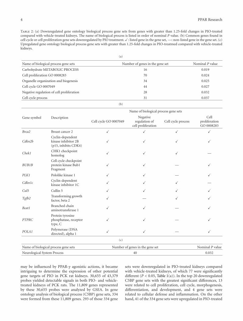
4 PPAR Research
Table 2: (a) Downregulated gene ontology biological process gene sets from genes with greater than 1.25-fold changes in PIO-treatedcompared with vehicle-treated kidneys. The name of biological process is listed in order of nominal P value. (b) Common genes found incell cycle or cell proliferation gene sets downregulated by PIO treatment. �: listed gene in the gene set, —: non-listed gene in the gene set. (c)Upregulated gene ontology biological process gene sets with greater than 1.25-fold changes in PIO-treatmed compared with vehicle-treatedkidneys.
(a)
Name of biological process gene sets Number of genes in the gene set Nominal P value
Carbohydrate METABOLIC PROCESS 16 0.019
Cell proliferation GO 0008283 70 0.024
Organelle organization and biogenesis 34 0.025
Cell cycle GO 0007049 44 0.027
Negative regulation of cell proliferation 28 0.032
Cell cycle process 31 0.037
(b)
Gene symbol Description
Name of biological process gene sets
Cell cycle GO 0007049Negative
regulation ofcell proliferation
Cell cycle processCell
proliferationGO 0008283
Brca2 Breast cancer 2 � � � �
Cdkn2bCyclin-dependentkinase inhibitor 2B(p15, inhibits CDK4)
� � � �
Chek1CHK1 checkpointhomolog
� � � —
BUB1BCell cycle checkpointprotein kinase Bub1Fragment
� � — �
PLK1 Pololike kinase 1 � � — �
Cdkn1cCyclin-dependentkinase inhibitor 1C
� � � —
Cul5 Cullin 5 � � � �
Tgfb2Transforming growthfactor, beta 2
� — � �
Bcat1Branched chainaminotransferase 1
� � — �
PTPRCProtein tyrosinephosphatase, receptortype, C
� � — �
POLA1Polymerase (DNAdirected), alpha 1
� � — �
(c)
Name of biological process gene sets Number of genes in the gene set Nominal P value
Neurological System Process 40 0.032
may be influenced by PPAR-γ agonistic actions, it becameintriguing to determine the expression of other potentialgene targets of PIO in PCK rat kidneys. 30,655 of 43,379probes yielded detectable signals in both PIO- and vehicle-treated kidneys of PCK rats. The 11,809 genes representedby these 30,655 probes were analyzed by GSEA. In geneontology analysis of biological process (C5BP) gene sets, 334were formed from these 11,809 genes. 293 of those 334 gene
sets were downregulated in PIO-treated kidneys comparedwith vehicle-treated kidneys, of which 77 were significantlydifferent (P < 0.05, Table 1(a)). In the top 20 downregulatedC5BP gene sets with the greatest significant differences, 13were related to cell proliferation, cell cycle, morphogenesis,differentiation, and development, and 4 gene sets wererelated to cellular defense and inflammation. On the otherhand, 41 of the 334 gene sets were upregulated in PIO-treated

PPAR Research 5
kidneys compared with vehicle-treated kidneys, of which 6were significantly different (P < 0.05, Table 1(b)). These genesets were related to catabolic and metabolic processes.
To examine the gene sets with the greatest changes,only 2,611 genes, which changed more than 1.25-fold inPIO-treated kidneys compared to vehicle-treated kidneys,were analyzed. 141 gene sets were formed from these 2,611genes. 112 of those 141 gene sets were downregulated inPIO-treated kidneys compared with vehicle-treated kidneysof PCK rats. Of these, 6 gene sets were significantly different(P < 0.05, Table 2(a)). 4 of these 6 gene sets are relatedto cell cycle and cell proliferation (Table 2(a)). Commongenes in these gene sets include G1/S or G2/M checkpointrelated genes, breast cancer 2 (Brca2), cyclin-dependentkinase inhibitor 2B (Cdkn2b), CHK1 checkpoint homolog(Chek1), cell cycle checkpoint protein kinase Bub1 fragment(BUB1B), pololike kinase 1 (PLK1), and cyclin-dependentkinase inhibitor 1C (Cdkn1c) (Table 2(b)). Of the remaining29 of the 141 gene sets that were upregulated in PIO-treatedkidneys compared with vehicle-treated kidneys, only one,related to neurological system processes, was significantlyelevated (P < 0.05) (Table 2(c)).
In curated gene sets of canonical pathways (C2CP), 257were formed from the 11,809 genes detected. 201 of these257 gene sets were downregulated in PIO-treated kidneyscompared with vehicle-treated kidneys, of which 33 weresignificantly lower (P < 0.05). From the 20 downregulatedC2CP gene sets with the highest significant differences(lowest P values), 6 gene sets were related to cell cycleand cell proliferation including c-Jun N-terminal kinase(JNK), epidermal growth factor (EGF), and platelet-derivedgrowth factor (PDGF) pathways, and 3 gene sets were relatedto inflammatory signals including interleukin-1 receptor(IL1R) and interleukin-6 (IL6) pathways (Table 3(a)). Onegene set, extracellular matrix (ECM) receptor interaction,also was in the top 20 downregulated in C2CP. On the otherhand, 56 of 257 gene sets were upregulated in PIO-treatedkidneys compared with vehicle-treated kidneys, of which 5gene sets were significantly higher (P < 0.05, Table 3(b)).3 of these 5 gene sets are related to glutamate, alanine, andaspartate metabolism.
GSEA is a computational method that determineswhether an a priori defined set of genes shows statisticallysignificant and concordant differences between two biolog-ical states and can detect important biological processesor canonical pathways by using the list rank informationwithout using a threshold [12]. Among the 43,379 probesspotted on the microarray slide, 189 probes were significantlychanged. From these 189 probes, 31 genes were identifiedby KEGG analysis. 23 of those 31 genes were downregu-lated in PIO-treated compared with vehicle-treated kidneys(Table 4(a)). Two key enzymes in fatty acid metabolism,stearoyl-coenzyme A desaturase 1 (Scd1) and uncouplingprotein 1 (Ucp1), which are involved in PPAR signalingwere in the top 15 genes downregulated by PIO treatment.On the other hand, 8 of the 31 genes were upregulated inPIO-treated kidneys compared with vehicle-treated kidneys(Table 4(b)).
Table 3: (a) Top 20 downregulated curated gene sets of canonicalpathways in PIO-treated compared with vehicle-treated kidneys.The name of biological process is listed in order of nominal Pvalue. (b) Upregulated curated gene sets of canonical pathways inPIO-treated compared with vehicle-treated kidneys. The name ofbiological process is listed in order of nominal P value.
(a)
Name of biological process gene sets
Number ofgenes inthe gene
set
NominalP value
HSA04640 hematopoietic cell lineage 32 0.000
HSA04610 complement and coagulationcascades
37 0.000
HSA04510 focal adhesion 110 0.001
Breast cancer estrogen signaling 60 0.001
HSA04060 cytokine cytokine receptorinteraction
99 0.002
HSA04912 GNRH Signaling Pathway 64 0.002
HSA04110 cell cycle 44 0.003
HSA01430 cell communication 39 0.004
IL1R pathway 15 0.007
Eicosanoid synthesis 15 0.009
HSA04512 ECM receptor interaction 41 0.009
Cell cycle KEGG 34 0.012
ST JNK MAPK pathway 17 0.017
EGF pathway 23 0.023
PDGF pathway 23 0.028
FCER1 pathway 26 0.029
GSK3 pathway 18 0.029
Prostaglandin and leukotrienemetabolism
19 0.032
IL6 pathway 17 0.032
HSA02010 ABC transporters general 21 0.033
(b)
Name of biological process gene sets
Number ofgenes inthe gene
set
NominalP value
HSA00190 oxidative phosphorylation 37 0.000
Glutamate metabolism 15 0.000
HSA00252 alanine and aspartatemetabolism
17 0.010
HSA00710 carbon fixation 15 0.019
HSA00251 glutamate metabolism 17 0.019
3.2. Cellular Expression and Distribution of Scd1 in RodentPolycystic Kidneys. For Scd1 and Ucp1, in order to confirmthe mRNA expression by DNA microarray screening above,real-time RT-PCR analysis was performed. The mRNA levelof Scd1 in the kidney was increased in PCK rats compared
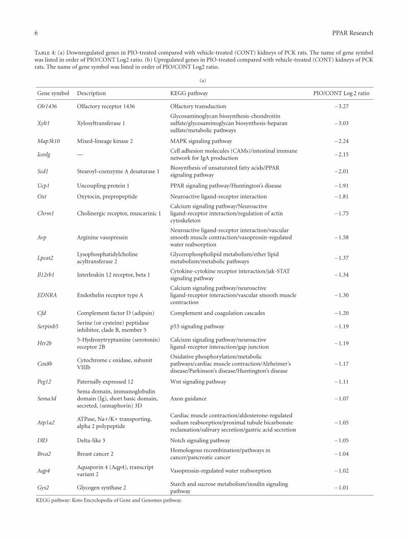
6 PPAR Research
Table 4: (a) Downregulated genes in PIO-treated compared with vehicle-treated (CONT) kidneys of PCK rats. The name of gene symbolwas listed in order of PIO/CONT Log2 ratio. (b) Upregulated genes in PIO-treated compared with vehicle-treated (CONT) kidneys of PCKrats. The name of gene symbol was listed in order of PIO/CONT Log2 ratio.
(a)
Gene symbol Description KEGG pathway PIO/CONT Log 2 ratio
Olr1436 Olfactory receptor 1436 Olfactory transduction −3.27
Xylt1 Xylosyltransferase 1Glycosaminoglycan biosynthesis-chondroitinsulfate/glycosaminoglycan biosynthesis-heparansulfate/metabolic pathways
−3.03
Map3k10 Mixed-lineage kinase 2 MAPK signaling pathway −2.24
Icoslg —Cell adhesion molecules (CAMs)/intestinal immunenetwork for IgA production
−2.15
Scd1 Stearoyl-coenzyme A desaturase 1Biosynthesis of unsaturated fatty acids/PPARsignaling pathway
−2.01
Ucp1 Uncoupling protein 1 PPAR signaling pathway/Huntington’s disease −1.91
Oxt Oxytocin, prepropeptide Neuroactive ligand-receptor interaction −1.81
Chrm1 Cholinergic receptor, muscarinic 1Calcium signaling pathway/Neuroactiveligand-receptor interaction/regulation of actincytoskeleton
−1.75
Avp Arginine vasopressinNeuroactive ligand-receptor interaction/vascularsmooth muscle contraction/vasopressin-regulatedwater reabsorption
−1.58
Lpcat2Lysophosphatidylcholineacyltransferase 2
Glycerophospholipid metabolism/ether lipidmetabolism/metabolic pathways
−1.37
Il12rb1 Interleukin 12 receptor, beta 1Cytokine-cytokine receptor interaction/jak-STATsignaling pathway
−1.34
EDNRA Endothelin receptor type ACalcium signaling pathway/neuroactiveligand-receptor interaction/vascular smooth musclecontraction
−1.30
Cfd Complement factor D (adipsin) Complement and coagulation cascades −1.20
Serpinb5Serine (or cysteine) peptidaseinhibitor, clade B, member 5
p53 signaling pathway −1.19
Htr2b5-Hydroxytryptamine (serotonin)receptor 2B
Calcium signaling pathway/neuroactiveligand-receptor interaction/gap junction
−1.19
Cox8bCytochrome c oxidase, subunitVIIIb
Oxidative phosphorylation/metabolicpathways/cardiac muscle contraction/Alzheimer’sdisease/Parkinson’s disease/Huntington’s disease
−1.17
Peg12 Paternally expressed 12 Wnt signaling pathway −1.11
Sema3dSema domain, immunoglobulindomain (Ig), short basic domain,secreted, (semaphorin) 3D
Axon guidance −1.07
Atp1a2ATPase, Na+/K+ transporting,alpha 2 polypeptide
Cardiac muscle contraction/aldosterone-regulatedsodium reabsorption/proximal tubule bicarbonatereclamation/salivary secretion/gastric acid secretion
−1.05
Dll3 Delta-like 3 Notch signaling pathway −1.05
Brca2 Breast cancer 2Homologous recombination/pathways incancer/pancreatic cancer
−1.04
Aqp4Aquaporin 4 (Aqp4), transcriptvariant 2
Vasopressin-regulated water reabsorption −1.02
Gys2 Glycogen synthase 2Starch and sucrose metabolism/insulin signalingpathway
−1.01
KEGG pathway: Koto Encyclopedia of Gene and Genomes pathway.
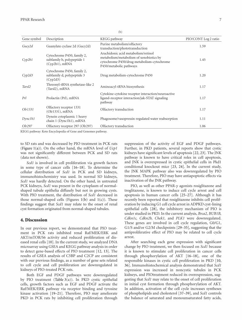
PPAR Research 7
(b)
Gene symbol Description KEGG pathway PIO/CONT Log 2 ratio
Gucy2d Guanylate cyclase 2d (Gucy2d)Purine metabolism/olfactorytransduction/phototransduction
1.59
Cyp2b1Cytochrome P450, family 2,subfamily b, polypeptide 1(Cyp2b1), mRNA
Arachidonic acid metabolism/retinolmetabolism/metabolism of xenobiotics bycytochrome P450/drug metabolism-cytochromeP450/metabolic pathways
1.45
Cyp2d3Cytochrome P450, family 2,subfamily d, polypeptide 3(Cyp2d3)
Drug metabolism-cytochrome P450 1.20
Tarsl2Threonyl-tRNA synthetase-like 2(Tarsl2), mRNA
Aminoacyl-tRNA biosynthesis 1.17
Prl Prolactin (Prl), mRNACytokine-cytokine receptor interaction/neuroactiveligand-receptor interaction/jak-STAT signalingpathway
1.17
Olr1331Olfactory receptor 1331(Olr1331), mRNA
Olfactory transduction 1.17
Dync1h1Dynein cytoplasmic 1 heavychain 1 (Dync1h1), mRNA
Phagosome/vasopressin-regulated water reabsorption 1.11
Olr297 Olfactory receptor 297 (Olr297) Olfactory transduction 1.06
KEGG pathway: Koto Encyclopedia of Gene and Genomes pathway.
to SD rats and was decreased by PIO treatment in PCK rats(Figure 1(a)). On the other hand, the mRNA level of Ucp1was not significantly different between PCK and SD rats(data not shown).
Scd1 is involved in cell proliferation via growth factorsin some type of cancer cells [16–18]. To determine thecellular distribution of Scd1 in PCK and SD kidneys,immunohistochemistry was used. In normal SD kidneys,Scd1 was hardly detected. On the other hand, in untreatedPCK kidneys, Scd1 was present in the cytoplasm of normal-shaped tubule epithelia diffusely but not in growing cysts.With PIO treatment, the distribution of Scd1 decreased inthose normal-shaped cells (Figures 1(b) and 1(c)). Thesefindings suggest that Scd1 may relate to the onset of renalcyst formation originated from normal-shaped tubules.
4. Discussion
In our previous report, we demonstrated that PIO treat-ment in PCK rats inhibited renal Raf/MEK/ERK andAKT/mTOR/S6 activity and reduced proliferation of dis-eased renal cells [10]. In the current study, we analyzed DNAmicroarray using GSEA and KEGG pathway analysis in orderto detect gene-based effects of PIO treatment [12, 13]. Theresults of GSEA analysis of C5BP and C2CP are consistentwith our previous findings, as a number of gene sets relatedto cell cycle and cell proliferation are downregulated inkidneys of PIO-treated PCK rats.
Both EGF and PDGF pathways were downregulatedby PIO treatment (Table 3(a)). In PKD cystic epithelialcells, growth factors such as EGF and PDGF activate theRaf/MEK/ERK pathway via receptor binding and tyrosinekinase activation [19–21]. Therefore, PIO may amelioratePKD in PCK rats by inhibiting cell proliferation through
suppression of the activity of EGF and PDGF pathways.Further, in PKD patients, several reports show that cystickidneys have significant levels of apoptosis [22, 23]. The JNKpathway is known to have critical roles in cell apoptosis,and JNK is overexpressed in cystic epithelial cells in Pkd1conditional knockout mice [23, 24]. In the current study,the JNK MAPK pathway also was downregulated by PIOtreatment. Therefore, PIO may have antiapoptotic effects viainactivation of the JNK pathway.
PIO, as well as other PPAR-γ agonists rosiglitazone andtroglitazone, is known to induce cell cycle arrest and cellapoptosis in human cancer cells [25–27]. Although it hasrecently been reported that rosiglitazone inhibits cell prolif-eration by inducing G1 cell cycle arrest in ADPKD cyst-liningepithelial cells [28], the inhibitory mechanism of PIO isunder studied in PKD. In the current analysis, Brca2, BUB1B,Cdkn1c, Cdkn2b, Chek1, and PLK1 were downregulated.These genes are involved in cell cycle regulation, G0/G1,G1/S and/or G2/M checkpoints [29–35], suggesting that theantiproliferative effect of PIO may be related to cell cyclearrest.
After searching each gene expression with significantchange by PIO treatment, we then focused on Scd1 becauseit is known to stimulate cell proliferation in cancer cellsthrough phosphorylation of AKT [16–18], one of theresponsible kinases in cystic cell proliferation in PKD [10,36]. Immunohistochemical analysis demonstrated that Scd1expression was increased in noncystic tubules in PCKkidneys, and PIOtreatment reduced its overexpression, sug-gesting that Scd1 may relate to the onset of cell proliferationin initial cyst formation through phosphorylation of AKT.In addition, activation of the cell cycle increases synthesesof phospholipids and cholesterol [37–39], and Scd1 controlsthe balance of saturated and monounsaturated fatty acids,
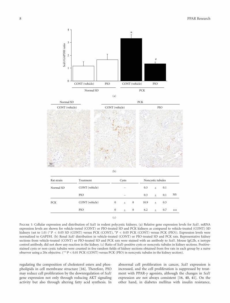
8 PPAR Research
Scd1
/GA
PD
H r
atio
PIO
#
CONT (vehicle)PIOCONT (vehicle)
PCKNormal SD
4
3
2
1
0
∗
(a)
PIOCONT (vehicle)
PCK Normal SD
CONT (vehicle)
50
(b)
Rat strain Treatment Cysts Noncystic tubules
Normal SD CONT (vehicle) 0.3 0.1
PIO 0.3 0.1 NS
PCK CONT (vehicle) 0 0 10.9 0.3
PIO 0 0 6.2 0.7 ∗∗
−
−
±
±
±
±±
±
(c)
Figure 1: Cellular expression and distribution of Scd1 in rodent polycystic kidneys. (a) Relative gene expression levels for Scd1. mRNAexpression levels are shown for vehicle-treted (CONT) or PIO-treated SD and PCK kidneys as compared to vehicle-treated (CONT) SDkidneys (set to 1.0) (∗P < 0.05 SD (CONT) versus PCK (CONT), #P < 0.05 PCK (CONT) versus PCK (PIO)). Expression levels werenormalized to GAPDH. (b) Renal Scd1 distribution in vehicle-treated (CONT) or PIO-treated SD and PCK rats. Representative kidneysections from vehicle-treated (CONT) or PIO-treated SD and PCK rats were stained with an antibody to Scd1. Mouse IgG2b, κ isotypecontrol antibody, did not show any reaction in the kidney. (c) Ratio of Scd1-positive cysts or noncystic tubules in kidney sections. Positive-stained cysts or non-cystic tubules were counted in five random fields of kidney sections obtained from five rats in each group by a naiveobserver using a 20x objective. (∗∗P < 0.01 PCK (CONT) versus PCK (PIO) in noncystic tubules in the kidney section).
regulating the composition of cholesterol esters and phos-pholipids in cell membrane structure [16]. Therefore, PIOmay reduce cell proliferation by the downregulation of Scd1gene expression not only through reducing AKT signalingactivity but also through altering fatty acid synthesis. In
abnormal cell proliferation in cancer, Scd1 expression isincreased, and the cell proliferation is suppressed by treat-ment with PPAR-γ agonists, although the changes in Scd1expression are not always consistent [16, 40, 41]. On theother hand, in diabetes mellitus with insulin resistance,
PPAR Research 9
adipose tissue or skeletal muscle Scd1 expression is decreasedand increased by PPAR-γ agonists [42–44]. Therefore, theexpression level of Scd1 and the effect of PPAR-γ ago-nists may depend on the disease and/or the state of cellproliferation.
Clinically, increased body weight, oedema, and urinarybladder tumors are concerned as possible side effects ofPPAR-γ agonists. Although those phenomena were notobserved in both genders of PCK rats in the current PIOtreatment, the effect of longer term treatment with differentdoses will need to be studied carefully. Since ameliorativeeffects are reported in several animal models of PKD [8–10,45], PPAR-γ agonists are thought to be a potential candidatefor therapeutic interventions in both ARPKD and ADPKDpatients.
5. Conclusions
In the current study, PIO reduced PKD progression andaltered the expression of renal genes involved in cell pro-liferation, cell cycle progression, and fatty acid metabolismin an orthologous rat model of human ARPKD. In additionto the previously demonstrated inhibition of Raf/MEK/ERKand AKT/mTOR/S6 signaling pathways by treatment of PCKrats with 10 mg/kg PIO for 16 weeks [10], suppression of cellproliferation may also be related to reductions in EGF, PDGF,and JNK pathways, cell cycle arrest related to Brca2, BUB1B,Cdkn1c, Cdkn2b, Chek1, and PLK1 genes, and alteration offatty acid metabolism related to Scd1.
Acknowledgments
This work was supported by Grant-in-Aid for scientificresearch (no. 22590351 to S. Nagao, and no. 23390033to T. Abe) and 21st Century COE Program (to FujitaHealth University) from the Ministry of Education, Culture,Sports, Science, and Technology of Japan and the CanadianInstitutes of Health Research (MOP230564 to H. Aukema).
References
[1] J. J. Grantham, S. Mulamalla, and K. I. Swenson-Fields, “Whykidneys fail in autosomal dominant polycystic kidney disease,”Nature Reviews Nephrology, vol. 7, no. 10, pp. 556–566, 2011.
[2] V. E. Torres, P. C. Harris, and Y. Pirson, “Autosomal dominantpolycystic kidney disease,” Lancet, vol. 369, no. 9569, pp. 1287–1301, 2007.
[3] P. C. Harris, “2008 Homer W. Smith award: insights intothe pathogenesis of polycystic kidney disease from genediscovery,” Journal of the American Society of Nephrology, vol.20, no. 6, pp. 1188–1198, 2009.
[4] W. E. Sweeney Jr and E. D. Avner, “Diagnosis and managementof childhood polycystic kidney disease,” Pediatric Nephrology,vol. 26, no. 5, pp. 675–692, 2011.
[5] B. Turkbey, I. Ocak, K. Daryanani et al., “Autosomal recessivepolycystic kidney disease and congenital hepatic fibrosis(ARPKD/CHF),” Pediatric Radiology, vol. 39, no. 2, pp. 100–111, 2009.
[6] A. Yessoufou and W. Wahli, “Multifaceted roles of peroxisomeproliferator-activated receptors (PPARs) at the cellular and
whole organism levels,” Swiss Medical Weekly, vol. 140, no.13071, pp. 4–10, 2010.
[7] C. Nofziger and B. L. Blazer-Yost, “PPARγ agonists, modula-tion of ion transporters, and fluid retention,” Journal of theAmerican Society of Nephrology, vol. 20, no. 12, pp. 2481–2483,2009.
[8] S. Muto, A. Aiba, Y. Saito et al., “Pioglitazone improves thephenotype and molecular defects of a targeted Pkd1 mutant,”Human Molecular Genetics, vol. 11, no. 15, pp. 1731–1742,2002.
[9] B. Dai, Y. Liu, C. Mei et al., “Rosiglitazone attenuates devel-opment of polycystic kidney disease and prolongs survival inHan:SPRD rats,” Clinical Science, vol. 119, no. 8, pp. 323–333,2010.
[10] D. Yoshihara, H. Kurahashi, M. Morita et al., “PPAR-γagonist ameliorates kidney and liver disease in an orthologousrat model of human autosomal recessive polycystic kidneydisease,” American Journal of Physiology, vol. 300, no. 2, pp.F465–F474, 2011.
[11] M. Kugita, K. Nishii, M. Morita et al., “Global gene expres-sion profiling in early-stage polycystic kidney disease inthe Han:SPRD Cy rat identifies a role for RXR signaling,”American Journal of Physiology, vol. 300, no. 1, pp. F177–F188,2011.
[12] A. Subramanian, P. Tamayo, V. K. Mootha et al., “Geneset enrichment analysis: a knowledge-based approach forinterpreting genome-wide expression profiles,” Proceedingsof the National Academy of Sciences of the United States ofAmerica, vol. 102, no. 43, pp. 15545–15550, 2005.
[13] H. Ogata, S. Goto, K. Sato, W. Fujibuchi, H. Bono, and M.Kanehisa, “KEGG: kyoto encyclopedia of genes and genomes,”Nucleic Acids Research, vol. 27, no. 1, pp. 29–34, 1999.
[14] W. Arozal, K. Watanabe, P. T. Veeraveedu et al., “Telmisartanprevents the progression of renal injury in daunorubicinrats with the alteration of angiotensin II and endothelin-1 receptor expression associated with its PPAR-γ agonistactions,” Toxicology, vol. 279, no. 1–3, pp. 91–99, 2011.
[15] M. Y. Chang and A. C. Ong, “Endothelin in polycystic kidneydisease,” Contributions to Nephrology, vol. 172, pp. 200–209,2011.
[16] R. A. Igal, “Stearoyl-coa desaturase-1: a novel key player in themechanisms of cell proliferation, programmed cell death andtransformation to cancer,” Carcinogenesis, vol. 31, no. 9, pp.1509–1515, 2010.
[17] D. Hess, J. W. Chisholm, and R. A. Igal, “Inhibition ofstearoylCoA desaturase activity blocks cell cycle progressionand induces programmed cell death in lung cancer cells,” PloSOne, vol. 5, no. 6, p. e11394, 2010.
[18] N. Scaglia, J. W. Chisholm, and R. A. Igal, “Inhibition ofstearoylCoA desaturase-1 inactivates acetyl-CoA carboxylaseand impairs proliferation in cancer cells: role of AMPK,” PLoSONE, vol. 4, no. 8, Article ID e6812, 2009.
[19] T. Yamaguchi, S. Nagao, D. P. Wallace et al., “Cyclic AMP acti-vates B-Raf and ERK in cyst epithelial cells from autosomal-dominant polycystic kidneys,” Kidney International, vol. 63,no. 6, pp. 1983–1994, 2003.
[20] T. Nakamura, I. Ebihara, I. Nagaoka et al., “Growth factor geneexpression in kidney of murine polycystic kidney disease,”Journal of the American Society of Nephrology, vol. 3, no. 7, pp.1378–1386, 1993.
[21] S. Nagao, T. Yamaguchi, M. Kusaka et al., “Renal activation ofextracellular signal-regulated kinase in rats with autosomal-dominant polycystic kidney disease,” Kidney International, vol.63, no. 2, pp. 427–437, 2003.
10 PPAR Research
[22] O. Ibraghimov-Beskrovnaya, “Targeting dysregulated cellcycle and apoptosis for polycystic kidney disease therapy,” CellCycle, vol. 6, no. 7, pp. 776–779, 2007.
[23] S. Nishio, M. Hatano, M. Nagata et al., “Pkd1 regulatesimmortalized proliferation of renal tubular epithelial cellsthrough p53 induction and JNK activation,” Journal of ClinicalInvestigation, vol. 115, no. 4, pp. 910–918, 2005.
[24] W. Yu, T. Kong, S. Beaudry et al., “Polycystin-1 protein leveldetermines activity of the Gα 12/JNK apoptosis pathway,”Journal of Biological Chemistry, vol. 285, no. 14, pp. 10243–10251, 2010.
[25] K. Yoshizawa, D. P. Cioca, S. Kawa, E. Tanaka, and K. Kiyosawa,“Peroxisome proliferator-activated receptor γ ligand troglita-zone induces cell cycle arrest and apoptosis of hepatocellularcarcinoma cell lines,” Cancer, vol. 95, no. 10, pp. 2243–2251,2002.
[26] M. Ming, J. P. Yu, X. Z. Meng, Y. H. Zhou, H. G. Yu, and H. S.Luo, “Effect of ligand troglitazone on peroxisome proliferator-activated receptor γ expression and cellular growth in humancolon cancer cells,” World Journal of Gastroenterology, vol. 12,no. 45, pp. 7263–7270, 2006.
[27] L. Cerquetti, C. Sampaoli, D. Amendola et al., “Rosiglitazoneinduces autophagy in H295R and cell cycle deregulation inSW13 adrenocortical cancer cells,” Experimental Cell Research,vol. 317, no. 10, pp. 1397–1410, 2011.
[28] Y. Liu, B. Dai, L. Fu, J. Jia, and C. Mei, “Rosiglitazoneinhibits cell proliferation by inducing g1 cell cycle arrest andapoptosis in ADPKD cyst-lining epithelia cells,” Basic andClinical Pharmacology and Toxicology, vol. 106, no. 6, pp. 523–530, 2010.
[29] E. Sagulenko, L. Savelyeva, V. Ehemann et al., “Suppression ofpolyploidy by the BRCA2 protein,” Cancer Letters, vol. 257, no.1, pp. 65–72, 2007.
[30] J. W. Davenport, E. R. Fernandes, L. D. Harris, G. A. M. Neale,and R. Goorha, “The mouse mitotic checkpoint gene Bub1b,a novel Bub1 family member, is expressed in a cell cycle-dependent manner,” Genomics, vol. 55, no. 1, pp. 113–117,1999.
[31] E. Myslinski, M. A. Gerard, A. Krol, and P. Carbon, “Tran-scription of the human cell cycle regulated BUB1B generequires hStaf/ZNF143,” Nucleic Acids Research, vol. 35, no. 10,pp. 3453–3464, 2007.
[32] S. Sengupta, J. Nie, R. J. Wagner, C. Yang, R. Stewart, and J. A.Thomson, “MicroRNA 92b controls the G1/S checkpoint genep57 in human embryonic stem cells,” Stem Cells, vol. 27, no. 7,pp. 1524–1528, 2009.
[33] Z. Ullah, C. de Renty, and M. L. DePamphilis, “Checkpointkinase 1 prevents cell cycle exit linked to terminal celldifferentiation,” Molecular and Cellular Biology, vol. 31, no. 19,pp. 4129–4143, 2011.
[34] D. T. Wu, M. Bitzer, W. Ju, P. Mundel, and E. P. Bottinger,“TGF-β concentration specifies differential signaling profilesof growth arrest/differentiation and apoptosis in podocytes,”Journal of the American Society of Nephrology, vol. 16, no. 11,pp. 3211–3221, 2005.
[35] Y. Degenhardt and T. Lampkin, “Targeting polo-like kinase incancer therapy,” Clinical Cancer Research, vol. 16, no. 2, pp.384–389, 2010.
[36] P. R. Wahl, M. L. Hir, A. Vogetseder et al., “Mitotic activationof Akt signalling pathway in Han:SPRD rats with polycystickidney disease,” Nephrology, vol. 12, no. 4, pp. 357–363, 2007.
[37] S. Jackowski, “Cell cycle regulation of membrane phospho-lipid metabolism,” Journal of Biological Chemistry, vol. 271, no.34, pp. 20219–20222, 1996.
[38] M. D. Siperstein, “Role of cholesterogenesis and isoprenoidsynthesis in DNA replication and cell growth,” Journal of LipidResearch, vol. 25, no. 13, pp. 1462–1468, 1984.
[39] L. S. Golfman, M. Bakovic, and D. E. Vance, “Transcriptionof the CTP:Phosphocholine Cytidylyltransferase α Gene IsEnhanced during the S Phase of the Cell Cycle,” Journal ofBiological Chemistry, vol. 276, no. 47, pp. 43688–43692, 2001.
[40] H. Koga, K. Selvendiran, R. Sivakumar et al., “PPARγ poten-tiates anticancer effects of gemcitabine on human pancreaticcancer cells,” International Journal of Oncology, vol. 40, no. 3,pp. 679–685, 2012.
[41] S. Takano, T. Kubota, H. Nishibori et al., “Pioglitazone, aligand for peroxisome proliferator-activated receptor-γ actsas an inhibitor of colon cancer liver metastasis,” AnticancerResearch, vol. 28, no. 6, pp. 3593–3599, 2008.
[42] A. Yao-Borengasser, N. Rassouli, V. Varma et al., “Stearoyl-coenzyme A desaturase 1 gene expression increases afterpioglitazone treatment and is associated with peroxisomalproliferator-activated receptor-γ responsiveness,” Journal ofClinical Endocrinology and Metabolism, vol. 93, no. 11, pp.4431–4439, 2008.
[43] K. Mai, J. Andres, T. Bobbert et al., “Rosiglitazone increasesfatty acid Δ9-desaturation and decreases elongase activityindex in human skeletal muscle in vivo,” Metabolism, vol. 61,no. 1, pp. 108–116, 2012.
[44] J. Song, H. Liu, H. W. Ressom, S. Tiwari, and C. M. Ecel-barger, “Chronic rosiglitazone therapy normalizes expressionof ACE1, SCD1 and other genes in the kidney of obese zuckerrats as determined by microarray analysis,” Experimental andClinical Endocrinology and Diabetes, vol. 116, no. 6, pp. 315–325, 2008.
[45] B. L. Blazer-Yost, J. Haydon, T. Eggleston-Gulyas et al.,“Pioglitazone attenuates cystic burden in the PCK rodentmodel of polycystic kidney disease,” PPAR Research, vol. 2010,Article ID 274376, 8 pages, 2010.

Hindawi Publishing CorporationPPAR ResearchVolume 2012, Article ID 371691, 7 pagesdoi:10.1155/2012/371691
Research Article
Fatty Acid Accumulation and Resulting PPARα Activation inFibroblasts due to Trifunctional Protein Deficiency
Masato Wakabayashi,1, 2 Yuji Kamijo,1, 3 Takero Nakajima,1 Naoki Tanaka,1
Eiko Sugiyama,1, 4 Tian Yangyang,1 Takefumi Kimura,1, 5 and Toshifumi Aoyama1
1 Department of Metabolic Regulation, Institute on Aging and Adaptation, Shinshu University Graduate School of Medicine,3-1-1 Asahi, Matsumoto 390-8621, Japan
2 Department of Pharmacy, Nagano Red Cross Hospital, Nagano 380-8582, Japan3 Department of Nephrology, Shinshu University School of Medicine, 3-1-1 Asahi, Matsumoto 390-8621, Japan4 Department of Nutritional Science, Nagano Prefectural College, Nagano 380-8525, Japan5 Department of Gastroenterology, Shinshu University School of Medicine, 3-1-1 Asahi, Matsumoto 390-8621, Japan
Correspondence should be addressed to Yuji Kamijo, [email protected] and Takero Nakajima, [email protected]
Received 31 January 2012; Accepted 24 February 2012
Academic Editor: Christopher J. Nicol
Copyright © 2012 Masato Wakabayashi et al. This is an open access article distributed under the Creative Commons AttributionLicense, which permits unrestricted use, distribution, and reproduction in any medium, provided the original work is properlycited.
To examine fatty acid accumulation and its toxic effects in cells, we analyzed skin fibroblasts from six patients with mitochondrialtrifunctional protein deficiency, who had abnormalities in the second through fourth reactions in fatty acid β-oxidation system. Wefound free fatty acid accumulation, enhanced three acyl-CoA dehydrogenases, catalyzing the first reaction in the β-oxidation sys-tem and being assumed to have normal activities in these patients, and PPARα activation that was confirmed in the experimentsusing MK886, a PPARα specific antagonist and fenofibrate, a PPARα specific agonist. These novel findings suggest that the fatty acidaccumulation and the resulting PPARα activation are major causes of the increase in the β-oxidation ability as probable compen-sation for fatty acid metabolism in the patients’ fibroblasts, and that enhanced cell proliferation and increased oxidative stressdue to the PPARα activation relate to the development of specific clinical features such as hypertrophic cardiomyopathy, slighthepatomegaly, and skeletal myopathy. Additionally, significant suppression of the PPARα activation by means of MK886 treatmentis assumed to provide a new method of treating this deficiency.
1. Introduction
The presence of an excessive level of fatty acids may causesignificant toxicity in many organs and tissues. We recentlyexamined the close relation between fatty acid toxicity andperoxisome proliferator-activated receptor (PPAR) func-tions. In some of our experiments, acute kidney injury wasinduced by albumin-overload nephropathy, in which PPARαprotected proximal tubular cells from acute toxicity inducedby fatty acids bound to albumin [1]; furthermore, pretreat-ment with low-dose fibrates protected against the fatty-acid-induced renal tubule toxicity by counteracting PPARαdeterioration [2]. In our other experiments, hepatic steatosisand hepatocellular carcinoma in hepatitis C virus core pro-tein transgenic mice were caused through fatty-acid-induced
PPARα activation [3, 4]. These in vivo experiments providedimportant results concerning fatty acid toxicity at the organand tissue levels; however, the degree of the toxicity differedgreatly, even among the same types of cells. We, therefore,undertook several experiments using cultured cells to eluci-date the detailed mechanisms in the cell toxicity. We adoptedfibroblasts from patients having a certain abnormality in themitochondrial fatty acid β-oxidation system.
Trifunctional protein (TP), catalyzing fatty acid β-oxida-tion in mitochondria, is a multienzyme complex com-posed of four molecules of the α-subunit containing theenoyl-CoA hydratase and 3-hydroxyacyl-CoA dehydrogenasedomains and four molecules of the β-subunit contain-ing the 3-ketoacyl-CoA thiolase domain. An inborn errorof this enzyme complex can cause sudden infant death
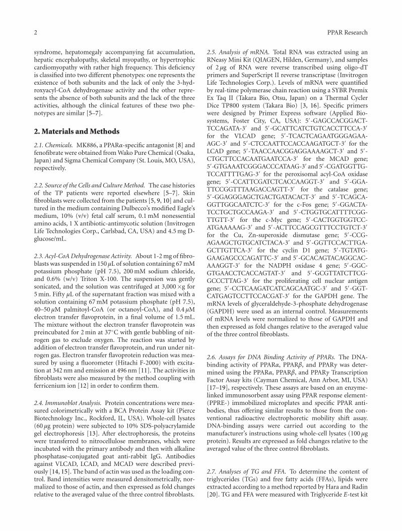
2 PPAR Research
syndrome, hepatomegaly accompanying fat accumulation,hepatic encephalopathy, skeletal myopathy, or hypertrophiccardiomyopathy with rather high frequency. This deficiencyis classified into two different phenotypes: one represents theexistence of both subunits and the lack of only the 3-hyd-roxyacyl-CoA dehydrogenase activity and the other repre-sents the absence of both subunits and the lack of the threeactivities, although the clinical features of these two phe-notypes are similar [5–7].
2. Materials and Methods
2.1. Chemicals. MK886, a PPARα-specific antagonist [8] andfenofibrate were obtained from Wako Pure Chemical (Osaka,Japan) and Sigma Chemical Company (St. Louis, MO, USA),respectively.
2.2. Source of the Cells and Culture Method. The case historiesof the TP patients were reported elsewhere [5–7]. Skinfibroblasts were collected from the patients [5, 9, 10] and cul-tured in the medium containing Dulbecco’s modified Eagle’smedium, 10% (v/v) fetal calf serum, 0.1 mM nonessentialamino acids, 1 X antibiotic-antimycotic solution (InvitrogenLife Technologies Corp., Carlsbad, CA, USA) and 4.5 mg D-glucose/mL.
2.3. Acyl-CoA Dehydrogenase Activity. About 1-2 mg of fibro-blasts was suspended in 150 μL of solution containing 67 mMpotassium phosphate (pH 7.5), 200 mM sodium chloride,and 0.6% (w/v) Triton X-100. The suspension was gentlysonicated, and the solution was centrifuged at 3,000×g for5 min. Fifty μL of the supernatant fraction was mixed with asolution containing 67 mM potassium phosphate (pH 7.5),40–50 μM palmitoyl-CoA (or octanoyl-CoA), and 0.4 μMelectron transfer flavoprotein, in a final volume of 1.5 mL.The mixture without the electron transfer flavoprotein waspreincubated for 2 min at 37◦C with gentle bubbling of nit-rogen gas to exclude oxygen. The reaction was started byaddition of electron transfer flavoprotein, and run under nit-rogen gas. Electron transfer flavoprotein reduction was mea-sured by using a fluorometer (Hitachi F-2000) with excita-tion at 342 nm and emission at 496 nm [11]. The activities infibroblasts were also measured by the method coupling withferricenium ion [12] in order to confirm them.
2.4. Immunoblot Analysis. Protein concentrations were mea-sured colorimetrically with a BCA Protein Assay kit (PierceBiotechnology Inc., Rockford, IL, USA). Whole-cell lysates(60 μg protein) were subjected to 10% SDS-polyacrylamidegel electrophoresis [13]. After electrophoresis, the proteinswere transferred to nitrocellulose membranes, which wereincubated with the primary antibody and then with alkalinephosphatase-conjugated goat anti-rabbit IgG. Antibodiesagainst VLCAD, LCAD, and MCAD were described previ-ously [14, 15]. The band of actin was used as the loading con-trol. Band intensities were measured densitometrically, nor-malized to those of actin, and then expressed as fold changesrelative to the averaged value of the three control fibroblasts.
2.5. Analysis of mRNA. Total RNA was extracted using anRNeasy Mini Kit (QIAGEN, Hilden, Germany), and samplesof 2 μg of RNA were reverse transcribed using oligo-dTprimers and SuperScript II reverse transcriptase (InvitrogenLife Technologies Corp.). Levels of mRNA were quantifiedby real-time polymerase chain reaction using a SYBR PremixEx Taq II (Takara Bio, Otsu, Japan) on a Thermal CyclerDice TP800 system (Takara Bio) [3, 16]. Specific primerswere designed by Primer Express software (Applied Bio-systems, Foster City, CA, USA): 5′-GAGCCACGGACT-TCCAGATA-3′ and 5′-GCATTCATCTGTCACCTTCCA-3′
for the VLCAD gene; 5′-TCACTCAGAATGGGAGAA-AGC-3′ and 5′-CTCCAATTCCACCAAGATGCT-3′ for theLCAD gene; 5′-TAACCAACGGAGGAAAAGCT-3′ and 5′-CTGCTTCCACAATGAATCCA-3′ for the MCAD gene;5′-GTGAAATCGGGACCCATAAG-3′ and 5′-CGATGGTTG-TCCATTTTGAG-3′ for the peroxisomal acyl-CoA oxidasegene; 5′-CCATTCGATCTCACCAAGGT-3′ and 5′-GGA-TTCCGGTTTAAGACCAGTT-3′ for the catalase gene;5′-GGAGGGAGCTGACTGATACACT-3′ and 5′-TCAGCA-GGTTGGCAATCTC-3′ for the c-Fos gene; 5′-GGACTA-TCCTGCTGCCAAGA-3′ and 5′-CTGGTGCATTTTCGG-TTGTT-3′ for the c-Myc gene; 5′-CACTGGTGGTCC-ATGAAAAAG-3′ and 5′-ACTTCCAGCGTTTCCTGTCT-3′
for the Cu, Zn-superoxide dismutase gene; 5′-CCG-AGAAGCTGTGCATCTACA-3′ and 5′-GGTTCCACTTGA-GCTTGTTCA-3′ for the cyclin D1 gene; 5′-TGTATG-GAAGAGCCCAGATTC-3′ and 5′-GCACAGTACAGGCAC-AAAGGT-3′ for the NADPH oxidase 4 gene; 5′-GGC-GTGAACCTCACCAGTAT-3′ and 5′-GCGTTATCTTCG-GCCCTTAG-3′ for the proliferating cell nuclear antigengene; 5′-CCTCAAGATCATCAGCAATGC-3′ and 5′-GGT-CATGAGTCCTTCCACGAT-3′ for the GAPDH gene. ThemRNA levels of glyceraldehyde-3-phosphate dehydrogenase(GAPDH) were used as an internal control. Measurementsof mRNA levels were normalized to those of GAPDH andthen expressed as fold changes relative to the averaged valueof the three control fibroblasts.
2.6. Assays for DNA Binding Activity of PPARs. The DNA-binding activity of PPARα, PPARβ, and PPARγ was deter-mined using the PPARα, PPARβ, and PPARγ TranscriptionFactor Assay kits (Cayman Chemical, Ann Arbor, MI, USA)[17–19], respectively. These assays are based on an enzyme-linked immunosorbent assay using PPAR response element-(PPRE-) immobilized microplates and specific PPAR anti-bodies, thus offering similar results to those from the con-ventional radioactive electrophoretic mobility shift assay.DNA-binding assays were carried out according to themanufacturer’s instructions using whole-cell lysates (100 μgprotein). Results are expressed as fold changes relative to theaveraged value of the three control fibroblasts.
2.7. Analyses of TG and FFA. To determine the content oftriglycerides (TGs) and free fatty acids (FFAs), lipids wereextracted according to a method reported by Hara and Radin[20]. TG and FFA were measured with Triglyceride E-test kit
PPAR Research 3
C16
C8
0
10
20
P1 P2 P3 P4 P5 P6 C1 C2 C3 P C
Acy
l-C
oA d
ehyd
roge
nas
eac
tivi
ty (
mU
/mg
prot
ein
)
∗
∗
(a)
0
50
100
0
50
100
P1 P2 P3 P4 P5
150
P6 C1 C2 C3 P C
FFA
TG
FFA
(n
Eq/
g ce
ll)
TG
(μg
/g c
ell)∗
(b)
Figure 1: Acyl-CoA dehydrogenase activity and FFA/TG contents in fibroblasts. Assay methods were, respectively, described in Section 2. (a)Indicates palmitoyl-CoA (open bar, C16) and octanoyl-CoA (closed bar, C8) dehydrogenase activities, respectively. (b) Indicates FFA (openbar) and TG (closed bar) contents, respectively. P1–P6, individual patient’s fibroblast; C1–C3, individual control fibroblast; P, means ± SDin six patients’ fibroblasts; C, means ± SD in three control fibroblasts. ∗P < 0.05 versus controls.
and an NEFAC-test kit (Wako Pure Chemical, Osaka, Japan),respectively.
2.8. Statistical Analysis. All data are expressed as mean ±standard deviation (SD). Statistical analysis was performedusing one-way ANOVA with Bonferroni correction (SPSSStatistics 17.0; SPSS Inc, Chicago, IL, USA). Correlation coef-ficients were calculated using Spearman’s rank correlationanalysis. A probability value of less than 0.05 was consideredto be statistically significant.
3. Results
3.1. Acyl-CoA Dehydrogenase Activity and the Content ofTG/FFA in Fibroblasts. Six strains of skin fibroblasts wereprepared from the individual TP patients, as well as the threestrains from the healthy adult men as described in Section 2.The reproductive rate of these fibroblasts was similar inall strains. TP deficiency is based on abnormalities in thesecond through fourth reactions in the mitochondrial fattyacid β-oxidation system; therefore, acyl-CoA dehydrogenase,catalyzing the first reaction in the β-oxidation system, wasassumed to be normal in the patients’ fibroblasts. Addition-ally, increased levels of FFA/TG due to the impaired β-oxi-dation ability in these fibroblasts were expected. Thus, theseparameters were analyzed first. Palmitoyl-CoA and octanoyl-CoA dehydrogenase activities in the patients’ fibroblasts were1.72- and 1.64-fold higher than those in the control fibro-blasts, respectively, (Figure 1(a)). FFA content in the patients’fibroblasts was 3.2-fold higher than that in the controlfibroblasts, while TG levels were similar in both fibroblasts.These FFA and TG levels were much lower than those inhuman serum (Figure 1(b)).
3.2. Expression of Three Acyl-CoA Dehydrogenases. Palmi-toyl-CoA and octanoyl-CoA dehydrogenation are catalyzed
by three forms of acyl-CoA dehydrogenase; therefore, theirexpression levels were examined. The protein levels ofVLCAD, LCAD, and MCAD in the patients’ fibroblasts were1.55-, 2.15-, and 1.97-fold higher than those in the controlfibroblasts, respectively, (Figure 2(a)). The mRNA contentsof VLCAD, LCAD, and MCAD in the patients’ fibroblastswere 2.00-, 2.92-, and 2.63-fold higher than those in thecontrol fibroblasts, respectively, (Figure 2(b)). These datawere consistent with the observations shown in Figure 1(a).The simultaneous increases in the expression levels of thethree forms of acyl-CoA dehydrogenase strongly suggestedthe presence of PPARα activation in the patients’ fibroblasts,since the three forms are known as PPARα target geneproducts [15]. The presence of PPARα activation was therebyexamined in detail.
3.3. Assays for DNA-Binding Activity of PPARs. Immunoblotanalysis using whole-cell lysates from the fibroblasts andspecific antibodies was performed and provided very faintbands for PPARβ and no bands for PPARα and PPARγ.mRNA analysis was also done as described in Section 2, andindicated that the PPARα, β, and γ mRNAs were 10−6∼10−4
levels for GAPDH mRNA in the fibroblasts, meaning thatthe data from the immunoblot and mRNA analyses wereunreliable for detecting PPAR activation. The PPRE-bindingassay was then done, which demonstrated an increase ofPPRE-binding activity only for PPARα in the whole-celllysates from the patients’ fibroblasts (Figure 3). These datasupported the presence of PPARα activation in the patients’fibroblasts.
3.4. Treatments with MK886 and Fenofibrate. To confirm theappearance of PPARα activation in the patients’ fibroblasts,the fibroblasts were treated with MK886, a PPARα-specificantagonist and fenofibrate, a PPARα specific agonist, respec-tively. The expression level of MCAD, a representative PPARα

4 PPAR Research
0
2
1
P1 P2 P3 P4 P5 P6 C1 C2 C3
VLCAD
LCAD
MCAD
Actin
VLCAD
LCAD
MCAD
P1 P2 P3 P4 P5 P6 C1 C2 C3 P C
Rel
ativ
e in
ten
sity
∗∗
∗
(a)
0
4
2
VLCAD
LCAD
MCAD
P1 P2 P3 P4 P5 P6 C1 C2 C3 P C
Rel
ativ
e m
RN
A e
xpre
ssio
n
∗∗
∗
(b)
Figure 2: Expression levels of three Species of acyl-CoA dehydrogenase. Assay methods were, respectively, described in Section 2. (a) Showsrelative quantification of expression levels of three acyl-CoA dehydrogenases. Upper panel indicates protein bands in immunoblot analysis.The band of actin was used as the loading control. Lower panel indicates relative protein amounts obtained by immunoblot anddensitometric analyses. (b) Shows relative mRNA expression. Open bar, VLCAD; gray bar, LCAD; closed bar, MCAD. P1–P6, individualpatient’s fibroblast; C1–C3, individual control fibroblast; P, means± SD in six patients’ fibroblasts; C, means± SD in three control fibroblasts.∗P < 0.05, versus controls.
0
2
1
P1 P2 P3 P4 P5 P6 C1 C2 C3 P CRel
ativ
e le
vel o
f P
PR
E-b
indi
ng
acti
vity
PPAR
PPAR
PPAR
∗
Figure 3: PPRE-binding activity. Assay methods were described inSection 2. Open bar, PPARα; gray bar, PPARβ; closed bar, PPARγ.P1–P6, individual patient’s fibroblast; C1–C3, individual controlfibroblast; P, means± SD in six patients’ fibroblasts; C, means± SDin three control fibroblasts. ∗P < 0.05, versus controls.
target gene product, was investigated. In the patients’ fibro-blasts, the MK886 treatment evidently reduced MCADexpression both in the protein and mRNA levels, and thefenofibrate treatment left this expression unchanged. Inthe control fibroblasts, the MK886 treatment did not affectthis expression, and the fenofibrate treatment increased itboth in the protein and mRNA levels (Figure 4). Thesedata demonstrated that a considerable level of PPARα acti-vation constitutively functioned in the patients’ fibroblasts.
4. Discussion
This study demonstrated the occurrence of FFA accumula-tion, increased palmitoyl-CoA and octanoyl-CoA dehydro-genase activities, coordinated enhancement in the expressionof three acyl-CoA dehydrogenases, a significant increase ofPPRE-binding activity only for PPARα, and reduced MCADexpression as a result of PPARα-specific antagonist treatmentin all of the fibroblasts from six patients with TP deficiencywho had abnormalities in the second through fourth reac-tions in the mitochondrial fatty acid β-oxidation system.These results demonstrated that a considerable level ofPPARα activation constitutively functioned in the patients’fibroblasts, in which FFA seemed to act as endogenous lig-ands toward PPARα as reported elsewhere [21–23]. FFAseems to work not toxically but protectively in the patients’fibroblasts, since the FFA accumulation and the resultingPPARα activation probably compensated for the impairedfatty acid metabolism in the patients’ fibroblasts. It would beinteresting to investigate whether this compensation appearsin the patients’ liver and heart, where considerable increasesof TG/FFA and much higher levels of PPARα expression areexpected [3, 24, 25]. From this viewpoint, the results ob-tained by using the patients’ fibroblasts in the current studyare useful for understanding the PPARα function.
This PPARα activation might induce cell proliferation inthe patients’ fibroblasts. To examine it, the mRNA levelswith several oncogene products and cell cycle regulators wereanalyzed. The mRNA levels in the patients’ fibroblasts were2.5±0.5-fold for c-Fos, 3.0±0.6-fold for c-Myc, 2.4±0.7-foldfor cyclin D1, and 2.1± 0.3-fold for proliferating cell nuclear
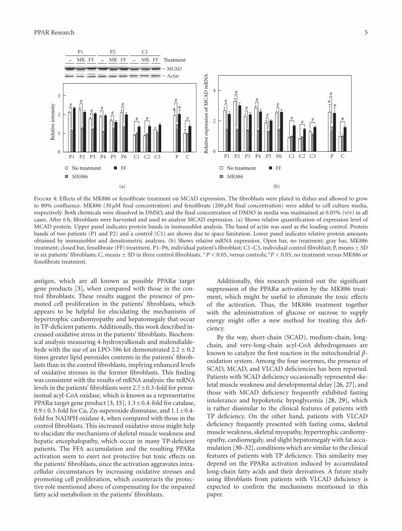
PPAR Research 5
0
3
2
P1 P2 P3 P4 P5 P6 C1 C2 C3 P C
##
# ##
#
# # #
#
1
MCAD Actin
– FFMK
P1
– FFMK
P2
– FFMK
C1
∗∗
∗
MK886
FFNo treatment
Treatment
Rel
ativ
e in
ten
sity
(a)
0
4
2
MK886
FF
P1 P2 P3 P4 P5 P6 C1 C2 C3 P C
#
#
#
#
#
#
## #
#
∗
∗∗
#
Rel
ativ
e ex
pres
sion
of
MC
AD
mR
NA
No treatment
(b)
Figure 4: Effects of the MK886 or fenofibrate treatment on MCAD expression. The fibroblasts were plated in dishes and allowed to growto 80% confluence. MK886 (30 μM final concentration) and fenofibrate (200 μM final concentration) were added to cell culture media,respectively. Both chemicals were dissolved in DMSO, and the final concentration of DMSO in media was maintained at 0.05% (v/v) in allcases. After 6 h, fibroblasts were harvested and used to analyze MCAD expression. (a) Shows relative quantification of expression level ofMCAD protein. Upper panel indicates protein bands in immunoblot analysis. The band of actin was used as the loading control. Proteinbands of two patients (P1 and P2) and a control (C1) are shown due to space limitation. Lower panel indicates relative protein amountsobtained by immunoblot and densitometric analyses. (b) Shows relative mRNA expression. Open bar, no treatment; gray bar, MK886treatment; closed bar, fenofibrate (FF) treatment. P1–P6, individual patient’s fibroblast; C1–C3, individual control fibroblast; P, means± SDin six patients’ fibroblasts; C, means ± SD in three control fibroblasts. ∗P < 0.05, versus controls; #P < 0.05, no treatment versus MK886 orfenofibrate treatment.
antigen, which are all known as possible PPARα targetgene products [3], when compared with those in the con-trol fibroblasts. These results suggest the presence of pro-moted cell proliferation in the patients’ fibroblasts, whichappears to be helpful for elucidating the mechanisms ofhypertrophic cardiomyopathy and hepatomegaly that occurin TP-deficient patients. Additionally, this work described in-creased oxidative stress in the patients’ fibroblasts. Biochem-ical analysis measuring 4-hydroxyalkenals and malondialde-hyde with the use of an LPO-586 kit demonstrated 2.2 ± 0.2times greater lipid peroxides contents in the patients’ fibrob-lasts than in the control fibroblasts, implying enhanced levelsof oxidative stresses in the former fibroblasts. This findingwas consistent with the results of mRNA analysis: the mRNAlevels in the patients’ fibroblasts were 2.7±0.3-fold for perox-isomal acyl-CoA oxidase, which is known as a representativePPARα target gene product [3, 15], 1.3±0.4-fold for catalase,0.9±0.3-fold for Cu, Zn-superoxide dismutase, and 1.1±0.4-fold for NADPH oxidase 4, when compared with those in thecontrol fibroblasts. This increased oxidative stress might helpto elucidate the mechanisms of skeletal muscle weakness andhepatic encephalopathy, which occur in many TP-deficientpatients. The FFA accumulation and the resulting PPARαactivation seem to exert not protective but toxic effects onthe patients’ fibroblasts, since the activation aggravates intra-cellular circumstances by increasing oxidative stresses andpromoting cell proliferation, which counteracts the protec-tive role mentioned above of compensating for the impairedfatty acid metabolism in the patients’ fibroblasts.
Additionally, this research pointed out the significantsuppression of the PPARα activation by the MK886 treat-ment, which might be useful to eliminate the toxic effectsof the activation. Thus, the MK886 treatment togetherwith the administration of glucose or sucrose to supplyenergy might offer a new method for treating this defi-ciency.
By the way, short-chain (SCAD), medium-chain, long-chain, and very-long-chain acyl-CoA dehydrogenases areknown to catalyze the first reaction in the mitochondrial β-oxidation system. Among the four isozymes, the presence ofSCAD, MCAD, and VLCAD deficiencies has been reported.Patients with SCAD deficiency occasionally represented ske-letal muscle weakness and developmental delay [26, 27], andthose with MCAD deficiency frequently exhibited fastingintolerance and hypoketotic hypoglycemia [28, 29], whichis rather dissimilar to the clinical features of patients withTP deficiency. On the other hand, patients with VLCADdeficiency frequently presented with fasting coma, skeletalmuscle weakness, skeletal myopathy, hypertrophic cardiomy-opathy, cardiomegaly, and slight hepatomegaly with fat accu-mulation [30–32], conditions which are similar to the clinicalfeatures of patients with TP deficiency. This similarity maydepend on the PPARα activation induced by accumulatedlong-chain fatty acids and their derivatives. A future studyusing fibroblasts from patients with VLCAD deficiency isexpected to confirm the mechanisms mentioned in thispaper.

6 PPAR Research
Abbreviations
FFA: Free fatty acidsGAPDH: Glyceraldehyde-3-phosphate dehydrogenaseLCAD: Long-chain acyl-CoA dehydrogenaseMCAD: Medium-chain acyl-CoA dehydrogenasePPAR: Peroxisome proliferator-activated receptorPPRE: PPAR response elementTG: TriglyceridesTP: Trifunctional proteinVLCAD: Very-long-chain acyl-CoA dehydrogenase.
Conflict of Interests
The authors have declared that no conflict of interests exists.
Acknowledgments
The authors thank Trevor Ralph for his English editorialassistance. The authors have declared that no financial sup-port was provided.
References
[1] Y. Kamijo, K. Hora, K. Kono et al., “PPARα protects proximaltubular cells from acute fatty acid toxicity,” Journal of the Ame-rican Society of Nephrology, vol. 18, no. 12, pp. 3089–3100,2007.
[2] K. Takahashi, Y. Kamijo, K. Hora et al., “Pretreatment by low-dose fibrates protects against acute free fatty acid-inducedrenal tubule toxicity by counteracting PPARα deterioration,”Toxicology and Applied Pharmacology, vol. 252, no. 3, pp. 237–249, 2011.
[3] N. Tanaka, K. Moriya, K. Kiyosawa, K. Koike, F. J. Gonzalez,and T. Aoyama, “PPARα activation is essential for HCV coreprotein-induced hepatic steatosis and hepatocellular carci-noma in mice,” Journal of Clinical Investigation, vol. 118, no.2, pp. 683–694, 2008.
[4] N. Tanaka, K. Moriya, K. Kiyosawa, K. Koike, and T. Aoyama,“Hepatitis C virus core protein induces spontaneous and per-sistent activation of peroxisome proliferator-activated recep-tor α in transgenic mice: implications for HCV-associatedhepatocarcinogenesis,” International Journal of Cancer, vol.122, no. 1, pp. 124–131, 2008.
[5] K. E. Orii, T. Aoyama, K. Wakui et al., “Genomic and muta-tional analysis of the mitochondrial trifunctional protein β-subunit (HADHB) gene in patients with trifunctional proteindeficiency,” Human Molecular Genetics, vol. 6, no. 8, pp. 1215–1224, 1997.
[6] S. Jackson, R. S. Kler, K. Bartlett et al., “Combined enzymedefect of mitochondrial fatty acid oxidation,” Journal of Cli-nical Investigation, vol. 90, no. 4, pp. 1219–1225, 1992.
[7] R. J. A. Wanders, L. Ijlst I, F. Poggi et al., “Human trifunctionalprotein deficiency: a new disorder of mitochondrial fatty acidβ-oxidation,” Biochemical and Biophysical Research Communi-cations, vol. 188, no. 3, pp. 1139–1145, 1992.
[8] J. P. Kehrer, S. S. Biswal, E. La et al., “Inhibition of peroxisome-proliferator-activated receptor (PPAR)α by MK886,” Biochem-ical Journal, vol. 356, no. 3, pp. 899–906, 2001.
[9] S. Ushikubo, T. Aoyama, T. Kamijo et al., “Molecular charac-terization of mitochondrial trifunctional protein deficiency:formation of the enzyme complex is important for stabiliza-tion of both α- and β-subunits,” American Journal of HumanGenetics, vol. 58, no. 5, pp. 979–988, 1996.
[10] L. Ijlst I, R. J. A. Wanders, S. Ushikubo, T. Kamijo, and T.Hashimoto, “Molecular basis of long-chain 3-hydroxyacyl-CoA dehydrogenase deficiency: identification of the majordisease-causing mutation in the α-subunit of the mitochon-drial trifunctional protein,” Biochimica Et Biophysica Acta, vol.1215, no. 3, pp. 347–350, 1994.
[11] S. Furuta, S. Mayazawa, and T. Hashimoto, “Purification andproperties of rat liver acyl-CoA dehydrogenases and electrontransfer flavoprotein,” Journal of Biochemistry, vol. 90, no. 6,pp. 1739–1750, 1981.
[12] T. C. Lehman, D. E. Hale, A. Bhala, and C. Thorpe, “An acyl-coenzyme A dehydrogenase assay utilizing the ferriceniumion,” Analytical Biochemistry, vol. 186, no. 2, pp. 280–284,1990.
[13] T. Aoyama, S. Yamano, D. J. Waxman et al., “Cytochrome P-450 hPCN3, a novel cytochrome P-450 IIIA gene product thatis differentially expressed in adult human liver. cDNA anddeduced amino acid sequence and distinct specificities ofcDNA-expressed hpcn1 and hPCN3 for the metabolism of ste-roid hormones and cyclosporine,” Journal of Biological Chem-istry, vol. 264, no. 18, pp. 10388–10395, 1989.
[14] T. Aoyama, I. Ueno, T. Kamijo, and T. Hashimoto, “Rat very-long-chain acyl-CoA dehydrogenase, a novel mitochondrialacyl-CoA dehydrogenase gene product, is a rate-limiting en-zyme in long-chain fatty acid β-oxidation system. cDNA anddeduced amino acid sequence and distinct specificities of thecDNA-expressed protein,” Journal of Biological Chemistry, vol.269, no. 29, pp. 19088–19094, 1994.
[15] T. Aoyama, J. M. Peters, N. Iritani et al., “Altered constitutiveexpression of fatty acid-metabolizing enzymes in mice lackingthe peroxisome proliferator-activated receptor α (PPARα),”Journal of Biological Chemistry, vol. 273, no. 10, pp. 5678–5684, 1998.
[16] Y. Kamijo, K. Hora, N. Tanaka et al., “Identification of func-tions of peroxisome proliferator-activated receptor α in prox-imal tubules,” Journal of the American Society of Nephrology,vol. 13, no. 7, pp. 1691–1702, 2002.
[17] B. Desvergne and W. Wahli, “Peroxisome proliferator-activa-ted receptors: nuclear control of metabolism,” Endocrine Re-views, vol. 20, no. 5, pp. 649–688, 1999.
[18] E. Duplus, M. Glorian, and C. Forest, “Fatty acid regulation ofgene transcription,” Journal of Biological Chemistry, vol. 275,no. 40, pp. 30749–30752, 2000.
[19] P. Gervois, I. P. Torra, J. C. Fruchart, and B. Staels, “Regulationof lipid and lipoprotein metabolism by PPAR activators,” Cli-nical Chemistry and Laboratory Medicine, vol. 38, no. 1, pp. 3–11, 2000.
[20] A. Hara and N. S. Radin, “Lipid extraction of tissues with alow-toxicity solvent,” Analytical Biochemistry, vol. 90, no. 1,pp. 420–426, 1978.
[21] S. A. Kliewer, S. S. Sundseth, S. A. Jones et al., “Fatty acidsand eicosanoids regulate gene expression through direct inter-actions with peroxisome proliferator-activated receptors α andγ,” Proceedings of the National Academy of Sciences of the UnitedStates of America, vol. 94, no. 9, pp. 4318–4323, 1997.
[22] B. M. Forman, J. Chen, and R. M. Evans, “Hypolipidemicdrugs, polyunsaturated fatty acids, and eicosanoids are ligands
PPAR Research 7
for peroxisome proliferator-activated receptors α and δ,” Pro-ceedings of the National Academy of Sciences of the United Statesof America, vol. 94, no. 9, pp. 4312–4317, 1997.
[23] H. E. Xu, M. H. Lambert, V. G. Montana et al., “Molecularrecognition of fatty acids by peroxisome proliferator-activatedreceptors,” Molecular Cell, vol. 3, no. 3, pp. 397–403, 1999.
[24] M. Rakhshandehroo, B. Knoch, M. Muller, and S. Ker-sten, “Peroxisome proliferator-activated receptor alpha targetgenes,” PPAR Research, vol. 2010, Article ID 612089, 2010.
[25] O. Braissant, F. Foufelle, C. Scotto, M. Dauca, and W. Wahli,“Differential expression of peroxisome proliferator-activatedreceptors (PPARs): tissue distribution of PPAR-α, -β, and -γin the adult rat,” Endocrinology, vol. 137, no. 1, pp. 354–366,1996.
[26] P. M. Coates, D. E. Hale, G. Finocchiaro, K. Tanaka, and S.C. Winter, “Genetic deficiency of short-chain acyl-coenzymeA dehydrogenase in cultured fibroblasts from a patient withmuscle carnitine deficiency and severe skeletal muscle weak-ness,” Journal of Clinical Investigation, vol. 81, no. 1, pp. 171–175, 1988.
[27] E. Naito, Y. Indo, and K. Tanaka, “Short chain acyl-co-enzyme A dehydrogenase (SCAD) deficiency. immunochem-ical demonstration of molecular heterogeneity due to variantSCAD with differing stability,” Journal of Clinical Investigation,vol. 84, no. 5, pp. 1671–1674, 1989.
[28] P. Rinaldo, J. J. O’Shea, P. M. Coates, D. E. Hale, C. A. Stanley,and K. Tanaka, “Medium-chain acyl-CoA dehydrogenasedeficiency: diagnosis by stable-isotope dilution measurementof urinary n-hexanoylglycine and 3-phenylpropionylglycine,”New England Journal of Medicine, vol. 319, no. 20, pp. 1308–1313, 1988.
[29] P. M. Coates, Y. Indo, D. Young, D. E. Hale, and K. Tanaka,“Immunochemical characterization of variant medium-chainacyl-CoA dehydrogenase in fibroblasts from patients withmedium-chain acyl-CoA dehydrogenase deficiency,” PediatricResearch, vol. 31, no. 1, pp. 34–38, 1992.
[30] T. Aoyama, Y. Uchida, R. I. Kelley et al., “A novel diseasewith deficiency of mitochondrial very-long-chain acyl-CoAdehydrogenase,” Biochemical and Biophysical Research Com-munications, vol. 191, no. 3, pp. 1369–1372, 1993.
[31] T. Aoyama, M. Souri, S. Ushikubo et al., “Purification ofhuman very-long-chain acyl-coenzyme A dehydrogenase andcharacterization of its deficiency in seven patients,” Journal ofClinical Investigation, vol. 95, no. 6, pp. 2465–2473, 1995.
[32] T. Aoyama, M. Souri, I. Ueno et al., “Cloning of human very-long-chain acyl-coenzyme A dehydrogenase and molecularcharacterization of its deficiency in two patients,” AmericanJournal of Human Genetics, vol. 57, no. 2, pp. 273–283, 1995.
Hindawi Publishing CorporationPPAR ResearchVolume 2012, Article ID 174932, 10 pagesdoi:10.1155/2012/174932
Research Article
Hepatic Cerebroside Sulfotransferase Is Induced by PPARαActivation in Mice
Takefumi Kimura,1, 2 Takero Nakajima,1 Yuji Kamijo,1, 3 Naoki Tanaka,1, 4 Lixuan Wang,1
Atsushi Hara,1 Eiko Sugiyama,5 Eiji Tanaka,2 Frank J. Gonzalez,4 and Toshifumi Aoyama1
1 Department of Metabolic Regulation, Institute on Aging and Adaptation, Shinshu University Graduate School of Medicine,3-1-1 Asahi, Matsumoto 390-8621, Japan
2 Department of Gastroenterology, Shinshu University School of Medicine, Matsumoto 390-8621, Japan3 Department of Nephrology, Shinshu University School of Medicine, Matsumoto 390-8621, Japan4 Laboratory of Metabolism, National Cancer Institute, National Institutes of Health, Bethesda, MD 20892, USA5 Department of Nutritional Science, Nagano Prefectural College, Nagano 380-8525, Japan
Correspondence should be addressed to Yuji Kamijo, [email protected]
Received 4 January 2012; Accepted 16 February 2012
Academic Editor: Christopher J. Nicol
Copyright © 2012 Takefumi Kimura et al. This is an open access article distributed under the Creative Commons AttributionLicense, which permits unrestricted use, distribution, and reproduction in any medium, provided the original work is properlycited.
Sulfatides are one of the major sphingoglycolipids in mammalian serum and are synthesized and secreted mainly from the liver asa component of lipoproteins. Recent studies revealed a protective role for serum sulfatides against arteriosclerosis and hypercoagu-lation. Although peroxisome proliferator-activated receptor (PPAR) α has important functions in hepatic lipoprotein metabolism,its association with sulfatides has not been investigated. In this study, sulfatide levels and the expression of enzymes related to sul-fatide metabolism were examined using wild-type (+/+), Ppara-heterozygous (+/−), and Ppara-null (−/−) mice given a controldiet or one containing 0.1% fenofibrate, a clinically used hypolipidemic drug and PPARα activator. Fenofibrate treatment increasedserum and hepatic sulfatides in Ppara (+/+) and (+/−) mice through a marked induction of hepatic cerebroside sulfotransferase(CST), a key enzyme in sulfatide synthesis, in a PPARα-dependent manner. Furthermore, increases in CST mRNA levels were cor-related with mRNA elevations of several known PPARα target genes, and such changes were not observed for other sulfatide-meta-bolism enzymes in the liver. These results suggest that PPARα activation enhances hepatic sulfatide synthesis via CST induction andimplicate CST as a novel PPARα target gene.
1. Introduction
Sulfatides are sphingoglycolipids composed of sphingoid,fatty acid, galactose, and sulfate [1] that are distributed invarious tissues such as the central nervous system, kidney,liver, and gastrointestinal tract [1–4]. Glycolipids are alsopresent in the serum as one of the major components of lipo-proteins [1]. Several studies have revealed a protective rolefor serum sulfatides against arteriosclerosis and hypercoagu-lation [5]. Serum levels of sulfatides are markedly decreasedin humans with end-stage renal failure [6] but normalizeafter renal transplantation [7]. However, the precise mech-anism regulating serum sulfatide concentrations in humansremains unclear. Previously studies demonstrated that serum
sulfatide levels were strongly correlated with hepatic, but notrenal, sulfatide levels in mice with protein overload nephro-pathy, and that decreased serum sulfatide levels were alsoassociated with the downregulation of hepatic expression ofcerebroside sulfotransferase (CST), a key enzyme in sulfatidesynthesis [8]. These and previous findings suggest the pos-sible participation of hepatic peroxisome proliferator-acti-vated receptor (PPAR) in the regulation of serum and liversulfatide metabolisms. To examine this possibility, serum andliver sulfatide concentrations and hepatic expression of a ser-ies of sulfatide-metabolizing enzymes were analyzed usingPpara-homozygous (+/+), Ppara-heterozygous (+/−), andPpara-null (−/−) mice fed a control diet or one containingfenofibrate, a typical PPARα activator.

2 PPAR Research
2. Materials and Methods
2.1. Mice and Treatment. All animal experiments were con-ducted in accordance with animal study protocols approvedby the Shinshu University School of Medicine. Wild-type(+/+), Ppara (+/−), and Ppara (−/−) mice on a 129/Svgenetic background were generated as described previously[9–11]. These mice were maintained in a pathogen-free envi-ronment under controlled conditions (25◦C; 12 h light/darkcycle) with tap water ad libitum and a standard rodent diet.Twelve-week-old male wild-type (+/+), Ppara (+/−), andPpara (−/−) mice weighing 25–30 g were used for the fol-lowing experiments. Mice of each genotype were randomlydivided into two groups (n = 6 in each group of the samegenotype). One mouse group was treated with a regular dietcontaining 0.1% fenofibrate (Wako Pure Chemical Industr-ies, Osaka, Japan), and the other group was continued on aregular diet as a control. In an additional experiment, Ppara(+/+), Ppara (+/−), and Ppara (−/−) mice were randomlydivided into two groups (n = 6 in each group of the samegenotype) and were treated with a regular diet with or with-out 0.5% clofibrate (Wako Pure Chemical Industries). Sevendays after commencing treatment, the mice were sacrificedunder anesthesia for collection of blood and tissues.
2.2. Extraction and Measurement of Lipids. Total lipids in theserum and liver were extracted using the hexane/isopropanolmethod [12], and serum/liver sulfatides were determined asforms of lysosulfatides (LS; sulfatides without fatty acids) bymatrix-assisted laser desorption ionization-time of flightmass spectrometry (MALDI-TOF MS) as previously describ-ed [13]. Sulfatides levels were calculated as the sum of thelevels of seven LS molecular species: LS-sphingadienine (LS-d18 : 2), LS-(4E)-sphingenine (LS-d18 : 1), LS-sphinganine(LS-d18 : 0), LS-4D-hydroxysphinganine (LS-t18 : 0), LS-(4E)-icosasphingenine (LS-d20 : 1), LS-icosasphinganine(LS-d20 : 0), and LS-4D-hydroxyicosasphinganine (LS-t20 :0). Triglyceride (TG) levels in the serum and liver were mea-sured using a Triglyceride E-test kit (Wako Pure ChemicalIndustries).
2.3. Immunoblot Analysis. Liver nuclear and cytosolic frac-tions were prepared from each mouse using NE-PER Nuclearand Cytoplasmic Extraction Regents (Thermo Fisher Scien-tific, Rockford, IL, USA) [14], and their protein concentra-tions were determined with a BCA protein assay kit (ThermoFisher Scientific) [15]. Nuclear fractions (10 μg protein) wereused for immunoblot analysis of PPARs and TATA box-bind-ing protein (TBP). For detection of other proteins, cytosolicfractions (5 μg protein) were employed. Proteins were se-parated using sodium dodecyl sulfate-polyacrylamide gelelectrophoresis and transferred to nitrocellulose membranes.After blocking, the membranes were incubated with primaryantibodies followed by alkaline phosphatase-conjugated sec-ondary antibodies [16–18]. Primary antibodies against long-chain acyl-CoA synthase (LACS), liver fatty acid-bindingprotein (L-FABP), and medium-chain acyl-CoA dehydro-genase (MCAD) were prepared as described previously
[19–21]. Antibodies against other proteins were purchasedcommercially: cerebroside sulfotransferase (CST) fromAbnova (Taipei, Taiwan), arylsulfatase A (ARSA) from Ever-est Biotech (Oxfordshire, UK), TBP from Abcam (Cam-bridge, UK), and ceramide galactosyltransferase (CGT),galactosylceramidase (GALC), microsomal transfer protein(MTP), PPARα, PPARβ/δ, PPARγ, and actin from SantaCruz Biotechnology (Santa Cruz, CA, USA). TBP and actinwere used as loading controls for nuclear and cytosolic pro-tein extracts, respectively. Band intensities were measureddensitometrically, normalized to those of TBP or actin, andthen expressed as fold changes relative to those of Ppara(+/+) mice treated with a control diet.
2.4. Analysis of mRNA. Total liver RNA was extracted usingan RNeasy Mini Kit (QIAGEN, Hilden, Germany), and sam-ples of 2 μg of RNA were reverse-transcribed using oligo-dTprimers and SuperScript II reverse transcriptase (InvitrogenCorporation, Carlsbad, CA, USA). Levels of mRNA werequantified by real-time polymerase chain reaction (PCR)using an SYBR Premix Ex Taq II (Takara Bio, Otsu, Japan) ona Thermal Cycler Dice TP800 system (Takara Bio) [10, 16].Specific primers were designed by Primer Express software(Applied Biosystems, Foster City, CA, USA) as shown inTable 1. The mRNA levels of glyceraldehyde-3-phosphate de-hydrogenase (GAPDH) were used as an internal control.Measurements of mRNA levels were normalized to those ofGAPDH and then expressed as fold changes relative to thoseof Ppara (+/+) mice treated with a control diet.
2.5. Assays for DNA-Binding Activity of PPARs. The DNA-binding activity of nuclear PPARαPPARβ/δ, and PPARγ wasdetermined using PPARα, PPARβ/δ, and PPARγ Transcrip-tion Factor Assay kits (Cayman Chemical, Ann Arbor, MI,USA) [22–24], respectively. These assays are based on an en-zyme-linked immunosorbent assay using PPAR responseelement (PPRE) immobilized microplates and specific PPARantibodies, thus offering similar results to those from theconventional radioactive electrophoretic mobility shift assay.DNA-binding assays were carried out according to the manu-facturer’s instructions using nuclear extracts (50 μg protein)prepared as described previously. Results are expressed asfold changes relative to those of Ppara (+/+) mice treatedwith a control diet.
2.6. Statistical Analysis. All data are expressed as mean ±standard deviation (SD). Statistical analysis was performedusing one-way ANOVA with Bonferroni correction (SPSSStatistics 17.0; SPSS Inc, Chicago, IL, USA). Correlation coef-ficients were calculated using Spearman’s rank correlationanalysis. A probability value of less than 0.05 was consideredto be statistically significant.
3. Results
3.1. Fenofibrate Increased Serum/Liver Sulfatides in a PPARα-Dependent Manner. Fenofibrate treatment increased serum,and more notably liver, sulfatide concentrations in Ppara

PPAR Research 3
Table 1: Primer pairs used for the RT-PCR.
Gene GeneBank accession number Primer sequence
ARSA NM 009713 F 5′-ACCACCCCTAACCTGGATCAGT-3′
R 5′-ATGGCGTGCACAGAGACACA-3′
CGT NM 011674 F 5′-TGGGTCCAGCCTATGGATGT-3′
R 5′-GCAGCGTTGGTCTTGGAAAC-3′
CST NM 016922 F 5′-ATGGCCTTCACGACCTCAGA-3′
R 5′-CGGTCTTGTGCGTCTTCATG-3′
GALC NM 008079 F 5′-GAGTGAGAATCATAGCGAGCGATA-3′
R 5′-AGTTCCTGGTCCAGCAGCAA-3′
GAPDH M32599 F 5′-TGCACCACCAACTGCTTAG-3′
R 5′-GGATGCAGGGATGATGTTCTG-3′
LACS NM 007981 F 5′-TCCTACGGCAGTGATCTGGTG-3′
R 5′-GGTTGCCTGTAGTTCCACTTGTG-3′
L-FABP NM 017399 F 5′-GCAGAGCCAGGAGAACTTTGAG-3′
R 5′-TTTGATTTTCTTCCCTTCATGCA-3′
MCAD NM 007382 F 5′-TGCTTTTGATAGAACCAGACCTACAGT-3′
R 5′-CTTGGTGCTCCACTAGCAGCTT-3′
MTP NM 008642 F 5′-GAGCGGTCTGGATTTACAACG-3′
R 5′-GTAGGTAGTGACAGATGTGGCTTTTG-3′
PPARα NM 011144 F 5′-CCTCAGGGTACCACTACGGAGT-3′
R 5′-GCCGAATAGTTCGCCGAA-3′
PPARβ/δ XM 128500 F 5′-TCAACATGGAATGTCGGGTT-3′
R 5′-ATACTCGAGCTTCATGCGGATT-3′
PPARγ NM 011146 F 5-TTCCACTATGGAGTTCATGCTTGT-3′
R 5′-TCCGGCAGTTAAGATCACACCTA-3′
F: forward sequence; R: reverse sequence.
(+/+) and (+/−) mice only (Figure 1(a)). However, the in-creases in serum/liver sulfatides were not detected in Ppara(−/−) mice with fenofibrate treatment. These results demon-strate that fenofibrate increases serum/liver sulfatide levels ina PPARα-dependent manner. The treatment did not affectthe composition of sulfatides (Table 2). Fenofibrate also de-creased serum/liver TG levels in a PPARα-dependent manner(Figure 1(b)), which was in agreement with previous reports[25, 26].
3.2. Fenofibrate Upregulated Hepatic CST in a PPARα-Depen-dent Manner. We assessed several major hepatic sulfatide-metabolizing enzymes to determine the mechanistic basis ofthe changes observed in sulfatide concentrations. CST andARSA, respectively, catalyze the forward and reverse reac-tions from galactosylceramides to sulfatides, and a similarrelationship exists for CGT and GALC in the synthesis ofgalactosylceramides from ceramides [8]. Fenofibrate treat-ment significantly increased levels of mRNA encoding CSTin Ppara (+/+) and (+/−) mice (Figure 2(a)), with the extentof induction higher in the Ppara (+/+) group. Upregulationof CST expression by fenofibrate was not observed in Ppara(−/−) mice. PPARα-dependent increases in CST mRNA cor-responded to increases in CST protein levels (Figure 2(b)).Fenofibrate treatment did not affect expression of the other
sulfatide-metabolizing enzymes, ARSA, CGT, and GALC, ateither the mRNA or the protein level (Figure 2). Since hepaticCST mRNA levels were strongly correlated with sulfatidelevels in the serum (r = 0.886, P = 0.019) and liver (r =0.943, P = 0.005), the increased serum/liver sulfatide levelsfound after treatment were viewed as mainly due to the sig-nificant induction of hepatic CST.
3.3. Hepatic CST Was Induced by PPARα Activation. As ex-pected, fenofibrate treatment significantly enhanced hepaticexpression of PPARα and several representative PPARα targetgenes, including LACS, MCAD, L-FABP, and MTP (Figures 3and 4) [27–29]. The DNA binding activity levels of PPARαwere also elevated by fenofibrate (Figure 3(b)). The treat-ment did not influence the expression and activity ofPPARβ/δ or PPARγ (Figure 3), nor did it affect levels ofCST mRNA or protein in the livers of Ppara (−/−) mice(Figure 2). The mRNA levels of CST were strongly correlatedwith those of PPARα target gene products (r = 0.886, P =0.019 for LACS; r = 0.928, P = 0.008 for MCAD; r = 0.943,P = 0.005 for L-FABP; and r = 0.943, P = 0.005 for MTP).PPARα-dependent induction of CST mRNA levels was alsoobserved in mice treated with clofibrate, another typicalPPARα activator (Figure 5). These results indicated that
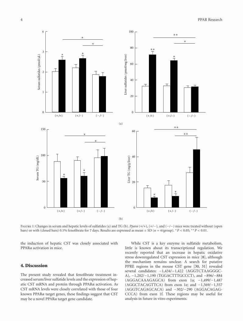
4 PPAR Research
0
1
2
3
4
Seru
m s
ulf
atid
es (
pmol
/μL)
(+/+) (+/−) (−/−) (+/+) (+/−) (−/−)
∗
∗
∗
∗
∗
∗
Live
r su
lfat
ides
(pm
ol/m
g liv
er)
∗∗
∗∗
0
20
40
60
80
100
(a)
(+/+) (+/−) (−/−)
∗
Seru
m T
G (
mg/
dL)
∗
∗∗
∗∗
0
50
100
150
(+/+) (+/−) (−/−)
Live
r T
G (
mg/
g liv
er)
0
20
40
60∗∗
∗∗
(b)
Figure 1: Changes in serum and hepatic levels of sulfatides (a) and TG (b). Ppara (+/+), (+/−), and (−/−) mice were treated without (openbars) or with (closed bars) 0.1% fenofibrate for 7 days. Results are expressed as mean ± SD (n = 6/group). ∗P < 0.05; ∗∗P < 0.01.
the induction of hepatic CST was closely associated withPPARα activation in mice.
4. Discussion
The present study revealed that fenofibrate treatment in-creased serum/liver sulfatide levels and the expression of hep-atic CST mRNA and protein through PPARα activation. AsCST mRNA levels were closely correlated with those of fourknown PPARα target genes, these findings suggest that CSTmay be a novel PPARα target gene candidate.
While CST is a key enzyme in sulfatide metabolism,little is known about its transcriptional regulation. Werecently reported that an increase in hepatic oxidativestress downregulated CST expression in mice [8], althoughthe mechanism remains unclear. A search for putativePPRE regions in the mouse CST gene [30, 31] revealedseveral candidates: −1,434/−1,422 (AGGTCTAAGGGC-A), −1,202/−1,190 (TGGACTTTGCCCT), and −896/−884(AGGACAAAGAGCA) from exon 1a; −1,499/−1,487(AGGCTACAGTTCA) from exon 1e; and −1,569/−1,557(AGGTCAGAGCACA) and −302/−290 (AGGACAGAG-CCCA) from exon 1f. These regions may be useful foranalysis in future in vitro experiments.

PPAR Research 5
0
2
4
6
8
CST
(re
lati
ve m
RN
A le
vels
)
0
2
4
6
8
AR
SA (
rela
tive
mR
NA
leve
ls)
0
2
4
6
8
CG
T (
rela
tive
mR
NA
leve
ls)
0
2
4
6
8
GA
LC (
rela
tive
mR
NA
leve
ls)
∗∗∗∗
∗∗∗∗
∗
∗
∗
∗
(+/+) (+/−) (−/−)
(+/+) (+/−) (−/−)
(+/+) (+/−) (−/−) (+/+) (+/−) (−/−)
(+/+) (+/−) (−/−)
(+/+) (+/−) (−/−)
(a)
0
1
2
3
4
0
1
2
3
4
CST
(re
lati
ve p
rote
in le
vels
)
AR
SA (
rela
tive
pro
tein
leve
ls)
0
1
2
3
4
CA
LC (
rela
tive
pro
tein
leve
ls)
0
1
2
3
4
CG
T (
rela
tive
pro
tein
leve
ls)
(+/+) (+/−) (−/−) (+/+) (+/−) (−/−)
(b)
(+/+) (+/−) (−/−)
(−) (+) (−) (+) (−) (+) Fenofibrate
CST
ARSA
CGT
GALC
Actin
Figure 2: Changes in hepatic expression of sulfatide-metabolizing enzymes by fenofibrate treatment. Open and closed bars indicate micetreated without or with 0.1% fenofibrate, respectively. Data are expressed as mean ± SD (n = 6/group). ∗P < 0.05; ∗∗P < 0.01. (a) ThemRNA levels of CST, ARSA, CGT, and GALC. Hepatic mRNA levels were normalized to those of GAPDH and then expressed as fold changesrelative to those of Ppara (+/+) mice treated with a control diet. (b) Immunoblot analysis. Actin was used as the loading control. Band inten-sities were measured densitometrically, normalized to those of actin, and then expressed as fold changes relative to those of Ppara (+/+) micetreated with a control diet.

6 PPAR Research
0
1
2
3
0
1
2
3
0
1
2
3∗∗∗∗ ∗
∗
(+/+) (+/−) (−/−) (+/+) (+/−) (−/−) (+/+) (+/−) (−/−)
PPA
R
(rel
ativ
e m
RN
A le
vels
)
PPA
R
(rel
ativ
e m
RN
A le
vels
)
PPA
R/
(rel
ativ
e m
RN
A le
vels
)
(a)
0
1
2
3 ∗∗∗∗ ∗
∗
(+/+) (+/−) (−/−)
PPA
R
(rel
ativ
e bi
ndi
ng
acti
vity
leve
ls)
0
1
2
3
(+/+) (+/−) (−/−)
(rel
ativ
e bi
ndi
ng
acti
vity
leve
ls)
(rel
ativ
e bi
ndi
ng
acti
vity
leve
ls)
PPA
R/
0
1
2
3
(+/+) (+/−) (−/−)
PPA
R(b)
0
1
2
3
4
∗∗∗∗ ∗
∗
(+/+) (+/−) (−/−)
PPA
R(r
elat
ive
prot
ein
leve
ls)
0
1
2
3
4
(+/+) (+/−) (−/−)
(rel
ativ
e pr
otei
n le
vels
)
0
1
2
3
4
(+/+) (+/−) (−/−)
(rel
ativ
e pr
otei
n le
vels
)
PPA
R/
PPA
R
(+/+) (+/−) (−/−)
(−) (+) (−) (+) (−) (+) Fenofibrate
TBP
PPAR /
PPAR
PPAR
(c)
Figure 3: Changes in hepatic expression of PPARs by fenofibrate treatment. Open and closed bars indicate mice treated without or with 0.1%fenofibrate, respectively. Data are expressed as mean ± SD (n = 6/group). ∗P < 0.05; ∗∗P < 0.01. (a) The mRNA levels of PPARs. HepaticmRNA levels were normalized to those of GAPDH and then expressed as fold changes relative to those of Ppara (+/+) mice treated witha control diet. (b) PPAR-binding activity based on an enzyme-linked immunosorbent assay. Detailed protocols are described in Section 2.Results are expressed as fold changes relative to those of Ppara (+/+) mice treated with a control diet. (c) Immunoblot analysis. TBP was usedas the loading control. Band intensities were measured densitometrically, normalized to those of TBP, and then expressed as fold changesrelative to those of Ppara (+/+) mice treated with a control diet.

PPAR Research 7
0
(a)
1
2
3
4
5
L-FA
BP
(re
lati
ve m
RN
A le
vels
)0
1
2
3
4
5
0
1
2
3
4
5
LAC
S (r
elat
ive
mR
NA
leve
ls)
0
1
2
3
4
5
MC
AD
(re
lati
ve p
rote
in le
vels
)
0
1
2
3
4
5
MT
P (
rela
tive
mR
NA
leve
ls)
0
1
2
3
4
5
LAC
S (r
elat
ive
prot
ein
leve
ls)
MC
AD
(re
lati
ve m
RN
A le
vels
)∗∗
∗∗
∗∗∗∗∗
∗
∗∗
∗∗∗∗ ∗
∗∗
∗∗
∗∗
∗∗∗
∗
∗
∗
∗∗
∗∗
∗
(+/+) (+/−) (−/−)
(+/+) (+/−) (−/−)
(+/+) (+/−) (−/−) (+/+) (+/−) (−/−)
(+/+) (+/−) (−/−)
(+/+) (+/−) (−/−)L-
FAB
P (
rela
tive
pro
tein
leve
ls)
0
1
2
3
4
5 ∗∗
∗∗∗
∗
(+/+) (+/−) (−/−)0
1
2
3
4
5
MT
P (
rela
tive
pro
tein
leve
ls) ∗
∗∗
∗
(+/+) (+/−) (−/−)
(b)
LACS
MCAD
L-FABP
MTP
Fenofibrate
(+/+) (+/−) (−/−)
(−) (+) (−) (+) (−) (+)
Figure 4: Changes in hepatic expression of conventional PPARα target genes by fenofibrate treatment. Open and closed bars indicate micetreated without or with fenofibrate, respectively. Data are expressed as mean ± SD (n = 6/group). ∗P < 0.05; ∗∗P < 0.01. (a) Analysis ofmRNA. Hepatic mRNA levels were normalized to those of GAPDH and then expressed as fold changes relative to levels of Ppara (+/+) micetreated with a control diet. (b) Immunoblot analysis. Band intensities were measured densitometrically, normalized to those of actin, andthen expressed as fold changes relative to those of Ppara (+/+) mice treated with a control diet.
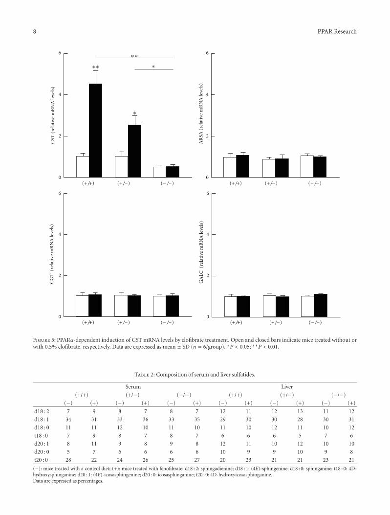
8 PPAR Research
0
2
4
6C
ST (
rela
tive
mR
NA
leve
ls)
∗∗
∗∗ ∗
∗
(+/+) (+/−) (−/−)
AR
SA (
rela
tive
mR
NA
leve
ls)
0
2
4
6
(+/+) (+/−) (−/−)
CG
T (
rela
tive
mR
NA
leve
ls)
0
2
4
6
(+/+) (+/−) (−/−)
GA
LC (
rela
tive
mR
NA
leve
ls)
0
2
4
6
(+/+) (+/−) (−/−)
Figure 5: PPARα-dependent induction of CST mRNA levels by clofibrate treatment. Open and closed bars indicate mice treated without orwith 0.5% clofibrate, respectively. Data are expressed as mean ± SD (n = 6/group). ∗P < 0.05; ∗∗P < 0.01.
Table 2: Composition of serum and liver sulfatides.
Serum Liver
(+/+) (+/−) (−/−) (+/+) (+/−) (−/−)
(−) (+) (−) (+) (−) (+) (−) (+) (−) (+) (−) (+)
d18 : 2 7 9 8 7 8 7 12 11 12 13 11 12
d18 : 1 34 31 33 36 33 35 29 30 30 28 30 31
d18 : 0 11 11 12 10 11 10 11 10 12 11 10 12
t18 : 0 7 9 8 7 8 7 6 6 6 5 7 6
d20 : 1 8 11 9 8 9 8 12 11 10 12 10 10
d20 : 0 5 7 6 6 6 6 10 9 9 10 9 8
t20 : 0 28 22 24 26 25 27 20 23 21 21 23 21
(−): mice treated with a control diet; (+): mice treated with fenofibrate; d18 : 2: sphingadienine; d18 : 1: (4E)-sphingenine; d18 : 0: sphinganine; t18 : 0: 4D-hydroxysphinganine; d20 : 1: (4E)-icosasphingenine; d20 : 0: icosasphinganine; t20 : 0: 4D-hydroxyicosasphinganine.Data are expressed as percentages.
PPAR Research 9
The degree of increases in serum sulfatides was lowerthan that in hepatic sulfatides by fenofibrate treatment (1.27-fold in the serum versus 2.20-fold in liver in Ppara (+/+) miceand 1.22-fold in the serum versus 1.95-fold in the liver ofPpara (+/−) mice). Sulfatides synthesized in the liver aresecreted into the blood together with TG as a component ofvery-low-density lipoprotein (VLDL) [32]. Thus, hepatic TGcontent was reduced by fenofibrate treatment probably dueto the enhanced of mitochondrial β-oxidation ability result-ing in a reduction of hepatic VLDL synthesis as seen in otherexperiments using cultured hepatocytes [33]. Further studiesare required to determine sulfatide metabolism in the serumand liver since they are significantly influenced by numerouspathophysiological events and treatments, including acutekidney injury [8, 34], clofibrate pretreatment [35], chronickidney disease [6], and kidney transplantation [7].
The role of PPARα has been clarified in several liver dis-eases. For instance, PPARα is downregulated in alcoholic liverdisease [11, 36] as well as after liver transplantation[37]. Per-sistent activation of PPARα ameliorates hepatic steatosis andinflammation in mice but may also induce hepatocarcino-genesis [10]. The association between liver disease and sulfa-tide metabolism may be of interest for further research.
Lastly, several animal studies have uncovered a protectiverole for serum sulfatides against arteriosclerosis and hyperco-agulation [5]. We also reported a close relationship betweenlower serum sulfatide concentrations and higher incidencesof cardiovascular disease in patients with end-stage renal fail-ure [6], in whom sulfatide levels returned to normal follow-ing kidney transplantation [7]. Accordingly, increasing ormaintaining serum sulfatide levels using fibrates may be use-ful in reducing the risk of cardiovascular events, which isconsistent with the known beneficial effect of fibrates seen inrandomized controlled studies [38]. Furthermore, thesefindings show a need to examine sulfatide metabolism in car-diomyocytes, endothelial cells, and vascular smooth cells todisclose any novel protective roles of PPARα in cardiovascu-lar inflammation and atherosclerosis, particularly in relationto CST upregulation.
Abbreviations
ARSA: Arylsulfatase ACGT: Ceramide galactosyltransferaseCST: Cerebroside sulfotransferaseGALC: GalactosylceramidaseGAPDH: Glyceraldehyde-3-phosphate dehydrogenaseLACS: Long-chain acyl-CoA synthaseL-FABP: Liver fatty acid-binding proteinLS: LysosulfatidesMCAD: Medium-chain acyl-CoA dehydrogenaseMTP: Microsomal triglyceride transfer proteinPCR: Polymerase chain reactionPPAR: Peroxisome proliferator-activated receptorPPRE: PPAR response elementTG: Triglycerides.
Conflicts of Interests
The authors have declared that no conflict of interests exists.
Acknowledgment
The authors thank Trevor Ralph for his English editorial as-sistance.
References
[1] I. Ishizuka, “Chemistry and functional distribution of sulfo-glycolipids,” Progress in Lipid Research, vol. 36, no. 4, pp. 245–319, 1997.
[2] K. Honke, Y. Zhang, X. Cheng, N. Kotani, and N. Taniguchi,“Biological roles of sulfoglycolipids and pathophysiology oftheir deficiency,” Glycoconjugate Journal, vol. 21, no. 1-2, pp.59–62, 2004.
[3] K. I. Nagai, K. Tadano-Aritomi, Y. Niimura, and I. Ishizuka,“Higher expression of renal sulfoglycolipids in marine mam-mals,” Glycoconjugate Journal, vol. 25, no. 8, pp. 723–726,2008.
[4] M. Eckhardt, “The role and metabolism of sulfatide in the ner-vous system,” Molecular Neurobiology, vol. 37, no. 2-3, pp. 93–103, 2008.
[5] M. Kyogashima, “The role of sulfatide in thrombogenesis andhaemostasis,” Archives of Biochemistry and Biophysics, vol. 426,no. 2, pp. 157–162, 2004.
[6] R. Hu, G. Li, Y. Kamijo et al., “Serum sulfatides as a novel bio-marker for cardiovascular disease in patients with end-stagerenal failure,” Glycoconjugate Journal, vol. 24, no. 9, pp. 565–571, 2007.
[7] L. Wang, Y. Kamijo, A. Matsumoto et al., “Kidney transplanta-tion recovers the reduction level of serum sulfatide in ESRDpatients via processes correlated to oxidative stress and plateletcount,” Glycoconjugate Journal, vol. 28, no. 3-4, pp. 125–135,2011.
[8] X. Zhang, T. Nakajima, Y. Kamijo et al., “Acute kidney injuryinduced by protein-overload nephropathy down-regulatesgene expression of hepatic cerebroside sulfotransferase inmice, resulting in reduction of liver and serum sulfatides,” Bio-chemical and Biophysical Research Communications, vol. 390,no. 4, pp. 1382–1388, 2009.
[9] S. S. T. Lee, T. Pineau, J. Drago et al., “Targeted disruption ofthe α isoform of the peroxisome proliferator- activated recept-or gene in mice results in abolishment of the pleiotropic effectsof peroxisome proliferators,” Molecular and Cellular Biology,vol. 15, no. 6, pp. 3012–3022, 1995.
[10] N. Tanaka, K. Moriya, K. Kiyosawa, K. Koike, F. J. Gonzalez,and T. Aoyama, “PPARα activation is essential for HCV coreprotein-induced hepatic steatosis and hepatocellular carci-noma in mice,” Journal of Clinical Investigation, vol. 118, no. 2,pp. 683–694, 2008.
[11] T. Nakajima, Y. Kamijo, N. Tanaka et al., “Peroxisome prolife-rator-activated receptor α protects against alcohol-inducedliver damage,” Hepatology, vol. 40, no. 4, pp. 972–980, 2004.
[12] A. Hara and N. S. Radin, “Lipid extraction of tissues with alow toxicity solvent,” Analytical Biochemistry, vol. 90, no. 1, pp.420–426, 1978.
[13] G. Li, R. Hu, Y. Kamijo et al., “Establishment of a quantitative,qualitative, and high-throughput analysis of sulfatides fromsmall amounts of sera by matrix-assisted laser desorption
10 PPAR Research
ionization-time of flight mass spectrometry,” Analytical Bio-chemistry, vol. 362, no. 1, pp. 1–7, 2007.
[14] I. V. Smirnova, D. C. Bittel, R. Ravindra, H. Jiang, and G. K.Andrews, “Zinc and cadmium can promote rapid nucleartranslocation of metal response element-binding transcriptionfactor-1,” Journal of Biological Chemistry, vol. 275, no. 13, pp.9377–9384, 2000.
[15] T. Aoyama, S. Yamano, D. J. Waxman et al., “CytochromeP-450 hPCN3, a novel cytochrome P-450 IIIA gene productthat is differentially expressed in adult human liver. cDNAand deduced amino acid sequence and distinct specificitiesof cDNA-expressed hPCN1 and hPCN3 for the metabolismof steroid hormones and cyclosporine,” Journal of BiologicalChemistry, vol. 264, no. 18, pp. 10388–10395, 1989.
[16] Y. Kamijo, K. Hora, N. Tanaka et al., “Identification of fun-ctions of peroxisome proliferator-activated receptor α in prox-imal tubules,” Journal of the American Society of Nephrology,vol. 13, no. 7, pp. 1691–1702, 2002.
[17] T. Aoyama, I. Ueno, T. Kamijo, and T. Hashimoto, “Rat very-long-chain acyl-CoA dehydrogenase, a novel mitochondrialacyl- CoA dehydrogenase gene product, is a rate-limiting en-zyme in long-chain fatty acid β-oxidation system. cDNA anddeduced amino acid sequence and distinct specificities of thecDNA-expressed protein,” Journal of Biological Chemistry, vol.269, no. 29, pp. 19088–19094, 1994.
[18] T. Aoyama, Y. Uchida, R. I. Kelley et al., “A novel disease withdeficiency of mitochondrial very-long-chain acyl-CoA dehy-drogenase,” Biochemical and Biophysical Research Communi-cations, vol. 191, no. 3, pp. 1369–1372, 1993.
[19] Y. Shindo and T. Hashimoto, “Acyl-coenzyme A synthetaseand fatty acid oxidation in rat liver peroxisomes,” Journal ofBiochemistry, vol. 84, no. 5, pp. 1177–1181, 1978.
[20] N. Tanaka, X. Zhang, E. Sugiyama et al., “Eicosapentaenoicacid improves hepatic steatosis independent of PPARα activa-tion through inhibition of SREBP-1 maturation in mice,” Bio-chemical Pharmacology, vol. 80, no. 10, pp. 1601–1612, 2010.
[21] S. Furuta, S. Mayazawa, and T. Hashimoto, “Purification andproperties of rat liver Acyl-CoA dehydrogenases and electrontransfer flavoprotein,” Journal of Biochemistry, vol. 90, no. 6,pp. 1739–1750, 1981.
[22] B. Desvergne and W. Wahli, “Peroxisome proliferator-acti-vated receptors: nuclear control of metabolism,” Endocrine Re-views, vol. 20, no. 5, pp. 649–688, 1999.
[23] E. Duplus, M. Glorian, and C. Forest, “Fatty acid regulation ofgene transcription,” Journal of Biological Chemistry, vol. 275,no. 40, pp. 30749–30752, 2000.
[24] P. Gervois, I. P. Torra, J. C. Fruchart, and B. Staels, “Regulationof lipid and lipoprotein metabolism by PPAR activators,” Cli-nical Chemistry and Laboratory Medicine, vol. 38, no. 1, pp. 3–11, 2000.
[25] B. Staels, J. Dallongeville, J. Auwerx, K. Schoonjans, E. Leiters-dorf, and J. C. Fruchart, “Mechanism of action of fibrates onlipid and lipoprotein metabolism,” Circulation, vol. 98, no. 19,pp. 2088–2093, 1998.
[26] X. Zhang, N. Tanaka, T. Nakajima, Y. Kamijo, F. J. Gonzalez,and T. Aoyama, “Peroxisome proliferator-activated receptor α-independent peroxisome proliferation,” Biochemical and Bio-physical Research Communications, vol. 346, no. 4, pp. 1307–1311, 2006.
[27] S. Mandard, M. Muller, and S. Kersten, “Peroxisome prolife-rator-activated receptor α target genes,” Cellular and MolecularLife Sciences, vol. 61, no. 4, pp. 393–416, 2004.
[28] T. Aoyama, J. M. Peters, N. Iritani et al., “Altered constitutiveexpression of fatty acid-metabolizing enzymes in mice lacking
the peroxisome proliferator-activated receptor α (PPARα),”Journal of Biological Chemistry, vol. 273, no. 10, pp. 5678–5684, 1998.
[29] C. Ameen, U. Edvardsson, A. Ljungberg et al., “Activation ofperoxisome proliferator-activated receptor α increases the ex-pression and activity of microsomal triglyceride transfer pro-tein in the liver,” Journal of Biological Chemistry, vol. 280, no.2, pp. 1224–1229, 2005.
[30] M. Podvinec, M. R. Kaufmann, C. Handschin, and U. A. Mey-er, “NUBIScan, an in Silico approach for prediction of nuclearreceptor response elements,” Molecular Endocrinology, vol. 16,no. 6, pp. 1269–1279, 2002.
[31] Y. Hirahara, M. Tsuda, Y. Wada, and K. Honke, “cDNA clon-ing, genomic cloning, and tissue-specific regulation of mousecerebroside sulfotransferase,” European Journal of Biochem-istry, vol. 267, no. 7, pp. 1909–1916, 2000.
[32] A. Hara and T. Taketomi, “Occurrence of sulfatide as a majorglycosphingolipid in WHHL rabbit serum lipoproteins,” Jour-nal of Biochemistry, vol. 102, no. 1, pp. 83–92, 1987.
[33] S. E. Hahn and D. M. Goldberg, “Modulation of lipoproteinproduction in HEP G2 cells by fenofibrate and clofibrate,”Biochemical Pharmacology, vol. 43, no. 3, pp. 625–633, 1992.
[34] Y. Kamijo, K. Hora, K. Kono et al., “PPARα protects proximaltubular cells from acute fatty acid toxicity,” Journal of theAmerican Society of Nephrology, vol. 18, no. 12, pp. 3089–3100,2007.
[35] K. Takahashi, Y. Kamijo, K. Hora et al., “Pretreatment by low-dose fibrates protects against acute free fatty acid-inducedrenal tubule toxicity by counteracting PPARα deterioration,”Toxicology and Applied Pharmacology, vol. 252, no. 3, pp. 237–249, 2011.
[36] W. Okiyama, N. Tanaka, T. Nakajima et al., “Polyenephospha-tidylcholine prevents alcoholic liver disease in PPARα-nullmice through attenuation of increases in oxidative stress,”Journal of Hepatology, vol. 50, no. 6, pp. 1236–1246, 2009.
[37] K. Nakagawa, N. Tanaka, M. Morita et al., “PPARα is down-regulated following liver transplantation in mice,” Journal ofHepatology, vol. 56, no. 3, pp. 586–594, 2012.
[38] A. Keech, A. Simes, P. Barter et al., “Effects of long-term feno-fibrate therapy on cardiovascular events in 9795 people withtype 2 diabetes mellitus (the FIELD study): randomised con-trolled trial,” Lancet, vol. 366, no. 9500, pp. 1849–1861, 2005.


![PPAR: Bangladesh: Ganges-Kobadak Irrigation Rehabilitation ... · Title: PPAR: Bangladesh: Ganges-Kobadak Irrigation Rehabilitation Project (Loan 671-BAN[SF]) Author: Asian Development](https://static.documents.pub/doc/80x56/5f87bd77888d5524a31f3544/ppar-bangladesh-ganges-kobadak-irrigation-rehabilitation-title-ppar-bangladesh.jpg)


