203 University of Notre Dame College of Science (Department of Biology) Notre Dame, Indiana (U.S.A.) V. P. Shabma Pharmacology and histopathology of certain insecticides in grasshopper Poecilocerus pictus F abricius (With 31 textfigures) Out of the various branches of organic insecticides, histopathology has received comparatively little attention. In fact, some work exists on the histo- pathological effect of insecticides, but most of it centers round the pyrethrum. However, there also exist some scattered work on the histopathological effect of some of the modern insecticides. In most of the previous works there is striking lack of information about the dose of insecticide in relation to the induced pharmacology and histopathology. Speyer (1925) concluded that the response of insects to poisoning by pyr- ethrins is characterized by excitation, convulsion, paralysis and death; a se- quence typical of nerve poisons. E ckert (1948) observed that upon contact with parathion, honeybees immediately become widely agitated and bellicose, and perform cleaning movements; they become moribund in30mts. Salkeld (1951) concluded that the hyperactivity and circus movements of the parathion poisoned bees indicated that both insecticides were acting as nerve poisons. Chadbottrne & R ainwater (1953) observed that Heliothis larvae treated with DDT and dieldrin ceased feeding after two hours and become inactive; after three to four hours respectively, slight tremors and convulsions were evident and after several additional hours both were severe. K rüger (1931) was able to observe with pyrethrum treatment in living transparent Gorethra larvae, the appearance of vacuoles in the ganglion and connectives of the nerve cord within 10 to 20 mts. after the onset of convulsions. The vacuoles made their appearance in the nerve fibres rather then in the cells. It is significant that no vacuolization was observed even after one day following convulsions from sub-lethal doses, so that this phenomenon probably presents only the extreme manifestation of toxicity. H artzell & W ilcoxon (1934b) reported nerve lesions throughout the main part of the central nervous system, in the brain, sub-oesophageal ganglion, thoracic ganglion, abdominal ganglion and the connectives of the Mealworm larvae (Tenebrio molitor), and adult grasshopper (Melanoplus femur-rubrum), that had been killed by pyrethrum concentrates applied externally. He concluded that death is caused by the destruction of cells of the central nervous system accompanied by paralysis. DOI: 10.21248/contrib.entomol.16.1-2.203-212
Transcript
203
University of Notre DameCollege of Science (Department of Biology)Notre Dame, Indiana (U.S.A.)
V. P. Sh a b m a
Pharmacology and histopathology of certain insecticides in grasshopper Poecilocerus pictus Fabricius
(W ith 31 textfigures)
Out of the various branches of organic insecticides, histopathology has received comparatively little attention. In fact, some work exists on the histo- pathological effect of insecticides, but most of it centers round the pyrethrum. However, there also exist some scattered work on the histopathological effect of some of the modern insecticides. In most of the previous works there is striking lack of information about the dose of insecticide in relation to the induced pharmacology and histopathology.
Sp e y e r (1925) concluded that the response of insects to poisoning by pyr- ethrins is characterized by excitation, convulsion, paralysis and death; a sequence typical of nerve poisons. E c k e r t (1948) observed that upon contact with parathion, honeybees immediately become widely agitated and bellicose, and perform cleaning movements; they become moribund in30mts. Sa l k e l d (1951) concluded that the hyperactivity and circus movements of the parathion poisoned bees indicated that both insecticides were acting as nerve poisons. Chadbottrne & R a in w a t e r (1953) observed that Heliothis larvae treated with DDT and dieldrin ceased feeding after two hours and become inactive; after three to four hours respectively, slight tremors and convulsions were evident and after several additional hours both were severe.
K r ü g e r (1931) was able to observe with pyrethrum treatment in living transparent Gorethra larvae, the appearance of vacuoles in the ganglion and connectives of the nerve cord within 1 0 to 2 0 mts. after the onset of convulsions. The vacuoles made their appearance in the nerve fibres rather then in the cells. It is significant that no vacuolization was observed even after one day following convulsions from sub-lethal doses, so that this phenomenon probably presents only the extreme manifestation of toxicity. H a r t z e l l & W il c o x o n (1934b) reported nerve lesions throughout the main part of the central nervous system, in the brain, sub-oesophageal ganglion, thoracic ganglion, abdominal ganglion and the connectives of the Mealworm larvae (Tenebrio molitor), and adult grasshopper (Melanoplus femur-rubrum), that had been killed by pyrethrum concentrates applied externally. He concluded that death is caused by the destruction of cells of the central nervous system accompanied by paralysis.
DOI: 10.21248/contrib.entomol.16.1-2.203-212
Powered by TCPDF (www.tcpdf.org)
204 V. P. Sharma, Pharmacology and histopathology of certain insecticides
K l in g e r (1936) made a histological study of the nerve lesions of Gypsy moth Porthetria dispar after application of 24 hours of 15 percent pyrethrum extract. He observed that nerve appeared to be isolated and surrounded by spaces as contrasted with the check where the tissue was not dislocated. A similar histological change was observed in the moribund insect. H a r t z e l l & Sc u d d e r (1942) state that pyrethrum and its activators Isobutyl undecylene amide each was found to show rather distinct and characteristic effects upon the central nervous system and associated tissues of the housefly. Pyrethrum has a widespread clumping effect on the chromatin of the nuclei, while the activators seem to cause a chromatolysis or dissolution of the chromatin. A combination of these two agents shows a histological picture that is summation of the effects of both. The interaction of these two types of nuclear destruction was believed to be the true basis of activation. R ic h a r d s (1943) believes that the effects of some neurotoxic insecticides are due to the destruction of some bound lipid sheath of the nerve cells and their processes. R ic h a r d & Cu t k o m p (1945) using a variety of insecticides, found that nerve was paralysed and presumably dead prior to appearance of any abnormalities or lesions, except the possible chromatin clumping. They also observed that in dead nerve tissue the neurones become granular internally. S a l k e l d (1951) reported that the midgut from the parathion poisoned bee showed no abnormality, distinct histological differences from the normal structures were seen in midguts from DDT poisoned bees. Ch a d b o u r n e & R a i n w a t e r (1953) were unable to detect any histo- pathological changes in the nervous system of Bollworm Heliothis armigera under the effect of several insecticides, though changes in other tissues (midgut epithelium, muscles, fat bodies and Malpighian tubules) were observed. S o l im a n & So l iMa n (1958) observed histological changes brought about in the midgut, fat bodies, muscles, Malpighian tubules, ventral nerve ganglia and haemolymph of larvae of Prodenia litura by contact with DDT, toxaphene, parathion, and cotton dust (Containing 3% BHC, 10% DDT, and 40% sulphur) in comparison with normal tissues.
Material and MethodsPoecilocerus pictus Fa b r ic o ts were collected from the fields of the outskirts
of Allahabad and were reared in the laboratory. They were supplied with Calotropis leaves and water. Rearing of grasshoppers was easily carried out in big cages having a 4" deep layer of soil on the bottom of the cages, for the females to lay eggs.
Allethrin, dieldrin, rmvan, and parathion insecticides were used for the study having the following descriptions.
1. Allethrin, technical :PurityAllethrin (allyl homolog of Cinerin I) ........................................................................ 90%Inert material, petroleum base o i l ..................................................................................... 10%
DOI: 10.21248/contrib.entomol.16.1-2.203-212
Powered by TCPDF (www.tcpdf.org)
Beiträge zur Entomologie, Band 18, Nr. 1/2; 1966 205
2. Dieldrin, technical PurityHexachloroepoxyoctahydro-endo, exodimethanonepthalene...................................... 76.5%Insecticidally active, related compounds.....................................................................13.5%Other related compounds...................................................................................................10%
3. Nuvan 50 EC. One liter Nuvan 50 EC contains 500 gms of 0 — 0, dimethyl- 2,2-di-chlorovinyl phosphate as well as solvents and emulsifying agents.
These insecticides were dissolved in acetone, benzene and alcohol and diluted in volumetric flasks for various treatments. The applications were carried out with the help of a micro-applicator on the pronotum of the grasshoppers. Lethal doses were experimentally ascertained and the treatments were made in sub-lethal and lethal doses. Male grasshoppers were employed for the study to avoid any controversy on the sex of the insect.
To observe the symptoms of poisoning induced by the insecticides, treatments were made in known doses and a control was run concurrently. The untreated, control and treated grasshoppers were released in different cages and a comparative study of the hyperactivity of body appendages and hyperactive behaviour of treated insects was noted down till either the insects died or they behaved as the controls or untreated insects behaved. In all the experiments 15 to 20 insects were treated keeping a control, and the experiments were repeated for three to five times.
For histopathological studies, living insects were dissected out and fixed in Ca b n o y ’s and B otjin’s fixatives ; and in alcohol for the nervous tissues to be stained in specific stains. The slides were prepared for the microscopic examination following the usual procedure of paraffin imbedding. For nervous tissues the stains used were Bora: a n ’s gold chloride and silver albumose, Toluidine blue, Methylene blue, and Haemotoxylin eosin ; and for the rest of the tissues Delafield Haemotoxylin eosin and M a l l o b y ’s triple stains were used. Delafield Haemo- toxylin and Eosin gave very satisfactory results in nervous and other tissues and was most frequently used during these preparations.
T reatm entsMale grasshoppers were topically applied on the pronotum of the insects
206 Y. P. Sharha, Pharmacology and histopathology of certain insecticides
ObservationsPharmacology
Treatments in sub-lethal doses induce symptoms which last for two to three hours and the grasshoppers return to normal behaviour after this time. Application of insecticides with all the four poisons induce an usual sequence of nerve poisos i. e,, excitation, convulsion, paralysis, and death. However, with allethrin treatment the rapid contraction and extension of abdomen, stretching of head, and fast hopping movements are very conspicuous. With dieldrin poisoning, the extension and contraction of abdomen is equally conspicuous as with allethrin, the hind pair of legs are continuously struck in front of the head against the ground and the abdomen swells in between the 2nd and 3rd segmental membrane to the point of rupture. With nuvan treatment first pair of legs are rubbed against each other, the abdomen is curved upwards, at times hind pair of legs are struck before the head, and the telescopic movement of the abdomen is very conspicuous. With parathion poisoning the conspicuous symptoms are the striking of hind pair of legs against the ground in front of the head, abdomen is raised high and curved downwards, mouth parts pressed towards ground and the head is continuously stretched.
HistopathologyHistological changes induced by these insecticides are of similar nature.
The variation has been observed in the intensity of the pathology.
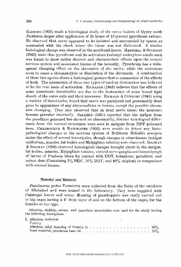
A lim en ta ry Canal Fore gut: Both the circular and longitudinal muscles of the foregut are vacuolated. These vacuoles lie scattered and occupy most part of the muscles and usually enclose the nuclei. The nuclei appear shrunk and pycnotic. The epithelial cells are badly shrunk, lie in a line, cytoplasm become granular, and the cell structure and cell boundary is completely lost. The nuclei at places are extruded, shrunk, pycnotic, the chromatins clump (the chromatins show break down with nuvan and parathion), at places the nuclei are vacoulated, and they lie displaced. The exfoliation of epithelium is, very marked and conspicuous. The epithelial cells almost cease to exist and the intima is largely separated from the epithelium. Hepatic ceaca develop varying sizes of vacuoles which lie scattered in the epithelial cells. The nuclei are degenerated and deeply stained (Figs. 1 to 6 ).M idgut: Histological degenerations are less pronounced in the midgut than in foregut. Scattered vacuoles appear in the longitudinal and circular muscles and the nuclei appear distorted. The epithelial cells are vacuolated and degenerated. The chromatin of the nuclei clump (show breakdown -with nuvan and parathion) and there is no exfoliation of epithelium. (Figs. 7 and 8 ).H indgut: There are no appreciable changes in the muscles of the hindgut. The epithelial cells are vacuolated and these vacuoles push the cytoplasm and the nuclei to one side. The nuclei are shrunk and become pycnotic. (Figs. 9 and 1 0 ).
DOI: 10.21248/contrib.entomol.16.1-2.203-212
Powered by TCPDF (www.tcpdf.org)
Beiträge zur Entomologie, Band 16, Nr. 1/2; 1Ô66 207
Table 1
Showing the com parative shrinkage in the tissues of crop with the effect of insecticides
Tissues Control Allethrin Dieldrin N ¡¡van Parathion
M alpighian tubu lesThe Malpighian tubules appear shrunk, vacuolated, and their surface become
uneven. The nuclei are deeply stained and dislocated (Figs. 11 and 12).
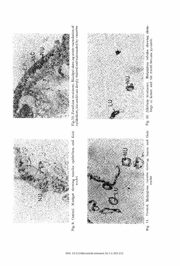
Fat bod iesSlight to moderate dissolution of fat bodies has been observed with the
application of these insecticides. The dissolution of fat cells varies from insecticide to insecticide and its dose (Figs. 13 and 14).
Central nervous systemB rain : Histological degenerations are less pronounced in the brain than in
the thoracic or abdominal ganglia. The neurilemma of the brain is not affected much except that it has been separated from the cortex at certain regions. The cortex showes acute vacuolation, cellular degeneration, presence of clear spaces and lysis. The first, second and third groups of globuli cells are shrunk, deeply stained, degenerated and displaced. The nuclei of the globuli cells are clumped with the allethrin and dieldrin, and show breakdown with the nuvan and parathion treatments. The nuclei of the neurosecretory cells appear vacuolated and the chromatin granules are thrown to one side; and the neurosecretory cells appear shrunk, vacuolated, their cytoplasm become ragged and degenerated.
DOI: 10.21248/contrib.entomol.16.1-2.203-212
Powered by TCPDF (www.tcpdf.org)
208 V. P. Sh a r m a , Pharmacology and histopathology of certain insecticides
The neurospongium showes slight tigrolysis, moderate vacuolation, cellular degeneration, partial dissolution of fibre tracts, other cell components and nerve tissue. The nuclei also gave degenerated appearance. Allethrin and dieldrin cause the formation of lesions in the unspecific regions of the neurospongium and no lesions are caused with the treatment of nuvan and parathion (Figs. 15 to 20).
Th ora cic gan glion : Histological degenerations are intense in the thoracic ganglion than in the brain. Neurilemma has been removed from many places. The cortex is vacuolated and there is complete degeneration of the tissue, presence of clear spaces and cytoplasm of the cortical cells become granular. The nuclei of the cortex show acute degeneration and the neurosecretory cells show moderate degeneration. The chromatins of the nuclei of cortex and the neurosecretory cells clump with the allethrin and dieldrin treatment and show breakdown with the nuvan and parathion treatment. The neurospongium show acute vacuolation, tigrolysis, acute cellular degeneration, and dissolution of the fibre tracts, cell components and nerve tissue. Lesions are formed with the treatment of allethrin and dieldrin (Figs. 21 to 25).
A bdom in al gan glion : Nature of the pathology remains the same, except that the pathology is reduced. In certain cases the neurilemma has been removed. The cortex show moderate to acute vacuolation and cellular degeneration. The nuclei and the neurosecretory cells show similar degenerative changes as we find in the thoracic ganglion. The chromatin of the nuclei are clumped with the allethrin and dieldrin treatment and show breakdown with the nuvan and parathion poisoning. The neurospongium show acute vacuolation, moderate dissolution of fibre tracts, and dissolution of the cell components and nerve tissue. Lesions lie in patches with allethrin and dieldrin treatments (Figs. 26 to 30).
DiscussionPharmacology of insecticides have been studied by Sp e y e r (1925), E c k e r t
(1948), Sa l k e l d (1951), and C h a d b o u r n e & R a in w a t e r (1953) etc. During these observations it was recorded that the symptoms induced in grasshoppers follow four stages i.e ., excitation, convulsion, paralysis, and death; a characteristic of nerve poisons as reported by B r o w n (1951). However, it was also observed that the histological degenerations are caused throughout the central part of the nervous system, alimentary canal, Malpighian tubules, fat bodies etc., with the application of these insecticides. Whether or not these histological degenerations and pharmacological symptoms are sufficient to classify these poisons as nerve poisons shall he for the future workers to decide on the basis of physiological and bio-chemical findings.
Histopathological studies were carried out with the alimentary canal, central nervous system, Malpighian tubules and fat bodies as it was supposed that the destruction in these major body organs would be sufficient to bring about death.
H a r t z e l l (1945) reported that haemotoxylin eosin y method was found to be a satisfactory general stain both for insect nerves and muscles, but did not
Fig. 29. Nuvan treatment. Abdominal ganglion showing vacuolation in cortex and neurospongium, the nuclei and neurosecretory cells are surrounded by vacuoles and appear shrunk
Fig. 30. Parathion treatment. Abdominal ganglion showing disappearance of neurilemma, vacuolation in cortex and neurospongium, and breakdown of chromatins
Beiträge zur Entomologie, Band 16, Nr. 1/2; 1966 209
differentiate nerve fibres as well visually as B o d ia n ’s method. He further reported that the Toluidine blue was found to be a inferior stain to the first two named methods. During these studies all these three methods were frequently used and it was found that haemotoxylin and eosin y gave satisfactory results with nervous tissues, alimentary canal, Malpighian tubules and fat bodies. Bo d ia n ’s and Toluidine blue gave equally good results.
Feeding technique, age of test insects, size of cage, number of insect per cage, and the nutrition of the test insects may all have an effect on the mortality figures in the laboratory insecticidal tests (M o r r iso n , 1943). During these investigations all test insects were of approximately same age group and were taken from the same population.
There was no difficulty in making comparison with sub-lethal or with lethal treatments where the insect could tolerate the toxic dose. However, care was taken to study the histological degenerations of the moribund than dead insects ; and in all the cases moribund insect’s tissues were examined in order to avoid any postmortem changes owing to death. It may be recalled that the term moribund has been used to indicate a condition of complete inability of insects to move and this condition is preceded by death.
Throughout these findings dose of treatment was recorded, as it was found that dose of insecticide is a material factor in bringing about pathology. Little care was taken by most of early workers to note the dose of treatments and as such pathology has been reported.
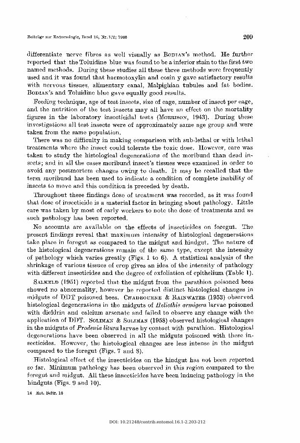
No accounts are available on the effects of insecticides on foregut. The present findings reveal that maximum intensity of histological degenerations take place in foregut as compared to the midgut and hindgut. The nature of the histological degenerations remain of the same type, except the intensity of pathology which varies greatly (Figs. 1 to 6). A statistical analysis of the shrinkage of various tissues of crop gives an idea of the intensity of pathology with different insecticides and the degree of exfoliation of epithelium (Table 1).
Sa l k e l d (1951) reported that the midgut from the parathion poisoned bees showed no abnormality, however he reported distinct histological changes in midguts of DDT poisoned bees. Ch a d b o u r n e & R a in w a t e r (1953) observed histological degenerations in the midguts of Heliothis armigera larvae poisoned with dieldrin and calcium arsenate and failed to observe any change with the application of DDT. So l im a n & So l im a n (1958) observed histological changes in the midguts of Prodenia litura larvae by contact with parathion. Histological degenerations have been observed in all the midguts poisoned with these insecticides. However, the histological changes are less intense in the midgut compared to the foregut (Figs. 7 and 8).
Histological effect of the insecticides on the hindgut has not been reported so far. Minimum pathology has been observed in this region compared to the foregut and midgut. All these insecticides have been inducing pathology in the hindguts (Figs. 9 and 10).14 Ent. Beitr. 16
DOI: 10.21248/contrib.entomol.16.1-2.203-212
Powered by TCPDF (www.tcpdf.org)
210 V . P. Sh akjia , Pharmacology and Mstopathology of certain insecticides
M alpigh ian tu bu les:Ch a d b o u r n e & R a in w a t Bit (1953) reported the degeneration of cell nuclei
of the Malpighian tubules of Heliothis armigera larvae with dieldrin poisoning. So l im a n & S o l iMa n (1958) reported degenerative changes in Malpighian tubules with parathion poisoning. Author observed the effect of these organic insecticides and found varying stages of degenerated tubules (Mgs. 11 and 12). A statistical analysis of the contraction has been made (Table 2), which gives an idea of the intensity of pathology.
Fat bod ies :Dissolution of fat bodies under the effect of dieldrin has been reported
by C h a d b o u rn e & R a i n w a t e r (1953) in Heliothis armigera larvae; and with parathion has been reported by S o lim an & S o lim an (1958) in Prodenia litura larvae. Such dissolution of fat cells and degeneration of their nuclei was also observed with these insecticides, however the intensity of dissolution varies greatly with the insecticide and its dose (Figs. 13 and 14).
C entral nervous system :Histological effect of pyrethrum has been studied by H a b t z e l l & W i lc o x o n
(1932), H a b t z e l l & W i lc o x o n (1934b), K li n g e r (1936), H a b t z e l l & S c u d d e r (1942), R ic h a r d s & C utkom p (1945) and K r u g e r (1931) etc., etc. Lesions appear to be characteristic of pyrethrum poisoning to the extent that they do not appear even in acute cases of nicotine and lead arsenate poisoning ( H a b t z e l l & W ilc o x o n , 1935) or by rotenone unless applied in very high concentrations sufficient to give a knockdown effect (H a b t z e l l , 1945). H a b t z e l l & W ilc o x o n (1934a) reported that three organic thiocyano (Lethane 384, Thanite, and gamma thiocyanopropylphenyl ether) contains insecticidal properties and cause lesions in the nerve tissue. During these findings author found lesions with allethrin poisoning throughout the main part of the central nervous system (Figs. 17, 22 and 27).
There is striking lack of literature on the neuropathological effect of Cyclo- diene insecticides on insects. Ch a d b o u r n e & R a in w a t e r (1953) were unable to detect any neuropathological degenerations with dieldrin poisoning. However, during the present investigations author observed histological degenerations throughout the central nervous system with dieldrin poisoning. Dieldrin cause chromatin clumping and lesion formation in brain, thoracic and abdominal ganglia along with other histological changes (Figs. 18, 23 and 28).
Histological degenerations are also observed throughout the central nervous system with nuvan treatment. Nuvan cause chromatolysis or the breakdown of chromatin granules along with other neuropathological changes and there was no lesion formation (Figs. 19, 24 and 29).
So l im a n & So l im a n (1958) reported histological degenerations in the ventral nerve ganglia of the Prodenia, litura by contact with parathion. It was observed that the application of parathion cause breakdown of chromatins, and lesions are not formed (Figs. 20, 25 and 30).
DOI: 10.21248/contrib.entomol.16.1-2.203-212
Powered by TCPDF (www.tcpdf.org)
Beiträge zur Entomologie, Band 16, Nr. 1/2; 1966 211
Conclusions1. The symptoms of poisoning induced by allethrin, dieldrin, nuvan and
parathion follow four stages i. e., excitation, convulsion, paralysis, and death.2. Histological degenerations have been observed throughout the alimentary
canal with the application of these insecticides, however maximum pathology has been observed in crop than in any other part of the alimentary canal.
3. Malpighian tubules and fat bodies show varying degrees of degenerations under the effect of these insecticides.
4. Neuropathological changes have been observed throughout the central nervous system with the treatment of all the insecticides. Maximum pathology has been observed in thoracic ganglion than in any other part of the nervous system.
5. Allethrin and dieldrin cause lesion formation and chromatin clumping.6. Nuvan and parathion cause breakdown of chromatin or chromatolysis and
lesions are not formed.7. Application of insecticides in sub-lethal dose cause little pathology moderate
to acute pathology was observed in insects that survived the lethal dose and maximum pathology was observed in moribund insect’s tissues.
SummaryAllethrin, dieldrin, nuvan and parathion were applied locally at the pronotum of male
grasshoppers in sub-lethal and lethal doses. The symptoms of poisoning were recorded and the tissues of the treated grasshoppers were prepared for histological study. Histological changes were observed in alimentary canal, central nervous system, Malpighian tubules and fat bodies as an effect of these insecticides. The induced pathology varied according to insecticide and dose; but its greater part did not depend on the choice of the insecticide. However, allethrin and dieldrin caused clumping of chromatins and formation of lesions, whereas nuvan and parathion caused chromatolysis but no lesions.
Zusammenfassung
Allethrin, Dieldrin, Nuvan und Parathion wurden in subletaler und letaler Dosis örtlich am Pronotum männlicher Heuschrecken angewendet. Es wird von den Vergiftungssymptomen berichtet. Die Gewebe der behandelten Heuschrecken wurden für histologische Beobachtungen präpariert. Unter der Wirkung dieser Insektizide wurden histologische Veränderungen im Verdauungskanal festgestellt, im Zentralnervensystem, in den Mal- pighischen Gefäßen und in den Fettkörpern. Der durch die Induktion hervorgerufene Krankheitsverlauf variiert von Insektizid zu Insektizid und dessen Dosis, ist aber vorwiegend unabhängig vom Insektizid selbst. Jedoch bewirken Allethrin und Dieldrin ein Zusammendrängen der Chromatine und die Bildung von Wunden, während Nuvan und Parathion eine Chromatolyse, aber keine Wunden verursachen.
P e 3 K ) M e
A jiJ ie T p H H , H H jm n p H H , H yB aH H n a p a T H O H npHJVieHJMHCb b jieT a jib H O Ä h c y ß n e - T a n b H o ii H 03e H a n p o H O T y M e M y * C K H X 3K 3eM iiJ iH poB ca p a H H H . O n n c b iB a io x c H cH M nTOM H OTpaBJieH H H . T K aH H o ß p a ß o x a H H H X 3 K 3 e M n jif lp o B n p H roT aB aiH B an H C b H im rH C T O JiorH uecK H X H c c jie a o B a H H Ä . E m jih ycT aH O B n eH bi r H c x o n o r a u e c K H e iraM eneH H H b H H m eB ap H xen bH O M x p a m r e , b H e H ip a a ib H o fi nepB H O H c n c x e M e , B M a u b n H rH e B b ix cocynax h b j k h p o b b ix x e n b p a x , B H 3B aH H bie 3xhmh h h c c k t h u h h -aM H . B b I3 B a H H b li i H H U y K H H eÜ XO H 60Jie3H H ÖblJI OTJIHHHHM HJIfl p a 3 H M X H H C 6K -
14*
DOI: 10.21248/contrib.entomol.16.1-2.203-212
Powered by TCPDF (www.tcpdf.org)
212 V. P. Shakma, Pharmacology and histopathology o f certain insecticides
THIJHHOB H H X H03BI, HO B fjO JILIIIH H C T B e C .n y 'ia eB H e 3aBHCHT OT CaMOrO H H CeK- THIIHHa. 0 ;U ia K O , aJIJieTpH H H iJHJIBapHH BBI3BIBaiOT CHiHMaHHe xpO M aTH H OB H o 6 p a 3 0 s a H H e p a H , a H yB aH h n a p a T H O H B H 3B iB ai0T xp oM a T O J iH 3 , h o H e a a rax p a n .
References
B r o w n , A. W. A., Insect control by chemicals. New York, 817 pp.,1951.C h a d b o u r n e , D. S. & R a i n w a t e r , C. T., Histological Effects of Calcium Arsenate, DDT,
and Dieldrin on Larval Tissues of the Bollworm. Journ. Econ. Ent., 46, 44—48; 1953.E c k e r t , J. E., Toxicity of some of the Newer Chemicals to the Honeybee. Journ. Econ.
Ent., 41, 487 -4 9 1 ; 1948.H a r t z e l l , A., Histological effects of certain sprays and activators on the nerves and
muscles of the housefly. Contrib. Boyce Thomp. Inst., 13, 443—454; 1945.H a r t z e l l , A. & S c i t d d b r , H. I., Histological effects of Pyrethrum and an Activator on
the Central Nervous System of the Housefly. Journ. Econ. Ent., 35, 428 — 433; 1942.H a r t z e l l , A. & S t r o n g , M ., Histological effects of Piperine on the central nervous system
of the housefly. Contrib. Boyce Thomp. Inst., 13, 253 — 257; 1944.H a r t z e l l , A. & W e x l b r , E., Histological effects of sesamin on the brain and muscles
of the housefly. Contrib. Boyce Thomp. Inst., 14, 123 — 125; 1946.H a r t z e l l , A. & W i l c o x o n , E., Some factors affecting the efficiency of contact insecticides.
II. Chemical and toxicological studies of Pyrethrum. Contrib. Boyce Thomp. Inst., 4, 107-117 ; 1932.
—, Experiments on the mode of action of Pyrethrum and its effects on the insect tissues. Ve Congr. Internat. Ent. Paris (1932), 2. Tavaux, 289 — 293; 1935.
—, Histopathology of nerve lesions of cicada after paralysis by the Killer-wasp. Contr. Boyce Thomp. Inst., 7, 421—425; 1935.
K l i n g e r , H., Die insectizide Wirkung von Pyrethrum- und Derrisgiften und ihre Abhängigkeit vom Insektenkörper. Arb. phys. angew. Ent. Bln-Dahlem, 3, 49 — 69, 115-151 ; 1936.
K r ü g e r , E., Untersuchungen über die Giftwirkung von dalmatischem Insektenpulver auf die Larven von Corethra plumicornis. Ztschr. angew. Ent., 18, 344— 353; 1931.
M o r r i s o n , F. O., The standardizing of a laboratory method for comparing the toxicity of contact insecticides. Can. Journ. Res. (Ser. C), 21, 35 — 75; 1943.
R i c h a r d s , A. G. J r . , Differentiation between toxic and suffocating effects of petroleum oils on larvae of the house mosquito (Culex pipens). Trans. Amer. Ent. Soc., 67, 161-196 ; 1941.
—, Lipid nerve sheaths in insects and their probable relation to insecticide action. Journ. New York Ent. Soc., 51, 55—69; 1943.
R i c h a r d s , A. G. J r . & C it t k o m p , L. K., Neuropathology in insects. Journ. New York Ent. Soc., 53, 3 1 3 -35 4 ; 1945.
S a l k e l d , E. H., A toxicological and histophysiological study of certain new insecticides as “ Stomach poisons” to the honey bee Apis mellifera L. Canad. Ent., 83, 39 — 52; 1951.
S o l i m a n , S. A. & S o l i m a n , A. A., Histopathological destruction caused to the cotton leafworm, Prodenia litura E .; by some of the newer insecticides. Bull. Soc. Ent. Egypt., 42, 199 -228 ; 1958.
S p e y e r , W., Beitrag zur Wirkung von Arsenverbindungen auf Lepidopteren. Ztschr. angew. Ent., 11, 395 -399 ; 1925.