2 Different Routes of Administration with its Advantages & Disadvantages Route of Administrati on Advantages Disadvantages Oral Safe Convenient Does not require assistance Often painless Medicament need not be sterile, hence, cheaper Slow onset of action Not for emergencies Not for uncooperative, unconscious or vomiting patient Absorption of drug may be variable and erratic where certain drugs are not absorbed Has first pass effect Not 100% bioavailability of drugs Irritation to gastric mucosa- nausea and vomiting Route of Administration Advantages Disadvantages Sublingual / Buccal Absorption -rapid (fast onset of action) Avoids first pass effect in liver Drugs with high first pass metabolism can be absorbed directly into systemic circulation Inconvenient Unpleasant taste of some drugs Drugs are usually available in small doses that requires repetitive administration Route of Administration Advantages Disadvantages Rectal Used for systemic effect and also for patient that is vomiting, unconscious or children Avoids first pass Inconvenient Drug absorption is often irregular and difficult to predict
Transcript
2
Different Routes of Administration with its Advantages & Disadvantages
Route of Administration
Advantages Disadvantages
Oral SafeConvenientDoes not require assistanceOften painlessMedicament need not be sterile, hence, cheaper
Slow onset of action Not for emergenciesNot for uncooperative, unconscious or vomiting patientAbsorption of drug may be variable and erratic where certain drugs are not absorbedHas first pass effectNot 100% bioavailability of drugsIrritation to gastric mucosa- nausea and vomiting
Route of Administration Advantages DisadvantagesSublingual / Buccal Absorption -rapid (fast
onset of action)Avoids first pass effect in liverDrugs with high first pass metabolism can be absorbed directly into systemic circulation
Inconvenient Unpleasant taste of some drugsDrugs are usually available in small doses that requires repetitive administration
Route of Administration
Advantages Disadvantages
Rectal Used for systemic effect and also for patient that is vomiting, unconscious or childrenAvoids first pass effect as drug enter the systemic circulation without passing liverGood for drugs affecting the bowel such as laxatives
InconvenientDrug absorption is often irregular and difficult to predict
Route of Administration Advantages Disadvantages
Respiratory/ Inhalation Large surface area provided by lungs for absorption
Action of onset is very rapid Fastest method ( 7- 10
seconds for drug to reach brain)
Inflammation of respiratory tract may be caused by irritant vapours
Drugs do not stay long in blood stream, so repetitive dose needed
11
Route of Administration
Advantages Disadvantages
Parenteral
Intravenous infusions
Intravenousinjection
Preferred when rapid absorption is essentialUsed in cases of emergency or when patients are unconscious or unable to accept oral medicationGastric irritation and vomiting are not provokedAvoids first pass effect 100% bioavailability
Preparation is sterilisedMore expensiveInvasive and painfulHave are chances of local tissue injuryAssistance of another person might be needed (e.g. insulin by diabetic patients)
Used for longer period of drug administrationLarge volumes can be infused Usually for supplement or cell or tissue activation purpose
Vital organs like heart and brain get exposed to high concentration of the drugNot for treatment purpose
100% bioavailability of drug as whole drug enters blood streamAccurate dose is delivered quicklyCan be used for irritating solutions (if given by subcutaneous or intramuscular injection they cause pain and tissue damage)
Difficult to administer than a subcutaneous or intramuscular injection because inserting a needle into a vein may be difficult, especially if people are obese
Route of Administration
Advantages Disadvantages
Topical Avoid the GI tract and hepatic first pass metabolismReduces systematic side effectsImproves patient complianceFast action onsetAllows higher concentration of action at side of applicationHave low toxicity level that gives a wide therapeutic index
Cannot deliver drugs with high dose efficacyOnly a small amount of drug can be applied at one timeCauses rash as common side effectRate of absorption may varyDosing deliver may require adjustment
11
Route of Administration Advantages Disadvantages
Miscellaneous
Ocular
Otic
Vaginal
Urethra
Application of the drug on site of action directly ensures a higher concentration of the drug that will be absorbed into the body.
Lower chances of side effects as its treated locally
Easy administration of drug into eye or ear upon repeated training
Retention of drug at site of action is poor due to low tear volume
The application of ointment formulations to the eye mayresult in a temporary blurring of vision.
Ocular formulations are sterile and hence require detailed preparation
Figure 1: Vascular pathway of drugs absorb from various systemic routes of administration and sites of first pass metabolism.
11
Cells have a phospholipid bilayer membrane that serves as the cell membrane that differentiates the cell into its intracellular and also extracellular region. Most cells consist of charged ions and hence, they have an electrical potential gradient across the plasma membrane where there is a difference in the distribution of ions in the intracellular and extracellular fluid. The intracellular fluid and extracellular fluid is found in the intracellular and extracellular region respectively.there are several different charged ions that are unevenly distributed in the intracellular and extracellular fluid. Among the ions that are distributed in the cell across its semipermeable membrane along with its concentration is as shown below:
Table 1: Distribution of ions across the cell membrane [2]
Figure 2: A patch of membrane of an excitable cell at rest with part of the surrounding intracellular and extracellular media. At the sides of the figure, the sizes of the symbols reflect the proportions of the corresponding ion concentration. The intracellular anion (A -) is important to the achievement of electroneutrality; however, A- is derived from large immobile and impermeable molecules (KA), and thus A- does not contribute to ionic flow. At rest, the membrane behaves as if it were permeable only to potassium. [3]
Na+, Cl- and bicarbonate (HCO3-) are the main solutes in the extracellular fluid and K+,Mg2+,
phosphate and proteins are the dominant solutes in the cell. [1] The distribution of ions are influenced by:
The Semipermable Membrane Concept Electrochemical Gradient
Pumps
Component
(mmol/L)
Outside Inside
K+ 4.5 140( varies among cells)
Na+ 140 10
Ca2+ total 3 1
Ca2+ free 1 0.1
Cl- 110 3
HCO3- 24 10
pH 7.35 7
Amino acids, proteins
10 120
11
The Semipermeable Membrane Concept
Polar molecules cannot diffuse through the lipid bilayer and the presence of proteins embeeded in the membrane is relied on to allow permeability to these ions.
The proteins tend to form channels that may be selective in nature.
For instance, some channels (K+ channels) allow only potassium ions to pass, while others are specific for sodium (Na+ channels).
For this reason, two membranes that have the same permeability to potassium because they have the same number of K+ channels may have quite different permeability to sodium if they contain different number of Na+ channels. [4]
Electrochemical gradient
The membrane potential acts like a battery, an energy source that affects the traffic of all charged substances across the membrane. The two forces that drive the diffusion of ions across a membrane are:
chemical force ( following the ion’s concentration gradient)
electrical force ( effect of the membrane potential on the ion’s movement)
These both combinations of forces acting on an ion is called the electrochemical gradient whereby it moves from areas of high concentration to low concentration down its gradient.
Sodium ions in the extracellular fluid are attracted by the excess of negative charges on the inner surface of the cell membrane, so both chemical and electrical forces drive Na + into the cell.
The chemical gradient for potassium ions tends to drive them out of the cell, but the movement is opposed by (1) the attraction between K+ and the negative charges on the inside of the cell membrane and (2) the repulsion between K+ and the positive charges on the outside of the membrane. [5]
Pumps : The Sodium–Potassium Pump
11
[6]
The Sodium-Potassium Pump Cycle
This pump accomplishes several vital functions:
Figure 3: The Sodium- Potassium Pump
In this new conformation, the protein has a low affinity for sodium ions and the 3 bound sodium ions dissociate from the protein and diffuse into the extracellular fluid
In this new conformation, the protein has a low affinity for sodium ions and the 3 bound sodium ions dissociate from the protein and diffuse into the extracellular fluid
The new conformation has a high affinity for potassium ions, two of which bind to the extracellular side of the protein
The new conformation has a high affinity for potassium ions, two of which bind to the extracellular side of the protein
This pump is an active transport mechanism that is driven by the breakdown of ATP and works through a series of conformational changes in a trans-membrane protein
This pump is an active transport mechanism that is driven by the breakdown of ATP and works through a series of conformational changes in a trans-membrane protein
Three sodium ions bind to the cytoplasmic side of the protein, causing the protein to change its conformation protein
Three sodium ions bind to the cytoplasmic side of the protein, causing the protein to change its conformation protein
In its new conformation, the molecule becomes phosphorylated at the expense of a molecule of ATP
In its new conformation, the molecule becomes phosphorylated at the expense of a molecule of ATP
The phosphorylation induces a second conformational change that translocates the 3 sodium ions across the membrane
The phosphorylation induces a second conformational change that translocates the 3 sodium ions across the membrane
The bound phosphate now dissociates and the protein reverts to its original conformation, exposing the two potassium ions to the cytoplasm on the inside of the cell
The bound phosphate now dissociates and the protein reverts to its original conformation, exposing the two potassium ions to the cytoplasm on the inside of the cell
This conformation has a low affinity for potassium ions, so the 2 bound potassium ions dissociate from the protein and diffuse into the interior of the cell.
[7]
This conformation has a low affinity for potassium ions, so the 2 bound potassium ions dissociate from the protein and diffuse into the interior of the cell.
[7]
11
It helps establish a net charge across the plasma membrane with the interior of the cell being negatively charged with respect to the exterior. This resting potential prepares nerve and muscle cells for the propagation of action potentials leading to nerve impulses and muscle contraction.
The accumulation of sodium ions outside of the cell draws water out of the cell and thus enables it to maintain osmotic balance
The gradient of sodium ions is harnessed to provide the energy to run several types of indirect pumps such as symport pumps and antiport pumps. [8]
The Resting Membrane Potential
It is the membrane potential of a neuron that is not transmitting signalsIs the result from the diffusion of K+ and Na+ through ion channels that are always open and ungated .
The Na+ and K+ gradients are maintained by the sodium-potassium pump
At the resting potential, most gated channels are closed. The opening of gated channels alters the rate of ion movement across the cell membrane and thus changes the transmembrane potential.
In neurons, the membrane potential is between -60 and -80 mV when the cell is not transmitting signals.
The Em is dependant and proportional to the concentration of K+ on each side of the membrane.
Membrane potential is highly sensitive to the concentration of K+ and the membrane contains many channels that allow only K+ to diffuse across the membrane and this prevent Cl - ions to pass causing cytosol region to be negatively charged.
The action of sodium-potassium pump only results in slight difference in Em while indirect action of the pump and movement of K+ and Na+ down its already established concentration gradient contributes to majority of membrane potential.
Action Potential
If a cell has gated ion channels, its membrane potential may change in response to stimuli that open or close those channels.The distribution of membrane channels can vary from one region of the cell membrane to another; this variation can affect how and where a cell responds to specific stimuli.
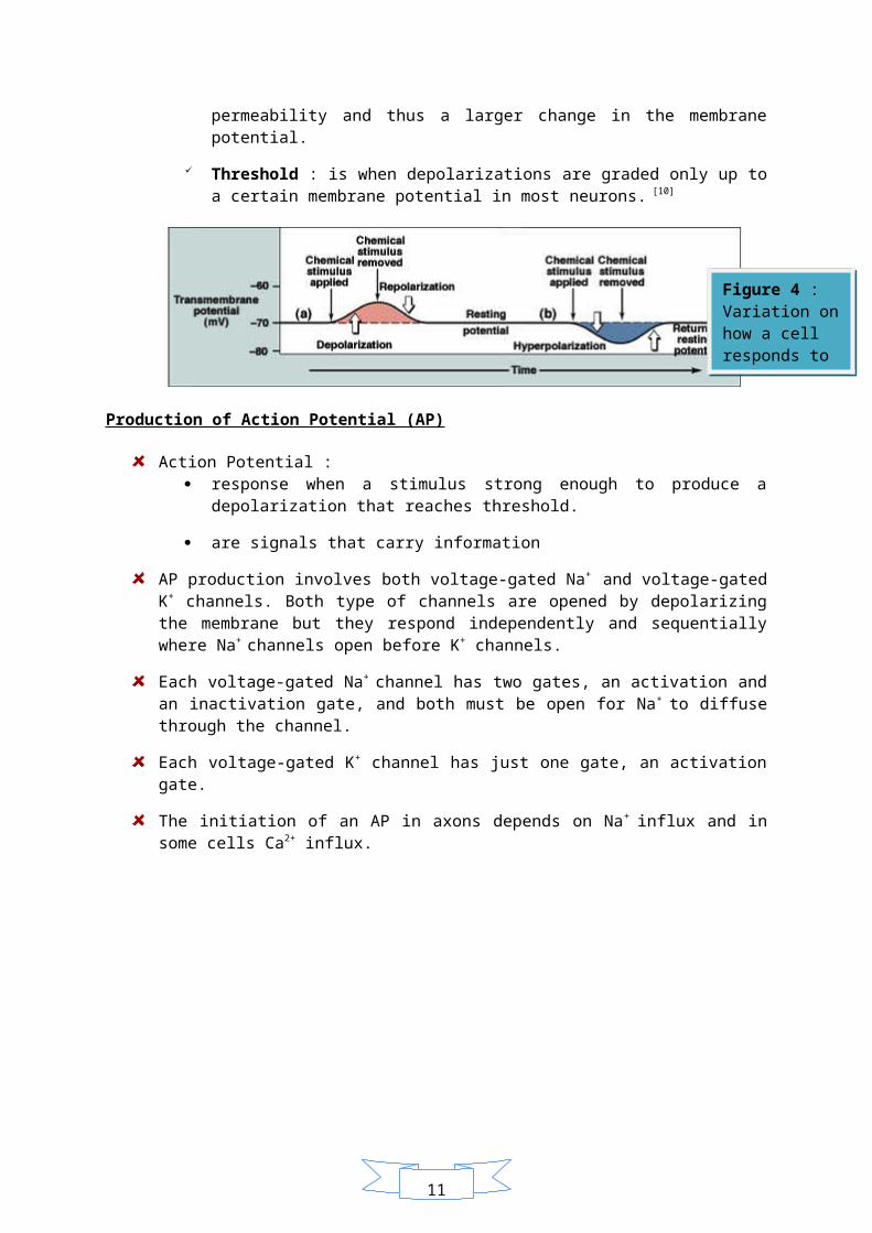
Hyperpolarization : an increase in the magnitude of the membrane potential ( the inside becomes more negatively charged) is triggered. This may be caused by the opening of K+ channels, which increases the membrane’s permeability to K+.
Depolarization : a reduction in the magnitude of the membrane potential ( the inside of the membrane becomes less negative). This may be due to the opening of gated Na+ channels, which increases the membrane’s permeability to Na+.
Graded potential : are the changes in membrane potential because the magnitude of the hyperpolarization or depolarization varies with the strength of the stimulus where a larger stimulus causes a larger change in permeability and thus a larger change in the membrane potential.
Threshold : is when depolarizations are graded only up to a certain membrane potential in most neurons. [10]
Production of Action Potential (AP)
Action Potential : response when a stimulus strong enough to produce a depolarization that reaches
threshold.
are signals that carry information
AP production involves both voltage-gated Na+ and voltage-gated K+ channels. Both type of channels are opened by depolarizing the membrane but they respond independently and sequentially where Na+ channels open before K+ channels.
Each voltage-gated Na+ channel has two gates, an activation and an inactivation gate, and both must be open for Na+ to diffuse through the channel.
Each voltage-gated K+ channel has just one gate, an activation gate.
The initiation of an AP in axons depends on Na+ influx and in some cells Ca2+ influx.
Figure 4 : Variation on how a cell responds to a specific stimuli.
Figure 5: Role of voltage-gated ion channels in generation of Action Potential
11
1 : At the resting potential, the activation gate on Na+ and K+ channels are closed and an inactivation gate on Na+ channel is open. The membrane’s resting potential is maintained.
2: When a stimulus depolarizes the membrane, the activation gates on some Na+ channels open, allowing more Na+ to diffuse into the cell. The Na+ influx causes further depolarization, which opens the activation gates on still more Na+ channels, allowing even more Na+ to diffuse into the cell. If the depolarization reaches the threshold, it triggers an action potential.
3: Once the threshold is crossed, depolarization opens the activation gates on most Na + channels, while the K+ channels’ activation gates remain closed. Na+ influx makes the inside of the membrane positive with respect to the outside.
4: The inactivation gates on most Na+ channels close, blocking Na+ influx. The activation gates on most K+ channels open, permitting K+ rapid efflux which again makes the inside of the cell negative.
5: It is the final phase of an action potential where both gates of the Na+ channels are closed, but the activation gates on some K+ channels are still open. As these gates close on most K+ channels, and the inactivation gates open on Na+ channels, the membrane returns to its resting state. The AP-induced depolarization of cell membrane spreads a small distance in either direction inside the axon. [9]
References
[1] Eeshils, Maurice,et. al.,Modern Nutrition in health and disease,10th ed,2006