Pharmacologyonline 3: 1021-1032 (2011) Sharma and Kumar 1021 HEPATOPROTECTIVE EFFECT OF CHLOROPHYTUM BORIVILIAUM ROOT EXTRACT AGAIST ARSEIC ITOXICATIO Sunil Kumar Sharma and Madhu Kumar* Cell & Molecular Biology Lab, Deptt of Zoology, University of Rajasthan, Jaipur, India *Corresponding author: Tel: +91 9829324629. e-mail : [email protected]Summary The present study was conducted to find out the hepatoprotective effect of Chlorophytum borivilianum root extract against arsenic induced toxicity. Three groups were made: Control(DDW), Arsenic intoxicated group (4 mg/kg b.w) and Combination group (NaAsO 2 (4 mg/kg b.w)+ cb root extract(800 mg/kg b.w). Animals received their respective doses daily for 30 days orally. Body weight, liver weight, heptopathological changes and level of ATPase were observed. Result showed significant decrease in body and liver weight along with disturbed hepatoarchitecture and decline in ATPase activity in arsenic intoxicated group as compared to control. In combination group, increased body and liver weight along with increased ATPase levels and almost normal hepatoarchitecture were observed as compared to arsenic treated group. Thus it can be concluded that Chlorophytum borivilianum root extract has potential to decrease the toxic effects of arsenic. Keywords: Cb; sodium arsenite; body weight; liver weight; ATPase . Introduction Arsenic, a ubiquitous metalloid which occurs naturally is a toxic pollutant. It is ranked first in a list of 20 hazardous substances 1 . Its exposure occurs from inhalation, absorption through skin, by ingestion of contaminated drinking water and by food 2 . Exposure to arsenic leads its accumulation in tissues such as skin, hair and nails, resulting in various clinical symptoms such as hyperpigmentation and keratosis 3 . It affects nearly all organs. Inorganic arsenic exposure may lead to cancer of liver, kidney, bladder, prostrate and skin as well as to Black foot disease, heart disease, maningioma and other adverse health effects 4,5,6 .
Transcript
Pharmacologyonline 3: 1021-1032 (2011) Sharma and Kumar
1021
HEPATOPROTECTIVE EFFECT OF CHLOROPHYTUM
BORIVILIA�UM ROOT EXTRACT AGAI�ST ARSE�IC
I�TOXICATIO�
Sunil Kumar Sharma and Madhu Kumar*
Cell & Molecular Biology Lab, Deptt of Zoology, University of
The present study was conducted to find out the hepatoprotective effect of
Chlorophytum borivilianum root extract against arsenic induced toxicity.
Three groups were made: Control(DDW), Arsenic intoxicated group (4 mg/kg
b.w) and Combination group (NaAsO2(4 mg/kg b.w)+ cb root extract(800
mg/kg b.w). Animals received their respective doses daily for 30 days orally.
Body weight, liver weight, heptopathological changes and level of ATPase
were observed. Result showed significant decrease in body and liver weight
along with disturbed hepatoarchitecture and decline in ATPase activity in
arsenic intoxicated group as compared to control. In combination group,
increased body and liver weight along with increased ATPase levels and
almost normal hepatoarchitecture were observed as compared to arsenic
treated group. Thus it can be concluded that Chlorophytum borivilianum root
extract has potential to decrease the toxic effects of arsenic.
Keywords: Cb; sodium arsenite; body weight; liver weight; ATPase .
Introduction
Arsenic, a ubiquitous metalloid which occurs naturally is a toxic pollutant. It
is ranked first in a list of 20 hazardous substances1. Its exposure occurs from
inhalation, absorption through skin, by ingestion of contaminated drinking
water and by food2. Exposure to arsenic leads its accumulation in tissues such
as skin, hair and nails, resulting in various clinical symptoms such as
hyperpigmentation and keratosis3. It affects nearly all organs. Inorganic
arsenic exposure may lead to cancer of liver, kidney, bladder, prostrate and
skin as well as to Black foot disease, heart disease, maningioma and other
adverse health effects4,5,6
.
Pharmacologyonline 3: 1021-1032 (2011) Sharma and Kumar
1022
Arsenic causes a significant increase in the rate of formation of reactive
oxygen species (ROS) such as O2-,OH- and H2O2.
7 These are generated during
redox cycling and metabolic activation process.8 It also causes lipid
peroxidation,9 oxidation of proteins and enzymes as well as DNA and DNA
adducts.10
ATPase forms a large family of membrane proteins which
couple ATP hydrolysis to the active transport of cations or other compounds
such as phospholipids across cell membranes. 11,12
and is also considered as a
master enzyme that controls many important functions at cellular and organ
level including active tansport and electric potential across plasma membrane,
intracellular pH regulation, cell division and cell elongation. 13
Chlorophytum borivilianum (Safed musli) is a traditional rare Indian
medicinal herb.14Its roots are widely used for various therapeutic applications
in the Ayurvedic and Unani medicinal systems.15 Major phytochemical
cpmpounds reported from the roots of Chlorophytum borivilianum are
saponins, fructans, gallotanins, phenolic compounds and fructo-
oligosaccharides (FOS). 16,17,18
Saponins are steroid or triterpenoid glycosides,
common in a large number of plants and plant products that are important in
human and animal nutrition. Several biological effects have been ascribed to
saponins. Extensive research has been carried out into the membrane-
permeabilising, immunostimulant, hypocholesterolaemic and anticarcinogenic
properties of saponins and they have also been found to significantly affect
growth, feed intake and reproduction in animals. 19
In the present study, the objective is to elucidate hepatoprotective role Cb
root extract on arsenic intoxicated liver damage in mice.
Meterials and methods
Animals: Random-bred, male Swiss albino mice, (7-8 weeks) were used for
experiments. These animals were maintained in the animal house at
temperatures of 24 ± 3°C and a light of 12:12 hours of light and dark. These
animals were housed in polypropylene cages and fed standard mice feed from
Hindustan Lever Ltd. India. Tap water was provided to the animals ad libitum
and tetracycline was given to the animals against any infections.The ethical
committee of Department of Zoology, University of Rajasthan, Jaipur (India)
has approved to carry out the experiments.
Chemical:- Heavy metal Arsenic in the form of Sodium arsenite
(NaAsO2,trivalent) CAS No. 7784-465 used in the present study and was obtained
from Himedia, Mumbai, India (Batch No. 3-1621 RM-1847. The salts of Arsenic
was dissolved in double distilled water (DDW) and was administered orally. Different dose of arsenic were administered and dose was determined on basis of
LD50/30.(Fig.1)
Pharmacologyonline 3: 1021-1032 (2011) Sharma and Kumar
1023
Figure 1. Different dose of arsenic administration and the dose selection
determination on basis of LD50/30.
Preparation of Chlorophytum borivilianum root extract (drug):The roots
were collected locally, air dried in shade and powdered. Powder was distilled
in Soxhlet apparatus (for 36 hours using DDW) at 400 C. The remaining
material was dried in oven at 360 C and was used as drug.
Reducing power assay: Reducing power assay done by method of Oyaizu
(1986)20.The absorbance read at 700 nm.
Figure 2. The reducing power assay of Chlorophytum borivilianum
and its comparison with ascorbic acid. Ascorbic acid served as positive
control.
Pharmacologyonline 3: 1021-1032 (2011) Sharma and Kumar
1024
Drug Tolerance study and Selection of Dose: Mice were divided into
various groups to receive 100, 200, 400, 800 mg/kg body weight of root
extract orally for seven consecutive days. The animals were observed for 30
days. After 30 days, lipid per oxidation (LPO) and GSH content were
measured in the liver in all groups and according to the highest GSH and
minimum LPO level, the dose was decided.
Treatment of animals
The following experiment was designed to examine the effects of
Cholorophytum borivilianum on arsenic induced toxicity.
Group I (Control Group): - Animals in this group were not given plant
extract or heavy metal. Only vehicle (DDW) was given orally.
Group II (Arsenic treated group):- Arsenic 4mg/kg bodyweight was given
orally upto 30 days in the form of sodium arsenite (III).
Group III (Combination Group):- Sodium arsenite and Chlorophytum
borivilianum root extract both were given up to 30 days orally.
Autopsy Intervals: - The Animals from the above groups were autopsied at
various intervals i.e. 1, 3, 7, 15 and 30 days.
Parameters to studied: - Body weight and Liver weight changes : The animal from each group were
weighed and killed by cervical dislocation on days 1, 3, 7, 15 and 30 days
and liver was carefully excised, trimmed free of extraneous tissue, blotted dry
and weighed quickly and used for histopathological and biochemical studies (
for ATPase).
Histopathological preparations – Excised liver was fixed in Bouin's fixative
for 24 hrs. The fixed tissue was further processed by standard method and
sections were cut at 5 µ and stained with Haematoxyline and Eosine.
Total ATPase assay: - Liver homogenate was made. The total ATPase was
determined according to the method of Akagawa and Tsukada (1979).21
Statistical analysis
The statistical significance in the different parameters between control and
experimental were assessed by one way ANOVA.
Results
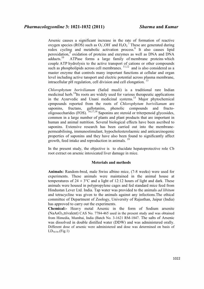
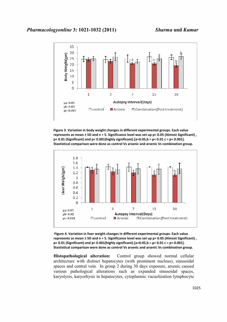
Body weight and Liver weight Changes :The body weight and liver weight in NaASO2 treated group were found to be significantly lower with respect to DDW (control) from day1 to 30 whereas both were recovered in combination group with respect to arsenic intoxicated group (Fig. 3, 4).
Pharmacologyonline 3: 1021-1032 (2011) Sharma and Kumar
1025
Figure 3. Variation in body weight changes in different experimental groups. Each value
represents as mean ± SD and n = 5. Significance level was set up p< 0.05 (Almost Significant) ,
p< 0.01 (Significant) and p< 0.001(highly significant).[a=0.05,b = p< 0.01 c = p< 0.001].
Stastistical comparison were done as control Vs arsenic and arsenic Vs combination group.
Figure 4. Variation in liver weight changes in different experimental groups. Each value
represents as mean ± SD and n = 5. Significance level was set up p< 0.05 (Almost Significant) ,
p< 0.01 (Significant) and p< 0.001(highly significant).[a=0.05,b = p< 0.01 c = p< 0.001].
Stastistical comparison were done as control Vs arsenic and arsenic Vs combination group.
Histopathological alteration: Control group showed normal cellular
architecture with distinct hepatocytes (with prominent nucleus), sinusoidal
spaces and central vein. In group 2 during 30 days exposure, arsenic caused
various pathological alterations such as expanded sinusoidal spaces,
karyolysis, karyorhysis in hepatocytes, cytoplasmic vacuolization lymphocyte
Pharmacologyonline 3: 1021-1032 (2011) Sharma and Kumar
1026
infiltration and enucleation (Fig 5, 6 & 7) in hepatocytes as compared to
control (DDW) group. In combination group, it showed recovery in the form
of maintained hepatic histoarchitecture.
Figure 5. Photomicrograph of arsenic treated group at 400X showing Cytoplasmic
Vacuolization, Expanded sinusoidal space.
Figure 6. Photomicrograph of control treated group at 400X showing central vein,
portal vein,binuceated cells and normal sinusoidal space.
Pharmacologyonline 3: 1021-1032 (2011) Sharma and Kumar
1027
Figure 7. . Photomicrograph of combination treated group at 400X showing
hepatocytes and sinusoidal space.
Total ATPase Assay: The total ATPase activity in NaASO2 treated group
was found to be significantly lower with respect to DDW (control), whereas
combination group showed significant elevation in total ATPase with respect
to their control (arsenic treated group) during 30 days experimental period
(Fig.8).
Figure 8.Variation in ATPase activity in different experimental groups. Each value represents as
mean ± SD and n = 5. Significance level was set up p< 0.05 (Almost Significant) , p< 0.01
(Significant) and p< 0.001(highly significant).[a=0.05,b = p< 0.01 c = p< 0.001]. Stastistical
comparison were done as control Vs arsenic and arsenic Vs combination group.
Pharmacologyonline 3: 1021-1032 (2011) Sharma and Kumar
1028
Discussion
Reduction in body weight is used as an indicator for the deterioration of
general health status. It has been reported that arsenic could induce
toxicological effects and biochemical dysfunctions representing serious health
hazards.22
The findings from the present study indicate that arsenic exposure
caused decrease in the body and liver weight which are in support of the
findings by Yousef et al. (2008) and El-Demerdash et al. (2009)23
who
reported that high arsenic exposure have significantly induced disturbances
the total body weight and liver weight. Oral administration of arsenic is
associated with severe gastrointestinal and liver side effects24
. Arsenic
increases permeability of intestinal lining and causes the gut leaky. This
increased permeability of intestinal lining may be responsible for improper
absorption of nutrients and loss of appetite and weakness25
. and reduction in
body weight26. Reduction in liver weight is due to damaged hepatic
histoarchitecture. Arteel et al.(2008) 27 suggested that arsenic exposure caused
a change in the balance between cell death and proliferation coupled with loss
of body weight. Arsenic produces ROS during its cycles between different
oxidation states,28
which appears to be involved in the mechanism of various
types of cell injury 29,7,9
. Liver cells have particularly high probability of being
subjected to ROS induced toxicity because hepatocytes produces large amount
of ROS during the dexoification of xenobiotics and toxic substances30
.
Arsenic induced reactive oxygen species and subsequent depletion of
antioxidant cell defenses can result in disruption of the pro-
oxidant/antioxidant balance in mammalian tissues31,32
. Consequently, ROS
directly react with cell biomolecules, causing damages to lipids, proteins and
DNA, and hence leading to cell death33,34
.
Present study revealed decreased ATPases activity after arsenic intoxication
at all autopsy intervals. Arsenic induced reactive oxygen species impairs cell
membrane stability7,8
and damages mitochondrial membrane severely. It is
well established that mitochondria are the major site of utilization of oxygen
and many of the mitochondrial enzymes contain essential sulfhydryl groups.
In addition, since the inner and outer mitochondrial membranes contain
unsaturated lipids, mitochondria are more susceptible to arsenic attack as well
as by the free radicals produced by it than other organelles35
.The damaged
membrane cannot develop a proton motive force that is preliminary
requirement of cellular energy production36
. Arsenic disrupts mitocondrial
membrane potential and increases ROS generation37
that causes depletion of
ATP.38
It uncouples oxidative phosphorylation, thus inhibiting energy-linked
reduction of NAD+, mitochondrial respiration, and ATP synthesis
39. Thus
ATPase activity is significantly reduced after arsenic intoxication.
In combination group modulation by Cb root extract was observed in terms
of increased body and liver weight, less damage in hepatocytes and increased
ATPase level. Cb root extract contains Saponins, Gallotannins and Fructans.
Saponins40,Gallotanins41 and Fructans18have antioxidant activity. Saponins
Pharmacologyonline 3: 1021-1032 (2011) Sharma and Kumar
1029
inhibit intracellular ROS formation, reduces the level of the lipid peroxidation
(MDA) and maintains cellular antioxidant enzymes activities 42. Saponins
have hypocholesteric properties 19. Cholesterol enrichment was shown to have
an inhibitory effect on many membrane ATPases, as it may directly interact
with the boundary lipids of ATPase and alter the intermolecular hydrogen
bonds of the protein.43,44 Saponin (Ginsenosides) interacts with membrane
cholesterol and displace it from surrounding lipids environment of ATPases.
Removal of cholesterol will lead to an increase in membrane fluidity which
facilitates conformational changes of ATPases during their transport cycle that
controls the enzyme activity of biological membrane and has important role in
ion transport.45
Saponins maintains Na+- K
+-ATPase activity against ROS
induced reaction.46
Saponins reduces ROS formation due to their antioxidant
property. Since cell membrane is protected, GIT is not leaky,So body weight
is increased. Hepatohistoarchitecture injuries induced by ROS, are reduced
due to antioxidant property of saponins.The body and liver weight are also
recovered. Thus it can be concluded that cb root extract can ameliorates
arsenic induced toxicity in terms of increased body and liver weight, lessens
the hepatotoxicity and maintains ATPase level.
References
1. ATSDR. Arsenic toxicity: What are the standards and regulation for arsenic exposure, 2011 (http://www.atsdr.cdc.gov).
2. Ratnaike RN. Acute and chronic arsenic toxicity. Postgrad Med J 2003;79:391–396.
3. Kapaj S, Peterson H, Liber K , Bhattacharya P. Human health effects from
chronic arsenic poisoning – A review. J of Environ Sci & Health Part A,
2006; 41:2399-2428.
4. Chan PC, Huff J. Arsenic carcinogenesis in animals and in humans:
mechanistic, experimental, and epidemiologic a l e v i d e n c e . J E n
v i r o n S c i H e a l t h, P a r t C : E n v i r o n Carcinog Ecotoxicol
Rev 1997; 15:83–122. 5. Smith AH, Goycolea M, Haque R, Biggs ML. Marked increase in bladder
and lung cancer mortality in a region of Northern Chile due to arsenic in drinking water. Am J Epidemiol 1998;147: 660-9.
6. Chiou HY, Wei ML, Tseng CH. Incidence of transitional cell carcinoma and arsenic in drinking water: a followup study of 8,102 residents in an arseniasis-endemic area in northeastern Taiwan. Am J Epidemiol 2001;153:411–18.
7. Liu SX, Athar M, Lippai I, Waldren C, Hei TK. Induction of oxyradicals by arsenic: implication for mechanism of genotoxicity. 2001;Proc Natl Acad Sci USa 98(4):1643-48.
8. Bashir S, Sharma Y, Irshad M, Dutta-Gupta S, Dogra TD. Arsenic-induced cell death in liver and brain of experimental rats. Basic Clin Pharmacol Toxicol 2006;98:38–43.
Pharmacologyonline 3: 1021-1032 (2011) Sharma and Kumar
1030
9. Sharma A, Sharma MK , Kumar M. Modulatory role of Emblica officinalis fruit extract against arsenic induced oxidative stress in Swiss albino mice. Chem Biol Interact 2009; 180 (1) :20-30.
10.Yamauchi H, Aminaka Y, Yoshida K, Sun G, Pi J , Waalkes M P.
Evaluation of DNA damage in patients with arsenic poisoning: urinary
8-hydroxyguanine.Toxicol and Appl Pharmacol 2004; 198( 3) 291-296.
11. Lutsenko S, Kaplan J. Organization of P-type ATPases: significance of
structural diversity. Biochem 1995; 34: 15607–15613. 12. Moller J P, Juul B, le Maire M. Structural organization, ion transport and
energy transduction of P-type ATPases. Biochim Biophys Acta 1996; 1286: 1–51.
13. Serrano R. Structure and function of plasma membrane ATP-ase. Annu Rev Plant Mol Biol 1989; 40: 61-94.
14. Thakur GS, Bag MS, Bhagwan S, Debnath M, Zacharia A, Bhaduria P, Prasad GBKS , Bisen PS. Chlorophytum bonvilianum: A white gold for biopharmaceuticals and nentraceuticals. Cur Pharmaceut Biotech 2009; 10: 650-666.
15. Oudhia P. Problems perceived by Safed Moosli (Chlorophytum borivilianum) growers of Chhattisgarh (India) region: A study. J. Medicinal and Aromatic Plant Sci 2001; 22/4A & 23/1A:396–399.
16. Acharya D, Mithanie–offer AC, Kaushik N, Miyamoto T, Paululat T, Mirjolet JF. Cytotoxic spirostane type saponins from the roots of Chlorophytum borivilianum. J Nat Prod 2009;11: 165-69.
17. Kaushik N. Saponins of Chlorophytum species. Phychem Rev 2005; 191-196.
18. Narashiman S, Govindarajan R, Madhavan V, Thakur M, Dixit VK, Mehrotra S , Madhusudanan K . Action of fructo-oligosccharide of Chlorophytum borivilianum against streptozotocin-induced oxidative stess. Planta Med 2006; 72(15) :1421-24.
19. Francis G, Keram Z, Makkart HPS , Becker K . The biological action of saponins in animal systems: A review. Br J Nutr 2002; 88 :587-605.
20. Oyaizu M. Studies on products of browning reaction. Antioxidative activities of products of browning reaction prepared from glucosamine. Jpn J Nutr 1986; 44: 307-315.
21. Akagawa K , Tsukada Y. Presence and characteristic of catechomin sensitive Na+ / K+ ATPase in rat stratum. J. Neurochem 1979; 32: 269-271.
22.Yousef IM, El-Demerdash MM, Radwan FME. Sodium arsenite induced biochemical perturbations in rats: ameliorating effect of curcumin. Food Chem Toxicol 2008;48:3506–11.
23. El-Demerdash FM, Yousef MI, Radwan FME. Ameliorating effect of curcumin on sodium arsenite-induced oxidative damage and lipid peroxidation in different rat organs. Food Chem Toxicol 2009; 47:249–54.
24. Sun HD,Ma L ,Hu XC, Zhang TD, Ai-Lin. I treated 32 cases of acute promyelocytic leukemia. Chin J Integrat Chin & West Med 1999;12: 2170.
25. Nutriwest. Healing the Leaky Gut. The Detox-Leaky Gut Connection 2003 (http://www.nutriwest.it).
26. Jadhav SH, Sarkar SN, Aggrawal M, Tripathi HC. Induction of oxidative stress in erythrocytes of male rats subcronically exposed to a mixture of
Pharmacologyonline 3: 1021-1032 (2011) Sharma and Kumar
1031
eight metals found as groundwater contaminants in different parts of India. Environ Contam Toxicol 2007; 52: 145-151.
27. Arteel GE, Guo L, Schlierf T, Beier JI, Kaiser JP, Chen TS, Liu M, Conklin DJ, Miller HL, Monfort C, States JC. Subhepatotoxic exposure to arsenic enhances lipopolysaccharide – induced liver injury in mice. Toxicol Appl Pharmacol 2008; 226(2) 128-139.
28. Ayala-Fierro F, Barber DS, Rael LT, and Carter DE. In vitro tissue specificity for arsine and arsenite toxicity in the rat. Tox Sci 1999; 52: 122–129.
29. Spector A. Oxidative stress and disease J Ocul Pharmacol Ther 2000;16:193-201.
30. Stohs SJ. The role of free radicals in toxicity and diseasein toxicity and disease. J Basic Clin Physiol Pharmacol 1995; 6:205 -228 .
31. Valko M, Rhodes CJ, Moncol J, Izakovic M, Mazur M. Free radicals, metals and antioxidants in oxidative stress-induced cancer. Chem-Biol Interact 2006; 160: 1–40
32. Hansen BH, Romma S, Garmo OA, Olsvik PO, Andersen RA. Antioxidative stress proteins and their gene expression in brown trout (Salmo trutta) from three rivers with different heavy metal levels. Comp Biochem Physiol Part C Toxicol Pharmacol 2006;143: 263–274.
33. Halliwell B, Gutteridge JMC.Free radicals in biology and medicine, Vol. 3. Oxford: University Press Inc 2002; pp 105–245.
34. Mo J, Xia Y, Wade TJ, Schmitt M, Le XC, Dang R, Mumford JL. Chronic arsenic exposure and oxidative stress: OGG1 expression and arsenic exposure, nail selenium, and skin hyperkeratosis in Inner Mongolia. Environ Health Perspect 2006;114:835–841.
35. Ramanathan K, Shila S, Kumaran S, Panneerselvam C. Ascorbic acid and α-tocopherol as potent modulators on arsenic induced toxicity in mitochondria. J Nutr Biochem 2003; 14:416–420.
36. Ye R, Han J, Kong X, Zhao L, Cao R, Rao Z, Zhao G. Protective Effects of Ginsenoside Rd on PC12 Cells against Hydrogen Peroxide. Biol Pharm Bull 2008; 31(10) 1923—1927.
37. Larochette N, Decaudin D, Jacotot E, Brenner C, Marzo I, Susin SA, Zamzami N, Xie Z, Reed J, Kroemer G. Arsenite induces apoptosis via a direct effect on the mitochondrial permeability transition pore. Exp Cell Res 1999; 249:413–421.
38. Csanaky J, Nemeti B, Gregus Z. Dose dependant biotransformation of arsenite in rats--not S-adenosylmethionine depletion impairs arsenic methylation at high dose. Toxicol. 2003; 183(1-3): 77-91.
39. Klaassen C. Watkins J. Casarett and Doull's Essentials of Toxicology. McGraw-Hill; 2003 512.
40. Qiang H, Gao P, Zhang C, Shi Z, Wang T, Wang L , Wang K. Effects of Panax notoginseng saponins on apoptosis induced by hydrogen peroxide in cultured rabbit bone marrow stromal cells via altering the oxidative level and down regulating caspase-3. J Nanj Med Uni 2009; 23(6): 373-379.
41. Bouchet N, Barrier L, Fauconneau B. Radical scavenging activity and antioxidant Properties of tannins from Guiera senegalensis (Combretaceae). Phytother Res 1998 12: 159–162.
42. Kaur R, Thukral AK , Arora S. Attenuation of free radicals by an aqueous extract of peels of Safed musli tubers (Chlorophytum borivilianum Sant et Fernand). J Chin Clin Med 2010; 1(5):1-8.
Pharmacologyonline 3: 1021-1032 (2011) Sharma and Kumar
1032
43. Yamasaki Y, Ito K, Enomoto Y, Sutko JL. Alterations by saponins of passive calcium permeability and sodium-calcium exchange activity of canine cardiac sarcolemnal vesicles. Biochimica et Biophysica Acta 1987; 897: 481– 487.
44. Choi S, Jung SY, Kim CH, Kim HS, Rhim H, Kim SC & Nah SY. Effect of Ginsenosides on voltage-dependent Ca2+channel subtypes in bovine chromaffin cells. J Ethnopharmacol 2001; 74, 75 – 81.
45. Ma LY, Xiao PG. Effects of Panax notoginseng saponins on platelet aggregation in rats with middle cerebral artery occlusion or in vitro and on lipid fluidity of platelet membrane. Phytother Res 1998; 12: 138– 140.
46. Manna P, Sinha M, Sil P C. Prophylactic role of arjunolic acid in response to streptozotocin mediated diabetic renal injury: Activation of polyol pathway and oxidative stress responsive signaling cascades. Chem-Biol Interact 2009; 181(3):297-308.