Page 1
Pharmacologyonline 3: 514-523 (2011) �ewsletter Dimova et al.
514
SPECIAL FEATURES OF PHOTOGRAPHIC DOCUME�TATIO� FOR PATIE�TS
WITH COMPLETE DE�TURES.
Mariana Dimova
1∗∗∗∗, Hrizdana Hadjieva2, Elena Hadjieva
3
1 Department of Prosthodontics- Faculty of Dental Medicine, Medical University –
Sofia and Department of Prosthodontics - Faculty of Dental Medicine, Medical
University – Varna, Bulgaria
2
Department of Prosthodontics- Faculty of Dental Medicine, Medical University –
Sofia, Bulgaria
3
General Practitioner
*Corresponding Author
Mariana Jordanova Dimova, D.D.S., Assoc. Prof.
Department of Prosthodontics- Faculty of Dental Medicine, Medical University – Sofia
Sv. Sv. G. Sofijski – Boul. 1, floor. 8, 1431 Sofia, Bulgaria
Telephone No: +359888872509
E- mail: [email protected]
Summary
Along with the development of photography and technologies, photo documentation in
dental practice is getting more and more important for documentation, discussion, saving and
exchange of information.
There is great amount of articles in the literature presenting and documenting different
clinical cases especially those with esthetic treatment with ceramic, metal-ceramic or
composite prostheses because of the good abilities and high resolution of dental photography.
The aim of the authors is to suggest an approach for documentation of clinical
findings, cast situation models, x-ray examination, and the old and new treatment in
completely edentulous patients.
The objects of our observation are 42 patients (26 women and 16 men) aged from 45
to 81 years. All patients are totally edentulous and are subjected to complete denture
treatment. Patients’ agreement is achieved for photo documentation of treatment process’
stages.
The individual characteristics and treatment stages in every dental treatment could be
documented and saved in database for educational and evidential purposes. The loss of teeth
doesn’t make the photo documentation easier or uninteresting but acquires skill, preparation
and conformity of its special features.
Keywords: Photo documentation, Completely edentulous patients.
Page 2
Pharmacologyonline 3: 514-523 (2011) �ewsletter Dimova et al.
515
Introduction
Before working out the treatment plan for complete dentures the prosthodontist should
observe, estimate, evaluate, summarize and save the information gathered from the clinical
examination, the x-ray examinations and situation cast models for an analysis of the case.
The detailed clinical examination of edentulous jaws at the first appointment and the
information transfer to the patients’ individual record 1, 2, 3, 4, 5, 6
allows to identify and record
the anatomical characteristics, intermaxillary correlations (due to the old prostheses) as well
as the following components: tongue’s features (impressions, changes in the epithelium,
plaque, size, mobility, and neoplastic changes), lips’ peculiarities (herpes, rhagades and
others), the tonus of cheeks and lips.
It should be stressed upon the fact that the clinical findings described in patient’s record
do not evoke identical understanding of the status 7, 8
in different clinicians, who read the
document. The subjective character of perception depends on the level of theoretical and
practical education, the individual clinical experience and postgraduate qualification of the
clinician.
Detailed observation of the old treatment (if there was any) possesses also an important
diagnostic value (9, 10)
. The inspection and evaluation of old dentures in respect to the
prosthetic margins, retention, stability, occlusal height and articulation helps to estimate and
make indirect conclusions for the chewing ability and effectiveness and gives trends for
subsequent investigation and treatment planning. In this sense the information gathered from
the old denture treatment gives additional characteristics to the whole “picture” and should be
saved in the patients’ personal record during the whole treatment and after that.
The situation casts of both jaws (2, 5)
has the advantage of presenting a 3D- prosthetic area,
but this kind of documentation doesn’t have informative value regarding color and texture of
the mucosa (inflammation, hyperkeratosis, desquamation etc.) .
Another important documental and diagnostic means, which gives more information
to the whole edentulous clinical case is the radiological investigation (11, 12)
, It is found quite
often impacted teeth, parts of roots, cysts and other problems in the jaw bones.
Therefore the information gathered from the above mentioned diagnostic methods
should be visualized, summarized and saved throughout the treatment which becomes
possible with the help of photo documentation13, 14, 15, 16, 17
. According to Krieger G. D.18
the
dentists should be taught that “a single picture is more informative than a thousand of
words”. T. Hedge 16
points out that the postgraduate educational programs in some dental
schools include preparing and presenting of a complete treatment plan of a case, including at
least 16 photos.
In this work the authors share their experience in photo documenting a great number of
edentulous patients and points out the characteristic features of that process as well as a
protocol for arranging a case of treatment with full dentures.
Aim: The aim of authors is to suggest a protocol for photo documentation of clinical findings,
situation cast models, x-ray examination, old and new treatment in completely edentulous
patients.
Materials and methods
The objects of our observation are 42 patients (26 women and 16 men) aged from 45
to 81 years. All patients are totally edentulous and are subjected to complete denture
treatment. It is achieved a patients’ information agreement for the purpose of photo
documentation of treatment process’ stages.
Page 3
Pharmacologyonline 3: 514-523 (2011) �ewsletter Dimova et al.
516
The photo documentation has been carried out with the help of a digital camera Nikon
Coolpix 4500 4 MPx-CCD sensor, max image size: 2272x1704 pixels, TIFF-RGB or JPEG
formatting. A ring light is used - SL-1 Macro cool light. The “white balance” has been set up.
For the intraoral pictures a set of mirrors are used, contrast plates and retractors (Nichrominox
- France). For the photo documentation of upper and lower prosthetic area middle or big
occlusal mirrors are used. For left and right occlusal shots there have been used big or middle
lateral mirrors.
The suggested from the authors’ protocol for photo documentation of the prosthetic
treatment of edentulous patients includes:
I. Photo documentation before the beginning of treatment:
• Extraoral pictures in full face and in half face (side view) of the patient without his old
dentures (Fig. 1, 2) at physiological rest and with the dentures (if there are any) in
central occlusion.
• Extraoral pictures in full face with the old dentures (if there are any)-smile
• Intraoral pictures of prosthetic area on upper jaw (Fig. 5, 6, 7, 8) and lower jaw (Fig.
9, 10, 11) - “occlusal view”.
• Focusing on special characteristics or peculiarities of prosthetic area and the adjacent
tissues. (Fig. 12, 13, 14, 15, 16).
• Extraoral pictures of old dentures (Fig. 20).
II. Photo documentation of situational cast models of upper and lower jaw- an “occlusal”
view (Fig. 18, 19) and a picture of the panoramic x-ray examination (Fig. 17)
III. Photo documentation after finishing the prosthetic treatment:
• Extraoral (not in the mouth) pictures of the new pair of dentures (Fig. 21)
• Intraoral pictures with the new dentures in central occlusion-frontal and side view
(Fig. 22, 23).
• Extraoral pictures in full face and in half face (sidevew) of the patient with the new
dentures in central occlusion (Fig. 3, 4).
• Extraoral picture with the old and new dentures-smile (24, 25).
Fig. 1, 2: Extraoral pictures in full face and in half face (side view) of the patient without dentures.
Fig. 3, 4: Extraoral pictures in full face and in half face (sidevew) of the patient with the new dentures
in central occlusion.
Page 4
Pharmacologyonline 3: 514-523 (2011) �ewsletter Dimova et al.
517
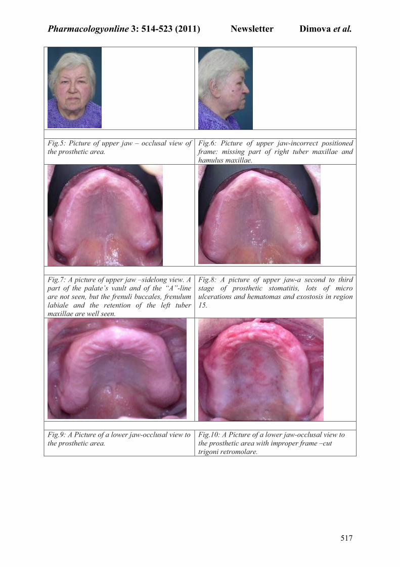
Fig.5: Picture of upper jaw – occlusal view of
the prosthetic area.
Fig.6: Picture of upper jaw-incorrect positioned
frame: missing part of right tuber maxillae and
hamulus maxillae.
Fig.7: A picture of upper jaw –sidelong view. A
part of the palate’s vault and of the “A”-line
are not seen, but the frenuli buccales, frenulum
labiale and the retention of the left tuber
maxillae are well seen.
Fig.8: A picture of upper jaw-a second to third
stage of prosthetic stomatitis, lots of micro
ulcerations and hematomas and exostosis in region
15.
Fig.9: A Picture of a lower jaw-occlusal view to
the prosthetic area.
Fig.10: A Picture of a lower jaw-occlusal view to
the prosthetic area with improper frame –cut
trigoni retromolare.
Page 5
Pharmacologyonline 3: 514-523 (2011) �ewsletter Dimova et al.
518
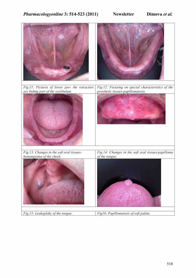
Fig.11: Pictures of lower jaw- the retractors
are hiding part of the vestibulum.
Fig.12: Focusing on special characteristics of the
prosthetic tissues-papillomatosis.
Fig.13: Changes in the soft oral tissues-
hemangioma of the cheek.
Fig.14: Changes in the soft oral tissues-papilloma
of the tongue.
Fig.15: Leukoplaky of the tongue. Fig16: Papillomatosis of soft palate.
Page 6
Pharmacologyonline 3: 514-523 (2011) �ewsletter Dimova et al.
519
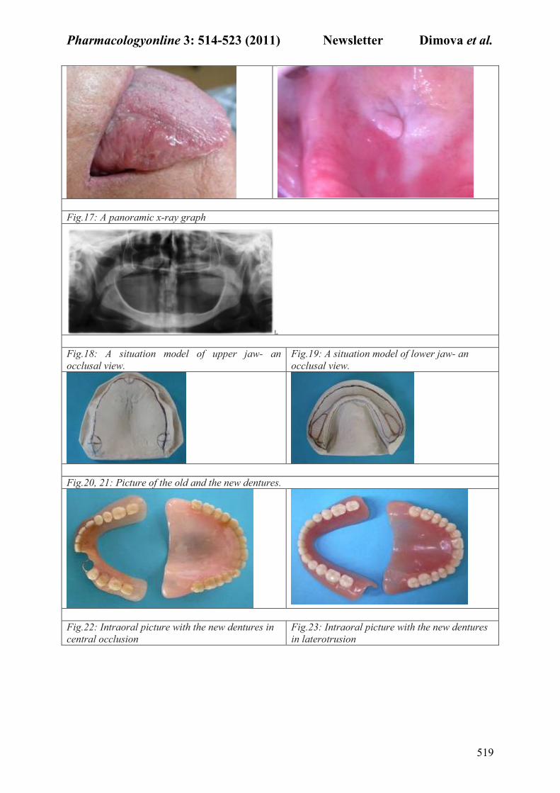
Fig.17: A panoramic x-ray graph
Fig.18: A situation model of upper jaw- an
occlusal view.
Fig.19: A situation model of lower jaw- an
occlusal view.
Fig.20, 21: Picture of the old and the new dentures.
Fig.22: Intraoral picture with the new dentures in
central occlusion
Fig.23: Intraoral picture with the new dentures
in laterotrusion
Page 7
Pharmacologyonline 3: 514-523 (2011) �ewsletter Dimova et al.
520
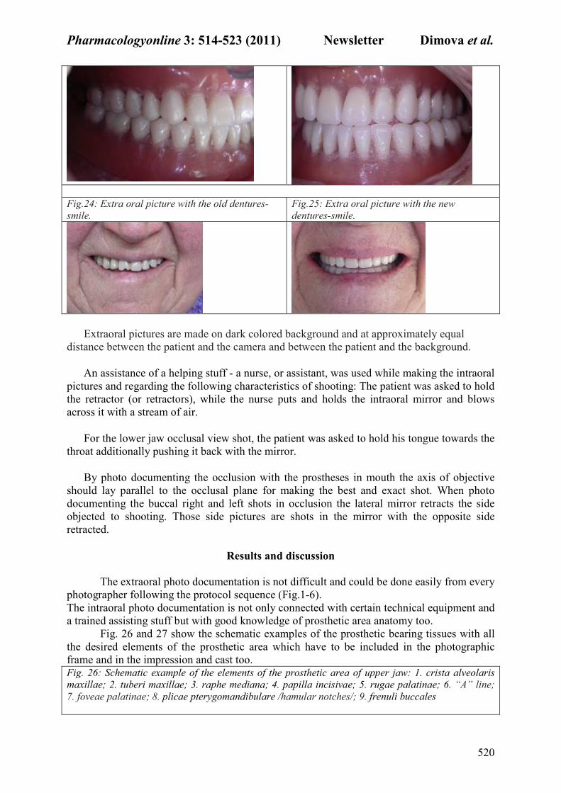
Fig.24: Extra oral picture with the old dentures-
smile.
Fig.25: Extra oral picture with the new
dentures-smile.
Extraoral pictures are made on dark colored background and at approximately equal
distance between the patient and the camera and between the patient and the background.
An assistance of a helping stuff - a nurse, or assistant, was used while making the intraoral
pictures and regarding the following characteristics of shooting: The patient was asked to hold
the retractor (or retractors), while the nurse puts and holds the intraoral mirror and blows
across it with a stream of air.
For the lower jaw occlusal view shot, the patient was asked to hold his tongue towards the
throat additionally pushing it back with the mirror.
By photo documenting the occlusion with the prostheses in mouth the axis of objective
should lay parallel to the occlusal plane for making the best and exact shot. When photo
documenting the buccal right and left shots in occlusion the lateral mirror retracts the side
objected to shooting. Those side pictures are shots in the mirror with the opposite side
retracted.
Results and discussion
The extraoral photo documentation is not difficult and could be done easily from every
photographer following the protocol sequence (Fig.1-6).
The intraoral photo documentation is not only connected with certain technical equipment and
a trained assisting stuff but with good knowledge of prosthetic area anatomy too.
Fig. 26 and 27 show the schematic examples of the prosthetic bearing tissues with all
the desired elements of the prosthetic area which have to be included in the photographic
frame and in the impression and cast too. Fig. 26: Schematic example of the elements of the prosthetic area of upper jaw: 1. crista alveolaris
maxillae; 2. tuberi maxillae; 3. raphe mediana; 4. papilla incisivаe; 5. rugae palatinae; 6. “А” line;
7. foveae palatinae; 8. plicae pterygomandibulare /hamular notches/; 9. frenuli buccales
Page 8
Pharmacologyonline 3: 514-523 (2011) �ewsletter Dimova et al.
521
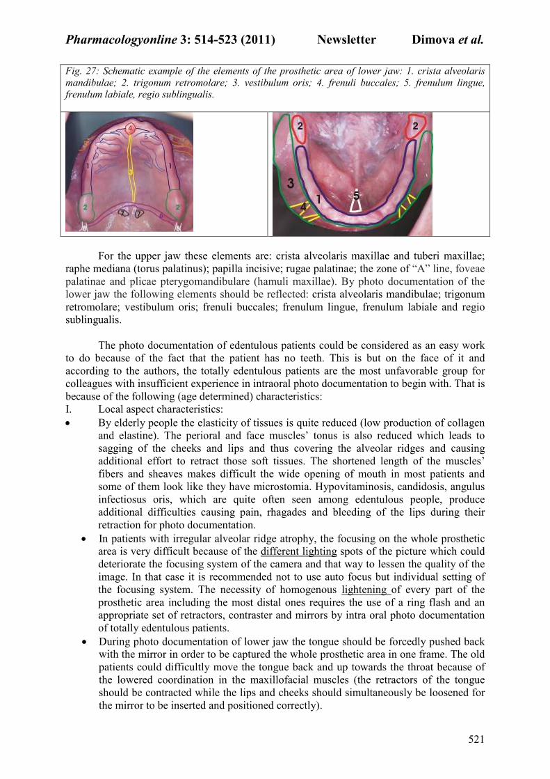
Fig. 27: Schematic example of the elements of the prosthetic area of lower jaw: 1. crista alveolaris
mandibulae; 2. trigonum retromolare; 3. vestibulum oris; 4. frenuli buccales; 5. frenulum lingue,
frenulum labiale, regio sublingualis.
For the upper jaw these elements are: crista alveolaris maxillae and tuberi maxillae;
raphe mediana (torus palatinus); papilla incisive; rugae palatinae; the zone of “А” line, foveae
palatinae and plicae pterygomandibulare (hamuli maxillae). By photo documentation of the
lower jaw the following elements should be reflected: crista alveolaris mandibulae; trigonum
retromolare; vestibulum oris; frenuli buccales; frenulum lingue, frenulum labiale and regio
sublingualis.
The photo documentation of edentulous patients could be considered as an easy work
to do because of the fact that the patient has no teeth. This is but on the face of it and
according to the authors, the totally edentulous patients are the most unfavorable group for
colleagues with insufficient experience in intraoral photo documentation to begin with. That is
because of the following (age determined) characteristics:
I. Local aspect characteristics:
• By elderly people the elasticity of tissues is quite reduced (low production of collagen
and elastine). The perioral and face muscles’ tonus is also reduced which leads to
sagging of the cheeks and lips and thus covering the alveolar ridges and causing
additional effort to retract those soft tissues. The shortened length of the muscles’
fibers and sheaves makes difficult the wide opening of mouth in most patients and
some of them look like they have microstomia. Hypovitaminosis, candidosis, angulus
infectiosus oris, which are quite often seen among edentulous people, produce
additional difficulties causing pain, rhagades and bleeding of the lips during their
retraction for photo documentation.
• In patients with irregular alveolar ridge atrophy, the focusing on the whole prosthetic
area is very difficult because of the different lighting spots of the picture which could
deteriorate the focusing system of the camera and that way to lessen the quality of the
image. In that case it is recommended not to use auto focus but individual setting of
the focusing system. The necessity of homogenous lightening of every part of the
prosthetic area including the most distal ones requires the use of a ring flash and an
appropriate set of retractors, contraster and mirrors by intra oral photo documentation
of totally edentulous patients.
• During photo documentation of lower jaw the tongue should be forcedly pushed back
with the mirror in order to be captured the whole prosthetic area in one frame. The old
patients could difficultly move the tongue back and up towards the throat because of
the lowered coordination in the maxillofacial muscles (the retractors of the tongue
should be contracted while the lips and cheeks should simultaneously be loosened for
the mirror to be inserted and positioned correctly).
Page 9
Pharmacologyonline 3: 514-523 (2011) �ewsletter Dimova et al.
522
• Patients who had not wear prostheses for a long time have their tongue usually
enlarged by hypertrophy and with hypertonic activity. If in such case the patient has
short frenulum lingue the retracting back of the tongue is not enough for a good
picture. The resolution is to ask the patient to swallow and to stay with open mouth
and the tongue put back and down. It is possible in the shot to present part of the
tongue, which is hiding parts the lingual slopes of the lower jaw. It is recommended in
such cases to use mirrors with lingual curve.
II. General aspect characteristics.
• Totally edentulous patients often are with lowered acoustic abilities or pressbyaccusis.
That forces the dentist to repeat several times the explanations and directions of what
they have to do before the photo documentation which lengthens the procedure in time.
• Usually old people get tired easily and when the photographing process gets longer the
muscles tiredness cause tremor, which damages the possibility of getting a good focused
picture. In other cases old patients’ general diseases as cardiovascular, respiratory,
neurological, psychological and others, demands a quick and dynamic protocol. On the
other hand in most cases there are no changes in the anatomy of the prosthetic area so it
is not necessary to make pictures in the middle stages of the treatment as it is with the
fixed prosthodontics’ patients. This allows us to suggest the photographic protocol only
in two parts: photo documentation of patient at the first appointment before beginning
the treatment, and documentation at the end of treatment. Exception of that could be
done in cases with oral mucosa inflammation or preprosthetic surgery corrections. It is
recommended to make an additional photo registration of treated areas.
• Sometimes local and general characteristic could interrupt and postpone photo
documentation.
• The upper listed old age characteristics should not unmotivate the dentists for photo
documentation of edentulous patients. There are also factors which facilitate this
procedure and one of them is the low production of saliva (hypo salivation), which
minimizes the need of constant evacuation of saliva.
• Another favorable factor is the availability of spare time of the patients, most of which
are in pension and even are satisfied of the additional attention during the photo
documentation.
• Patients readily sign the information agreement for photo documentation and collaborate
through it. These additional interrelations between the patients and the clinician who
direct the photo documentation has a good psycho-prophylactic effect and leads to a
positive attitude of patients to the prosthodontic treatment.
Conclusions
The photographic diagnosis is as important as any other diagnostic method. As a mode
of documenting information, photo documentation gives the greatest abundance of
information. While at observation appointment some characteristics of totally edentulous
prosthetic area can be missed the rational of photo documentation is that its information can
be repeatedly reviewed after the patient leaves the clinic.
The clinical significance of photo documentation of edentulous patients consists of the
following:
1. The ability of dental clinicians to distinguish and identify the anatomic parameters and
characteristic of prosthetic area, to recognize and concentrate on specific findings
which influence the diagnostics and future treatment plan are trained by the
photographic protocol.
2. The photo documentation ensures a well arranged visualization for the treatment
resolution of the case.
Page 10
Pharmacologyonline 3: 514-523 (2011) �ewsletter Dimova et al.
523
3. The data transfer between the clinics and the technical laboratories by means of photo
documentation (including casts and written directions) improves the collaboration and
create optimal prerequisite for successful treatment with full dentures.
4. The photo documentation allows creating a database of clinical cases with diagnostic,
prognostic and evidence value. The comparative characteristics between the old and
new prostheses give visualization of the success or failure of treatment.
The prosthetic treatment of a totally edentulous patient is a challenge for the clinicians
and photo documentation of such cases is an indication for the professional level and
qualification of practitioner. The protocol suggested for photo documentation of edentulous
patients is suitable for presenting and discussing of a clinical case during educational seminars
for students, postgraduates and colleagues.
References
1. Geering H, Kundert M. Total- und Hybridprothetik. Farbatlanten der Zahnmedizin,
Georg Thieme Verlag Stuttgart New York; 1986. p. 7-16
2. Golgstein R, M Lancaster. Survey of patient attitudes toward esthetic procedures.
J. P.D. 1984; 52: 775
3. Hupfauf L, Gernet W, Horn R, Jüde H, Kobes L, Landt H, et al. Totalprothesen.
Praxis der Zahnheilkunde. Urban & Schwarzenberg; 1987. 2: p. 3-11
4. Winkler S. Essentials of Complete Denture Prosthodontics. Philadelphia W. B.
Saunders; 1979. p. 88-141
5. Zarb G, Bolender C, Hickey J., Carlsson G. Boucher`s Prosthodontic Treatment
for Edentulous Patients. St. Louis CV Mosby; 1999. p. 10
6. Angulo F. Panoramic radiograph in edentulous and partially edentulous patients.
Acta Odontol. Venez. 1989; 27, 2-3: 60-67
7. Block N. Mental pictures and cognitive science. The Philosophical Review. XCII,
4, 1983; 21-24
8. Schneider F, Fink G. Funktionelle MRT in Psychiatrie und Neurologie Springer
Berlin – Heidelberg; 2007. p. 236-239
9. Alexander L. Effects of complete dentures on facial esthetics. J.P.D. 1964; 14, 2:
231-255
10. Brisman A. Esthetics: A comparison of dentist`s concepts. J. Am. Dent. Assoc.
1980, 100: 345
11. Keur, J., P. Campbell, J. Mc Carthy: Radiological findings in 1135 edentulous
patients, J. Oral Rehab., 1987,14, 183-91
12. Mohamed S, Hamouda A, El-Gheriani W. Panoramic radiographic findings of
edentulous patients prior to full denture. Ainshams Dental Journal 2005; VIII, 2:
255-259
13. Bengel W. Mastering Digital Dental Photography. Quintessence Publishing 2002;
249
14. Bang W. European mastering digital dental photography. Qintessence publishing,
New Malden, Surrey, UK; 2006. P. 56-60
15. Tatsuo, H. Photography in Medical and Dental Field. “A Medical Photograph” in
Clinical Application in Dentistry and its Digitization. J. of the Society of
Photographic Science and Technology of Japan 2003; 66, 1: 13-16,
16. Hedge T. Snapping Images: How To Get Full Use Out Of Your Digital Camera.
Dental Economics online 2002; 6
17. Ratcliff J, Fondriest J, Bush D. Digital dental photography: A Clinicians Guide.
Digital photography Course Manual. L.D. Rankey Institute; 2004
18. Krieger G. Continuum for complete care. Academy for general dentistry 2009


![Pharmacologyonline 2: 601-628 (2010) ewsletter Ladda et al. · Pharmacologyonline 2: 601-628 (2010) ewsletter Ladda et al. 604 2] The interest of the manufacturer to promote newly](https://static.documents.pub/doc/80x56/5f8c8434e51bb360d142b2a3/pharmacologyonline-2-601-628-2010-ewsletter-ladda-et-al-pharmacologyonline-2.jpg)


