Page 1
Pharmacologyonline 3: 935-943 (2011) ewsletter �agamani and Mahmood
935
Synthesis of SCL-LCL-PHA Co-Polymer from Starch by Pseudomonas
Aeruginosa OU67 Isolated from Polluted Water
P. Nagamani , Shaik.Mahmood*
Environmental Microbiology Lab., Department of Botany, Osmania University,
Hyderabad-500 007.AP, India.
*Corresponding Author: [email protected]
Summary
Pseudomonas aeruginosa OU67 was isolated from polluted water and found to produce short-
chain-length-long-chain-length polyhydroxyalkanoate (SCL-LCL-PHA) co-polymer and
characterized. Strain OU67 grew well on starch at temperatures 300 C, pH7 and accumulated
57.77 PHA%(wt) at 30 h of incubation. The purified polymer sample from cells was determined
as SCL-MCL-PHA co-polymer by gas chromatographic and nuclear magnetic resonance analysis
of polymers. The polymer extracted from P. aeruginosa OU67 exhibited comparable material
properties with the commercial polymer. As compared with heterotrophic bacteria, P. aeruginosa
OU67 was able to utilize a wide range of carbon sources with good enzymatic potentials, for
PHA production is thus, low-cost PHA production can be achieved. The 16S rDNA gene
sequences of P. aeruginosa OU67 have been deposited in the EMBL data library under
accession number FN663622.The type strain was submitted in the (JCM 17284T = CCTCC AB
2010470T)
Keywords: Pseudomonas aeruginosa OU67;starch;co-polymer;NMR.
Introduction
Polyhydroxyalkanoates (PHA) are a family of bio polyesters synthesized by many types of
bacteria as carbon and energy reserve materials[1].Due to increasing industrial interest exists in
the biotechnological production of Polyhydroxyalkanoates (PHA) from renewable resources to
arrive at bio-based and biodegradable polymeric materials that can act as alternatives for
common plastics derived from petrol [2,3].For these reasons, researchers have focused their
endeavours on the development of biomaterials (natural products that are synthesized and
catabolized by different organisms and that have broad biotechnological applications) to generate
fully biodegradable compounds with potential industrial applicability[4]. PHA can be divided
into three classes depending on the number of carbon atoms in their monomer units; shortchain-
length (SCL), medium-chain-length (MCL) and long-chain-length polyhydroxyalkanoates (LCL-
PHAs), composed by hydroxyacids with 3–5, 6–14 or more than 14 carbon atoms respectively.
About more than 100 different monomer units reported so far, none of them contain more than
14 carbon atoms as constituents of PHA [5].
Page 2
Pharmacologyonline 3: 935-943 (2011) ewsletter �agamani and Mahmood
936
Anderson and collaborators referred to the capability for PHA biosynthesis from unrelated
substrates [1,6] this is very important as it allows the synthesis of PHAs from simple substrates
rather than fromexpensive or relatively toxic substrates [6]Pseudomonas aeruginosa and other
fluorescent pseudomonads including P. putida, P. mendocina and P. stutzeri, p. fluorescens were
reported to produce mcl PHAs from the unrelated carbon source gluconate or glucose [7,8,9].
In the present study we have investigated the influence of cultural conditions by using starch as
carbon source, on the synthesis and accumulation of PHA by Pseudomonas aeruginosa
OU67.The potential of the isolate to accumulate copolyesters of short-chain-length-long-chain-
length polyhydroxyalkanoate(SCL-LCL-PHA) using starch as carbon substrate has also been
evaluated.
Materials and methods
Microorganism and cultural conditions
Polyhydroxyalkanoate accumulating strain P. aeruginosa sp. OU67 which was isolated from the
polluted water was used in this study. E2 medium [10] was used for PHA production. Aerobic
conditions were maintained by shaking the inoculated Erlenmeyer flasks at 300 C for 28 h. Pure
cultures were isolated on agar containing Nile blue .Cultures were directly monitored for the
fluorescent colonies by exposing to ultraviolet light to detect accumulation of lipid storage
compounds including PHA.
Estimation of viable cell count
To estimate viable cell count, the samples were serially diluted with sterile saline solution (1%%
w/v NaCl). The diluted samples (0.1 ml) were plated in triplicate, on nutrient agar plates and
incubated at 30 °C for 24 h to form fully developed colonies. The results were expressed as
colony forming units per ml (cfu/ml).
Characterization of P. aeruginosa OU67
Morphological, growth and biochemical studies were performed using standard methods [11,
12]. Nutrient agar was used for growth, maintenance of the strain and the determination
of the phenotypic characteristics. The isolate OU67 was characterized by its growth at various
temperatures (5, 30, 45 and 60°C), tolerance at different salt levels (2, 4 and 10 g NaCl/100 ml)
and reduction of nitrate. In addition, lecithinase, lipase, gelatinase , protease production and
starch hydrolysis were examined, and anaerobic growth of the isolate was also performed.
Biochemical characteristics were also checked with the Hi25 Enterobacteriaceae identification
kit (KB003) and Hi Carbohydrate kit parts A, B and C (KB009) (both from HiMedia) according
to the manufacturer’s protocol. The antibiotic sensitivity of the isolate OU67was tested against
different antibiotics. The DNA G+C content was determined by the method of Tamaoka &
Komagata (1984) with the modification that DNA was hydrolysed and the resultant nucleotides
were analysed by reversed-phase HPLC[13].
For phylogenetic characterization, the 16S rRNA gene was amplified [14], and the PCR product
was purifieded using the QIA quick PCR purification kit (Qiagen). Sequencing was performed
by using ABI PRISM model 3700 automatic DNA sequencer and the Big Dye Terminator cycle
sequencing kit (both from Applied Biosystems). The 16S rRNA gene sequence (1670
nucleotides) was submitted to the RDP website, aligned and used to build a phylogenetic tree of
the isolate OU67 by neighbor-joining method using the tree builder tool. [15].
Page 3
Pharmacologyonline 3: 935-943 (2011) ewsletter �agamani and Mahmood
937
Scanning Electron Microscopy
For microscopic studies sample was transferred to vials and fixed in 2.5% Glutaraldehyde in
0.05 M phosphate buffer (pH 7.2) for 24 hr at 40C and post fixed 2% aqueous Osmium Tetroxide
in the same buffer for 2 h. After the post fixation samples were dehydrated in series of graded
alcohol and dried to critical point drying with Electron Microscopy Science CPD unit. Then
dried samples were mounted over the stubs with double sided conductivity tape. Finally, applied
a thin layer of gold metal over the sample using an automated sputter coater (JEOL JFC-1600)
for about 3 min. Then scanned the samples in scanning electron microscope (Model: JOEL-JSM
5600, JAPAN).
ucleotide sequence accession number. The 16S rDNA gene sequences of P. aeruginosa
OU67 have been deposited in the EMBL data library under accession number FN663622.The
type strain was submitted in the (JCM 17284T = CCTCC AB 2010470
T).
Estimation of biomass
The microorganism was grown aerobically in 250 and 500 ml Erlenmeyer flasks with 50 ml of
the culture medium and incubated in a rotary shaker at 150 rev/min and 30 °C during 48 h.
Pha extraction and purification
For the extraction of PHA, 300mL of the cells were harvested by centrifugation at 5000×g and
then lyophilized. The following methods were then employed. The PHA extracted from the cell
pellet by the hypochlorite method[16],was washed with methanol and acetone consecutively and
centrifuged at 8000 rpm for 20 min. The polymers were then dissolved in hot chloroform (60°C)
and the solution poured onto glass trays. The chloroform was allowed to evaporate slowly by
placing the trays in the cold room at 4°C. The film of PHA so obtained was used for further
analysis.
Effect of different carbon sources
To increase the yields of polymer PHA, various carbon sources (2% w/v) such as sucrose,
fructose and lactose and starch were added to the nitrogen free medium with the inoculum of 2%
(v/v). Finally starch was selected with E2 mineral medium to optimize the studies.
Analytical methods
Microbial growth was monitored by measuring the cell density of the culture at 600 nm after
suitable dilution with distilled water. Organic nitrogen in the samples was estimated following its
mineralization with hot sulphuric acid. PHA quantification was quantitated according to the
method of Law and Slepecky (1961), whereby the dried pellets containing intracellular PHA
were hydrolysed using concentrated sulfuric acid for 1 h to obtain crotonic acid, which was
quantified by measuring absorbance at 235 nm. Analysis was performed in triplicates for shake
flask samples. Cell dry weight (cdw) was measured by lyophilizing harvested cells from 3 ml
culture broth. PHA content and its composition were determined by gas chromatography using
PHA standards. Cell concentration was defined as cell dry weight per litre of the culture
medium. The PHA content was defined as the ratio of PHA concentration to cell concentration
given as percentage [17].
Page 4
Pharmacologyonline 3: 935-943 (2011) ewsletter �agamani and Mahmood
938
Gas chromatography (GC)
PHA was quantified using a slight modification of the gas chromatographic method of
Riis and Mai (1988).Instead of whole cells, pure, extracted PHA was used. The precipated
polymer was weighed in tightly sealable vials (volume 10 ml). Two ml of 1,2-Dichloroethane
(DCE), 2 ml n-Propanol containing hydrochloric acid (HCl) (1 volume concentrated HCl + 4
volume n-Propanol) and 200 µl internal standard (2.0 g benzoic acid in 50 ml n-Propanol) were
added. The mix was incubated for 4 h in a water bath at 850C. The mixture was shaken
intermittently. After cooling to room temperature, 4 ml water were added and the mixture shaken
for 20 – 30 s. The heavier DCE-Propanol phase was collected and injected directly into the gas
chromatograph. Quantitative evaluation was affected by means of the peak areas of standard
polymer and benzoic acid. [18].
IR
For FT-IR analysis, the PHA was precipitated from the chloroform using cold ethanol. The
precipitated polymer was used to prepare KBr discs (sample: KBr, 1:100). An FT-IR spectrum
1720X spectrometer (Perkin Elmer, USA) was used under the following conditions: spectral
range, 4,000–400 cm–1
; window material, CsI; 16 scans; resolution 4 cm–1
; the detector was a
temperature-stabilized, coated FR-DTGS detector.
MR
The 1H NMR analysis of the polyester samples was carried out on Varian-300 spectrometer
(USA). The 300 MHz 1H NMR spectra were recorded at 24°C in CDCl3 solution of polyester (50
mg/ml) with a acquisition time of 2.0480 seconds, sweep width of 4000 Hz. Tetra methyl saline
was used as an internal chemical shift standard. The spectra was recorded for commercial PHA
(Sigma-Aldrich, USA) and for the polymer extracted from test strains. All experiments were
performed in triplicate to check the reproducibility.
Results and discussion
Characterization of P. aeruginosa OU6
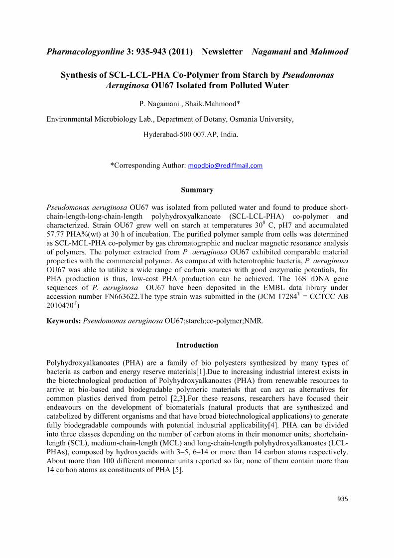
Gram –ve, motile, short rods(figure 1a), (1–1.5 µm×2.5–4.0 µm) occurring singly, 3 mm
diameter colonies on nutrient agar medium, greenish yellow pigmented colonies, diffusible
pigment, turns to dark green on aging and able to emit fluorescence under the UV light
(figure1b). Non spore are formers, colonies are circular glistening, growth good at 20 to 30oc and
pH 7.Catalase +ve, oxidase+, able to degrade caseine, esculin, melanin and starch. HCN +ve,
able to solubilise phosphate, cellulose and lipids. Utilizes L-arabinose, xylose, adonitol,
melibiose, glucose, lactose and citrate. Reduces nitrate to nitrite. Able o accumulate mcl-scl
PHA. Sensitive to Penicillin, novobiocin, nalidixicacid, nitrofurazone and kanamycin. The G+C
content of strain OU67 was 54 mol. %.The 16S rDNA gene sequence is deposited in gene bank
with accession number F 663622. A phylogenetic tree (Figure 5) demonstrated that the isolated
strain was a memberof the genus Pseudomonas , and it formed a monophyletic lineage.
Sequence similarity calculations after a neighbor-joining analysis indicated that the closest
relatives of strain OU67 was Pseudomonas aeruginosa LMG 1242T
(Z76651) with 99.5%
similarity. According to the phylogenetic tree (Figure 5), based on 16S rDNA sequences, the
new strain OU67was found to be affiliated to the genus Pseudomonas.
Page 5
Pharmacologyonline 3: 935-943 (2011) ewsletter �agamani and Mahmood
939
Biosynthesis of poly-β-hydroroxyalkanoates
In shaken flask cultures, the cell mass increased steadily, leading to a maximum cell density
within 30 h of cultivation after which there was a gradual decrease (Figure 4). Cell density of the
culture at 600 nm (OD600) was attained to maximum value of 0.573 after dilutions.. The pH of
the culture medium decreased during the growth, from its initial value of 7to a minimum of 5 .5.
PHA conc g/L was up to 1.755.Cessation of logarithmic growth coincided with the approach of
the pH minimum and rapid consumption of starch. PHA accumulated rapidly during the
stationary phase and reached a maximum concentration of 57.77% of dry cell weight (dcw) at 30
h of growth. It is well known that the ratio of carbon source to nitrogen source (C/N) affects
PHA accumulation; this was also known to be true for strain OU67 grown in glucose media.
However, the production of PHA obtained maximum at 300C, assumed that the temperature was
one of the significant factors to the PHA production for strain OU67. Previously the strain of
Haloferax Mediterranean and Azotobacter chroococcum were employed to produce PHA in a
starch medium [19,20] where hydrolysis of starch was carried out separately. Ramsay et al
reported the accumulation of only 27% medium-chain-length PHA (MCL-PHA)by using
nonanoic acid.[21]
Polymer analysis
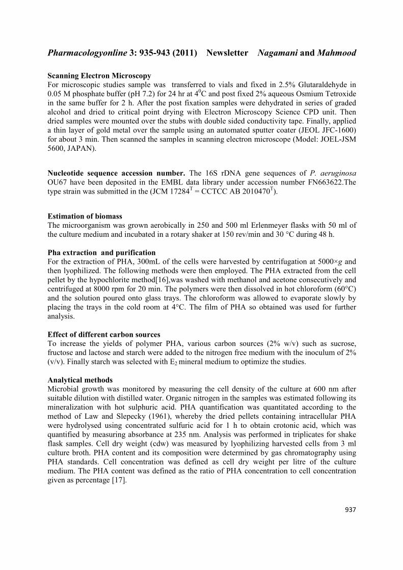
IR spectra were recorded for the polymers dissolved in chloroform. Spectra showed two intense
absorption bands at 1,730.22 and 1,280.66 cm¡1, corresponding to C = O and C–O stretching
groups, respectively. Other absorption bands at 1,379, 1,456, 2,924 and 3,437 cm- 1
corresponding to -CH3, -CH2, -CH and O–H groups are shown in Figure 2. The polymer
analysis revealed the presence of hetero polymer. Its composition was confirmed by 1HNMR
(Figuer 3). The resonance, as observed at0.880, 1.199, 1.622, 2.518 and 5.251 δ by 1H-NMR
analysis were, respectively, for CH3 (3HV, 3HHD and 3HOD side group), CH3 (3HB side
group), CH2 (3HV, 3HHD and 3HOD side group), CH2 (3HV, 3HB, 3HHD and 3HOD bulk
structures), CH (3HV, 3HB,3HHD and HHD bulk structures) of theCDCl3 -soluble fraction of
the polymer confirmed the presence of the copolymer consisting of 3HB, 3HV, 3HHD and
3HOD units in P. aeruginosa OU67(figure 3). In GC analysis, three major ester peaks were
found for the PHA isolated from starch (data not shown).Strain proved good capability to
synthesize SCL-LCL-PHA co-polymer from unrelated carbon sources. The reason for this may
be α-amylase production of the strain and the versatility of this bacterium in the selection of the
carbon source may provide an attractive alternative for the utilization of starch-derived raw
materials.
Conclusion
The results shown above demonstrated that the bacterium, which was isolated from polluted
water, identified as Pseudomonas aeruginosa OU67. It was able to produce good amount of
PHA (up to 57.77% of dry cell weight) in the presence of excesss starch as carbon source in the
E2 mineral medium (Fig. 4), is capable of accumulating LCL 3-hydroxyhexadecanoate and 3-
hydroxyoctadecanoate units with SCL 3-hydroxybutyrate and 3-hydroxyvalerate as constituents
of PHA. Use of inexpensive substrates such as starch could contribute to reducing the PHA
production cost.
Page 6
Pharmacologyonline 3: 935-943 (2011) ewsletter �agamani and Mahmood
940
a b
Figure 1: a Scanning electron micrograph showing morphology of P. aeruginosa OU67
b. Flourescence micrograph of P. aeruginosa OU67
Figure 2: IR spectra of polymer from isolate OU67, grown on starch
Page 7
Pharmacologyonline 3: 935-943 (2011) ewsletter �agamani and Mahmood
941
Figure 3: H1 NMR of polymer from isolate OU67 grown on starch.
8 10 12 14 16 18 20 22 24 26 28 30 32 34 36
0
1
2
3
4
5
6
7
pH
Time
OD at 600nm
% PHA g/L
Time(hr)
pH
,cell
dry
wt
0
2
PH
A(g
/L),
OD
at 6
00
nm
Figure 4: Cell growth and PHA accumulation by Pseudomonas aeruginosa OU6
grown on the starch containing medium. Each point represented an average of three test tubes.
Page 8
Pharmacologyonline 3: 935-943 (2011) ewsletter �agamani and Mahmood
942
Figure 5: Phylogenetic position of the bacterium OU67 with the genus Pseudomonas with
EMBL/GENE bank accession o F 663622
The evolutionary history was inferred using the Neighbor-Joining method [15] . The bootstrap
consensus tree inferred from 1000 replicates [22](Felsenstein, 1985) is taken to represent the
evolutionary history of the taxa analyzed All positions containing gaps and missing data were
eliminated from the dataset (Complete deletion option). Phylogenetic analyses were conducted in
MEGA4[23] (Tamura et al., 2007). The tree is drawn to scale, with branch lengths in the same
units as those of the evolutionary distances used to infer the phylogenetic tree. The evolutionary
distances were computed using the Jukes-Cantor method [24](Jukes and Cantor, 1969) and are in
the units of the number of base substitutions per site.
References
1. Anderson AJ, Dawes EA (1990). Occurrence, metabolism, metabolic role, and industrial use
of bacterial polyhydroxyalkanoates. Microbiol Rev. 54:450–472.
2. Braunegg G, Lefebvre G and Genser KF (1998). Polyalkanoates, Biopolyester from Renewable
Resources: Physiological and Engineering Aspects. J. Biotech. 65: 127-161. 3.Braunegg G, Bona R and
Koller M (2004). Sustainable polymer production. Polym-Plastics Technol Eng .4 3:1779–1793.
4.Kim DY, Rhee YH (2003). Biodegradation of microbial and synthetic polyesters by fungi. Appl.
Microbiol. Biotechnol. 61:300-308.
5.Reddy CSK, Ghai R, Rashmi and Kalia VC (2003) .Polyhydroxyalkanoates: an overview. Bioreso.
Technol .87:137-146.
Strain OU67(F 663622)
Pseudomonas aeruginosa LMG 1242T
(Z76651)
Pseudomonas otitidis MCC10330T
(AY953147)
Pseudomonas resinovorans LMG 2274T
(Z76668)
Pseudomonas alcaligenes LMG 1224T
(Z76653)
Pseudomonas stutzeri CCUG 11256T
(U26262)
Pseudomonas alcaliphila AL15-21T
(AB030583)
Pseudomonas mendocina LMG 1223T
(Z76664)
Pseudomonas pseudoalcaligenes subsp.pseudoalcaligenes
DSM 50188T
(Z76675) Pseudomonas indoloxydans IPL-1T
(DQ916277)
Pseudomonas citronellolis DSM 50332T
(Z76659)
Pseudomonas nitroreducens DSM 14399T
(AM088474)
Pseudomonas anguilliseptica NCIMB 1949T
(X99540)
Pseudomonas echinoides DSM 1805-T (AJ012461)
100
100
52
87
62
38
38
92
52
43
99
0.005
Page 9
Pharmacologyonline 3: 935-943 (2011) ewsletter �agamani and Mahmood
943
6.Steinbuechel A (1991). Polyhydroxyalkanoic acids. In: Byrom D, editor. Biomaterials. Basingstoke:
MacMillan. 125-213.
7. Timm A & Steinbüchel A (1992). Formation of polyesters consisting of medium-chain-length 3-
hydroxyalkanoic acids from gluconate by Pseudomonas aeruginosa and other fluorescent
pseudomonads. Appl. Environ. Microbiol. 56: 3360–3367.
8.He WN, TianWD, Zhang G, Chen GQ & Zhang ZM(1998). Production of novel polyhydroxy-alkanoates
by Pseudomonas stutzeri 1317 from glucose and soybean oil. FEMS Microbiol. Lett. 169: 45–49.
9.Yuji Jiang ,Xin Song, Lei Gong, Ping Li,Chuanchao Dai , Weilan Shao (2008). High poly(β_-
hydroxybutyrate) production by Pseudomonas fluorescens A2a5 from inexpensive substrates. Enzyme and
Microbial Technology .42:167–172.
10.Lageveen RG, Huisman GW, Preustig H, Ketelaar P, Eggink G, Witholt B (1988). Formation of
polyesters by Pseudomonas oleovorans; effect of substrates on formation and composition of poly- (R)-3-
hydroxyalkanoates and poly-(R) -3-hydroxyalkenoates. Appl Environ Microbiol. 54:2924–2932.
11.Holding AJ, Collee JG (1971) .Routine biochemical tests. Methods Microbiol. 6A:2–32.
12. Smibert RM, Krieg NR (1994) Phenotypic characterization. In: Gerhardt P, Murray RGE, Wood WA,
Krieg NR (eds) Methods for general and molecular bacteriology. American Society for Microbiology,
Washington DC, pp 607–654.
13.Komagata, K. & Suzuki, K. (1987). Lipid and cell-wall analysis in bacterial systematics. Methods
Microbiol .19, 161–207.
14. Weisburg WG, Barns SM, Pelletier DA, Lane DJ (1991) .16SRibosomal DNA amplifycation for
phylogenetic study. J Bacteriol.173.(2):697–703.
15. Saitou, N. and Nei, M. (1987) The neighbor-joining method a new method for reconstructing
phylogenetic trees, Mol. Biol. Evo.l 4: 406–425.
16. Rawte T, Mavinkurve S (2002). A rapid hypochlorite method for the extraction of
polyhydroxyalkanoates from bacterial cells. Indian J Exp Biol. 40:924–929.
17. Law, J.H. and Slepecky, R.A. (1961). Assay of poly-b-hydroxybutyric acid. J Bacteriol. 82: 33–36.
18. Riis V, Mai W (1988). Gas chromatographic determination of poly-β-hydroxybutyric acid in microbial
biomass after hydrochloric acid propanolysis. J.Chromatogr. 445: 285–289.
19. Chen CW, Don TM and Yen HF (2006). Enzymatic extruded starch as a carbon source for the
production of poly(3-hydroxybutyrateco- 3-hydroxyvalerate by Haloferax mediterranei. Process Biochem
.41:2289–2296.
20. Quillaguaman J, Hashim S, Bento F, Mattiasson B and Hatti-Kaul R, (2005).
Poly (β-hydroxybutyrate) production by a moderate halophile, Halomonas boliviensis LC1 using starch
hydrolysate as substrate. J Appl Microbiol 99:151–157 .
21. Zhiyong Sun & Juliana A. Ramsay & Martin Guay &Bruce A. Ramsay(2007) . Carbon-limited fed-
batch production of medium-chain-lengthpolyhydroxyalkanoates from nonanoic acid
by Pseudomonas putida KT2440 Appl Microbiol Biotechnol .74:69–77.
22. Felsestein J (1985). Confidence limits on phylogenies: an approach using the bootstrap.
Evolution. 39: 783–791.
23. Tamura, K., Dudley, J., Nei, M., Kumar, S. (2007). MEGA4: molecular evolutionary genetic analysis
(MEGA) software version 4.0. Mol Biol Evol. 24: 1596–1599.
24. Jukes, T. H. & Cantor, C. R. (1969). Evolution of protein molecules. In Mammalian Protein
Metabolism, vol. 3, pp. 21–132. Edited by H. N. Munro. New York: Academic Press.