Page 1
DESIGN, DEVELOPMENT AND EVALUATION OF
NANOSUSPENSIONS FOR ENHANCEMENT OF
ORAL BIOAVAILABILITY OF POORLY SOLUBLE
DRUGS
PH.D. SYNOPSIS
SUBMITTED TO
GUJARAT TECHNOLOGICAL UNIVERSITY
FOR THE DEGREE
OF
DOCTOR OF PHILOSOPHY IN PHARMACY
BY:
MS. JALPA S. PAUN,
ASST. PROFESSOR,
B.K. MODY GOVT. PHARMACY COLLEGE, RAJKOT.
(COLLEGE CODE - 212)
ENROLLMENT NO: 119997290038
SUPERVISOR:
DR. H. M. TANK, PRINCIPAL,
MATUSHREE V.B. MANVAR COLLEGE OF PHARMACY,
UPLETA, RAJKOT.
(COLLEGE CODE - 266)
Page 2
GUJARAT TECHNOLOGICAL UNIVERSITY
CERTIFICATE FROM RESEARCH SCHOLAR
I, Ms. Jalpa Shantilal Paun, herewith submit 6 (Six) copies of the synopsis of my PhD Thesis,
to The Controller of Examination, Gujarat Technological University, Ahmedabad through the
Supervisor, Dr. H. M. Tank, I have also enclosed the soft copy of the synopsis on a CD in the
Portable Document Format (PDF).
My address for communication will be as follows: (I hereby undertake to intimate the PhD section of any change of address)
Ms. Jalpa Shantilal Paun
‘Jalpa’,
5, Kidwainagar,
150ft Ring Road,
Rajkot-360005
Signature of Research Scholar :
Full Name of Research Scholar : Ms. Jalpa Shantilal Paun
Enrolment No. : 119997290038
Date : /10/2016
Page 3
GUJARAT TECHNOLOGICAL UNIVERSITY
CERTIFICATE FROM RESEARCH SCHOLAR & SUPPERVISOR Certified that the Research Scholar with details as above, in Certificate No.1 has carried out the
research work detailed in the Synopsis and Thesis being submitted, during the period 27/09/2011
to 10/10/2016.
Further certified that: 1. There is a prima facie case for consideration of the thesis.
2. The Research Scholar has published a minimum of 2 research papers out of which one
should be in referred journal such as SCI / SCOPUS / SSCI / ABDC / EMERALD. Copies
of the publications or acceptance letters are enclosed herewith (mandatory from Batch
2013).
3. To the best of our knowledge the synopsis / thesis does not include any work which has,
at any time, previously, been submitted for the award of a degree except to the extent of
point 4 below.
4. The following section(s) (if any) of the synopsis / thesis relate to collaborative work: None If patent is being filled, it is recommended that to please tick the following option:
The Thesis be sent for evaluation after the Non-Disclosure Agreement (NDA) has been signed by the examiner and there is a need to maintain the confidentiality of proprietary information (the student has been informed that obtaining NDA from prospective examiners may delay the thesis evaluation)
Signature of Research Scholar :
Name of Research Scholar : Ms. Jalpa Shantilal Paun
Signature of Supervisor :
Name of Supervisor : Dr. Hemraj M. Tank
Page 4
GUJARAT TECHNOLOGICAL UNIVERSITY
CERTIFICATE FROM SUPPERVISOR
The Synopsis and thesis with details as above, may be accepted by the University for
evaluation by external examiners.
__________________________________________
(Signature of Supervisor with date)
Name: Dr. Hemraj M. Tank
Page 5
i
Index
Sr. No. Title Page No.
1 Title of the Thesis 1
2 Abstract 1
3 Brief description of the state of the art of the research topic 1
4 Definition of the problem 5
5 Objective and scope of the work 5
6 Original contribution by the thesis 6
7 Methodology of the Research 7
8 Results of Candesartan Cilexetil Nanosuspension 11
9 Results of Telmisartan Nanosuspension 12
10 Results of Ziprasidone Hydrochloride Nanosuspension 13
11 Achievement with respect to objectives 15
12 Conclusion 15
13 Copies of papers published and a list of all publications arising from the
thesis
16
14 Patents 16
15 References 16
Page 6
PH.D. SYNOPSIS
Ms. Jalpa S. Paun (Enrollment No. 119997290038) 1
1. Title of the thesis
DESIGN, DEVELOPMENT AND EVALUATION OF NANOSUSPENSIONS FOR
ENHANCEMENT OF ORAL BIOAVAILABILITY OF POORLY SOLUBLE DRUGS.
2. Abstract
Drug which belongs to BCS Class-II, has poor oral bioavailability due to its limited aqueous
solubility. Antihypertensive agents (Candesartan Cilexetil and Telmisartan) as well as
atypical antipsychotic agent (Ziprasidone Hydrochloride Monohydrate) with poor water
solubility were selected as drug candidates for the research work. In this study, an attempt
was made to develop stable nanosuspensions to enhance oral bioavailability of selected
drugs. Analytical methods were developed for selected drugs for the estimation of drug in
formulations and plasma too. Received gratis samples of selected drugs and stabilizers were
subjected for identification and compatibility study by FTIR and DSC. Based on solubility
from different solvents and their combinations, methanol was identified as solvent and water
as an anti-solvent. Nanosuspensions were prepared using precipitation-ultrasonication
method using suitable stabilizers and lyophilized using mannitol as a cryoprotectant
according to physicochemical properties of drugs. Various formulation parameters like
amount of drug, amount of stabilizers, solvent to anti-solvent ratio as well as process
parameters like effect of stirring time, stirring speed, sonication time etc. were screened by
Plackett-Burman design to identify key factors producing maximum effect on quality of
nanosuspension. Maximum impact producing two factors were considered for further study to
optimize the formulation by 32factorial design. The optimized formulations of selected drugs
were evaluated by various parameters like particle size and size distribution, polydispersity
index, zeta potential, solubility study, in-vitro dissolution study, gas chromatography for
presence of residual solvent and scanning electron microscopy. Optimized formulations were
subjected to accelerated stability study according to ICH guidelines. In-vivo bioavailability
study was also carried out to compare optimized nanosuspensions with available marketed
preparations.
3. Brief description on the state of the art of the research topic
In pharmaceutical field, formulation of poorly water-soluble drug has always been a
challenging problem and it is a major issue for the development of new dosage form. Around
Page 7
PH.D. SYNOPSIS
Ms. Jalpa S. Paun (Enrollment No. 119997290038) 2
10% of the present drugs, 40% of the research drugs and 60% of drugs coming directly from
synthesis have low solubility 1–10 μg/ml [1-3]. If drug solubility cannot be improved [4], the
drug cannot be absorbed through GI tract upon oral administration and cannot exert its
pharmacological action on the target tissue. It is due to the phospholipidic nature of cell
membranes, thus certain degree of lipophilicity is required for those drug compounds, while
in terms of permeability high lipophilicity is beneficial. In most of the cases it translates into
poor aqueous solubility [5]. This creates delivery problems such as low oral bioavailability
and erratic absorption. Drug solubility can be enhanced using traditional approaches such as
co-solvents, salt formation, complexation, or delivery through carriers like liposome, solid-
dispersions or micronization [6]. However, in many cases they cannot solve the
bioavailability problem. For example, micronization of poorly soluble drugs has been applied
for many years to improve dissolution velocity of poorly soluble drugs, but reducing the drug
to micron size does not increase the saturation solubility of the drug, and at such a low
saturation solubility, as generally observed in the BCS class II drugs, the increment in the
dissolution characteristics does not help to a great extent, nanonization has been employed for
treating the BCS class II drugs.
When the drug being reduced to nanosized level, there is an increase in the saturation
solubility assisted by improvement in dissolution characteristics, which could be attributed to
the effective increase in the particle surface area, according to Ostwald–Freundlich equation
and Noyes-Whitney equation. Ostwald–Freundlich equation expresses how particle size
influences on saturation solubility (Cs), a compound-specific constant relying only on
temperature in a given solvent. Accordingly, Cs of the drug increases substantially with a
decrease of particle size [2,7]. Nanosuspensions have emerged as a promising strategy for an
efficient delivery of hydrophobic drugs because of their versatile features such as very small
particle size [8].
It is generally considered that compounds with very low aqueous solubility shows dissolution
rate-limited absorption. Improvement of aqueous solubility in such case is a valuable goal to
improve therapeutic efficacy. The dissolution rate is a function of the solubility and the
surface area of the drug, thus, dissolution rate will increase if the solubility of the drug is
increased, and it will also increase with an increase in the surface area of the drug [9,10].
Candesartan cilexetil, one of the selected antihypertensive drugs, is an ester prodrug that is
hydrolyzed during absorption from the gastrointestinal tract to the active form candesartan.
Page 8
PH.D. SYNOPSIS
Ms. Jalpa S. Paun (Enrollment No. 119997290038) 3
The absolute bioavailability for candesartan is about 40% when candesartan cilexetil is given
as a solution and about 14% when given as tablets. Peak plasma concentrations of
candesartan occur about 3 to 4 hours after oral doses as tablets. Candesartan is more than
99% bound to plasma proteins. It is excreted in urine and bile mainly as unchanged drug and
a small amount of inactive metabolites. The terminal elimination half-life is about 9 hours.
Candesartan is not removed by haemodialysis [11]. Candesartan Cilexetil is categorized
under Angiotensin II Receptor Antagonist, which is white to off-white crystalline powder. It
is practically insoluble in water, sparingly soluble in methanol [12].
Telmisartan is categorized under antihypertensive agent - angiotensin II receptor antagonists.
It is white or slightly yellowish, crystalline powder, practically insoluble in water, sparingly
soluble in strong acid (except insoluble in HCl), soluble in strong base, slightly soluble in
methyl alcohol, sparingly soluble in dichloromethane, having melting range 261-263°C. It is
considered as BCS Class II drug having low solubility and high permeability. The absolute
oral bioavailability is dose dependent about 42% after a 40-mg dose. Telmisartan is rapidly
absorbed from gastrointestinal tract with peak plasma concentration 350ng/ml being reached
0.5 to 1hour after oral dose. It is metabolized by conjugation to form pharmacologically
inactive acyl glucuronide; the glucuronide of the parent compound is the only metabolite that
has been identified in human plasma and urine and also excreted entirely in the feces via bile,
as unchanged drug. 99% of drug is bound to plasma proteins. Terminal elimination is
reported to about 24 hours [13-16].
Ziprasidone Hydrochloride is categorized under an atypical antipsychotic agent. It is white or
slightly pink powder, practically insoluble in water, slightly soluble in methanol and
methylene chloride, having melting point 300°C. It is considered as BCS Class II drug having
low solubility and high permeability. The absolute bioavailability of 20 mg dose under fed
conditions is reported approximately 60%. Ziprasidone Hydrochloride is well absorbed from
the gastrointestinal tract with peak plasma concentrations being reached 6 to 8 hours after
oral dose. Ziprasidone Hydrochloride is metabolized by aldehyde oxidase and by the
cytochrome P450 iso-enzyme CYP3A4. It is excreted mainly as metabolites in the faeces
(about 66%) and urine (about 20%); less than 5% of a dose appears as unchanged drug. 99%
of drug is bound to plasma proteins. Mean terminal elimination half-life is reported to about 7
hours and volume of distribution is 1.5 L/kg. Peak plasma concentration of Ziprasidone
Hydrochloride is about 89ng/ml reaching 2 to 3 hours after oral dose [17-22].
Page 9
PH.D. SYNOPSIS
Ms. Jalpa S. Paun (Enrollment No. 119997290038) 4
3.1 Research work done related to Nanosuspension by precipitation method
Ref.
No.
Drug
Name
Author Reference Conclusion
23 Nateglinide Papdiwal
A. et. al.
Formulation and
Characterization of
Nateglinide
Nanosuspension by
Precipitation Method.
International Journal of
Pharmaceutical Sciences
and Nanotechnology,
2014; 7(4): 2685-2691.
Solubility and dissolution rate
of Nateglinide was improved
by the preparation of
nanosuspension using
nanoprecipitation technique.
24 Meloxicam Raval A.
J. et.al.
Preparation and
Characterization of
Nanoparticles for
Solubility and Dissolution
Rate Enhancement of
Meloxicam. International
Research Journal of
Pharmaceuticals, 2011;
1(2): 42-49.
Dissolution was improved by
preparing stable nanoparticles
by combining anti-solvent
precipitation and high pressure
homogenization approaches in
presence of stabilizers and
converting into dry powders by
spray-drying.
3.2 Research Paper related to Nanosuspension for bioavailability enhancement
Ref.
No.
Drug
Name
Author Reference Conclusion
25 Nitrendipine Xia D.
et. al.
Preparation of stable
nitrendipine nanosuspensions
using the precipitation–
ultrasonication method for
enhancement of dissolution and
oral bioavailability. European
Journal of Pharmaceutical
Sciences, 2010; 40(4):325–334.
Nanosuspensions by the
precipitation–ultrasonication
method demonstrated that
Cmax and AUC0→12 values of
nanosuspension in rats were
approximately 6.1-fold and
5.0-fold greater than that of
commercial tablets.
26 Carvedilol Liu D.
et. al.
Fabrication of Carvedilol
Nanosuspensions through the
Anti-Solvent Precipitation–
Ultrasonication Method for the
Improvement of Dissolution
Rate and Oral Bioavailability.
AAPS Pharm SciTech, 2012;
13(1), 295-304.
The in-vivo test
demonstrated that Cmax and
AUC0→36 values of
nanosuspensions were
approximately 3.3-fold and
2.9-fold greater than that of
the commercial tablets,
Page 10
PH.D. SYNOPSIS
Ms. Jalpa S. Paun (Enrollment No. 119997290038) 5
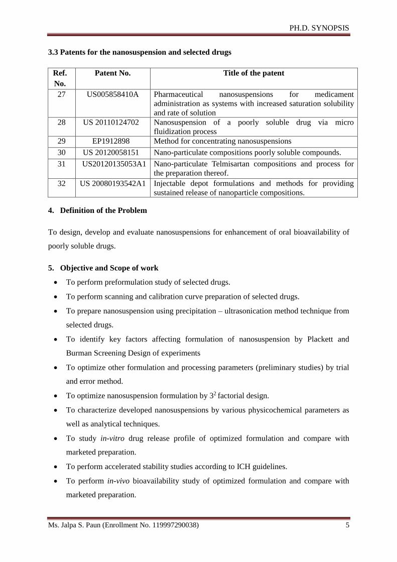
3.3 Patents for the nanosuspension and selected drugs
Ref.
No.
Patent No. Title of the patent
27 US005858410A Pharmaceutical nanosuspensions for medicament
administration as systems with increased saturation solubility
and rate of solution
28 US 20110124702 Nanosuspension of a poorly soluble drug via micro
fluidization process
29 EP1912898 Method for concentrating nanosuspensions
30 US 20120058151 Nano-particulate compositions poorly soluble compounds.
31 US20120135053A1 Nano-particulate Telmisartan compositions and process for
the preparation thereof.
32 US 20080193542A1 Injectable depot formulations and methods for providing
sustained release of nanoparticle compositions.
4. Definition of the Problem
To design, develop and evaluate nanosuspensions for enhancement of oral bioavailability of
poorly soluble drugs.
5. Objective and Scope of work
To perform preformulation study of selected drugs.
To perform scanning and calibration curve preparation of selected drugs.
To prepare nanosuspension using precipitation – ultrasonication method technique from
selected drugs.
To identify key factors affecting formulation of nanosuspension by Plackett and
Burman Screening Design of experiments
To optimize other formulation and processing parameters (preliminary studies) by trial
and error method.
To optimize nanosuspension formulation by 32 factorial design.
To characterize developed nanosuspensions by various physicochemical parameters as
well as analytical techniques.
To study in-vitro drug release profile of optimized formulation and compare with
marketed preparation.
To perform accelerated stability studies according to ICH guidelines.
To perform in-vivo bioavailability study of optimized formulation and compare with
marketed preparation.
Page 11
PH.D. SYNOPSIS
Ms. Jalpa S. Paun (Enrollment No. 119997290038) 6
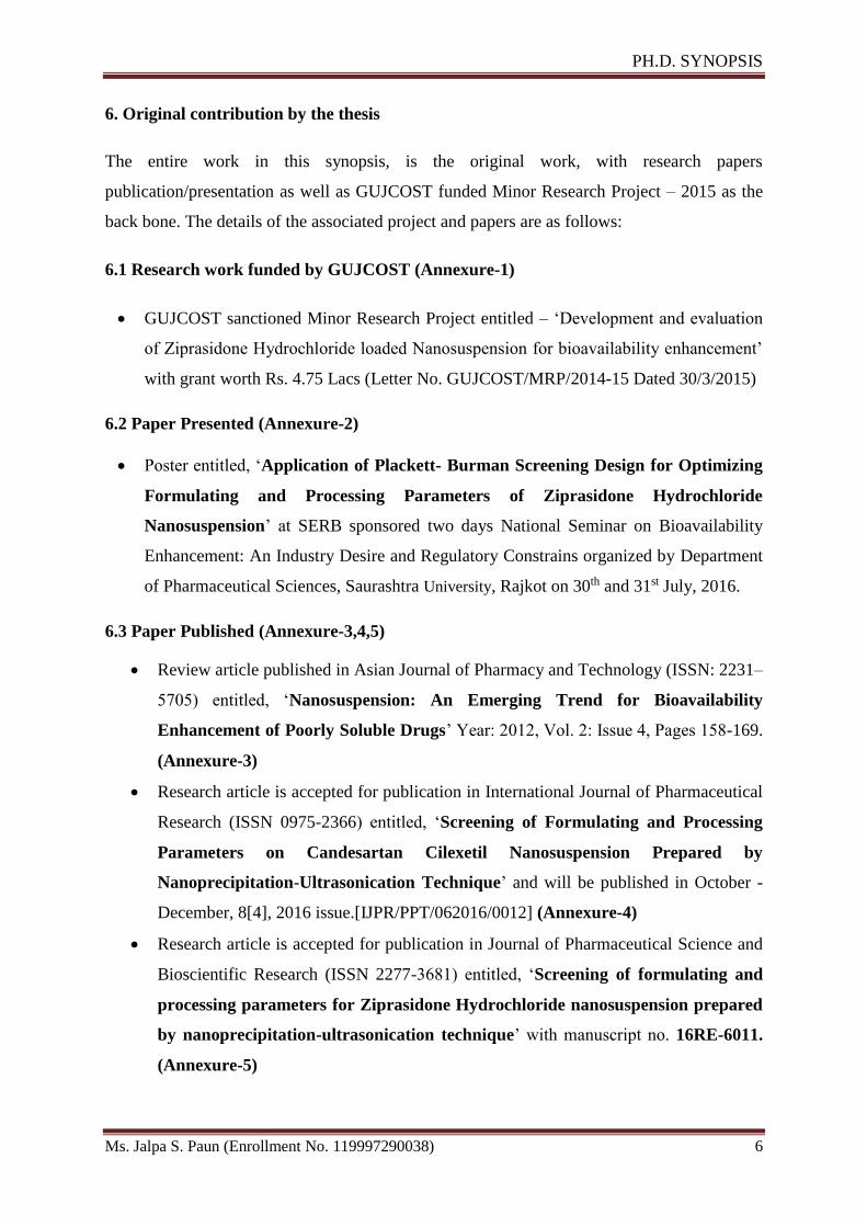
6. Original contribution by the thesis
The entire work in this synopsis, is the original work, with research papers
publication/presentation as well as GUJCOST funded Minor Research Project – 2015 as the
back bone. The details of the associated project and papers are as follows:
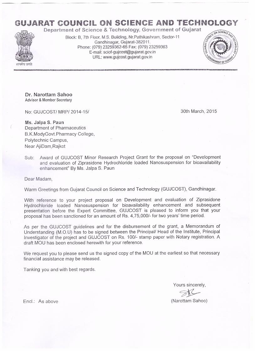
6.1 Research work funded by GUJCOST (Annexure-1)
GUJCOST sanctioned Minor Research Project entitled – ‘Development and evaluation
of Ziprasidone Hydrochloride loaded Nanosuspension for bioavailability enhancement’
with grant worth Rs. 4.75 Lacs (Letter No. GUJCOST/MRP/2014-15 Dated 30/3/2015)
6.2 Paper Presented (Annexure-2)
Poster entitled, ‘Application of Plackett- Burman Screening Design for Optimizing
Formulating and Processing Parameters of Ziprasidone Hydrochloride
Nanosuspension’ at SERB sponsored two days National Seminar on Bioavailability
Enhancement: An Industry Desire and Regulatory Constrains organized by Department
of Pharmaceutical Sciences, Saurashtra University, Rajkot on 30th and 31st July, 2016.
6.3 Paper Published (Annexure-3,4,5)
Review article published in Asian Journal of Pharmacy and Technology (ISSN: 2231–
5705) entitled, ‘Nanosuspension: An Emerging Trend for Bioavailability
Enhancement of Poorly Soluble Drugs’ Year: 2012, Vol. 2: Issue 4, Pages 158-169.
(Annexure-3)
Research article is accepted for publication in International Journal of Pharmaceutical
Research (ISSN 0975-2366) entitled, ‘Screening of Formulating and Processing
Parameters on Candesartan Cilexetil Nanosuspension Prepared by
Nanoprecipitation-Ultrasonication Technique’ and will be published in October -
December, 8[4], 2016 issue.[IJPR/PPT/062016/0012] (Annexure-4)
Research article is accepted for publication in Journal of Pharmaceutical Science and
Bioscientific Research (ISSN 2277-3681) entitled, ‘Screening of formulating and
processing parameters for Ziprasidone Hydrochloride nanosuspension prepared
by nanoprecipitation-ultrasonication technique’ with manuscript no. 16RE-6011.
(Annexure-5)
Page 12
PH.D. SYNOPSIS
Ms. Jalpa S. Paun (Enrollment No. 119997290038) 7
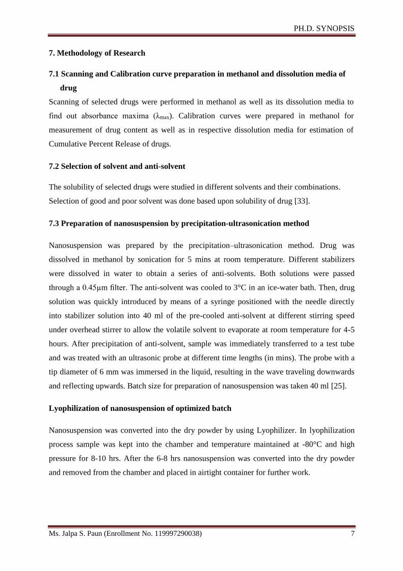
7. Methodology of Research
7.1 Scanning and Calibration curve preparation in methanol and dissolution media of
drug
Scanning of selected drugs were performed in methanol as well as its dissolution media to
find out absorbance maxima (λmax). Calibration curves were prepared in methanol for
measurement of drug content as well as in respective dissolution media for estimation of
Cumulative Percent Release of drugs.
7.2 Selection of solvent and anti-solvent
The solubility of selected drugs were studied in different solvents and their combinations.
Selection of good and poor solvent was done based upon solubility of drug [33].
7.3 Preparation of nanosuspension by precipitation-ultrasonication method
Nanosuspension was prepared by the precipitation–ultrasonication method. Drug was
dissolved in methanol by sonication for 5 mins at room temperature. Different stabilizers
were dissolved in water to obtain a series of anti-solvents. Both solutions were passed
through a 0.45µm filter. The anti-solvent was cooled to 3°C in an ice-water bath. Then, drug
solution was quickly introduced by means of a syringe positioned with the needle directly
into stabilizer solution into 40 ml of the pre-cooled anti-solvent at different stirring speed
under overhead stirrer to allow the volatile solvent to evaporate at room temperature for 4-5
hours. After precipitation of anti-solvent, sample was immediately transferred to a test tube
and was treated with an ultrasonic probe at different time lengths (in mins). The probe with a
tip diameter of 6 mm was immersed in the liquid, resulting in the wave traveling downwards
and reflecting upwards. Batch size for preparation of nanosuspension was taken 40 ml [25].
Lyophilization of nanosuspension of optimized batch
Nanosuspension was converted into the dry powder by using Lyophilizer. In lyophilization
process sample was kept into the chamber and temperature maintained at -80°C and high
pressure for 8-10 hrs. After the 6-8 hrs nanosuspension was converted into the dry powder
and removed from the chamber and placed in airtight container for further work.
Page 13
PH.D. SYNOPSIS
Ms. Jalpa S. Paun (Enrollment No. 119997290038) 8
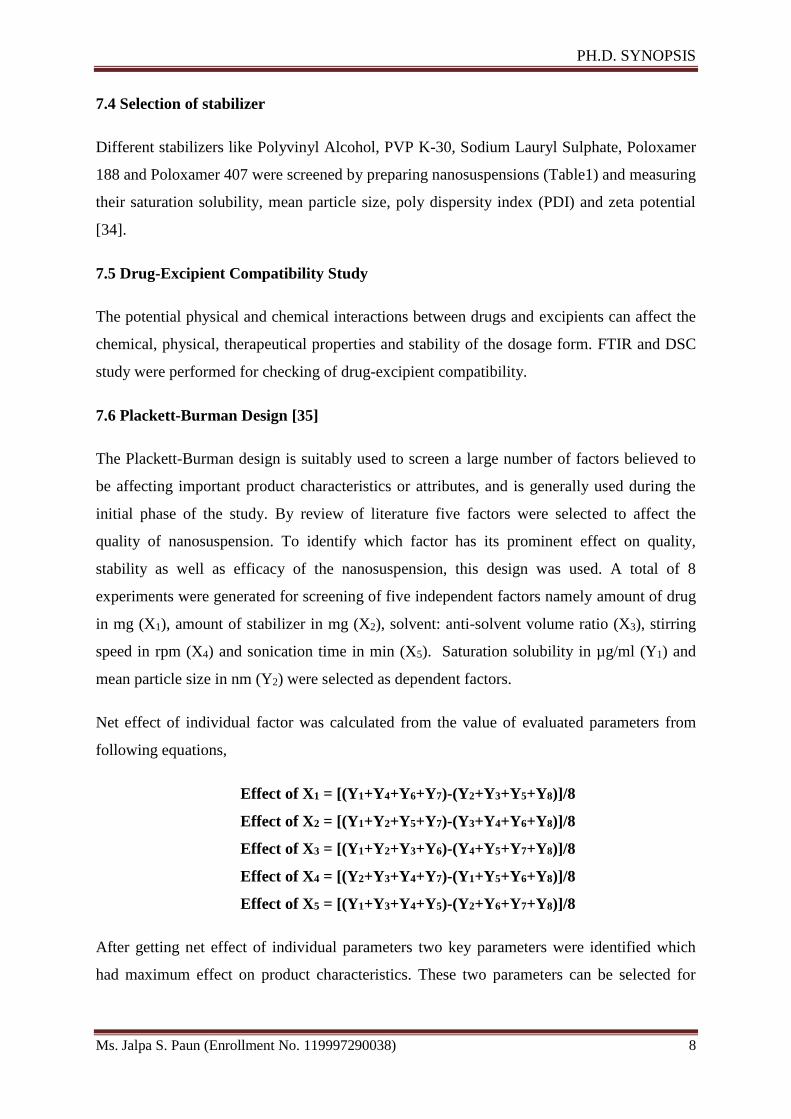
7.4 Selection of stabilizer
Different stabilizers like Polyvinyl Alcohol, PVP K-30, Sodium Lauryl Sulphate, Poloxamer
188 and Poloxamer 407 were screened by preparing nanosuspensions (Table1) and measuring
their saturation solubility, mean particle size, poly dispersity index (PDI) and zeta potential
[34].
7.5 Drug-Excipient Compatibility Study
The potential physical and chemical interactions between drugs and excipients can affect the
chemical, physical, therapeutical properties and stability of the dosage form. FTIR and DSC
study were performed for checking of drug-excipient compatibility.
7.6 Plackett-Burman Design [35]
The Plackett-Burman design is suitably used to screen a large number of factors believed to
be affecting important product characteristics or attributes, and is generally used during the
initial phase of the study. By review of literature five factors were selected to affect the
quality of nanosuspension. To identify which factor has its prominent effect on quality,
stability as well as efficacy of the nanosuspension, this design was used. A total of 8
experiments were generated for screening of five independent factors namely amount of drug
in mg (X1), amount of stabilizer in mg (X2), solvent: anti-solvent volume ratio (X3), stirring
speed in rpm (X4) and sonication time in min (X5). Saturation solubility in µg/ml (Y1) and
mean particle size in nm (Y2) were selected as dependent factors.
Net effect of individual factor was calculated from the value of evaluated parameters from
following equations,
Effect of X1 = [(Y1+Y4+Y6+Y7)-(Y2+Y3+Y5+Y8)]/8
Effect of X2 = [(Y1+Y2+Y5+Y7)-(Y3+Y4+Y6+Y8)]/8
Effect of X3 = [(Y1+Y2+Y3+Y6)-(Y4+Y5+Y7+Y8)]/8
Effect of X4 = [(Y2+Y3+Y4+Y7)-(Y1+Y5+Y6+Y8)]/8
Effect of X5 = [(Y1+Y3+Y4+Y5)-(Y2+Y6+Y7+Y8)]/8
After getting net effect of individual parameters two key parameters were identified which
had maximum effect on product characteristics. These two parameters can be selected for
Page 14
PH.D. SYNOPSIS
Ms. Jalpa S. Paun (Enrollment No. 119997290038) 9
product optimization by factorial design and other three parameters can be optimized by trial
and error method.
7.7 Optimization of other Preliminary Parameters
Preliminary parameters were optimized by varying one parameter at a time, while keeping
others constant, so that effect of varied parameters could be evaluated. Each batch was
repeated thrice (n=3) for the confirmation of repeatability. The parameters were optimized to
achieve minimum particle size and maximum saturation solubility. Optimized parameters
were solvent to anti-solvent volume ratio (1:4, 1:6, 1:8), stabilizer concentration (30mg,
40mg, 50mg), stirring speed (800RPM, 1000RPM, 1200RPM), sonication time (10min,
20min, 30min)etc.
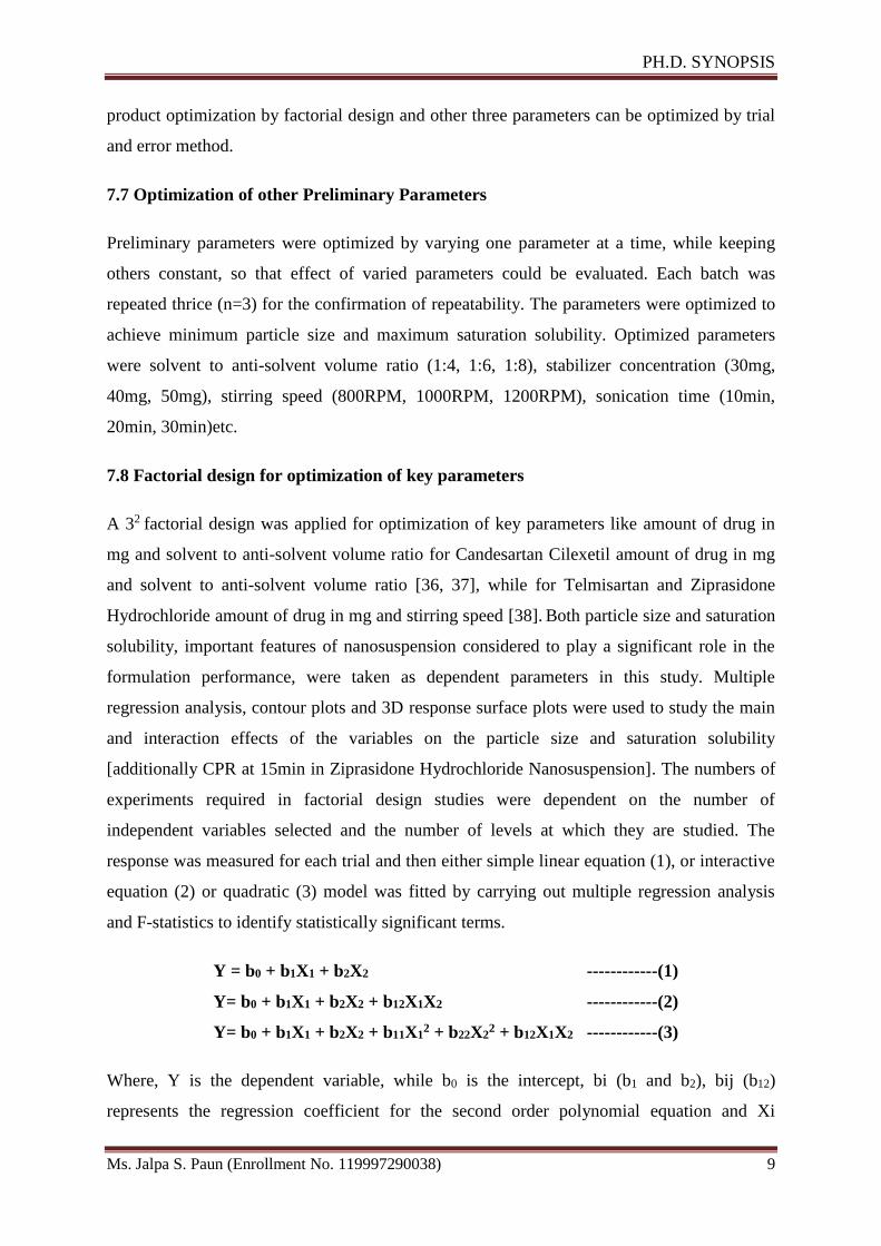
7.8 Factorial design for optimization of key parameters
A 32 factorial design was applied for optimization of key parameters like amount of drug in
mg and solvent to anti-solvent volume ratio for Candesartan Cilexetil amount of drug in mg
and solvent to anti-solvent volume ratio [36, 37], while for Telmisartan and Ziprasidone
Hydrochloride amount of drug in mg and stirring speed [38]. Both particle size and saturation
solubility, important features of nanosuspension considered to play a significant role in the
formulation performance, were taken as dependent parameters in this study. Multiple
regression analysis, contour plots and 3D response surface plots were used to study the main
and interaction effects of the variables on the particle size and saturation solubility
[additionally CPR at 15min in Ziprasidone Hydrochloride Nanosuspension]. The numbers of
experiments required in factorial design studies were dependent on the number of
independent variables selected and the number of levels at which they are studied. The
response was measured for each trial and then either simple linear equation (1), or interactive
equation (2) or quadratic (3) model was fitted by carrying out multiple regression analysis
and F-statistics to identify statistically significant terms.
Y = b0 + b1X1 + b2X2 ------------(1)
Y= b0 + b1X1 + b2X2 + b12X1X2 ------------(2)
Y= b0 + b1X1 + b2X2 + b11X12 + b22X2
2 + b12X1X2 ------------(3)
Where, Y is the dependent variable, while b0 is the intercept, bi (b1 and b2), bij (b12)
represents the regression coefficient for the second order polynomial equation and Xi
Page 15
PH.D. SYNOPSIS
Ms. Jalpa S. Paun (Enrollment No. 119997290038) 10
represents the levels of independent formulation variables. Mathematical modeling was
carried out by using equation 3 to obtain a second order polynomial equation [39]. The values
of dependent variable obtained at various levels of two independent variables (X1 and X2)
were subjected to multiple regressions to yield a second order polynomial equation. The main
effects of X1 and X2 represent the average result of changing one variable at a time from its
low to high value. The interaction (X1X2) shows how the particle size and saturation
solubility changed when two variables were simultaneously changed. The larger the
magnitude of the t value and the smaller the p value, the more significant is the
corresponding coefficient.
7.9 Checkpoint analysis
A checkpoint analysis was performed to confirm the utility of established response surface
plots and contour plots in the preparation of nanosuspension. Values of independent variables
(X1 and X2) were selected and corresponding values of dependent variables were calculated
by substituting the values in the reduced polynomial equation. Nanosuspensions were
prepared experimentally by taking the amounts of the independent variables (X1 and X2) on
the same checkpoints. Checkpoint cum optimized batch was prepared three times and mean
values were determined. Difference of theoretically computed values of particle size as well
as saturation solubility and the mean values of experimentally obtained for both responses
were compared.
7.10 Evaluation of Nanosuspensions
Saturation solubility [40]
Particle size and PDI [41]
Zeta potential [41]
Dissolution study [42]
Drug content
Scanning electron microscopy (SEM)
Accelerated stability study as per ICH Guidelines [43]
Residual solvent by Gas – Chromatography
In-vivo Bioavailability study
Page 16
PH.D. SYNOPSIS
Ms. Jalpa S. Paun (Enrollment No. 119997290038) 11
8. Results of Candesartan Cilexetil Nanosuspension
Scanning and calibration curve was prepared in methanol in the range of 5-30μg/ml by UV-
Visible spectrophotometer showed λmax at 254 nm and regression equation was found to be
Y = 0.0295X + 0.011 with regression co-efficient 0.9994 for UV absorption spectrum of
Candesartan Cilexetil. Scanning and calibration curve was prepared in 0.05 M Phosphate
Buffer, pH 6.5 in the range of 4-16μg/ml by UV-Visible spectrophotometer showed λmax at
259 nm and regression equation was found to be Y = 0.0501X - 0.0116 with regression co-
efficient 0.9992. Selection of solvent and anti-solvent showed that drug had highest solubility
(5.31mg/ml) in methanol and least solubility (0.00119 mg/ml) in water, so they were
selected as solvent and anti-solvent respectively. Different stabilizers like Polyvinyl Alcohol,
PVP K-30, Sodium Lauryl Sulphate, Poloxamer 188 and Poloxamer 407 were screened from
which PVP K-30 was selected by subjecting nanosuspension for measurement of their
saturation solubility, mean particle size, poly dispersity index (PDI) and zeta potential. FTIR
and DSC study of drug, stabilizer and physical mixture indicated compatibility of ingredients.
Results of Plackett-Burman screening design revealed that solvent: anti-solvent ratio as
well as amount of drug were found to be promising formulating parameters having
prominent effect on quality of Candesartan Cilexetil nanosuspension, so they were selected as
independent factors X1 and X2 respectively. Mean particle size (Y1) and saturation solubility
(Y2) were selected as dependent factors for 32 factorial design for optimization of the
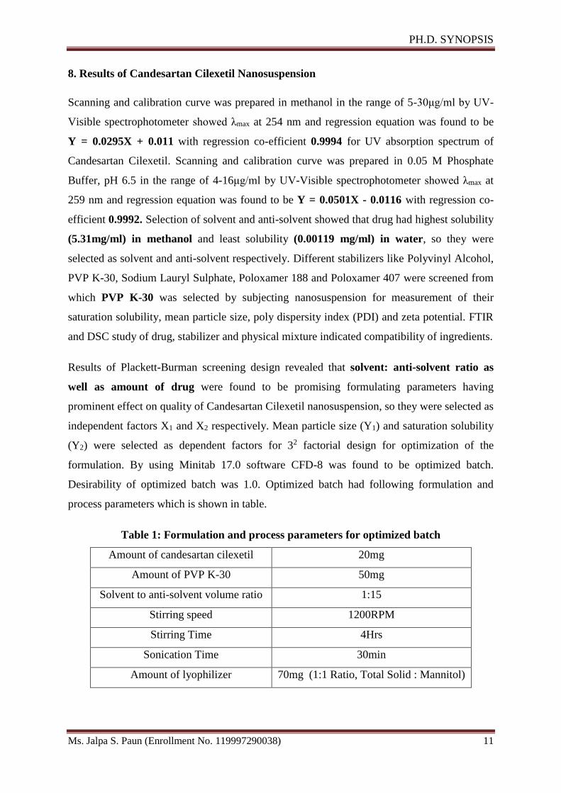
formulation. By using Minitab 17.0 software CFD-8 was found to be optimized batch.
Desirability of optimized batch was 1.0. Optimized batch had following formulation and
process parameters which is shown in table.
Table 1: Formulation and process parameters for optimized batch
Amount of candesartan cilexetil 20mg
Amount of PVP K-30 50mg
Solvent to anti-solvent volume ratio 1:15
Stirring speed 1200RPM
Stirring Time 4Hrs
Sonication Time 30min
Amount of lyophilizer 70mg (1:1 Ratio, Total Solid : Mannitol)
Page 17
PH.D. SYNOPSIS
Ms. Jalpa S. Paun (Enrollment No. 119997290038) 12
Results were obtained for evaluation parameters of optimized batch like, the mean particle
size and PDI of nanosuspension were 242.7nm and 0.345 respectively while the zeta potential
and saturation solubility were −32.98mVand 111.85µg/ml, respectively. Drug content was
found to be 101.01%w/w. The in-vitro dissolution of candesartan cilexetil was 97.13%w/w
was obtained within 2 min. Residual solvent methanol was observed 171.87ppm which was
less than 3000ppm described by ICH guidelines for class-2 solvent. The Surface Topology as
measured by Scanning Electron Microscopy of pure drug was found to be long, thin and flat
with particles larger (5-32µm) in size. However after formulation, particles became smaller
(about 300nm) which were adsorbed on the surface of mannitol used as cryoprotectant may
be by hydrophobic interaction. The in-vitro dissolution rate of candesartan cilexetil was
significantly increased as compared to marketed formulation by reducing the particle size.
Stability study, according to ICH guideline (25°C ± 2°C and 65%RH ± 5%RH) revealed that
there is no significant physical and chemical change after 6 months evaluated by mean
particle size, zeta potential, CPR at 2 min and drug content of the lyophilized formulation
The in-vivo test demonstrated that the Cmax and AUC0→24 values of nanosuspension in rats
were greater than that of marketed formulation respectively.
9. Results of Telmisartan Nanosuspension
Scanning and calibration curve was prepared in methanol in the range of 2-20μg/ml by UV-
Visible spectrophotometer showed λmax at 296 nm and regression equation was found to be Y
= 0.51X + 0.011 with regression co-efficient 0.999 by UV absorption spectrum of
Telmisartan. Scanning and calibration curve was prepared in Phosphate Buffer, pH 7.5 in the
range of 2-20μg/ml by UV-Visible spectrophotometer showed λmax at 296 nm and regression
equation was found to be Y = 0.0422X - 0.0038 with regression co-efficient 0.999. Selection
of solvent and anti-solvent showed that drug has the highest solubility (3.329mg/ml) in
methanol and least solubility (0.012mg/ml) in water, so they were selected as solvent and
anti-solvent respectively. Different stabilizers like Polyvinyl Alcohol, PVP K-30, Sodium
Lauryl Sulphate, Poloxamer 188 and Poloxamer 407 were screened from which Poloxamer
407 was selected by subjecting nanosuspension for measurement of their saturation solubility,
mean particle size, poly dispersity index (PDI) and zeta potential. FTIR and DSC study of
drug, stabilizer and physical mixture indicated compatibility of ingredients.
Results of Plackett-Burman screening design revealed that amount of drug as well as
stirring speed were found to be promising formulating and processing parameters having
Page 18
PH.D. SYNOPSIS
Ms. Jalpa S. Paun (Enrollment No. 119997290038) 13
prominent effect on quality of Telmisartan nanosuspension, so they were selected as
independent factors X1 and X2 respectively. Mean particle size (Y1) and saturation solubility
(Y2) were selected as dependent factors for 32 factorial design for optimization of
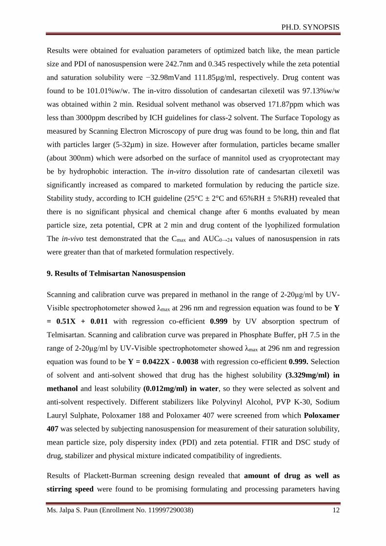
formulation. By using Minitab 17.0 software TFD-6 was found to be optimized batch.
Desirability of optimized batch was 0.9629. Optimized batch had following formulation and
process parameters which is shown in table.
Table 2: Formulation and process parameters for optimized batch
Amount of Telmisartan 15mg
Amount of Poloxamer 407 50mg
Solvent to anti-solvent volume ratio 1:8
Stirring speed 1200RPM
Stirring Time 4Hrs
Sonication Time 30min
Amount of lyophilizer 65mg (1:1 Ratio, Total Solid : Mannitol)
Results were obtained for evaluation parameters of optimized batch like, the mean particle
size and PDI of nanosuspension were 328.0 nm and 0.477 respectively while the zeta
potential and saturation solubility were −30.36mVand 100.16µg/ml, respectively. Drug
content was found to be 99.54%w/w. The in-vitro dissolution of telmisartan was 98.24%w/w
was obtained within 2 min. Residual solvent methanol was observed 192.27ppm which was
less than 3000ppm described by ICH guidelines for class-2 solvent. The Surface Topology as
measured by Scanning Electron Microscopy of pure drug was found to be long, thin and flat
with particles larger (0.5-15µm) in size. However after formulation, particles became smaller
(about 300nm) which were adsorbed on the surface of mannitol used as cryoprotectant by
hydrophobic interaction. Stability study, according to ICH guideline (25°C ± 2°C and
65%RH ± 5%RH) revealed that there is no significant physical and chemical change after 6
months evaluated by mean particle size, zeta potential, CPR at 2 min and drug content of the
lyophilized formulation The in-vivo test demonstrated that the Cmax and AUC0→24 values of
nanosuspension in rats were greater than that of marketed formulation respectively.
10. Results of Ziprasidone Hydrochloride Nanosuspension
Scanning and calibration curve was prepared in methanol in the range of 10-60μg/ml by UV-
Visible spectrophotometer showed λmax at 317 nm and regression equation was found to be Y
= 0.0149X + 0.0133 with regression co-efficient 0.999 by UV absorption spectrum of
Page 19
PH.D. SYNOPSIS
Ms. Jalpa S. Paun (Enrollment No. 119997290038) 14
Ziprasidone Hydrochloride. Scanning and calibration curve was prepared in 0.05M Phosphate
Buffer, pH 7.5 in the range of 10-60μg/ml by UV-Visible spectrophotometer showed λmax at
318 nm and regression equation was found to be Y = 0.0142X - 0.0012 with regression
co-efficient 0.9994. Selection of solvent and anti-solvent showed that drug has highest
solubility (2.443mg/ml) in methanol and least solubility (0.022mg/ml) in water, so they
were selected as solvent and anti-solvent respectively. Different stabilizers like Polyvinyl
Alcohol, PVP K-30, Sodium Lauryl Sulphate, Poloxamer 188 and Poloxamer 407 were
screened from which Poloxamer 407 was selected by subjecting nanosuspension for
measurement of their saturation solubility, mean particle size, poly dispersity index (PDI) and
zeta potential. FTIR and DSC study of drug, stabilizer and physical mixture indicated
compatibility of ingredients.
Results of Plackett-Burman screening design revealed that amount of drug as well as
stirring speed were found to be promising formulating parameters having prominent effect
on quality of Ziprasidone Hydrochloride nanosuspension, so they were selected as
independent factors X1 and X2 respectively. Mean particle size (Y1), saturation solubility (Y2)
and CPR at 15min (Y3) were selected as dependent factors for 32 factorial for optimization of
formulation. By using Minitab 17.0 software ZFD-6 was found to be optimized batch.
Desirability of optimized batch was 0.9035. Optimized batch had following formulation and
process parameters which is shown in table.
Table 3: Formulation and process parameters for optimized batch
Amount of Ziprasidone Hydrochloride
(Equivalent to 15mg of Ziprasidone Base)
16.95mg
Amount of Poloxamer 407 50mg
Solvent to anti-solvent volume ratio 1:8
Stirring speed 1200RPM
Stirring Time 4Hrs
Sonication Time 30min
Amount of lyophilizer
(1:1 Ratio, Total Solid : Mannitol)
66.95 mg
Results were obtained for evaluation parameters of optimized batch like, the mean particle
size and PDI of nanosuspension, were 218.0 nm and 0.456 respectively while the zeta
potential and saturation solubility were −32.1mV and 76.25µg/ml respectively. Drug content
Page 20
PH.D. SYNOPSIS
Ms. Jalpa S. Paun (Enrollment No. 119997290038) 15
was found to be 100.46%w/w. The in-vitro dissolution of Ziprasidone Hydrochloride was
obtained 96.61%w/w within 15 min. Residual solvent methanol was observed 73.53ppm in
optimized batch which was less than 3000ppm described by ICH guidelines for class-2
solvent. The Surface Topology as measured by Scanning Electron Microscopy of pure drug
was found to be long, thin and flat with particles larger (2-27µm) in size. However after
formulation, particles became smaller (about 200nm) which were adsorbed on the surface of
mannitol used as cryoprotectant may be by hydrophobic interaction. Stability study,
according to ICH guideline (25°C ± 2°C and 65%RH ± 5%RH) revealed that there is no
significant physical and chemical change after 6 months evaluated by mean particle size, zeta
potential, CPR at 15 min and drug content of the lyophilized formulation. The in-vivo test
demonstrated that the Cmax and AUC0→12 values of nanosuspension in rats were greater than
that of marketed formulation respectively.
11. Achievements with respect to objectives
Selected drugs were BCS Class II drugs so problem was there with poor saturation
solubility which was increased with nanosuspension formulations
Similarly BCS Class II drugs shows dissolution as rate limiting step but here
nanosuspensions revealed maximum drug release obtained within 2 – 20 mins and
indicating better performance compared to marketed formulations.
The in-vivo bioavailability study proved that the Cmax and AUC0→24 values of
nanosuspension in rats were greater than that of commercial formulations.
The optimized lyophilized product was physically and chemically stable upto 6
months when tested according to ICH guidelines.
12. Conclusion
Nanosuspension was prepared successfully from selected drugs by combination of
precipitation and ultrasonication technique and lyophilized using mannitol as a cryoprotectant
according to physicochemical properties of drugs. Analytical methods were developed for
selected drugs for the estimation of drug in formulations and dissolution media. Solvents and
anti-solvents were selected based upon solubility of drug in respective solvents and their
combination. Stabilizers were selected by preparing nanosuspension and evaluated with
suitable parameters. Compatibility study were carried out using FTIR and DSC studies and
showed drug- excipient compatibility. Various formulation parameters like effect of
Page 21
PH.D. SYNOPSIS
Ms. Jalpa S. Paun (Enrollment No. 119997290038) 16
stabilizers, solvent to anti-solvent ratio as well as processing parameters like effect of stirring
time, stirring speed, sonication time etc. were screened by Plackett-Burman design to identify
key factors producing maximum effect on quality of nanosuspension. Maximum impact
producing two factors were considered for further study to optimize the formulation by 32
factorial design. The optimized formulations were evaluated by various parameters like mean
particle size, polydispersity index, zeta potential, saturation solubility study, in-vitro
dissolution study, gas chromatography for presence of residual solvent and scanning electron
microscopy. Optimized formulations were subjected to accelerated stability study according
to ICH guidelines and found to be physically and chemically stable for 6 months. In-vivo
bioavailability study of optimized nanosuspensions was also carried out and results revealed
improved Cmax and AUC compared with marketed preparations.
13. Copies of papers published and a list of all publications arising from the thesis
13.1 Published / Accepted Papers----------As per point 6.2 and 6.3-----------
13.2 Papers arising from thesis
Sr.
No.
Probable Title Probable Journal
1 Application of Plackett- Burman Screening Design for
Optimizing Formulating and Processing Parameters of
Ziprasidone Hydrochloride Nanosuspension
International Journal of
Pharmaceutical Sciences
and Nanotechnology
2 Formulation and Evaluation of Candesartan Cilexetil
Loaded Nanosuspension for Bioavailability
Enhancement.
Indo American Journal of
Pharmaceutical Research
3 Design, development and evaluation of nanosuspension
for enhancement of oral bioavailability of telmisartan.
AAPS SciTech
4 Development and evaluation of nanosuspension
formulation for oral bioavailability enhancement of
Ziprasidone Hydrochloride.
Drug Development and
Industrial Pharmacy
14. Patents (if any) ------------- NA------------
15. References
1. Keck CM and Müller RH, 2006, Drug nanocrystals of poorly soluble drugs produced by
high pressure homogenisation, European Journal of Pharmaceutics and Biopharmaceutics,
62(1), 3–16, ISSN: 0939-6411.
Page 22
PH.D. SYNOPSIS
Ms. Jalpa S. Paun (Enrollment No. 119997290038) 17
2. Kesisoglou F and Mitra A, 2012, Crystalline nanosuspensions as potential toxicology and
clinical oral formulations for BCS II / IV compounds, AAPS Journal, 14(4), 677-687,
ISSN: 1550-7416.
3. Verma S, Kumar S, Gokhale R and Burgess DJ, 2011, Physical stability of
nanosuspensions: investigation of the role of stabilizers on ostwald ripening, International
Journal of Pharmaceutics, 406(1-2), 145–52, ISSN: 0378-5173.
4. Junghanns AH, 2008, Nanocrystal technology, drug delivery and clinical applications,
International Journal of Nanomedicine, 3(3), 295–309, ISSN: 1176-9114.
5. Kesisoglou F, Panmai S and Wu Y, 2007, Nanosizing--oral formulation development and
biopharmaceutical evaluation, Advanced Drug Delivery Reviews, 59(7), 631–44, ISSN:
0169-409X.
6. Che E, Zheng X, Sun C, Chang D, Jiang T and Wang S, 2012, Drug nanocrystals : a state
of the art formulation strategy for preparing the poorly water-soluble drugs, Asian Journal
of Pharmaceutical Sciences, 7(2), 85–95, ISSN: 1818-0876.
7. Gao L, Zhang ED and Chen EM, 2008, Drug nanocrystals for the formulation of poorly
soluble drugs and its application as a potential drug delivery system, Journal of
Nanoparticle Research, 10(5), 845–62, ISSN: 1388-0764.
8. Patil MN and Pandit AB, 2007, Cavitation-a novel technique for making stable nano-
suspensions, Ultrasonics Sonochemistry, 14(5), 519–530, ISSN: 1350-4177.
9. Hassan MA, Suleiman MS, Najib NM, 1990, Improvement of the in vitro dissolution
characteristics of famotidine by inclusion in β- cyclodextrin, International Journal of
Pharmaceutics, 58, 19–24, ISSN: 0378-5173.
10. Rania HF, Mohammed AK, 2008, Enhancement of famotidine dissolution rate through
liquisolid tablets formulation: In vitro and in vivo evaluation, European Journal of
Pharmaceutics and Biopharmaceutics, 69, 993–1003, ISSN: 0939-6411.
11. O’Neil MJ, Heckelman PE, Koch CB, Roman KJ, Kenny CM, D’Arecca MR,
Candesartan Cilexetil, In: The Merck Index – An encyclopedia of chemicals, drugs and
biological, 14th Edition, Merck Research Laboratory, Division of Merck & Co., Inc.,
Whitehouse Station, New Jersey; 2006, pp 1739.
12. Sweetman SC, Candesartan, In: Martindale - The Complete Drug Reference, 36th
Edition, Pharmaceutical Press, London; 2009, pp 1238.
13. O’ Neil MJ, Heckelman PE, Koch CB, Roman KJ, Kenny Cm and D’Arecca MR (Eds).
(2006) Telmisartan, In: The Merck Index – An encyclopedia of chemicals, drugs and
Page 23
PH.D. SYNOPSIS
Ms. Jalpa S. Paun (Enrollment No. 119997290038) 18
biological, 14th Edn, Merck Research Laboratory, Division of Merck & Co., Inc.,
Whitehouse Station, New Jersey, pp 9129.
14. Sweetman SC (Eds). (2009) Telmisartan, In: Martindale - The Complete Drug Reference,
36th Edn, Pharmaceutical Press, London, pp 1409.
15. Stangier J, Schmid J, Türck D et. al., 2000, Absorption, metabolism, and excretion of
intravenouslyand orally administered telmisartan in healthy volunteers, Journal of
Clinical Pharmacology, 40, 1312–1322, ISSN: 1552-4604.
16. Stangier J, Su CA, Hendriks MG et. al., 2000, The effect of telmisartan on the steady-
state pharmacokinetics of digoxin in healthy male volunteers, Journal of Clinical
Pharmacology, 40, 1373–1379, ISSN: 1552-4604.
17. O’ Neil MJ, Heckelman PE, Koch CB, Roman KJ, Kenny Cm and D’Arecca MR (Eds).
(2006) Ziprasidone Hydrochloride, In: The Merck Index – An encyclopedia of chemicals,
drugs and biological, Merck Research Laboratory, Division of Merck & Co., Inc.,
Whitehouse Station, New Jersey, 14th Edn, pp 10307.
18. Sweetman SC (Eds), (2009) Ziprasidone Hydrochloride, In: Martindale - The Complete
Drug Reference, Pharmaceutical Press, London, 36th Edition. 2009, pp 1036.
19. Miceli JJ, Wilner KD, Swan SK, Tensfeldt TG, 2005, Pharmacokinetics, safety, and
tolerability of intramuscular Ziprasidone in healthy volunteers, Journal of Clinical
Pharmacology, 45, 620–30, ISSN: 1552-4604.
20. Preskorn SH, 2005, Pharmacokinetics and therapeutics of acute intramuscular
ziprasidone, Clinical Pharmacokinetics, 44, 1117–33, ISSN: 0312-5963.
21. Miceli JJ, Smith M, Robarge L, Morse T, Laurent A, 2000, The effects of ketoconazole
on ziprasidone pharmacokinetics - a placebo-controlled crossover study in healthy
volunteers, British Journal of Clinical Pharmacology, 49(S1), 71–76, ISSN: 1365-2125.
22. Martini LG, Crowley PJ. (2011) Controlling drug release in oral product development
programs : An industrial Perspective, In: Controlled release in oral drug delivery.
Springer, New York, 14th Edition, pp 49-69.
23. Papdiwal A, Sagar K and Pande V, 2014, Formulation and Characterization of
Nateglinide Nanosuspension by Precipitation Method, International Journal of
Pharmaceutical Sciences and Nanotechnology, 7(4), 2685-2691, ISSN: 0974-3278.
24. Raval AJ and Patel MM, 2011, Preparation and Characterization of Nanoparticles for
Solubility and Dissolution Rate Enhancement of Meloxicam, International Research
Journal of Pharmacy, 1(2), 42-49, ISSN: 2230-8407.
Page 24
PH.D. SYNOPSIS
Ms. Jalpa S. Paun (Enrollment No. 119997290038) 19
25. Xia D, Quan P, Piao H et. al., 2010, Preparation of stable nitrendipine nanosuspensions
using the precipitation–ultrasonication method for enhancement of dissolution and oral
bioavailability, European Journal of Pharmaceutical Sciences, 40(4), 325–334, ISSN:
0928-0987.
26. Liu D, Xu H, Tian B et. al., 2012, Fabrication of Carvedilol Nanosuspensions through the
Anti-Solvent Precipitation–Ultrasonication Method for the Improvement of Dissolution
Rate and Oral Bioavailability, AAPS PharmSciTech, 13(1), 295-304, ISSN: 1530-9932.
27. Robert B, Bernd K, Muller RH, Peters K (1999) Pharmaceutical nanosuspensions for
medicament administration as systems with increased saturation solubility and rate of
solution, US 5858410.
28. Chen MJ, Hui HW, Lee T, Paul K, Surapaneni S (2011) Nanosuspension of a poorly
soluble drug via microfluidization process, US 20110124702.
29. Thomas L (2010) Method for concentrating nanosuspensions, EP1912898.
30. Ferreiro MG, Dunmann C, Kroehne L, Voigt A (2012) Nano-particulate compositions
poorly soluble compounds, US 20120058151.
31. Filipcsei G, Otvos Z, Pongracz K, Darvas F (2012) Nano-particulate telmisartan
compositions and process for the preparation thereof, US 20120135053A1.
32. Shah JC, Shah PS, Wisniecki P, Wagner DR (2008) Injectable depot formulations and
methods for providing sustained release of nanoparticle compositions, US
20080193542A1.
33. Shivakumar HG, Ramalingaraju G, Siddaramaiah, 1999, Influence of solvents on crystal
habit and properties of paracetamol crystals, Indian Journal of Pharmaceutical Sciences,
61(2), 100-104, ISSN: 0250-474X.
34. Pandya VM, Patel JK and Patel DJ, 2011, Formulation, optimization and characterization
of Simvastatin Nanosuspension prepared by nanoprecipitation technique, Der Pharmacia
Lettre, 3(2), 129-140, ISSN: 0975-5071.
35. Gacula MC. (1993) Product Optimization. In: Design and Analysis of Sensory
Optimization Food and Nutrition Press, Trumbull: Connecticut USA, pp137.
36. Kakran M, Sahoo NG, Li L et. al., 2010, Fabrication of drug nanoparticles by evaporative
precipitation of nanosuspension, International Journal of Pharmaceutics, 383, 285–292,
ISSN: 0378-5173.
37. Das S, Suresh PK, 2011, Nanosuspension: a new vehicle for the improvement of the
delivery of drugs to the ocular surface. Application to amphotericin B, Nanomedicine:
Nanotechnology, Biology, and Medicine, 7, 242–247, ISSN: 1549-9634.
Page 25
PH.D. SYNOPSIS
Ms. Jalpa S. Paun (Enrollment No. 119997290038) 20
38. Pignatello R, Bucolo C, Spedalieri G, Maltese A and Puglisi G, “Flurbiprofen-loaded
acrylate polymer nanosuspensions for ophthalmic application.” Biomaterials, 2002, 23,
3247–3255.
39. Armstrong NC, James KC (1996) Pharmaceutical experimental design and
interpretation. Bristol, PA, USA: Taylor and Francis Publications, pp. 131-92.
40. Muller RH, Jacobs C, Kayser O, 2001, Nanosuspensions as particulate drug formulations
in therapy rationale for development and what we can expect for the future, Advanced
Drug Delivery Reviews, 47(1), 3–19, ISSN: 0169-409X.
41. Shinde SS, Hosmani AH, 2014, Preparation and evaluation of nanosuspensions for
enhancing the dissolution of lornoxicam by anti-solvent precipitation technique, Indo-
American Journal of Pharmaceutical Research, 4(1), 398-405, ISSN: 2231-6876.
42. Li W, Yang Y, Tian Y et.al., 2011, Preparation and in-vitro/in-vivo evaluation of
Revaprazan Hydrochloride nanosuspension, International Journal of Pharmaceutics, 408,
157–162, ISSN: 0378-5173.
43. ICH Harmonised Tripartite Guideline, Stability Testing of New Drug Substances and
Products, Q1A (R2). [Online]
http://www.ich.org/fileadmin/Public_Web_Site/ICH_Products/Guidelines/Quality/Q1A_
R2/Step4/Q1A_R2__Guideline.pdf [Accessed on 15 March 2014]
Page 30
Asian J. Pharm. Tech. 2012; Vol. 2: Issue 4, Pg 158-169 [AJPTech.]
158
ISSN- 2231–5705 (Print) www.asianpharmaonline.org
ISSN- 2231–5713 (Online)
REVIEW ARTICLE
Nanosuspension: An Emerging Trend for Bioavailability Enhancement of
Poorly Soluble Drugs
Paun J.S.1*
and Tank H.M.
2
1Department of Pharmaceutics, S.J. Thakkar Pharmacy College, Rajkot
2Department of Pharmaceutics, Matushree V.B. Manvar College of Pharmacy, Dumiyani
*Corresponding Author E-mail: [email protected]
ABSTRACT: Drug effectiveness is influenced by a crucial factor like solubility of drug, independence of the route of administration.
Most of the newly discovered drugs coming out from High-throughput screening are failing due to their poor water
solubility which is major problem for dosage form design. Now a day, nanoscale systems for drug delivery have
gained much interest as a way to improve the solubility problems. Nanosuspension technology is a unique and
economical approach to overcome poor bioavailability that is related with the delivery of hydrophobic drugs, including
those that are poorly soluble in aqueous media. Design and development of nanosuspension of such drugs is an
attractive alternative to solve this problem. Preparation of nanosuspension is simple and applicable to all poorly
soluble drugs. A nanosuspension not only solves the problem of solubility and bioavailability but also alters
pharmacokinetic profile of the drug which may also improve safety and efficacy. This review article takes account of
introduction, advantages, properties, formulation consideration, preparation, characterization and application of the
nanosuspensions.
.
KEYWORDS: Nanosuspensions, Poorly soluble drugs, Drug Delivery, Bioavailability, Solubility enhancement.
INTRODUCTION: Bioavailability is defined as the rate and extent to which the
active ingredient is absorbed from a drug product and
becomes available at the site of action.1
From a
pharmacokinetic perspective, bioavailability data for a
given formulation provide an estimate of the relative
fraction of the orally administered dose that is absorbed into
the systemic circulation when compared to the
bioavailability data for a solution, suspension or
intravenous dosage form. In addition, bioavailability studies
provide other useful pharmacokinetic information related to
distribution, elimination, effects of nutrients on absorption
of the drug, dose proportionality and linearity in
pharmacokinetics of the active and inactive moieties.
Bioavailability data can also provide information indirectly
about the properties of a drug substance before entry into
the systemic circulation, such as permeability and the
influence of pre-systemic enzymes and/or transporters.
Received on 28.10.2012 Accepted on 12.11.2012
© Asian Pharma Press All Right Reserved Asian J. Pharm. Tech. 2(4): Oct. - Dec. 2012; Page 158-169
Bioavailability of a drug is largely determined by the
properties of the dosage form, rather than by the drug's
physicochemical properties, which determine absorption
potential. Differences in bioavailability among formulations
of a given drug can have clinical significance; thus,
knowing whether drug formulations are equivalent is
essential.
Poorly water soluble drugs are increasingly becoming a
problem in terms of obtaining satisfactory dissolution
within the gastrointestinal tract that is necessary for good
oral bioavailability. It is not only existing drugs that cause
problems but it is the challenge to ensure that new drugs are
not only active pharmacologically but have enough
solubility to ensure fast enough dissolution at the site of
administration, often the gastrointestinal tract.2
FACTORS AFFECTING BIOAVAILABILITY:
Low bioavailability is most common with oral dosage
forms of poorly water-soluble, slowly absorbed drugs. Solid
drugs need to dissolve before they are exposed to be
absorbed. If the drug does not dissolve readily or cannot
penetrate the epithelial membrane (eg, if it is highly ionized
Page 31
Asian J. Pharm. Tech. 2012; Vol. 2: Issue 4, Pg 158-169 [AJPTech.]
159
and polar), time at the absorption site may be insufficient.
In such cases, bioavailability tends to be highly variable as
well as low3. Age, sex, physical activity, genetic phenotype,
stress, disorders (eg, achlorhydria, malabsorption
syndromes), or previous GI surgery (eg, bariatric surgery)
can also affect drug bioavailability.
IMPROVEMENT OF BIOAVAILABILITY:
Improvement of bioavailability of poorly water soluble drug
remains one of the most challenging aspects of drug
development. By many estimates up to 40% of new
chemical entities discovered by the pharmaceutical industry
today are poorly water soluble compounds.4
Together with the permeability, the solubility behavior of a
drug is a key determinant of its bioavailability. There have
always been certain drugs for which solubility has
presented a challenge to the development of a suitable
formulation for oral administration. Examples are
griseofulvin, digoxin, phenytoin, sulphathiazole etc. With
the recent arrival of high throughput screening of potential
therapeutic agents, the number of poorly soluble drug
candidates has risen sharply and the formulation of poorly
soluble compounds for delivery now presents one of the
most frequent and greatest challenges to formulation
scientists in the pharmaceutical industry.
Consideration of the modified Noyes-Whitney equation
provides some hints as to how the dissolution rate of even
very poorly soluble compounds might be improved to
minimize the limitations to oral availability.5
The main
possibilities for improving dissolution according to this
analysis are:
• To increase the surface area available for dissolution
by decreasing the particle size of the solid compound,
• By optimizing the wetting characteristics of the
compound surface,
• To decrease the diffusion layer thickness,
• To ensure sink conditions for dissolution and,
• To improve the apparent solubility of the drug under
physiologically relevant conditions.6
A fundamental step in the solubilization of drug compound
is the selection of an appropriate salt form, or for liquid
drugs, adjustment of pH of the solution. Traditional
approaches to drug solubilization include either chemical or
mechanical modification of the drug molecule, or
physically altering the macromolecular characteristics of
aggregated drug particles.
Improvement of bioavailability can be obtained by
following measures:
• Addition of solubilizing excipients
• Inclusion complexes
• Polymorphism
• Lipid-based emulsion systems
• Salt form
• Solid dispersions
• Particle size reduction etc.
NEED OF NANOSUSPENSION FOR
BIOAVAILABILITY ENHANCEMENT:
Nevertheless, pharmacokinetic studies of BCS class – II
drugs showed that they have a low oral bioavailability,
which may be due to poor water solubility of drug. There
are many classical pharmaceutical ways to improve drug
dissolution rate such as dissolution in aqueous mixtures
with an organic solvent,7 formation of ß-cyclodextrin
complexes,8 solid dispersions
9 and drug salt form.
10
During last 20 years a new technology, reducing drug
particle size, has been developed to increase drug
dissolution rate. According to Noyes–Whitney equation,
drugs with smaller particle size have enlarged surface areas
which lead to increase dissolution velocity. Higher the
dissolution rate together with the resulting higher
concentration gradient between gastrointestinal lumen and
systemic circulation could further increase oral
bioavailability of drugs.11
A nanosuspension is a submicron
colloidal dispersion of drug particles which are stabilized
by surfactants. A pharmaceutical nanosuspension is defined
as very finely dispersed solid drug particles in an aqueous
vehicle for oral, topical, parenteral or pulmonary
administration. The particle size distribution of the solid
particles in nanosuspensions is usually less than one micron
with an average particle size ranging between 200 and 600
nm.12
In nanosuspension technology, the drug is maintained
in the required crystalline state with reduced particle size,
leading to an increased dissolution rate and therefore
improved bioavailability. An increase in the dissolution rate
of micronized particles (particle size < 10 µm) is related to
an increase in the surface area and consequently the
dissolution velocity. Nanosized particles can increase
solution velocity and saturation solubility because of the
vapor pressure effect. In addition; the diffusional distance
on the surface of drug nanoparticles is decreased, thus
leading to an increased concentration gradient. Increase in
surface area as well as concentration gradient lead to a
much more pronounced increase in the dissolution velocity
as compared to a micronized product. Another possible
explanation for the increased saturation solubility is the
creation of high energy surfaces when disrupting the more
or less ideal drug microcrystals to nanoparticles.
Dissolution experiments can be performed to quantify the
increase in the saturation solubility of a drug when
formulated into a nanosuspension.13
The stability of the particles obtained in the nanosuspension
is attributed to their uniform particle size which is created
by various manufacturing processes. The absence of
particles with large differences in their size in
nanosuspensions prevents the existence of different
saturation solubilities and concentration gradients;
Page 32
Asian J. Pharm. Tech. 2012; Vol. 2: Issue 4, Pg 158-169 [AJPTech.]
160
consequently preventing the Oswald ripening effect.
Ostwald ripening is responsible for crystal growth and
subsequently formation of microparticles. It is caused by a
difference in dissolution pressure/saturation solubility
between small and large particles. Molecules diffuse from
the higher concentration area around small particles which
have higher saturation solubility to an area around larger
particles possessing a lower drug concentration. This leads
to the formation of a supersaturated solution around the
large particles and consequently to drug crystallization and
growth of the large particles.
ADVANTAGES OF NANOSUSPENSIONS:
The major advantages of nanosuspension technology are:14
• Provides ease of manufacture and scale-up for large
scale production,
• Long-term physical stability due to the presence of
stabilizers,
• Oral administration of nanosuspensions provide rapid
onset, reduced fed/fasted ratio and improved
bioavailability,
• Rapid dissolution and tissue targeting can be achieved
by IV route of administration,
• Reduction in tissue irritation in case of
subcutaneous/intramuscular administration,
• Higher bioavailability in case of ocular administration
and inhalation delivery,
• Drugs with high log P value can be formulated as
nanosuspensions to increase the bioavailability of such
drugs,
• Improvement in biological performance due to high
dissolution rate and saturation solubility of the drug,
• Nanosuspensions can be incorporated in tablets,
pellets, hydrogels and suppositories are suitable for
various routes of administration,
• The flexibility offered in the modification of surface
properties and particle size, and ease of post-
production processing of nanosuspensions enables
them to be incorporated in various dosage forms for
various routes of administration, thus proving their
versatility.
INTERESTING SPECIAL FEATURES OF
NANOSUSPENSIONS: 15
• Increase in saturation solubility and consequently an
increase in the dissolution rate of the drug.
• Increase in adhesive nature, thus resulting in enhanced
bioavailability.
• Increasing the amorphous fraction in the particles,
leading to a potential change in the crystalline structure
and higher solubility.
• Absence of ostwald ripening, producing physical long
term stability as an aqueous suspension.
• Possibility of surface-modification of nanosuspensions
for site specific delivery.
CRITERIA FOR SELECTION OF DRUG FOR
NANOSUSPENSIONS:
Nanosuspension can be prepared for the API that is having
either of the following characteristics:16
� Water insoluble but which are soluble in oil (high log
P) OR API are insoluble in both water and oils
� Drugs with reduced tendency of the crystal to dissolve,
regardless of the solvent
� API with very large dose
METHODS OF PREPARATION FOR
NANOSUSPENSIONS:
Milling techniques (Nanocrystals or Nanosystems)
Media milling:
Media milling is a technique used to prepare
nanosuspensions.11,12, 17-19
Nanocrystal is a patent protected
technology developed by Liversidge et al. In this technique,
the drug nanoparticles are obtained by subjecting the drug
to media milling. High energy and shear forces generated as
a result of impaction of the milling media with the drug
provide the necessary energy input to disintegrate the
microparticulate drug into nanosized particles. In the media
milling process, the milling chamber is charged with the
milling media, water or suitable buffer, drug and stabilizer.
Then the milling media or pearls are rotated at a very high
shear rate. The major concern with this method is the
residues of milling media remaining in the finished product
could be problematic for administration.17
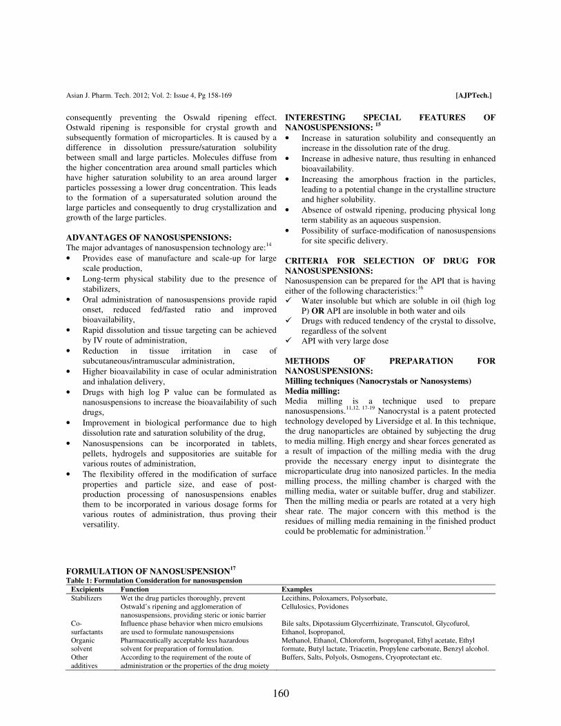
FORMULATION OF NANOSUSPENSION17
Table 1: Formulation Consideration for nanosuspension
Excipients Function Examples
Stabilizers Wet the drug particles thoroughly, prevent
Ostwald’s ripening and agglomeration of
nanosuspensions, providing steric or ionic barrier
Lecithins, Poloxamers, Polysorbate,
Cellulosics, Povidones
Co-
surfactants
Influence phase behavior when micro emulsions
are used to formulate nanosuspensions
Bile salts, Dipotassium Glycerrhizinate, Transcutol, Glycofurol,
Ethanol, Isopropanol,
Organic
solvent
Pharmaceutically acceptable less hazardous
solvent for preparation of formulation.
Methanol, Ethanol, Chloroform, Isopropanol, Ethyl acetate, Ethyl
formate, Butyl lactate, Triacetin, Propylene carbonate, Benzyl alcohol.
Other
additives
According to the requirement of the route of
administration or the properties of the drug moiety
Buffers, Salts, Polyols, Osmogens, Cryoprotectant etc.
Page 33
Asian J. Pharm. Tech. 2012; Vol. 2: Issue 4, Pg 158-169 [AJPTech.]
161
Nanosuspensions are produced by using high-shear media
mills or pearl mills. The mill consists of a milling chamber,
milling shaft and a recirculation chamber. An aqueous
suspension of the drug is then fed into the mill containing
small grinding balls/pearls. As these balls rotate at a very
high shear rate under controlled temperature, they fly
through the grinding jar interior and impact against the
sample on the opposite grinding jar wall. The combined
forces of friction and impact produce a high degree of
particle size reduction. The milling media or balls are made
of ceramic-sintered aluminium oxide or zirconium oxide or
highly cross-linked polystyrene resin with high abrasion
resistance. Planetary ball mill is one example of the
equipment that can be used to achieve a grind size below
0.1 µm.
Dry co-grinding:
Nanosuspensions prepared by high pressure
homogenization and media milling using pearl-ball mill are
wet–grinding processes. Recently, nanosuspensions can be
obtained by dry milling techniques. Successful work in
preparing stable nanosuspensions using dry-grinding of
poorly soluble drugs with soluble polymers and copolymers
after dispersing in a liquid media has been reported.20-22
Itoh et al reported the colloidal particles formation of many
poorly water soluble drugs; griseofulvin, glibenclamide and
nifedipine obtained by grinding with polyvinyl pyrrolidone
(PVP) and sodium dodecyl sulfate (SDS). Many soluble
polymers and co-polymers such as PVP, polyethylene
glycol (PEG), hydroxyl propyl methylcellulose (HPMC)
and cyclo-dextrin derivatives have been used.23-25
Physico-
chemical properties and dissolution of poorly water soluble
drugs were improved by co-grinding because of an
improvement in the surface polarity and transformation
from a crystalline to an amorphous drug.26,27
Dry co-
grinding can be carried out easily and economically and can
be conducted without organic solvents. The co-grinding
technique can reduce particles to the submicron level and a
stable amorphous solid can be obtained.
Advantages:
• Media milling is applicable to the drugs that are poorly
soluble in both aqueous and organic media.
• Very dilute as well as highly concentrated
nanosuspensions can be prepared by handling 1mg/ml
to 400mg/ml drug quantity.
• Nanosize distribution of final nanosized products.
Disadvantages:
• Nanosuspensions contaminated with materials eroded
from balls may be problematic when it is used for long
therapy. (Wet milling technique)
• The media milling technique is time consuming.
• Some fractions of particles are in the micrometer
range.
• Scale up is not easy due to mill size and weight.
High Pressure Homogenization:
Homogenization in Aqueous media (Dissocubes)
Homogenization involves the forcing of the suspension
under pressure through a valve having a narrow aperture.
Dissocube technology was developed by Muller et al. in
which, the suspension of the drug is made to pass through a
small orifice that results in a reduction of the static pressure
below the boiling pressure of water, which leads to boiling
of water and formation of gas bubbles. When the
suspension leaves the gap and normal air pressure is
reached again, the bubbles shrink and the surrounding part
containing the drug particles rushes to the center and in the
process colloids, causing a reduction in the particle size.
Most of the cases require multiple passes or cycles through
the homogenizer, which depends on the hardness of drug,
the desired mean particle size and the required
homogeneity.
Scholer et al. prepared atovaquone nanosuspensions using
this technique.28
To produce a nanosuspension with a higher
concentration of solids, it is preferred to start
homogenization with very fine drug particles, which can be
accomplished by pre-milling.
Homogenization in Non Aqueous Media (Nanopure):
Nanopure is the technology in which suspension is
homogenized in water-free media or water mixtures.29
In
the Dissocubes technology the cavitation is the determining
factor of the process. But, in contrast to water, oils and oily
fatty acids have very low vapor pressure and a high boiling
point. Hence, the drop of static pressure will not be
sufficient enough to initiate cavitation.
Patents covering disintegration of polymeric material by
high- pressure homogenization mention that higher
temperatures of about 80°C promoted disintegration, which
cannot be used for thermo labile compounds. In nanopure
technology, the drug suspensions in the non- aqueous media
were homogenized at 0°C or even below the freezing point
and hence are called "deep-freeze" homogenization. The
results obtained were comparable to Dissocubes and hence
can be used effectively for thermo labile substances at
milder conditions.
Advantages:
• Drugs that are poorly soluble in both aqueous and
organic media can be easily formulated into
nanosuspensions.
• Ease of scale-up and little batch-to-batch variation.30
• Narrow size distribution of the nanoparticulate drug
present in the final product 31
• Allows aseptic production of nanosuspensions for
parenteral administration.
• Flexibility in handling the drug quantity, ranging from
1 to 400mg mL-1
, thus enabling formulation of very
dilute as well as highly concentrated nanosuspensions.
Page 34
Asian J. Pharm. Tech. 2012; Vol. 2: Issue 4, Pg 158-169 [AJPTech.]
162
Disadvantages:
• Prerequisite of micronized drug particles.
• Prerequisite of suspension formation using high-speed
mixers before subjecting it to homogenization.
Precipitation Method:
Using a precipitation technique, the drug is dissolved in an
organic solvent and this solution is mixed with a miscible
anti-solvent. In water-solvent mixture the solubility is low
and the drug precipitates. Mixing processes vary
considerably. Precipitation has also been coupled with high
shear processing. The nanoedge process (is a registered
trademark of Baxter International Inc. and its subsidiaries)
relies on the precipitation of friable materials for
subsequent fragmentation under conditions of high shear
and/or thermal energy.32
Nanoedge:
The basic principles of Nanoedge are the same as that of
precipitation and homogenization. A combination of these
techniques results in smaller particle size and better stability
in a shorter time. The major drawback of the precipitation
technique, such as crystal growth and long-term stability,
can be resolved using the Nanoedge technology. Rapid
addition of a drug solution to an anti-solvent leads to
sudden super-saturation of the mixed solution, and
generation of fine crystalline or amorphous solids.
Precipitation of an amorphous material may be favored at
high super-saturation when the solubility of the amorphous
state is exceeded. The success of drug nanosuspensions
prepared by precipitation techniques has been reported.32-35
In this technique, the precipitated suspension is further
homogenized, leading to reduction in particle size and
avoiding crystal growth. Precipitation is performed in water
using water-miscible solvents such as methanol, ethanol
and isopropanol. It is desirable to remove those solvents
completely, although they can be tolerated to a certain
extent in the formulation. For an effective production of
nanosuspensions using the Nanoedge technology, an
evaporation step can be included to provide a solvent-free
modified starting material followed by high-pressure
homogenization.
Nanojet technology:
This technique, called opposite stream or nanojet
technology, uses a chamber where a stream of suspension is
divided into two or more parts, which colloid with each
other at high pressure. The high shear force produced
during the process results in particle size reduction.
Equipment using this principle includes the M110L and
M110S microfluidizers (Microfluidics).
The major disadvantage of this technique is the high
number of passes through the microfluidizer and the
product obtained contains a relatively larger fraction of
microparticles.
Emulsions as templates:
Apart from the use of emulsions as a drug delivery vehicle,
they can also be used as templates to produce
nanosuspensions. The use of emulsions as templates is
applicable for those drugs that are soluble in either volatile
organic solvent or partially water-miscible solvent. Such
solvents can be used as the dispersed phase of the emulsion.
There are two ways of fabricating drug nanosuspensions by
the emulsification method. In the first method, an organic
solvent or mixture of solvents loaded with the drug is
dispersed in the aqueous phase containing suitable
surfactants to form an emulsion. The organic phase is then
evaporated under reduced pressure so that the drug particles
precipitate instantaneously to form a nanosuspension
stabilized by surfactants. Since one particle is formed in
each emulsion droplet, it is possible to control the particle
size of the nanosuspension by controlling the size of the
emulsion droplet. Optimizing the surfactant composition
increases the intake of organic phase and ultimately the
drug loading in the emulsion. Originally, organic solvents
such as methylene chloride and chloroform were used.36
However, environmental hazards and human safety
concerns about residual solvents have limited their use in
routine manufacturing processes. Relatively safer solvents
such as ethyl acetate and ethyl formate can still be
considered for use.37,38
The emulsion is formed by the conventional method and the
drug nanosuspension is obtained by just diluting the
emulsion. Dilution of the emulsion with water causes
complete diffusion of the internal phase into the external
phase, leading to instantaneous formation of a
nanosuspension. The nanosuspension thus formed has to be
made free of the internal phase and surfactants by means of
di-ultrafiltration in order to make it suitable for
administration. However, if all the ingredients that are used
for the production of the nanosuspension are present in a
concentration acceptable for the desired route of
administration, then simple centrifugation or
ultracentrifugation is sufficient to separate the
nanosuspension.
Advantages:
• Use of specialized equipment is not necessary.
• Particle size can easily be controlled by controlling the
size of the emulsion droplet.
• Ease of scale-up if formulation is optimized properly.
Disadvantages:
• Drugs that are poorly soluble in both aqueous and
organic media cannot be formulated by this technique.
• Safety concerns because of the use of hazardous
solvents in the process.
• Need for di-ultrafiltration for purification of the drug
nanosuspension, which may render the process costly.
Page 35
Asian J. Pharm. Tech. 2012; Vol. 2: Issue 4, Pg 158-169 [AJPTech.]
163
• High amount of surfactant / stabilizer is required as
compared to the production techniques described
earlier.
The production of drug nanosuspensions from emulsion
templates has been successfully applied to the poorly water-
soluble and poorly bioavailable anti-cancer drug mitotane,
where a significant improvement in the dissolution rate of
the drug (five-fold increase) as compared to the commercial
product was observed.39
Microemulsions as templates:
Microemulsions are thermodynamically stable and iso-
tropically clear dispersions of two immiscible liquids, such
as oil and water, stabilized by an interfacial film of
surfactant and co surfactant.40
Their advantages, such as high drug solublization, long
shelf life and ease of manufacture, make them an ideal drug
delivery vehicle. Recently, the use of microemulsions as
templates for the production of solid lipid nanoparticles41
and polymeric nanoparticles42
has been described. Taking
advantage of the micro emulsion structure, one can use
microemulsions even for the production of
nanosuspensions.43
The drug can be either loaded in the
internal phase or preformed microemulsions can be
saturated with the drug by intimate mixing. The suitable
dilution of the microemulsion yields the drug
nanosuspension by the mechanism described earlier. The
influence of the amount and ratio of surfactant to co
surfactant on the uptake of internal phase and on the
globule size of the microemulsion should be investigated
and optimized in order to achieve the desired drug loading.
The nanosuspension thus formed has to be made free of the
internal phase and surfactants by means of di-ultrafiltration
in order to make it suitable for administration. However, if
all the ingredients that are used for the production of the
nanosuspension are present in a concentration acceptable
for the desired route of administration, then simple
centrifugation or ultracentrifugation is sufficient to separate
the nanosuspension. The advantages and disadvantages are
the same as for emulsion templates. The only added
advantage is the need for less energy input for the
production of nanosuspensions by virtue of
microemulsions.
Supercritical fluid method:
Supercritical fluid technology can be used to produce
nanoparticles from drug solutions. The various methods
attempted are rapid expansion of supercritical solution
process (RESS), supercritical anti-solvent process and
precipitation with compressed anti-solvent process (PCA).
The RESS involves expansion of the drug solution in
supercritical fluid through a nozzle, which leads to loss of
solvent power of the supercritical fluid resulting in
precipitation of the drug as fine particles. In the PCA
method, the drug solution is atomized into a chamber
containing compressed CO2. As the solvent is removed, the
solution gets supersaturated and thus precipitates as fine
crystals. The supercritical anti- solvent process uses a
supercritical fluid in which a drug is poorly soluble and a
solvent for the drug that is also miscible with the
supercritical fluid. The drug solution is injected into the
supercritical fluid and the solvent gets extracted by the
supercritical fluid and the drug solution gets supersaturated.
The drug is then precipitated as fine crystals. The
disadvantages of the above methods are use of hazardous
solvents and use of high proportions of surfactants and
stabilizers as compared with other techniques, particle
nucleation overgrowth due to transient high supersaturation,
which may also result in the development of an amorphous
form or another undesired polymorph.44
POST-PRODUCTION PROCESSING:
Post-production processing of nanosuspensions becomes
essential when the drug candidate is highly susceptible to
hydrolytic cleavage or chemical degradation. Processing
may also be required when the best possible stabilizer is not
able to stabilize the nanosuspension for a longer period of
time or there are acceptability restrictions with respect to
the desired route. Considering these aspects, techniques
such as lyophillization or spray drying may be employed to
produce a dry powder of nano-sized drug particles. Rational
selection has to be made in these unit operations
considering the drug properties and economic aspects.17
CHARACTERIZATION OF NANOSUSPENSION:
Mean particle size and particle size distribution
The mean particle size and particle size distribution are
important characterization parameters as they influence the
saturation solubility, dissolution velocity, physical stability
as well as biological performance of nanosuspensions. It
has been indicated by Muller and Peters (1998) that
saturation solubility and dissolution velocity show
considerable variation with the changing particle size of the
drug.13
Photon correlation spectroscopy (PCS) can be used
for rapid and accurate determination of the mean particle
diameter of nanosuspensions. Moreover, PCS can even be
used for determining the width of the particle size
distribution (polydispersity index, PI). The PI is an
important parameter that governs the physical stability of
nanosuspensions and should be as low as possible for the
long-term stability of nanosuspensions. A PI value of 0.1–
0.25 indicates a fairly narrow size distribution whereas a PI
value greater than 0.5 indicates a very broad distribution.
No logarithmic normal distribution can definitely be
attributed to such a high PI value. Although PCS is a
versatile technique, because of its low measuring range
(3nm to 3µm) it becomes difficult to determine the
possibility of contamination of the nanosuspension by
micro particulate drugs (having particle size greater than
3µm). Hence, in addition to PCS analysis, laser
diffractometry (LD) analysis of nanosuspensions should be
carried out in order to detect as well as quantify the drug
Page 36
Asian J. Pharm. Tech. 2012; Vol. 2: Issue 4, Pg 158-169 [AJPTech.]
164
microparticles that might have been generated during the
production process.
Various methods are available for particle size
measurement.45
Laser diffractometry yields a volume size
distribution and can be used to measure particles ranging
from 0.05–80 µm and in certain instruments particle sizes
up to 2000µm can be measured. The typical LD
characterization includes determination of diameter 50%
LD (50) and diameter 99% LD (99) values, which indicate
that either 50 or 99% of the particles are below the
indicated size. The LD analysis becomes critical for
nanosuspensions that are meant for parenteral and
pulmonary delivery. Even if the nanosuspension contains a
small number of particles greater than 5–6 µm, there could
be a possibility of capillary blockade or emboli formation,
as the size of the smallest blood capillary is 5–6 µm. It
should be noted that the particle size data of a
nanosuspension obtained by LD and PCS analysis are not
identical as LD data are volume based and the PCS mean
diameter is the light intensity weighted size. The PCS mean
diameter and the 50 or 99% diameter from the LD analysis
are likely to differ, with LD data generally exhibiting higher
values. The nanosuspensions can be suitably diluted with
deionized water before carrying out PCS or LD analysis.
Crystalline state and particle morphology:
The assessment of the crystalline state and particle
morphology together helps in understanding the
polymorphic or morphological changes that a drug might
undergo when subjected to nano sizing. Additionally, when
nanosuspensions are prepared drug particles in an
amorphous state are likely to be generated. Hence, it is
essential to investigate the extent of amorphous drug
nanoparticles generated during the production of
nanosuspensions. The changes in the physical state of the
drug particles as well as the extent of the amorphous
fraction can be determined by X-ray diffraction analysis30,31
and can be supplemented by differential scanning
Calorimetry.46
In order to get an actual idea of particle
morphology, scanning electron microscopy is preferred.31
Particle charge (zeta potential):
The particle charge is of importance in the study of the
stability of the suspensions. Usually the zeta potential of
more than ±40mV will be considered to be required for the
stabilization of the dispersions. For electrostatically
stabilized nanosuspension a minimum zeta potential of
±30mV is required and in case of combined steric and
electrostatic stabilization it should be a minimum of ±
20mV of zeta potential is required.
Surface charges can arise from (i) ionization of the particle
surface or (ii) adsorption of ions (such as surfactants) onto
the surface. Typically, the surface charge is assessed
through measurements of the zeta potential. Zeta potential
is the potential at the hydrodynamic shear plane and can be
determined from the particle mobility under an applied
electric field.47
The mobility will depend on the effective
charge on the surface. Zeta potential is also a function of
electrolyte concentration.
Solubility study:
The solubility can also define as the ability of one substance
to form a solution with another substance. The substance to
be dissolved is called as solute and the dissolving fluid in
which the solute dissolve is called as solvent, which
together form a solution.
The main advantage associated with the nanosuspensions is
improved saturation solubility. This is studied in different
physiological solutions at different pH. Kelvin equation and
the Ostwald-Freundlich equations can explain increase in
saturation solubility. Determination of this parameter is
useful to assess in vivo performance of the formulation
also.48
In vitro dissolution study:
Dissolution rate may be defined as amount of drug
substance that goes in the solution per unit time under
standard conditions of liquid/solid interface, temperature
and solvent composition. It can be considered as a specific
type of certain heterogeneous reaction in which a mass
transfer results as a net effect between escape and
deposition of solute molecules at a solid surface.49
In vitro dissolution screening should be the first line of
biopharmaceutical evaluation of nano-formulations. Since
oral nano-formulations are designed to disperse in the
stomach contents, dissolution in Simulated Gastric Fluid
(SGF) should provide an initial estimate of the dissolution
rate enhancement. For insoluble compounds, where
dissolution is expected to mainly occur in the intestinal
region, further in vitro testing in simulated intestinal media
will provide additional insight on expected bio-
performance. Several reports in the literature report an
increased in vitro dissolution rate for nanosized APIs.
However one should keep in mind that the small particle
size for nano-formulations may pose additional needs in
terms of analytical sample handling and processing to
ensure that no undissolved API is assayed during the
dissolution test. Filtering through smaller pore size filters or
(ultra)centrifugation to separate un-dissolved API has been
implemented in the literature to address this issue.50
Stability of Nanosuspensions:
Stability of the suspensions is dependent on the particle
size. As the particle size reduces to the nanosize the surface
energy of the particles will be increased and they tend to
agglomerate. So stabilizers are used which will decrease the
chances of Ostwald ripening effect and improving the
stability of the suspension by providing a steric or ionic
barrier. Typical examples of stabilizers used in
Page 37
Asian J. Pharm. Tech. 2012; Vol. 2: Issue 4, Pg 158-169 [AJPTech.]
165
nanosuspensions are cellulosics, poloxamer, polysorbates,
lecithin, polyoleate and povidones etc.51
In-vivo biological performance:
The establishment of an in-vitro/in-vivo correlation and the
monitoring of the in-vivo performance of the drug is an
essential part of the study, irrespective of the route and the
delivery system employed. It is of the utmost importance in
the case of intravenously injected nanosuspensions since the
nanosuspensions: a promising drug delivery strategy in-vivo
behaviour of the drug depends on the organ distribution,
which in turn depends on its surface properties, such as
surface hydrophobicity and interactions with plasma
proteins.52-55
In fact, the qualitative and quantitative
composition of the protein absorption pattern observed after
the intravenous injection of nanoparticles is recognized as
the essential factor for organ distribution.52-56
Hence,
suitable techniques have to be used in order to evaluate the
surface properties and protein interactions to get an idea of
in-vivo behaviour. Techniques such as hydrophobic
interaction chromatography can be used to determine
surface hydrophobicity,57
whereas 2-D PAGE52
can be
employed for the quantitative and qualitative measurement
of protein adsorption after intravenous injection of drug
nanosuspensions in animals.
APPLICATIONS OF NANOSUSPENSIONS IN DRUG
DELIVERY:
Parenteral administration:
From the formulation perspective, nanosuspensions meet
almost all the requirements of an ideal drug delivery system
for the parenteral route. Since the drug particles are directly
nanosized, it becomes easy to process almost all drugs for
parenteral administration. Hence, nanosuspensions enable
significant improvement in the parenterally tolerable dose
of the drug, leading to a reduction in the cost of the therapy
and also improved therapeutic performance. The maximum
tolerable dose of paclitaxel nanosuspension was found to be
three times higher than the currently marketed Taxol, which
uses Cremophore EL and ethanol to solubilize the drug.58
Nanosuspensions can be administered via different
parenteral administration routes ranging from intra-articular
via intra peritonal to intravenous injection. For
administration by the parenteral route, the drug either has to
be solubilized or has particle/globule size below 5µm to
avoid capillary blockage. In this regard, liposomes are
much more tolerable and versatile in terms of parenteral
delivery. However, they often suffer from problems such as
physical instability, high manufacturing cost and difficulties
in scale-up. Nanosuspensions would be able to solve the
problems mentioned above. In addition, nanosuspensions
have been found to increase the efficacy of parenterally
administered drugs.29
Oral administration:
The oral route is the preferred route for drug delivery
because of its numerous well-known advantages. The
efficacy or performance of the orally administered drug
generally depends on its solubility and absorption through
the gastrointestinal tract. Hence, a drug candidate that
exhibits poor aqueous solubility and / or dissolution rate
limited absorption is believed to possess slow and/or highly
variable oral bioavailability. Danazol is poorly bioavailable
gonadotropin inhibitor, showed a drastic improvement in
bioavailability when administered as a nanosuspension as
compared to the commercial danazol macrosuspension
Danocrine. Danazol nanosuspension led to an absolute
bioavailability of 82.3%, where as the marketed danazol
suspension Danocrine was 5.2% bioavailable.11
Nanosizing of drugs can lead to a dramatic increase in their
oral absorption and subsequent bioavailability. Improved
bioavailability can be explained by the adhesiveness of drug
nanoparticles to the mucosa, the increased saturation
solubility leading to an increased concentration gradient
between gastrointestinal tract lumen and blood as well as
the increased dissolution velocity of the drug. Aqueous
nanosuspensions can be used directly in a liquid dosage
form and a dry dosage form such as tablet or hard gelatin
capsule with pellets. The aqueous nanosuspension can be
used directly in the granulation process or as a wetting
agent for preparing the extrusion mass pellets. A similar
process has been reported for incorporating solid lipid
nanoparticles into pellets. Granulates can also be produced
by spray drying of nanosuspensions.29
Ophthalmic drug delivery:
Nanosuspensions could prove to be vital for drugs that
exhibit poor solubility in lachrymal fluids. Suspensions
offer advantages such as prolonged residence time in a cul-
de-sac, which is desirable for most ocular diseases for
effective treatment and avoidance of high tonicity created
by water soluble drugs. Their actual performance depends
on the intrinsic solubility of the drug in lachrymal fluids.
Thus the intrinsic dissolution rate of the drug in lachrymal
fluids controls its release and ocular bioavailability.
However, the intrinsic dissolution rate of the drug will vary
because of the constant inflow and outflow of lachrymal
fluids. One example of a nanosuspension intended for
ophthalmic controlled delivery was developed as a
polymeric nanosuspension of ibuprofen.59
This
nanosuspension is successfully prepared using Eudragit
RS100 by a quasi-emulsion and solvent diffusion method.
Nanosuspensions of glucocorticoid drugs; hydrocortisone,
prednisolone and dexamethasone enhance rate, drug
absorption and increase the duration of drug action.60
To
achieve sustained release of the drug for a stipulated time
period, nanosuspensions can be incorporated in a suitable
hydro-gel base or mucoadhesive base or even in ocular
inserts. The bio-erodible as well as water soluble/permeable
polymers possessing ocular tolerability61
could be used to
sustain the release of the medication. The polymeric
nanosuspension of flurbiprofen has been successfully
Page 38
Asian J. Pharm. Tech. 2012; Vol. 2: Issue 4, Pg 158-169 [AJPTech.]
166
formulated using acrylate polymers such as Eudragit RS
100 and Eudragit RL 100.62-64
The polymeric
nanosuspensions have been characterized for drug loading,
particle size, zeta potential, in-vitro drug release, ocular
tolerability and in-vivo biological performance. The
designed polymeric nanosuspensions revealed superior in-
vivo performance over the existing marketed formulations
and could sustain drug release for 24 h. The scope of this
strategy could be extended by using various polymers with
ocular tolerability.
Pulmonary drug delivery:
Nanosuspensions may prove to be an ideal approach for
delivering drugs that exhibit poor solubility in pulmonary
secretions. Currently such drugs are delivered as suspension
aerosols or as dry powders by means of dry powder
inhalers. The drugs used in suspension aerosols and dry
powder inhalers are often jet milled and have particle sizes
of microns.
Because of the microparticulate nature and wide particle
size distribution of the drug moiety present in suspension
aerosols and dry powder inhalers, some disadvantages are
encountered: like limited diffusion and dissolution of the
drug at the site of action, rapid clearance of the drug from
the lungs, less residence time for the drugs, unwanted
deposition of the drug particles in pharynx and mouth. 65,66
The ability of nanosuspensions to offer quick onset of
action initially and then controlled release of the active
moiety is highly beneficial and is required by most
pulmonary diseases. Moreover, as nanosuspensions
generally contain a very low fraction of microparticulate
drug, they prevent unwanted deposition of particles in the
mouth and pharynx, leading to decreased local and systemic
side-effects of the drug. Additionally, because of the
nanoparticulate nature and uniform size distribution of
nanosuspensions, it is very likely that in each aerosol
droplet at least one drug nanoparticle is contained, leading
to even distribution of the drug in the lungs as compared to
the microparticulate form of the drug. In conventional
suspension aerosols many droplets are drug free and others
are highly loaded with the drug, leading to uneven delivery
and distribution of the drug in the lungs. Nanosuspensions
could be used in all available types of nebulizer. However,
the extent of influence exerted by the nebulizer type as well
as the nebulization process on the particle size of
nanosuspensions should be ascertained.
Bioavailability enhancement:
Drug with poor solubility or permeability in gastrointestinal
tract leads to poor oral bioavailability. Nanosuspension
resolves the problem of poor bioavailability by solving the
problem of poor solubility, and poor permeability across the
membranes. Dissolution rate was increased in diclofenac
when formulated in nanosuspension form from 25% to 50%
in SGF and H2O while in case of SIF it was increased from
10% to 35% as compared to coarse suspension.67
Bioavailability of poorly soluble, a COX-2 inhibitor,
celecoxib was improved using a nanosuspension
formulation. The crystalline nanosized celecoxib alone or in
tablet showed a dramatic increase of dissolution rate and
extent compared to micronized tablet. Spironolactone and
budesonide are poorly soluble drugs. The higher flux
contributes to the higher bioavailability of nanosuspension
formulation. The bioavailability of poorly soluble
fenofibrate following oral administration was increased
compared to the suspensions of micronized fenofibrate.68
Significant difference (p< 0.05) was observed between the
fluxes from saturated solution Vs nanosuspension at all
concentrations of surfactant. Oral administration of
micronized Amphotericin B did not show any significant
effect. However administration in nanosuspension form,
showed a significant reduction (P < 0.5%) of the liver
parasite load by 28.6%, it indicates that the nanosuspension
of amphotericin B has high systemic effect and superior
oral uptake in nanosuspension form.69
The poor oral bioavailability of the drug may be due to poor
solubility, poor permeability or poor stability in the
gastrointestinal tract (GIT). Nanosuspensions resolve the
problem of poor bioavailability by solving the twin
problems of poor solubility and poor permeability across
the membrane. Bioavailability of poorly soluble oleanolic
acid, a hepato-protective agent, was improved using a
nanosuspension formulation. The therapeutic effect was
significantly enhanced, which indicated higher
bioavailability. This was due to the faster dissolution (90%
in 20 min) of the lyophilized nanosuspension powder when
compared with the dissolution from a coarse powder (15%
in 20 min).29
Target drug delivery:
Nanosuspensions can also be used for targeted delivery as
their surface properties and in vivo behavior can easily be
altered by changing either the stabilizer or the milieu. Their
versatility, ease of scale up and commercial product enable
the development of commercial viable nanosuspensions for
targeted delivery. The engineering of stealth
nanosuspensions by using various surface coatings for
active or passive targeting of the desired site is the future of
targeted drug delivery systems. Targeting of
Cryptosporidium parvum, the organism responsible for
cryptosporidiosis, was achieved by using surface modified
mucoadhesive nanosuspensions of bupravaquone.70,71
Similarly, conditions such as pulmonary aspergillosis can
easily be targeted by using suitable drug candidates, such as
amphotericin B, in the form of pulmonary nanosuspensions
instead of using stealth liposomes.72
(Review 8)
Nanosuspensions can also be used for targeting as their
surface properties and changing of the stabilizer can easily
alter the in vivo behavior. The drug will be up taken by the
mononuclear phagocytic system to allow regional-specific
delivery. This can be used for targeting anti-mycobacterial,
Page 39
Asian J. Pharm. Tech. 2012; Vol. 2: Issue 4, Pg 158-169 [AJPTech.]
167
fungal or leishmanial drugs to the macrophages if the
infectious pathogen is persisting intracellularly.73
Topical formulations:
Drug nanoparticles can be incorporated into creams and
water-free ointments. The nanocrystalline form leads to an
increased saturation solubility of the drug in the topical
dosage form, thus enhancing the diffusion of the drug into
the skin.74-78
Mucoadhesion of the nanoparticles:
Nanosuspension containing drug nanoparticles orally
diffuse into the liquid media and rapidly encounter the
mucosal surface. The particles are immobilized at the
intestinal surface by an adhesion mechanism referred to as
"bioadhesion." From this moment on, the concentrated
suspension acts as a reservoir of particles and an adsorption
process takes place very rapidly. The direct contact of the
particles with the intestinal cells through a bioadhesive
phase is the first step before particle absorption.66
The
adhesiveness of the nanosuspensions not only helps to
improve bioavailability but also improves targeting of the
parasites persisting in the GIT.
MARKETED PRODUCTS BASED ON
NANOSUSPENSION:
All the products based on nanosuspension have been
approved by the FDA from the year 2000 on. All listed
products are based on top-down approaches, eight relying
on media milling and one on high- pressure
homogenization. Although the bottom-up approaches hold
tremendous potential with respect to improving
bioavailability in obtaining smaller particle sizes (< 100nm)
and amorphous drug particles, no commercial application of
these systems has yet been realized. A third remarkable
point is that all commercial products are intended for oral
delivery. This is an illustration of the general preference of
the oral route, since it avoids the pain and discomfort
associated with injections and is more attractive from a
marketing and patient compliance perspective. Finally, the
major advantage of nanocrystals for oral delivery is
generally regarded as being on the increased specific
surface area of the particles. However, EMEND® and
TriglideTM
are formulated as nanosuspension to reduce
fed/fasted variability.79
CONCLUSION: Nanotechnology is an incredible field in the medicine.
Since solubility is a crucial factor for drug effectiveness, it
is a challenging task to formulate any poorly soluble drug in
the industry in conventional dosage forms. Nano-technique
is simple; fewer requirements of excipients are there for
formulation of dosage form. Attractive features, such as
reduction of particles size up to submicron level lead to a
significant increase in dissolution velocity as well as
saturation solubility. Improved bio-adhesiveness, versatility
in surface modification and ease of post-production
processing have widened the applications of
nanosuspensions for various routes. Nanosuspension
technology can be combined with traditional dosage forms:
tablets, capsules, pellets and also can be used for parenteral
products. Production techniques such as media milling and
high-pressure homogenization have been successfully
employed for large scale production of nanosuspensions.
The advances in production methodologies using emulsions
or micro emulsions as templates and precipitation method
have provided still simpler approaches for production but
with limitations. Further investigation in this regard is still
essential. Some of the patented commercially productive
technologies have been reviewed and if the patent period
ends for such techniques there would be a revolutionary
advancement in formulation of poorly water soluble drugs.
Table 2: Current marketed pharmaceutical products based on nanocrystals.80
Product Drug Compound Company Manufacturing
Technique
Technology
RAPAMUNE® Sirolimus Wyeth MM Elan Nanocrystals®
EMEND® Aprepitant Merck MM Elan Nanocrystals®
TriCor® Fenofibrate Abbott MM Elan Nanocrystals®
MEGACE®ES Megestrol Acetate PAR Pharmaceutical MM Elan Nanocrystals®
Avinza® Morphine Sulphate King Pharmaceutical MM Elan Nanocrystals®
Focalin®XR Dexmethylphenidate Hydrochloride Novartis MM Elan Nanocrystals®
Ritalin®LA Methylphenidate Hydrochloride Novartis MM Elan Nanocrystals®
Zanaflex CapsulesTM Tizanidine Hydrochloride Acorda MM Elan Nanocrystals®
TriglideTM Fenofibrate First Horizon Pharmaceutical HPH Skye Pharma IDD® -
P Technology
Page 40
Asian J. Pharm. Tech. 2012; Vol. 2: Issue 4, Pg 158-169 [AJPTech.]
168
REFERENCES: 1. Makoid CM, Vuchetich PJ, Banakar UV. Basic
Pharmacokinetics. First Edition. The Virtual University Press.
1999.
2. Aulton ME. Pharmaceutics - The Science and Dosage Form
Design. Second Edition. Churchill Livingstone. 2007.
3. Russell TL et al. Influence of gastric pH and emptying on
dipyridamole absorption. Pharm Res. 1994;11:136-143.
4. Lipinski CA. Avoiding investment in doomed drugs, is poor
solubility an industry wide problem? Curr Drug Discov. 2001;17-
19.
5. Noyes AA, Whitney WR. The rate of solution of solid substances
in their own solutions. J Am Chem Soc. 1897;19:930–934.
6. Galia E et al. Evaluation of various dissolution media for
predicting in vivo performance of class I and II drugs. Pharm
Res. 1998;15:698-705.
7. Stovall DM et al. Solubility of crystalline nonelectrolyte solutes
in organic solvents: mathematical correlation of 4-chloro-3-
itrobenzoic acid and 2-chloro-5-nitrobenzoic acid solubilities
with the Abraham solvation parameter model. Phys Chem Liq.
2005;43:351–360.
8. Makhlof A et al. Cyclodextrins as stabilizers for the preparation
of drug nanocrystals by the emulsion solvent diffusion method.
Int J Pharm. 2008;357:280–285.
9. Park YJ, Hyun CK. Revaprazan-containing solid dispersion and
process for the preparation thereof. WO Patent 078922. 2008.
10. Tao T et al. Preparation and evaluation of Itraconazole
dihydrochloride for the solubility and dissolution rate
enhancement. Int J Pharm. 2009;367:109–114.
11. Liversidge GG, Conzentino P. Drug particle size reduction for
decreasing gastric irritancy and enhancing absorption of naproxen
in rats. Int J Pharm. 1995;125:309–313.
12. Muller RH, Jacobs C, Kayer O. Nanosuspensions for the
formulation of poorly soluble drugs. In: F Nielloud, G Marti-
Mestres (ed). Pharmaceutical emulsion and suspension. Marcel
Dekker, New York. 2000,383-407.
13. Müller RH, Peters K. Nanosuspensions for the formulation of
poorly soluble drug I: Preparation by size reduction technique. Int
J Pharm. 1998;160:229-37.
14. Nagaraju P et al. Nanosuspensions: Promising Drug Delivery
Systems. International Journal of Pharmaceutical Sciences and
Nanotechnology. 2010;2(4):679-684.
15. Dhiman S., Thakur GS, Dharmila. Nanosuspension: A recent
approach for nano drug delivery system. International Journal of
Current Pharmaceutical Research. 2011;3(4):96-101.
16. Patil SA, Rane BR, Bakliwal SR, Pawar SP. Nano Suspension:
At a glance. Int J Ph Sci. 2011; 3(1): 947-960.
17. Patravale VB, Date AA, Kulkarni RM. Nanosuspension: a
promising drug delivery strategy. J Pharm Pharmacol. 2004;
56:827-40.
18. Rabinow BE. Nanosuspensions in drug delivery. Nat Rev Drug
Discov. 2004;3:785-96.
19. Shah T et al. Nanosuspensions as a drug delivery system: A
comprehensive review. Drug Deliv Tech. 2007;7:42-53.
20. Wongmekiat A et al. Formation of fine drug particles by co-
grinding with cyclodextrin. I. the use of β-cyclodextrin anhydrate
and hydrate. Pharm Res. 2002;19:1867-72.
21. Itoh K et al. Nanoparticle formation of poorly water soluble drugs
from ternary ground mixtures with PVP and SDS. Chem Pharm
Bull. 2003;51:171-4.
22. Mura P et al. Investigation of the effects of grinding and co-
grinding on physicochemical properties of glisentide. J Pharm
Biomed Anal. 2002;30:227-37.
23. Mura P et al. The influence of polyvinylpyrrolidone on naproxen
complexation with hydroxyl propyl- β-cyclodextrin. Eur J Pharm
Sci. 2001;13:187-94.
24. Otsuka M and MatsudaY. Effect of co-grinding with various
kinds of surfactants on the dissolution behavior of phenytoin. J
Pharm Sci. 1995;84:1434-37.
25. Sugimoto M et al. Improvement of dissolution characteristics and
bioavailability of poorly water-soluble drugs by novel co-
grinding method using water soluble polymer. Int J Pharm.
1998;160:11-9.
26. Yonemochi E et al. Physicochemical properties of amorphous
clarithromycin obtained by grinding and spray drying. Eur J
Pharm Sci. 1999;7:331-8.
27. Watanabe T et al. Stabilization of amorphous indomethacin by
co-grinding in a ternary mixture. Int J Pharm. 2002;241:103-11.
28. Scholer N et al. Atovaquone nanosuspensions show excellent
therapeutic effect in a new murine model of reactivated
toxoplasmosis. Antimicrob Agents Chemother. 2001;45:1771–
1779.
29. Venkatesha T et al. Nanosuspensions: Ideal Approach for the
Drug Delivery of Poorly Water Soluble Drugs. Der Pharmacia
Lettre. 2011;3(2):203-213.
30. Muller RH, Bohm BHL, Grau J. Nanosuspensions: a formulation
approach for poorly soluble and poorly bioavailable drugs. In D.
Wise (Ed.) Handbook of pharmaceutical controlled release
technology, 2000, 345- 357.
31. Jahnke S. The theory of high-pressure homogenization. In:
Muller RH, Benita S, Bohm BHL, Emulsions and nano
suspensions for the formulation of poorly soluble drugs,
Medpharm Scientific Publishers, Stuttgart, 1998:177–200.
32. Kipp JE et al. Microprecipitation method for preparing submicron
suspensions. US Patent 6,607,784 2003.
33. Zili Z, Sfar S and Fessi H. Preparation and characterization of
poly-ε- carprolactone nanoparticles containing griseofulvin. Int J
Pharm. 2005;294:261-7.
34. Trotta M et al. Emulsions containing partially water-miscible
solvents for the preparation of dry nanosuspensions. J Control
Rel. 2001;76:119-28.
35. Zhang X, Xia Q and Gu N. Preparation of all-trans retinoic acid
nanosuspensions using a modified precipitation method. Drug
Dev Ind Pharm. 2006;32:857-63.
36. Bodmeier R, McGinity JM. Solvent selection in the preparation
of poly (DL-lactide) microspheres prepared by solvent
evaporation method. Int J Pharm. 1998;43:179–186.
37. Sah H. Microencapsulation technique using ethyl acetate as a
dispersed solvent: effects on its extraction rate on the
characteristics of PLGA microspheres. J Control Rel.
1997;47:233–245.
38. Sah H. Ethyl formate–alternative dispersed solvent useful in
preparing PLGA microspheres. Int J Pharm. 2000;195:103–113.
39. Trotta M, Gallarate M, Pattarino F, Morel S, Emulsions
containing partially water miscible solvents for the preparation of
drug nanosuspensions, J Control Rel. 2001;76:119–128.
40. Eccleston GM, Microemulsions. In: Swarbrick S, Boylan CJ,
(eds) Encyclopedia of pharmaceutical technology, Vol.9, Marcel
Dekker, New York. 1992:375–421.
41. Gasco MR, Solid lipid nanospheres form warm micro-emulsions,
Pharm Technol Eur. 1997;9:32–42.
42. Rades T et al. Effects of formulation variables on characteristics
of poly (ethylcyanoacrylates) nanocapsules prepared from w/o
micro-emulsions, Int J Pharm. 2002;235:237– 246.
43. Trotta M et al. Preparation of Griseofulvin nanoparticles from
water-dilutable microemulsions. Int J Pharm. 2003;254:235–242.
44. Kamble V et al. Nanosuspension a novel drug delivery system.
International Journal of Pharma and Bio Sciences. 2010;1(4):352-
360.
45. Allen T, Particle Size Measurement, 5th edition. Springer, 2004.
46. Shanthakumar TR et al. Comparative pharmacokinetic data of
DRF-4367 using nanosuspension and HP-β-CD formulation.
Proceedings of the International Symposium on Advances in
Page 41
Asian J. Pharm. Tech. 2012; Vol. 2: Issue 4, Pg 158-169 [AJPTech.]
169
Technology and Business Potential of New Drug Delivery
Systems, Mumbai. 2004; 5:75.
47. Hunter RJ. Foundations of Colloid Science. 2nd Edition. Oxford
University Press, New York. 2001.
48. Banavath H et al. Nanosuspension: an attempt to enhance
bioavailability of poorly soluble drugs International Journal of
Pharmaceutical Sciences and Rresearch. 2010;1(9):1-11.
49. Prasanna L, Giddam AK. Nanosuspension Technology: A
Review. International Journal of Pharmacy and Pharmaceutical
Sciences. 2010;2(4):35-40.
50. Keck CM, Muller RH. Drug nanocrystals of poorly soluble drugs
produced by high pressure homogenization. Eur J Pharm
Biopharm. 2006;62(1):3–16.
51. Tejal S et al. Nanosuspensions as a drug delivery system: A
comprehensive review. Drug Deliv Tech. 2007; 7:42-53.
52. Blunk T et al. Colloidal carriers for intravenous drug targeting:
Plasma protein adsorption patterns on surface-modified latex
particles evaluated by two-dimensional polyacrylamide gel
electrophoresis. Electrophoresis 1993;14:1382–1387.
53. Blunk T et al. Kinetics of plasma protein adsorption on model
particles for controlled drug delivery and drug targeting. Eur J
Pharm Biopharm. 1996;42:262–268.
54. Luck M et al. Identification of plasma proteins facilitated by
enrichment on particulate surfaces: Analysis by two-dimensional
electrophoresis and N-terminal micro sequencing. Electrophoresis
1997a;18:2961–2967.
55. Luck M et al. Analysis of plasma protein adsorption on polymeric
nanoparticles with different surface characteristics. J Biomed
Mater Res. 1997b;1:478–485.
56. Muller RH. Differential opsonization: A new approach for the
targeting of colloidal drug carriers. Arch Pharm. 1989;322:700.
57. Wallis KH and Muller RH. Determination of the surface
hydrophobicity of colloidal dispersions by mini-hydrophobic
interaction chromatography. Pharm Ind. 1993;55:1124–1128.
58. Merisko L et al. Formulation and anti-tumor activity evaluation
of nanocrystalline suspensions of poorly soluble anti-cancer
drugs. Pharm Res. 1996;13:272–278.
59. Pignatello R et al. Eudragit RS100® nanosuspensions for the
ophthalmic controlled delivery of ibuprofen. Eur J Pharm Sci.
2002;16,53-61.
60. Kassem MA et al. Nanosuspension as an opthamic delivery
system for certain glucocorticoid drugs. Int J Pharm.
2007;340,126-33.
61. Pignatello R et al. Flurbiprofen-loaded acrylate polymer
nanosuspensions for ophthalmic application. Biomaterials.
2002a;23:3247–3255.
62. Bucolo C et al. Enhanced ocular anti-inflammatory activity of
ibuprofen carried by an Eudragit RS 100 nanoparticle suspension.
Ophthalmic Res. 2002;34:319–323.
63. Pignatello R et al. Eudragit RS100 nanosuspensions for the
ophthalmic controlled delivery of ibuprofen. Eur. J. Pharm. Sci.
2002b;16:53–61.
64. Pignatello R, Bucolo C, Puglisi G. Ocular tolerability of Eudragit
RS 100 and RL 100 nanosuspensions as carrier for ophthalmic
controlled delivery. J Pharm Sci. 2002c;91:2636–2641.
65. Muller RH, Jacobs C. Production and characterization of a
budesonide nanosuspension for pulmonary administration. Pharm
Res. 2002b;19:189–194.
66. Ponchel M et al. Mucoadhesion of colloidal particulate systems
in the gastrointestinal tract. Eur J Pharm Biopharm. 1997;4:25–
31.
67. Francesco L et al. Diclofenac nanosuspensions. Influence of
preparation procedure and crystal form on drug dissolution
behavior. Int J Pharm 2009;373:124–132.
68. Hanafy A et al. Pharmacokinetic evaluation of oral fenofibrate
nanosuspension and SLN in comparison to conventional
suspension of micronized drug. Adv Drug Del Rev.
2007;59(6):419‐426.
69. Kayser O et al. Formulation of amphotericin B as nanosuspension
for oral administration. Int J Pharm. 2003;254:73–75.
70. Müller RH and Jacobs C. Buparvaquone mucoadhesive
nanosuspension: preparation, optimization and long-term
stability. Int J Pharm. 2002;237:151-61.
71. Kayser O. A new approach for targeting to Cryptosporidium
parvum using mucoadhesive nanosuspensions: research and
applications. Int J Pharm. 2001;214:83-5.
72. Kohno S et al. Amphotericin B encapsulated in polyethylene
glycol immunoliposomes for infectious diseases. Adv Drug Del
Rev. 1997;24:325-9.
73. Kayser O et al. The impact of Nanobiotechnology on the
development of new drug delivery systems. Current Pharm
Biotech. 2005;6:3-5.
74. Müller RH, Böhm BHL and Grau MJ. Nanosuspensions-
Formulations for poorly soluble drugs with poor bioavailability
/2nd communication: Stability, biopharmaceutical aspects,
possible drug forms and registration aspects. Pharm Ind.
1999;61:175-8.
75. Shim J et al. Transdermal delivery of mixnoxidil with block
copolymer nanoparticles. J Control Rel. 2004;97:477-84.
76. Kohli AK and Alpar HO. Potential use of nanoparticles for
transcutaneous vaccine delivery: Effect of particle size and
charge. Int J Pharm. 2004;275:13-7.
77. Yamaguchi Y et al. Successful treatment of photo-damaged skin
of nano-scale at RA particles using a novel transdermal delivery.
J Control Rel. 2005;104:29-40.
78. ChenX et al. Ketoprofen nanoparticle gels formed by evaporative
precipitation into aqueous solution. AIChE J. 2006;52:2428-35.
79. Eerdenbrugh BV, Mooter GV and Augustijns P. Top-down
production of drug nanocrystals: Nanosuspension stabilization,
miniaturation and transformation into solid products. Int J Pharm.
2008;364: 64-75
80. Mauludin R. Nanosuspension of poorly soluble drugs for oral
administration. Ph D Thesis. Free University of Berlin.
Page 42
INTERNATIONAL JOURNAL OF PHARMACEUTICAL RESEARCHA Step Towards ExcellenceISSN - 0975-2366
Date: 13.08.16
TO WHOM SO EVER IT MAY CONCERN
This is to certify that research article entitled “Screening of Formulating andProcessing Parameters on Candesartan Cilexetil Nanosuspension Prepared byNanoprecipitation-Ultrasonication Technique.” by Jalpa S. Paun,and Hemraj M.Tank, have been accepted by journal and published in October - December, 8[4] 2016 issue.
With Regards
Dr. Vineet C JainEditor-in-Chief,IJPR, Surat.
Page 43
This is computer generated receipt and requires no signature. Copyright © 2011-16 JPSBR Publications, All Rights Reserved. www.jpsbr.org
JOURNAL OF PHARMACEUTICAL SCIENCE AND
BIOSCIENTIFIC RESEARCH (ISSN 2277–3681)
(An International Peer Reviewed Pharmaceutical Journal that Encourages Innovation and Creativities)
___________________________________________________________________
Reference No. 16RE-6011 Date: 28/08/2016
TO WHOM SO EVER IT MAY CONCERN
This is to certify that research manuscript of Ms./Mr. Jalpa S. Paun entitled
‘Screening of Formulating and Processing Parameters for Ziprasidone
Hydrochloride Nanosuspension Prepared by Nanoprecipitation-
Ultrasonication Technique’ with manuscript number: JPSBR16RS6012, has
been received, reviewed and accepted for publication in Journal of
Pharmaceutical Science and Bioscientific Research (ISSN 2277–3681), in
Volume 6, Issue 6: November 2016 after getting reviewed by two reviewers of
journal. The JPSBR Impact Factor is 2.633.
Author: Jalpa S. Paun
Co-author/s: H. M. Tank
Thank You,
Dr. N. V. Shah
Editor In Chief
Journal of Pharmaceutical Science and Bioscientific Research
email:[email protected] , [email protected]
www.jpsbr.org