www.mdpi.org/molecules Review Phenolic Molecules in Virgin Olive Oils: a Survey of Their Sensory Properties, Health Effects, Antioxidant Activity and Analytical Methods. An Overview of the Last Decade Alessandra Bendini 1,*, Lorenzo Cerretani 1, Alegria Carrasco-Pancorbo 2, Ana Maria Gómez-Caravaca 2, Antonio Segura-Carretero 2, Alberto Fernández-Gutiérrez 2 and Giovanni Lercker 1
1 Department of Food Science, University of Bologna. P.zza Goidanich 60, I-47023 Cesena (FC),
Italy; E-mails: [email protected]; [email protected] 2 Department of Analytical Chemistry, Faculty of Sciences, University of Granada, c/Fuentenueva
* Author to whom correspondence should be addressed; E-mail: [email protected]; Tel. (+39) 0547338121; Fax: (+39) 0547382348
Received: 9 June 2007; in revised form: 2 August 2007/ Accepted: 2 August 2007 / Published: 6 August 2007
Abstract: Among vegetable oils, virgin olive oil (VOO) has nutritional and sensory characteristics that to make it unique and a basic component of the Mediterranean diet. The importance of VOO is mainly attributed both to its high content of oleic acid a balanced contribution quantity of polyunsaturated fatty acids and its richness in phenolic compounds, which act as natural antioxidants and may contribute to the prevention of several human diseases. The polar phenolic compounds of VOO belong to different classes: phenolic acids, phenyl ethyl alcohols, hydroxy-isochromans, flavonoids, lignans and secoiridoids. This latter family of compounds is characteristic of Oleaceae plants and secoiridoids are the main compounds of the phenolic fraction. Many agronomical and technological factors can affect the presence of phenols in VOO. Its shelf life is higher than other vegetable oils, mainly due to the presence of phenolic molecules having a catechol group, such as hydroxytyrosol and its secoiridoid derivatives. Several assays have been used to establish the antioxidant activity of these isolated phenolic compounds. Typical sensory gustative properties of VOO, such as bitterness and pungency, have been attributed to secoiridoid molecules. Considering the importance of the phenolic fraction of VOO,
Molecules 2007, 12 1680
high performance analytical methods have been developed to characterize its complex phenolic pattern. The aim of this review is to realize a survey on phenolic compounds of virgin olive oils bearing in mind their chemical-analytical, healthy and sensory aspects. In particular, starting from the basic studies, the results of researches developed in the last ten years will be focused. Keywords: Phenols; Virgin olive oil; Sensory properties; Antioxidant activity; Analytical techniques.
Phenolic molecules in virgin olive oil
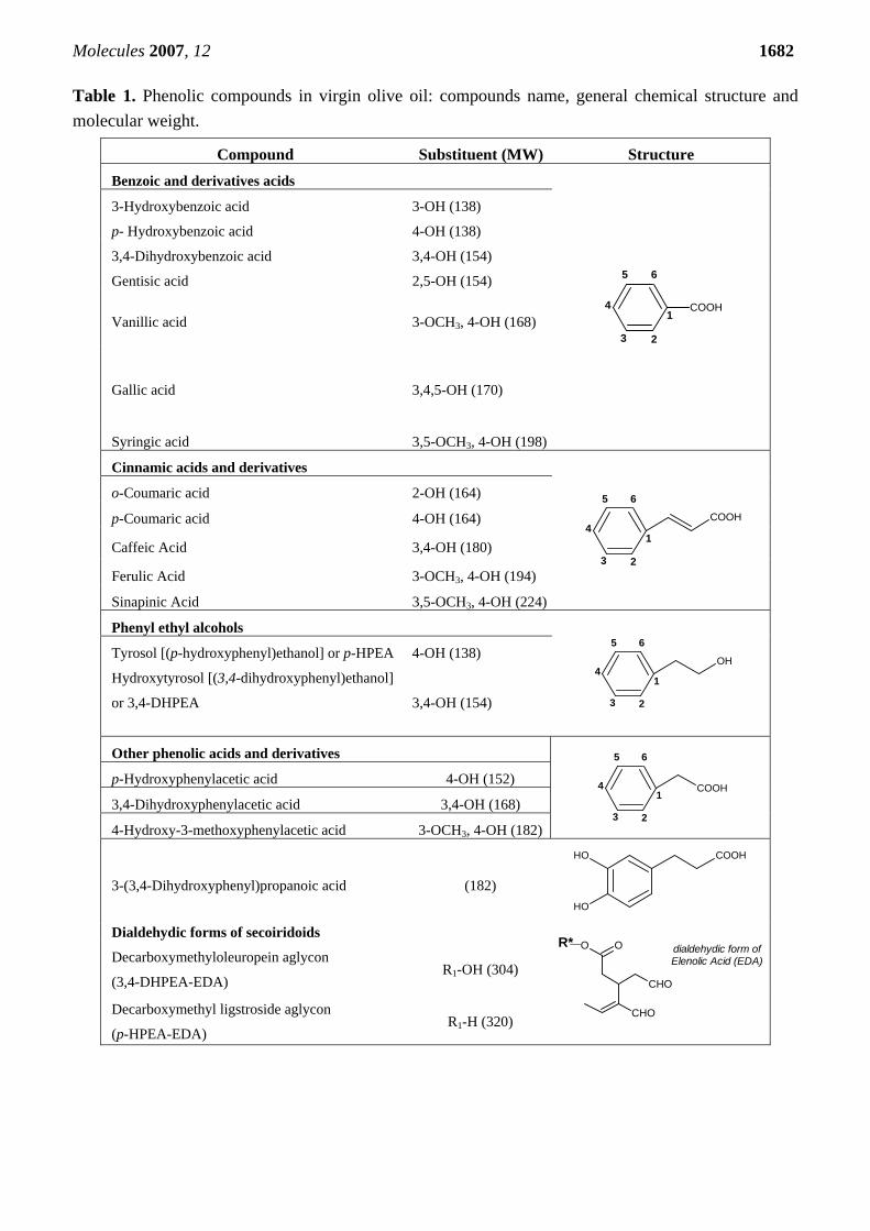
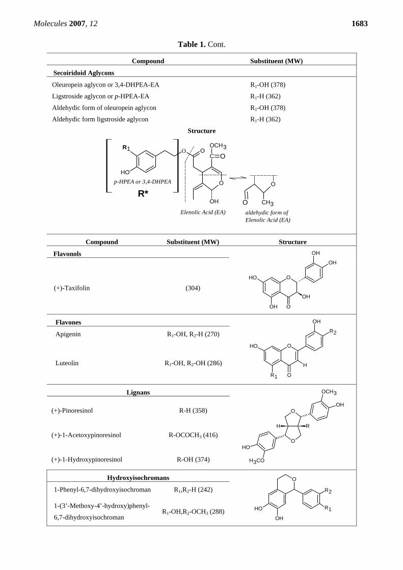
Oleuropein belongs to a specific group of coumarin-like compounds, the secoiridoids, which are
abundant in Oleaceae. Secoiridoids are compounds that are usually glycosidically bound and produced from the secondary metabolism of terpenes. The secoiridoids, found only in plants belonging to the family of Olearaceae that includes Olea europaea L., are characterised by the presence of elenolic acid in its glucosidic or aglyconic form, in their molecular structure. In particular, they are formed from a phenyl ethyl alcohol (hydroxytyrosol and tyrosol), elenolic acid and, eventually, a glucosidic residue. Oleuropein is an ester of hydroxytyrosol (3,4-DHPEA) and the elenolic acid (EA) glucoside (oleosidic skeleton common to the secoiridoid glucosides of Oleaceae) [1-5]. Secoiridoids of VOO in aglyconic forms arise from glycosides in olive fruits by hydrolysis of endogenous β-glucosidases during crushing and malaxation. These newly formed substances, having amphiphilic characteristics, are partitioned between the oily layer and the vegetation water, and are more concentrated in the latter fraction because of their polar functional groups. During storage of VOO hydrolytic mechanisms that lead to release of simple phenols, such as hydroxytyrosol and tyrosol, from complex phenols as secoiridoids may be involved [6-8]. The most abundant secoiridoids of VOO, identified for the first time by Montedoro et al. [1-3, 9] and confirmed also by other authors [10-13], are the dialdehydic form of elenolic acid linked to hydroxytyrosol or tyrosol (p-HPEA) respectively termed 3,4-DHPEA-EDA and p-HPEA-EDA, and an isomer of the oleuropein aglycon (3,4-DHPEA-EA) (Table 1). In 1999 another hydroxytyrosol derivative, hydroxytyrosol acetate (3,4-DHPEA-AC) was found in virgin olive oil [14].
Phenolic acids are secondary aromatic plant metabolites that are widely spread throughout the plant kingdom [15-17]. These naturally occurring phenolic acids contains two distinguishing constitutive carbon frameworks, namely the hydroxycinnamic and hydroxybenzoic structures. Elucidation of their roles in plant life is only one of the many ongoing investigations regarding phenolic acids: one vast area of interest lies in food quality [18-20]. Phenolic acids have been associated with color and sensory qualities, as well as with the health-related and antioxidant properties of foods [21-22]. One impetus for analytical investigations has been the role of phenolics in the organoleptic properties (flavor, astringency, and hardness) of foods [23-24]. Additionally, the content and profile of phenolic acids, their effect on fruit maturation, prevention of enzymatic browning, and their roles as food preservatives has been evaluated [25]. Recent interest in phenolic acids stems from their potential protective role, through ingestion of fruit and vegetables, against diseases that may be related to oxidative damage (coronary heart disease, stroke, and cancers) [26-28].
Molecules 2007, 12 1681 In particular, several phenolic acids such as gallic, protocatechuic, p-hydroxybenzoic, vanillic, caffeic, syringic, p- and o-coumaric, ferulic and cinnamic acid have been identified and quantified in VOO (in quantities lower than 1 mg of analyte kg-1 of olive oil). In this regard two research groups have extensively analyzed samples of VOO for these types of compounds [29-32]. In one of these mentioned articles, for instance, the authors found that trans-cinnamic acid, sinapinic acid, caffeic acid and 3,4-dihydroxyphenylacetic acid were present in several monovarietal VOO of the six Spanish olive cultivars analyzed [31]; therefore, these compounds might be potential markers of geographical origin or the olive fruit variety.
(+)-Pinoresinol is a common component of the lignan fraction of several plants such as Forsythia species [33] and Sesamum indicum seeds, whereas (+)-1-acetoxypinoresinol and (+)-1-hydroxy-pinoresinol and their respective glucosides have been detected in the bark of the olive tree (Olea europaea L.). According to Owen et al. [34], the quantity of lignans in VOO may be up to 100 mg kg-1, but as with the simple phenols and SIDs, considerable inter-oil variation exists. As suggested by Brenes et al. [35], the amount of lignans may be used as varietal marker, and they reported a method to authenticate VOO produced by Picual olives based on the very low content of the lignan (+)-1-acetoxypinoresinol in these oils.
A few years ago, Bianco et al. [36] investigated the presence of hydroxy-isochromans in VOO. In fact, during the malaxation step of VOO extraction, hydrolytic processes through the activity of glycosidases and esterases augment the quantity of hydroxytyrosol and carbonylic compounds, thus favouring the presence of all compounds necessary for the formation of isochroman derivatives. Two hydroxy-isochromans, formed by the reaction between hydroxytyrosol and benzaldehyde or vanillin, have been identified by HPLC-MS/MS technique and quantified in commercial VOOs.
Flavonoids are widespread secondary plant metabolites. During the past decade, an increasing number of publications on the health beneficial effects of flavonoids have appeared, such those related to cancer and coronary heart diseases [37-40]. Flavonoids are largely planar molecules and their structural variation comes in part from the pattern of modification by hydroxylation, methoxylation, prenylation, or glycosylation. Flavonoid aglycones are subdivided into flavones, flavonols, flavanones, and flavanols depending upon the presence of a carbonyl carbon at C-4, an OH group at C-3, a saturated single bond between C-2 and C-3, and a combination of no carbonyl at C-4 with an OH group at C-3, respectively. Several authors have reported that flavonoids such as luteolin and apigenin are also phenolic components of VOO [41-46]. Luteolin may originate from rutin or luteolin-7-glucoside, and apigenin from apigenin glucosides. There are also several interesting studies in which several flavonoids have been found in olive leaves and fruits [47-50].
Molecules 2007, 12 1682 Table 1. Phenolic compounds in virgin olive oil: compounds name, general chemical structure and molecular weight.
Compound Substituent (MW) Structure Benzoic and derivatives acids
COOH1
23
4
5 6
3-Hydroxybenzoic acid 3-OH (138)
p- Hydroxybenzoic acid 4-OH (138)
3,4-Dihydroxybenzoic acid 3,4-OH (154)
Gentisic acid 2,5-OH (154)
Vanillic acid 3-OCH3, 4-OH (168)
Gallic acid 3,4,5-OH (170)
Syringic acid 3,5-OCH3, 4-OH (198)
Cinnamic acids and derivatives
COOH
1
23
4
5 6
o-Coumaric acid 2-OH (164)
p-Coumaric acid 4-OH (164)
Caffeic Acid 3,4-OH (180)
Ferulic Acid 3-OCH3, 4-OH (194)
Sinapinic Acid 3,5-OCH3, 4-OH (224)
Phenyl ethyl alcohols
OH
1
23
4
5 6
Tyrosol [(p-hydroxyphenyl)ethanol] or p-HPEA 4-OH (138)
Molecules 2007, 12 1684 Why are the phenolic compounds present in virgin olive oil so important? Why is their determination so interesting and difficult?
Last year, Boskou published an interesting review [51] wherein the sources of natural phenolic
antioxidants were discussed, and the following idea was highlighted: “Widely distributed in the plant kingdom and abundant in our diet, plant phenols are today among the most talked about classes of phytochemicals”. To answer to the question of “why are phenolic compounds so interesting?”, the author of the review summarized several issues which have been studied in depth during the last decade:
– The levels and chemical structure of antioxidant phenols in different plant foods, aromatic plants and various plant materials.
– The probable role of plant phenols in the prevention of various diseases associated with oxidative stress such as cardiovascular and neurodegenerative diseases and cancer.
– The ability of plant phenols to modulate the activity of enzymes, a biological action not yet understood.
– The ability of certain classes of plant phenols such as flavonoids (also called polyphenols) to bind to proteins. Flavonol–protein binding, such as binding to cellular receptors and transporters, involves mechanisms which are not related to their direct activity as antioxidants.
– The stabilization of edible oils, protection from formation of off-flavors and stabilization of flavours.
– The preparation of food supplements. Focusing on phenolic compounds of virgin olive oil and bearing in mind the reasons for being so
important, attention must be paid to the fact that this class of compounds has not been completely characterized due to the complexity of their chemical nature and the complexity of the matrix in which they are found. Moreover, one of the current problems for developing rapid and reproducible analysis of phenolic compounds is the absence of suitable pure standards, in particular secoiridoid molecules and lignans.
Health aspects linked to phenols in VOO
VOO is an integral ingredient of the Mediterranean diet and accumulating evidence suggests that it
may have health benefits which include reduction of risk factors of coronary heart disease, prevention of several types of cancers, and modification of immune and inflammatory responses. VOO can be considered as example of a functional food, with a variety of components that may contribute to its overall therapeutic characteristics [52]. Its nutritional and healthy values and pleasant flavour have contributed to an increase in consumption of VOO which has fostered cultivation of olives outside the traditional olive oil producing region of the Mediterranean basin into newer areas such as Australia, Argentina and South Africa. The nutritional value of VOO arises from high levels of oleic acid, and from minor components such as phytosterols, carotenoids, tocopherols and hydrophilic phenols [53].
VOO contains at least 30 phenolic compounds. The major phenolic compounds are oleuropein derivatives, based on hydroxytyrosol which are strong antioxidants and radical scavengers. Recently there has been a surge in the number of publications that has investigated their biological properties. Bisignano et al. [54] found that hydroxytyrosol and oleuropein have antimicrobial activity against
Molecules 2007, 12 1685 several bacterial strains that are causal agents of intestinal or respiratory tract infections in humans. In a more recent in-vivo study, Glatzle and co-workers [55] demonstrated that enteral immunonutrition with VOO more effectively reduced septic pulmonary dysfunction compared to a fish oil-enriched lipid formula at the same concentration.
It has recently been found that hydroxytyrosol is renally excrete: while some of hydroxytyrosol is unchanged, some is also metabolized to the following metabolites: glucuronide conjugate, sulphate conjugate, homovanillic acid, homovanillic alcohol, 3,4-dihydroxyphenylacetic acid and 3,4-dihydroxyphenylacetaldehyde [56-57]. The radical scavenging potencies of these metabolites of hydroxytyrosol have also been investigated using the radical assay DPPH. The glucuronide conjugate was more potent than hydroxytyrosol while the sulphate conjugate was nearly devoid of radical scavenging activity. When phenol-rich VOO characterized by increasing concentrations of catecholic compounds were administered to human volunteers, Visioli and co-authors [58] observed a dose-dependent urinary excretion of hydroxytyrosol and its metabolite homovanillic alcohol. In a later study, the same authors [59] noticed that the urinary levels of unconjugated tyrosol and hydroxytyrosol correlated with their intake, except at the highest dose, which increased the quantity of glucuronide conjugate. Tuck et al. investigated the in vivo fate of hydroxytyrosol and tyrosol after intravenous and oral dosing of these tritium labelled compound to rats [60]. No significant differences in the amount of phenolic compounds eliminated in urine between the intravenous dosing method and the oral oil-based dosing method for either tyrosol or hydroxytyrosol were found.
Phytochemical compounds such as oleuropein and oleuropein aglycon have been intensively studied for some promising results with respect to their effects on human health and their potential medicinal properties. It has been found that diets containing olive oil phenols may increase in vivo resistance of LDLs to oxidation; the effectiveness of oleuropein has been explained in part through its ability to act as an antioxidant and in part through a hypocholesterolaemic effect [61]. In an investigation by Coni et al. [62] it was found that when fed a diet rich in oleuropein to rabbits, the ability of LDL to resist to oxidation increased, thanks to its antioxidant capacity; moreover, they found a significant reduction of the plasmatic levels of total, free and ester-derivatives of cholesterol. Oleuropein aglycon, the bitter component of olives and olive oil, is among the first example of how selected nutrients from an VOO-rich “Mediterranean diet” can directly regulate HER2-driven breast cancer disease [63]. As oleuropein aglycon exhibits synergistic anti-tumor effects when concurrently given to breast cancer cells chronically exposed to trastuzumab (Tzb; Herceptin™) for several months, this further underscores the potential clinical relevance of these findings.
On the basis of their shared throat irritant properties (pungency), Beauchamp et al. [64-65] examined whether p-HPEA-EDA, now referred to as “oleocanthal”, might mimic the pharmacological effects of ibuprofen, a potent modulator of inflammation and analgesia. It was found that, like ibuprofen, both enantiomers of p-HPEA-EDA caused dose-dependent inhibition of COX-1 and COX-2 activities (cyclo-oxygenase enzymes that catalyze key steps in the biochemical inflammation pathways derived from arachidonic acid) but had no effect on lipoxygenase in vitro. These authors hypothesized that long-term consumption of oleocanthal may help to protect against some diseases by virtue of its ibuprofen-like COX-inhibiting activity (by reducing the risk of developing some cancers and by lowering platelet aggregation in the blood). However, as noted by Fogliano and Sacchi [66], no data is available about the concentration of the various aglycons, including oleocanthal in plasma and urine after VOO consumption; absorption and bioavailability studies indicate however that tyrosol and
Molecules 2007, 12 1686 hydroxytyrosol are likely to be bio-available. It is worth mentioning that acid hydrolysis of oleocanthal would produce the elenolic acid, a dialdehyde compound even more similar to ibuprofen than oleocanthal itself.
In the context of the Mediterranean diet and coronary heart diseases, it has also been shown that VOO rich in phenols increases the resistance of LDL to oxidation, both in vitro and ex vivo [67-69]. The study carried out by Bogani et al. [70] confirmed the anti-thrombotic and anti-inflammatory effects of VOO phenolic components, in a postprandial setting; in fact, the results showed significant reductions in serum concentration of inflammatory markers (TXB2 and LTB4) at 2 and 6 h after consumption of VOO, but not after consumption of either olive oil or corn oil. They also evaluated the effects of these different oils on in vivo indexes of oxidative stress (plasma antioxidant capacity and urinary hydrogen peroxide levels) and showed the antioxidant activity of VOO phenolics after ingestion (increased plasma antioxidant capacity after 2 h of VOO consumption).
Foods containing high amounts of lignans such as flaxseed have been found to be protective against breast cancer, and in particular, to exert an anti-estrogenic effect; this latter observation might be explained by considering the structural similarities between the lignans and the synthetic antiestrogen tamoxifen [71].
Two hydroxy-isochromans, 1-(3'-methoxy-4'-hydroxyphenyl)-6,7-dihydroxyisochroman and 1-phenyl-6,7-dihydroxyisochroman, are formed by reaction between hydroxytyrosol and vanillin and benzaldehyde, respectively, (under very mild conditions). They have only recently been discovered in VOO [36] and are active in inhibited platelet aggregation and thromboxane release evoked by agonists (sodium arachidonate and collagen) that induce reactive oxygen species-mediated platelet activation [72].
A large number of studies, mainly experimental models, have been performed on certain minor components of olive oil. However, as commented in an excellent review by Covas et al. [73], the precepts of evidence-based medicine require high-level scientific evidence to be provided before nutritional recommendations for the general public can be formulated. Scientific evidence required is provided by randomized, controlled, double-blind clinical trials (level I evidence), and to some extent by large cohort studies (level II evidence). Basic research, despite its usefulness in permitting adoption on a mechanistic approach, does not provide evidence for nutritional recommendations. Of course, the level of evidence of a particular study depends not only on its design, but also on its quality (external and internal validity, homogeneity of the sample, and statistical power). Finally, evidence is built by the agreement of the results of several similar studies. In the same review, the authors highlighted that in experimental studies, olive oil phenols have been shown to:
1) have antioxidant effects, greater than those of vitamin E, on lipid and DNA oxidation [74-77]; 2) prevent endothelial dysfunction by decreasing the expression of cell adhesion molecules [78],
increasing nitric oxide (NO) production and inducible NO synthesis [79] and quenching vascular endothelium intracellular free radicals [80];
3) inhibit platelet-induced aggregation [81]; 4) enhance the mRNA transcription of the antioxidant enzyme glutathione peroxidase (GSH-Px). It
should be mentioned however that to regard this last point controversial results have been obtained depending on the tissue in which the gene expression was evaluated [75, 82].
Other potential activities of VOO phenolic compounds include chemopreventive activity [77]. The anticarcinogenic activity of phenols may be due not only to their antioxidant properties, but also to
Molecules 2007, 12 1687 their ability to reduce the bioavailability of food carcinogens and to inhibit their metabolic activation [83-84]. There are several mechanistic considerations of the role of phenolic compounds as anticarcinogens, as reviewed by Yang et al. [85]. Phenols as related to oxidative stability of VOO
Oxidation is an inevitable process that starts after the VOO has been extracted and leads to
deterioration that becomes more pronounced during oil storage. Initially lipids are radically oxidised to hydroperoxides, which are odourless and tasteless [86] and do not account for sensory changes. However, decomposition occurs through homolytic cleavage of the hydroperoxide group with production of various volatile compounds, known as secondary oxidation products, which are responsible for typical unpleasant sensory characteristics. Oxygen, light, temperature, metals, pigments, unsaturated fatty acid composition, as well as the quantity and kind of natural antioxidants, are all factors that can influence the free radical mechanism of the autoxidation process in a different manner [87-88].
Natural antioxidants exhibit complex properties between air-oil and oil-water interfaces that significantly affect their relative activities in different lipidic systems. The presence of hydrophilic phenolic compounds in VOO and their high antioxidant activity can be explained by the so-called “polar paradox” [89] which dictates that “polar antioxidants are more effective in non polar lipids, whereas non-polar antioxidants are more active in polar lipid emulsions”. According to Frankel [90] in a bulk oil system the hydrophilic antioxidants, such as polar phenols, are oriented in the air-oil interface (a low quantity of air is always trapped in the oil) and become more protective against oxidation than the lipophilic antioxidants, like tocopherols, which remain in solution in the oil.
In a study carried out by Paiva-Martins and co-workers [91] it was found that when food is processed with VOO in the presence of water, olive phenolic extracts with higher quantities of 3,4-DHPEA-EA and 3,4-DHPEA-EDA would be better than VOO extracts with higher quantities of hydroxytyrosol, despite the higher antioxidant activity of hydroxytyrosol in bulk oil.
Moreover, the orientation of phenolic compounds in the oil-water interface and the active surface of water droplets influence the protection against the oxidation of oil. Recently, some researchers [92-95] have determined that VOO contains a low quantity of water (ranging from 450 mg kg-1 to 3,000 mg kg-1 depending on the extraction technology), that increases when samples were not filtered. Part of the total water content presents in VOO is free and available for chemical and enzymatic reactions and also keeps hydrophilic phenols in solution. This can explain the hydrolytic process that occurs both to phenols (by esterases) and triacylglycerols (by lipase) during prolonged VOO storage. Thus, more rapid oxidation of the unfiltered oil could be expected. Instead, according to Gomez-Caravaca et al. [95] and Tsimidou et al. [96], stability of unfiltered samples, when measured in terms of resistance to accelerated oxidation (value by OSI or Rancimat instruments) was in all cases significantly higher than that of the corresponding filtered oils. This coincided with a higher total phenolic content in unfiltered VOO. Undoubtedly, a loss of a significant fraction of phenols during filtration is related to the reduction of oxidative stability.
Chain-breaking antioxidants, such as phenolic compounds, react with lipid radicals to form non-reactive radicals, interrupting the propagation chain. In fact, these compounds are able to donate an electron or a hydrogen atom to the lipid radical formed during the propagation phase of lipid oxidation
Molecules 2007, 12 1688 and stabilize the resulting phenoxyl radical by delocalizing the unpaired electron [97-100]. Phenolic compounds exert their antioxidant abilities in VOO by scavenging peroxyl and alkoxyl radicals, and by chelation of transition metals ions present in trace quantities [101]. Paiva-Martins and Gordon [102-103] have studied the antioxidant effects of pure phenolic compounds (hydroxytyrosol, hydroxytyrosol acetate, oleuropein, 3,4-DHPEA-EA, and 3,4-DHPEA-EDA) by both the diphenylpicrylhydrazyl (DPPH) assay and the ferric reducing antioxidant potential (FRAP) assay in bulk oil and in emulsions (both with and without ferric ions). The compounds showing the best antioxidant activity in oil in water emulsions in the presence of iron were 3,4-DHPEA-EA and 3,4-DHPEA-EDA, which in contrast to hydroxytyrosol and oleuropein did not show pro-oxidant activity. However, when the radical scavenging activity was measured for these compounds, 3,4-DHPEA-EA showed a much higher activity than 3,4-DHPEA-EDA, suggesting that chelation of iron was of major significance in determining the antioxidant activity of these compounds in the presence of iron and water. According to this latter behaviour, Bendini et al. [104] showed evidence that 3,4-DHPEA-EDA has the ability to chelate copper in bulk oil.
The stability of VOO is improved by synergistic interactions between various antioxidants present (both phenolic and non-phenolic) and the lipid composition. Such cooperative activity seems to explain the antioxidant synergism observed when α-tocopherol and ascorbic acid or ubiquinol are used in combination. Recent investigations have also demonstrated an antioxidant synergism between α-tocopherol and some phenolics (green tea catechins and quercetin) [105-106]. Bendini et al. [104] hypothesized that phenols having an ortho-dihydroxyl structure, and in particular an isomer of 3,4-DHPEA-EDA, were able to reduce the oxidized forms of tocopherols (tocopheryl radicals and quinones). This was substantiated by Pazos et al. [107] who demonstrated that phenols, as well as several benzoic acids and epicatechin gallates, were potentially active in the regeneration of α-tocopherol via reduction of α-tocopheroxyl radical. Moreover this capacity was found to be directly proportional to the ability of phenolic compounds to transfer a single H atom.
There have been numerous studies on the relative antioxidant potency of the individual olive oil phenols, although it may vary depending on the methods used for evaluation. Many authors have frequently studied the ability of antioxidant molecules or extracts to scavenge some free radicals, and in this regard, several stable, coloured free radicals (DPPH and ABTS) are widely used due to their intense absorbance in the visible region. In this case, the hydrogen-donating activity can be determined.
However, as a general guide to their potency, oleuropein and hydroxytyrosol have been shown to be more effective than vitamin E [108] and butylated hydroxytoluene (BHT) or other synthetic antioxidants approved for use in foods [109-111].
From comparison with the principal phenolic constituents of VOO, it has been claimed that hydroxytyrosol is the most active antioxidant compound [112]. Both hydroxytyrosol and oleuropein have been shown to be scavengers of superoxide anions, and inhibitors of the hypochlorous acid-derived radicals, but hydroxytyrosol was more effective than oleuropein in this regard [113]. Both compounds also scavenged hydroxyl radicals, but in this case oleuropein showed greater activity [114]. Gordon et al. [115] investigated the antioxidant activity of hydroxytyrosol acetate by scavenging of DPPH radicals in comparison with that of the phenolic extract from VOO and the pure components hydroxytyrosol, oleuropein, 3,4-DHPEA-EA, and α-tocopherol in bulk oil and oil-in-water emulsions. In this study the authors showed that hydroxytyrosol acetate had a weaker DPPH radical scavenging
Molecules 2007, 12 1689 activity than hydroxytyrosol and 3,4-DHPEA-EA was slightly less effective than hydroxytyrosol acetate in oil but was the most effective hydroxytyrosol derivative in an emulsion oil in water.
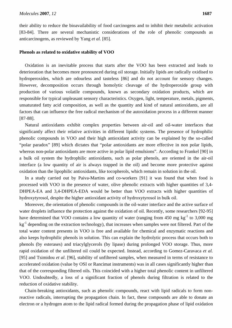
It is well known that the high oxidative stability of VOO is primarily due to o-diphenols such as hydroxytyrosol and its oleosidic forms [108]. Aparicio et al. [116], using statistical analysis of data relative to 79 VOO of olives cv. Hojiblanca and Picual, measured correlations between oxidative stability (valued by Rancimat) and several compositional variables. The phenols (R2=0.87), o-diphenols (R2=0.77), and the oleic/linoleic ratio (R2=0.71) had the highest values, followed by chlorophylls (R2=0.68), total tocopherols (R2=0.65) and carotenoids (R2=0.59). Principal components analysis confirmed that phenols, oleic/linoleic ratio, and tocopherols had the maximum correlation with oxidative stability. From these results, the phenolic content would contribute around 51% of the stability of VOO, and particularly 30% for phenols and 21% for o-diphenols whereas, the oleic/linoleic ratio would account for only 27%. Since a hypothetical synergy effect was detected between these chemical variables, it is more prudent to conclude that 78% of the stability is due to the combined effect of both variables. The authors surmised that the contribution of total tocopherols was around 9%, whereas the remaining percentage of 13% could be attributed to chlorophylls and carotenoids.
In experiments carried out by Carrasco-Pancorbo and co-authors [117], the antioxidant activity of several single phenolic compounds of VOO (hydroxytyrosol, tyrosol, elenolic acid, 3,4-DHPEA-EDA, (+)-pinoresinol, (+)-1-acetoxypinoresinol, oleuropein aglycon and ligstroside aglycon) was evaluated by different chemical approaches: radical assay (DPPH), accelerated oxidation in a lipid model system (OSI, oxidative stability index), and an electrochemical method (flow injection analysis FIA-amperometry and cyclic voltammetry). These authors verified that, as is generally assumed, the presence of a single hydroxyl group on benzenic ring conferred only limited antioxidant activity. On the other hand, the presence of a catechol moiety enhances the ability of the phenolic compounds to act as antioxidants. The results obtained in all three tests showed that hydroxytyrosol, 3,4-DHPEA-EDA and oleuropein aglycon were the strongest in terms of antioxidant power. Elenolic acid, which does not have a phenolic ring, was one of the compounds that presented the weakest antioxidant activity, as also reported by Briante et al. [118]; this compound together with (+)-pinoresinol, tyrosol, ligstroside aglycon and (+)-1-acetoxypinoresinol showed pro-oxidant effect when tested by OSI. Similar results were found by Nenadis et al. [119]: tyrosol, hydroxytyrosol and their secoiridoid derivatives were examined calculating the bond dissociation enthalpy (BDE) of phenolic hydroxyl groups and the ionization potential (IP) as descriptors to predict the H-atom-donating and electron-donating abilities of antioxidants, respectively. Catechol derivatives had the lowest BDE values (77.7-80.1 kcal mol-1), whereas the lignans, pinoresinol and 1-acetoxypinoresinol, and other monophenols had much higher BDE values (85.1-88.0 kcal mol-1), which suggested a lower potential for radical scavenging.
In a recent work, Lorenz et al. [120] investigated the antioxidant and radical scavenging properties of several phenolic isochromans. All hydroxy-isochromans tested exceeded the scavenging effect of trolox (an hydrophilic analogue of α-tocopherol). They found excellent ROS/RNS (reactive species of oxygen/nitrogen) scavenging features of the hydroxy-isochromans and also concluded that their simple synthesis added to their interest as candidates for pharmaceutical interventions that protect against the deleterious action of ROS/RNS.
Molecules 2007, 12 1690
Figure 1 Correlations among OSI values (in hours), phenolic amounts and antioxidant activity (DPPH test) by spectrophotometric assays. a, OSI vs Total Phenols (mg gallic acid kg-1 VOO); b, OSI vs DPPH (mmol trolox kg-1 VOO); c, OSI vs o-diphenols (mg gallic acid kg-1 VOO). Analyses were carried out over three years; in each figure the number of samples is reported (N). Three replicates were prepared and analyzed for each sample.
OSI vs Total Phenols (by spectrophotometric assay)
y = 12.133x + 51.756R2 = 0.6523
r=0.8077N=76
0
100
200
300
400
500
600
700
0 5 10 15 20 25 30 35 40
a
OSI vs DPPH (antiradical test)
y = 0.0615x - 0.2062R2 = 0.707r=0.8408
N=64
0.0
0.5
1.0
1.5
2.0
2.5
0 5 10 15 20 25
b
OSI vs o -diphenols (by spectrophotometric assay)
y = 5.9318x + 8.5033R2 = 0.7302
r=0.8545N=58
0
50
100
150
200
250
300
350
0 5 10 15 20 25 30 35 40
c
Molecules 2007, 12 1691
Franconi et al. [121] tested the antioxidant activity of two VOO (Seggianese and Taggiasca characterized by different quali-quantitative phenolic profiles) on human LDL by measuring malondialdehyde and conjugate diene generation induced by copper ions. In both tests antioxidant potency correlated with total phenols; moreover, reduction of malondialdehyde and generation of conjugate diene was dependent on the amount of total phenols. High levels of secoiridoids enhance the antioxidant activity, suggesting that VOO rich in these compounds could have health-protecting properties consistent with a low extent of LDL oxidation.
Recently, the interest in oxidized forms of VOO phenols has significantly increased, especially in relation to determination of freshness/ageing status [122-123]. Moreover, characterization of these oxidized phenolics could represent an analytical instrument to investigate the thermal processes of the oils during refinement [124]; this could also provide the means to verify fraudulent practices such as "gentle deodorization" (under soft refining conditions) or blending of VOO with other oils. In 2005 Rios et al. [122] compared the performance of HPLC-APCI-MS and GC-IT-MS analytical techniques to evaluate the oxidation products of elenolic acid, oleuropein and ligstroside aglycons. Five oxidation phenols were identified with gas-chromatography. Armaforte et al. [125] showed that SPE procedure (usually used to extract the phenolic fraction from VOO) may be a not appropriate analytical step when VOO contains significant polar oxidation products (from phenols or lipids); in fact, these latter compounds could interfere with the retention mechanism of phenols during their extraction. These authors also proposed an index to establish the degree of freshness of VOO. This value, or TPAR (ratio between total peak area of reduced and oxidized forms of phenols) is close to 1 for fresh samples whereas it decreases rapidly in VOO with an increasing content of oxidized phenols.
Carrasco-Pancorbo and co-authors [124] by studying the phenolic profiles of the oils after a drastic heating treatment (at 180°C) found several “unknown” compounds, (by using HPLC-UV, HPLC-MS and CE-UV) that were probably linked to phenol oxidation. In particular, seven peaks significantly increased when the thermal treatment was longer (from 1 to 3 h) and their presence was also confirmed in refined olive oils. The concentration of hydroxytyrosol, elenolic acid, 3,4-DHPEA-EDA and 3,4-DHPEA-EA decreased more quickly with the thermal treatment than other phenolic compounds present in olive oil, confirming their high antioxidant power; moreover 3,4-DHPEA-AC and p-HPEA-EA were more resistant to heat treatment, whereas the amount of (+)-pinoresinol and (+)-1-acetoxypinoresinol were almost unchanged. Sensory properties elicited by phenols in VOO
Virgin olive oil is a natural fruit juice obtained directly from olives without any further refining
process. Its flavour is characteristic and is markedly different from those of other edible fats and oils. The combined effect of the taste, odor (directly via the nose or indirectly through the retronasal path via the mouth) and chemical responses (pungency, astringency, metallic, cooling or burning) gives rise to the sensation generally perceived as “flavor” [126]. VOO, when extracted from fresh and healthy olive fruits (Olea europaea L.) and properly processed and stored, is characterized by an unique combination of aroma and taste that is highly appreciated [127-128]. The sensory aspect, due to the use of VOO as a seasoning on cooked and especially raw foods, has great repercussions on its acceptability. Thus, since sensory quality plays an important role in directing the preference of consumers, many attempts have been made to clarify the relationships between the sensory attributes
Molecules 2007, 12 1692 in a VOO as perceived by assessors and its volatile and phenol profiles, which are responsible for aroma and taste, respectively [128].
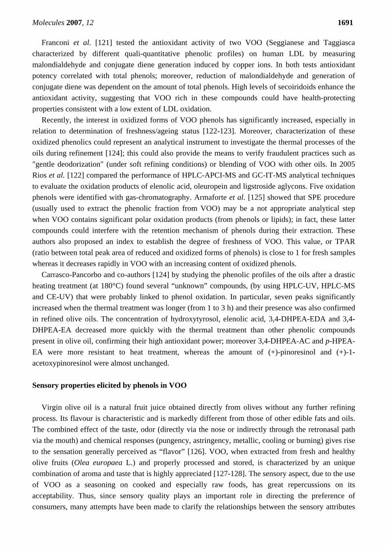
Few individuals, except for trained assessors of VOO, know that bitterness and pungency perceived by taste are positive attributes for a VOO. These two sensory characteristics are strictly connected by the quali-quantitative phenolic profile of the product. An example of the positive correlations between amount of phenols and bitter and pungent intensities is shown in Figures 2a and 2b.
Some phenols mainly elicit the tasting perception of bitterness; however, other phenolic molecules can stimulate the free endings of the trigeminal nerve located in the palate and also in the gustative buds giving rise to the chemesthetic perceptions of pungency, astringency and metallic attributes. Thus, the intensity of bitterness and pungency is mainly related to the olives cultivar and the ripening stage and, as reported by many authors, are especially abundant in oils obtained from unripe fruits. For instance, Caponio et al. [129] showed that in Coratina and Oliarola Salentina VOO, oleuropein and its aglycon form both decrease as ripening of the olives progressed. From this data, the bitter to pungent taste would appear to be mainly ascribable to oleuropein aglycon since greater amounts of this phenolic compound are present in the Coratina oils with respect to O. Salentina oils, which are known to have a sweet taste. In order to attenuate such these taste sensations, the authors suggested the need to postpone harvesting of Coratina olives.
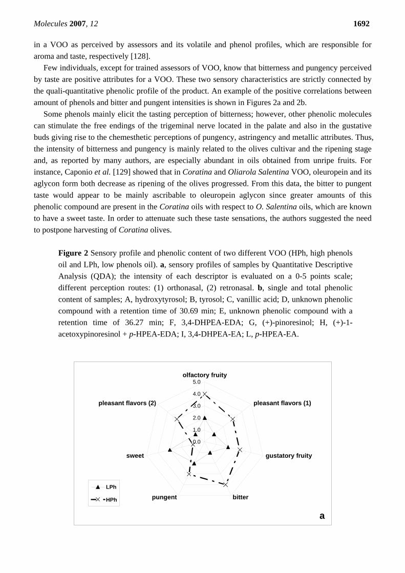
Figure 2 Sensory profile and phenolic content of two different VOO (HPh, high phenols oil and LPh, low phenols oil). a, sensory profiles of samples by Quantitative Descriptive Analysis (QDA); the intensity of each descriptor is evaluated on a 0-5 points scale; different perception routes: (1) orthonasal, (2) retronasal. b, single and total phenolic content of samples; A, hydroxytyrosol; B, tyrosol; C, vanillic acid; D, unknown phenolic compound with a retention time of 30.69 min; E, unknown phenolic compound with a retention time of 36.27 min; F, 3,4-DHPEA-EDA; G, (+)-pinoresinol; H, (+)-1-acetoxypinoresinol + p-HPEA-EDA; I, 3,4-DHPEA-EA; L, p-HPEA-EA.
0.0
0.1
0.2
0.3
0.4
0.5ytiurfyrotcaflo
)1(srovalftnasaelp
ytiurfyrotatsug
rettibtnegnup
teews
)2(srovalftnasaelp
hPL
hPH
a
Molecules 2007, 12 1693
latoTLIHGFEDCBA0
2
4
6
8
05001051002052
mg
3,4-
DH
PAA
/kg
ofoi
lhPHhPL
bFigure 2. Cont.
A Quantitative Descriptive sensory Analysis (QDA) carried out by Rotondi et al. [130] confirmed a decreasing trend of the positive olive oil descriptors, such as bitterness and pungency, when Nostrana di Brisighella olives ripened. The highest statistically significant intensity was at the beginning of fruit skin pigmentation. The decrease in bitterness and pungency was also related to a reduction in total phenols and o-diphenols levels. In particular, a positive correlation between the secoiridoids content and bitterness and pungency was observed.
With the aim of reducing the bitterness intensity in VOOs, disfavored by many consumers when present at high intensity, some authors [131] developed postharvest technology based on hot-water treatments of olive fruits (cultivars Manzanilla, Picual, and Verdial) in the temperature range of 60-68°C or [132] with air-heating (40°C during 24, 48, and 72 h). These treatments promote a reduction in bitterness that is directly related to the time and temperature of treatment, probably due to a partial inhibition of glycosidases and esterases; in fact, these enzymes are involved in the release of secoiridoid derivatives from oleuropein during the crushing malaxation process. However, this heat treatment also affected other quality traits such as oxidative stability and color and could produce a change in the aroma profile of the VOO as well.
The standard method of analyzing the bitter taste of olive oil is by sensory analysis using a panel of tasters [133]. However, an analytical panel is often not likely to be available, since a permanent staff of trained tasters and a highly specialized panel chief is necessary. Many consumers from extra-European countries are not accustomed to the typical high intensity of bitterness or pungency of fresh VOO and, consequently, must be blended with less bitter VOO. For this reason, methods for the evaluation of the bitterness level based on physical-chemical determinations would be very useful for the industry. Several authors have found a strong relationship between these sensory attributes and the content of phenolic compounds in the olive oils. In 1992 Gutiérrez et al. [134] proposed an analytical method for
Molecules 2007, 12 1694 measurement of bitterness, based on solid phase extraction (SPE) of phenols and their spectrophotometric detection at 225 nm; this parameter termed IB or index of bitterness, was highly correlated to the sensory intensity of bitterness and is still the most widely used for its determination. Some years later, Mateos and co-workers [135] showed that several non-bitter phenolic compounds could also absorb at 225 nm; consequently, they asserted that this index was not appropriate for comparing bitterness of VOO obtained from blend of olive varieties characterized by very different phenolic profiles (e.g. Picual and Arbequina). Moreover, Mateos suggested that evaluation of the bitterness level of a VOO could be described by the experimental equation obtained from the regressions between intensity of bitterness and the concentrations of oleuropein aglycon using a Panel test and chromatographic analysis, respectively.
Some researchers suggest that secoiridoid derivatives of hydroxytyrosol are the main contributors to olive oil bitterness. Recently, a procedure called taste dilution analysis (TDA) was reported by Frank and co-authors to underlie the sensory threshold of bitter for oleuropein derivatives [136]. Bitterness was assessed by preparing serial dilutions of samples in water and then tasting in order of increasing concentration until the concentration at which the diluted sample can be differentiated from water as judged in a triangle test is found. When an isomer (or isomers) of oleuropein aglycon was prepared by β-glucosidase hydrolysis of oleuropein isolated from olives and evaluated by assessors, it was found to be bitter with a threshold of 50 μmol. Using the same evaluation technique, no bitterness was observed for hydroxytyrosol or elenolic acid.
Andrewes et al. [137] assessed the relationship between polyphenols and olive oil pungency. p-HPEA-EDA was the key source of the burning sensation found in many olive oils. In contrast, 3,4-DHPEA-EDA, tasted at an equivalent concentration, produced very little burning sensation. This is a clear example of the different sensory properties of a secoiridoid derivative of hydroxytyrosol and tyrosol. In 2005, Beauchamp and co-authors [64] measured the pungent intensity of p-HPEA-EDA isolated from different VOO confirming this molecule is the principal agent in VOO responsible for throat irritation. These researchers also tested the throat-irritant properties of its synthetic form (named “oleocanthal”, with oleo- for olive,-canth- for sting, and -al for aldehyde) dissolved in non-irritating corn oil. They found an effect comparable to that of the purified compound from VOO and a dose-dependent activity.
In 2003, Gutierrez-Rosales and co-authors [138] isolated the major peaks found in the phenolic profile of VOO using preparative HPLC; after dissolving in water these molecules purified were then tasted to evaluate the intensity of bitterness. It was concluded that the peaks corresponding to the 3,4-DHPEA-EDA, 3,4-DHPEA-EA and p-HPEA-EDA were those mainly responsible for the bitter taste of VOO. As previously reported, Mateos et al. [135] verified the better correlation between the aldehydic form of oleuropein aglycon and bitterness.
Recently some researchers [139] have studied the temporal perception of bitterness and pungency in monovarietal VOOs; analyses were performed by a trained sensory panel utilizing a time–intensity (TI) evaluation technique; bitterness curves had a faster rate of rising and declining than pungency curves: the curves for bitterness reached a maximum after approximately 16–20 s, whereas the maximum of the perception of pungency is registered between 26 and 29 s and is independent of the maximum intensity of the perception.
As already discussed, several authors have associated some phenols with bitterness, thus obtaining models and relationships between individual phenols separated by HPLC and bitterness intensity [135,
Molecules 2007, 12 1695 138,140]. In these reports, bitterness was measured by a panel test or calculated from K225 values. Moreover tyrosinase- and peroxidase-based biosensors are being developed for the bitterness assessment [141], and are showing interesting possibilities. However, HPLC is often not available in many olive oil mill laboratories because of economic reasons, as well as specialized technical staff and biosensors. As an alternative, Beltrán and co-authors [142] proposed that measurement of phenol content can be used. This is a simple analytical method that involves liquid-liquid extraction and colorimetric measurement using Folin-Ciocalteau reagent [143]. In their experimental work, the authors analyzed the relationship between phenol content and K225 for oils from four of the most important olive cultivars worldwide (Frantoio, Hojiblanca, Picual, Arbequina); 360 samples were used to develop the model. As a practical application, bitterness intensities were evaluated by sensory analysis of 25 VOO samples, and were then estimated by applying the prediction model. In order to provide an easy tool for bitterness estimation, VOO bitterness was classified by its phenol content into four categories (results expressed as mg of caffeic acid per kg of oil and intensity of bitterness between 0 and 5 values): phenol contents equal or lower than 220 mg kg-1 corresponded to non-bitter oils or oils with almost imperceptible bitterness (intensities 0–1.5); slight bitterness corresponded to 220–340 mg kg-1 (intensities 1.6–2.5); bitter oils have a phenol contents ranging from 340 to 410 mg kg-1 (intensities 2.5–2.99); and a phenol contents higher than 410 mg kg-1 corresponds to quite bitter or very bitter oils (intensities higher than 3). In general, the authors determined that the oils were classified correctly into the same bitterness categories by both methods at 92%, achieving 100% of correct classification for the lowest and highest bitterness categories. New analytical approaches to characterization of the phenolic profile and applied studies during the last decade
In order to utilize VOO as a source of phenolic compounds, to develop complete compositional
databases and to obtain more accurate data about the intake of antioxidants further chemical characterization is needed. Identification and quantitation, based traditionally on HPLC (with different detectors, such as UV, fluorescence, coulometric electrode array detection, amperometric detector) [144-150], GC-FID [151-154] and, more recently CE-UV, can be aided today by MS and NMR, which is a focus of the present review.
Liquid chromatography/mass spectrometry (LC-MS) has been widely accepted as the main tool in identification, structural characterization and quantitative analysis of phenolic compounds in olive oil. Using a mass spectrometer for detection offers some undoubted advantages, such as independence of a chromo- or fluorophore, lower LOD than UV in most cases [155], the possibility to obtain structural information and easy separation of coeluting peaks using the information about mass as a second dimension.
The sensitivity of response in MS is clearly dependent on the interface technology employed. In LC-MS analysis of phenolic compounds, atmospheric pressure ionization interfaces, i.e. APCI and electrospray ionization (ESI), are used almost exclusively today, and both positive and negative ionization are applied. In general, phenolic compounds are detected with a greater sensitivity in the negative ion mode, but the results from positive and negative ion modes are complementary, and the positive ion mode shows structurally significant fragments [156].
Molecules 2007, 12 1696
On the other hand, optimal ionization depends not only on the interface parameters, but also on the mobile phase of the liquid chromatography. As a first rule, the use of non-volatile salts in the mobile phase (common in other chromatographic methods) should be avoided, as they would interfere with the ionization source. The mobile-phase composition and its pH need also careful optimization as they may influence the ionization efficiency of the analytes.
The selection of the analyzer, apart from its accessibility, is determined by the required sensitivity and selectivity and the general objectives. LC-atmospheric pressure ionization (API)-MS typically only yields a single strong ion, which reduces its ability to make analyte accurate identifications. In the most cases, single-stage MS is used in combination with UV detection to facilitate the identification of phenolic compounds in olive oil samples with the help of standards and/or reference data. Ion Trap or QqQ provide the possibility of doing MS/MS or MSn, which can be used for structure elucidation or for additional selectivity to gain sensitivity by reducing the chemical noise [157]. MS/MS and MSn involve two (or more) stages of mass analysis, separated by a fragmentation step. TOF MS, which is one of the most advanced MS analyzers, provides excellent mass accuracy [158] over a wide dynamic range if a modern detector technology is chosen. The latter, moreover, allows measurements of the correct isotopic pattern [159], providing important additional information for the determination of elemental composition [160].
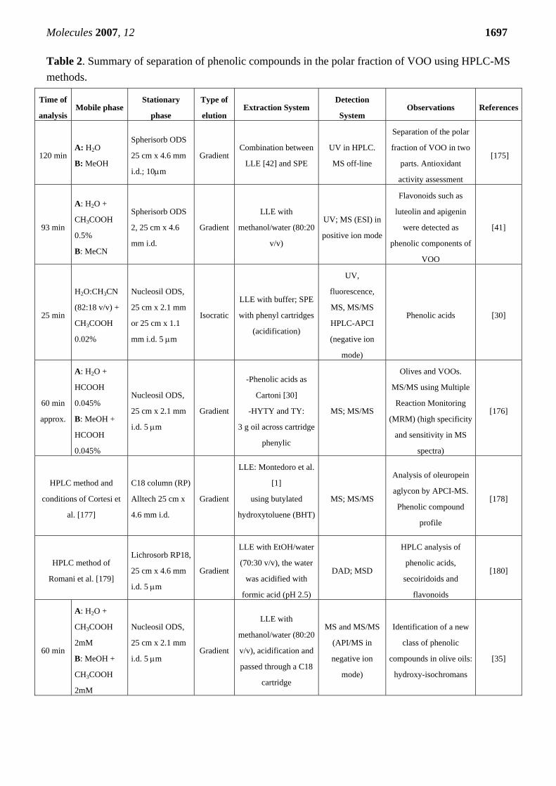
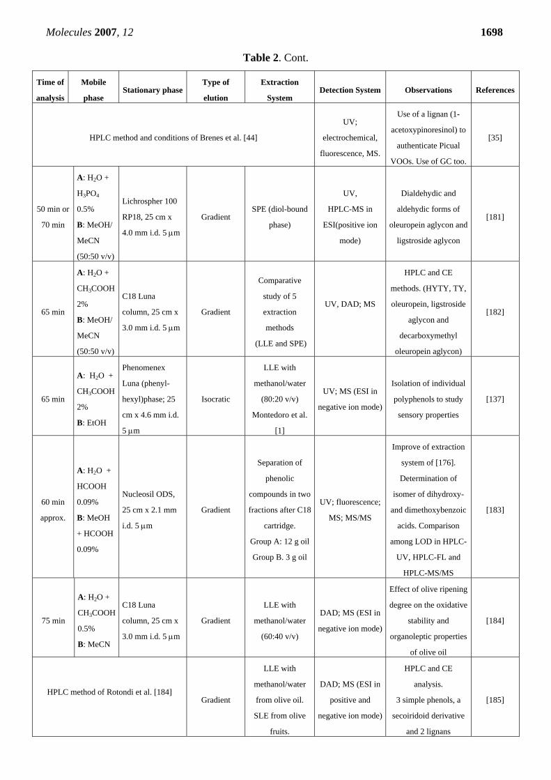
Table 2 provides an overview of methodologies based on LC-tandem mass spectrometry used for the analysis of phenolic compounds in olive oil. The table does not include several publications in which the analysis of olive fruit, leaves, pulp and pomace, olive tree wood, as well as olive oil waste waters were carried out by using HPLC-MS [14, 49, 156, 161-168]. Other important issues are the presence of phenolic metabolites of VOO in the human low density lipoprotein fraction [169-170].
High-resolution spectroscopic techniques, and particularly NMR spectroscopy, are finding interesting applications in the analysis of complex mixtures of various food extracts that contain phenols.
During the past decade proton nuclear magnetic resonance spectroscopy (NMR) has been successfully used in olive oil analysis [171-172]. Currently available high-resolution spectroscopic techniques, coupled with the facilities of computerized mathematical or other treatment of data have found interesting applications in the field of agricultural and food science without the necessity for a separative technique coupled with NMR, as commented by Gerothanassis [173]. Additionally the usefulness of 1H NMR spectroscopy has been increasingly recognized for its non-invasiveness, rapidity, and sensitivity for a wide range of compounds in a single measurement. However, difficulties may arise in relation to the information obtained from spectra of multicomponent mixtures such as olive oil. Strong signal overlap, dynamic range problems, diversity of intensities due to various concentrations of the food constituents, and the inherent lack of scalar coupling information between different moieties lead to ambiguous or incomplete assignments, thus hindering detection even with the use of multidimensional NMR [174]. One possible approach to these problems involves the combination of the advantages of NMR spectroscopy with those of chromatography. Coupled techniques such as LC-NMR or LC-NMR/MS may provide information on overall composition and enable the identification of individual phenols in complex matrices. Moreover, on-line solid phase extraction (SPE) in LC-NMR for peak storage after the liquid chromatography separation prior to NMR analysis or similar techniques have been recently applied.
Molecules 2007, 12 1697 Table 2. Summary of separation of phenolic compounds in the polar fraction of VOO using HPLC-MS methods.
Time of
analysis Mobile phase
Stationary
phase
Type of
elution Extraction System
Detection
System Observations References
120 min A: H2O
B: MeOH
Spherisorb ODS
25 cm x 4.6 mm
i.d.; 10μm
Gradient Combination between
LLE [42] and SPE
UV in HPLC.
MS off-line
Separation of the polar
fraction of VOO in two
parts. Antioxidant
activity assessment
[175]
93 min
A: H2O +
CH3COOH
0.5%
B: MeCN
Spherisorb ODS
2, 25 cm x 4.6
mm i.d.
Gradient
LLE with
methanol/water (80:20
v/v)
UV; MS (ESI) in
positive ion mode
Flavonoids such as
luteolin and apigenin
were detected as
phenolic components of
VOO
[41]
25 min
H2O:CH3CN
(82:18 v/v) +
CH3COOH
0.02%
Nucleosil ODS,
25 cm x 2.1 mm
or 25 cm x 1.1
mm i.d. 5 μm
Isocratic
LLE with buffer; SPE
with phenyl cartridges
(acidification)
UV,
fluorescence,
MS, MS/MS
HPLC-APCI
(negative ion
mode)
Phenolic acids [30]
60 min
approx.
A: H2O +
HCOOH
0.045%
B: MeOH +
HCOOH
0.045%
Nucleosil ODS,
25 cm x 2.1 mm
i.d. 5 μm
Gradient
-Phenolic acids as
Cartoni [30]
-HYTY and TY:
3 g oil across cartridge
phenylic
MS; MS/MS
Olives and VOOs.
MS/MS using Multiple
Reaction Monitoring
(MRM) (high specificity
and sensitivity in MS
spectra)
[176]
HPLC method and
conditions of Cortesi et
al. [177]
C18 column (RP)
Alltech 25 cm x
4.6 mm i.d.
Gradient
LLE: Montedoro et al.
[1]
using butylated
hydroxytoluene (BHT)
MS; MS/MS
Analysis of oleuropein
aglycon by APCI-MS.
Phenolic compound
profile
[178]
HPLC method of
Romani et al. [179]
Lichrosorb RP18,
25 cm x 4.6 mm
i.d. 5 μm
Gradient
LLE with EtOH/water
(70:30 v/v), the water
was acidified with
formic acid (pH 2.5)
DAD; MSD
HPLC analysis of
phenolic acids,
secoiridoids and
flavonoids
[180]
60 min
A: H2O +
CH3COOH
2mM
B: MeOH +
CH3COOH
2mM
Nucleosil ODS,
25 cm x 2.1 mm
i.d. 5 μm
Gradient
LLE with
methanol/water (80:20
v/v), acidification and
passed through a C18
cartridge
MS and MS/MS
(API/MS in
negative ion
mode)
Identification of a new
class of phenolic
compounds in olive oils:
hydroxy-isochromans
[35]
Molecules 2007, 12 1698
Table 2. Cont.
Time of
analysis
Mobile
phase Stationary phase
Type of
elution
Extraction
System Detection System Observations References
HPLC method and conditions of Brenes et al. [44]
UV;
electrochemical,
fluorescence, MS.
Use of a lignan (1-
acetoxypinoresinol) to
authenticate Picual
VOOs. Use of GC too.
[35]
50 min or
70 min
A: H2O +
H3PO4
0.5%
B: MeOH/
MeCN
(50:50 v/v)
Lichrospher 100
RP18, 25 cm x
4.0 mm i.d. 5 μm
Gradient SPE (diol-bound
phase)
UV,
HPLC-MS in
ESI(positive ion
mode)
Dialdehydic and
aldehydic forms of
oleuropein aglycon and
ligstroside aglycon
[181]
65 min
A: H2O +
CH3COOH
2%
B: MeOH/
MeCN
(50:50 v/v)
C18 Luna
column, 25 cm x
3.0 mm i.d. 5 μm
Gradient
Comparative
study of 5
extraction
methods
(LLE and SPE)
UV, DAD; MS
HPLC and CE
methods. (HYTY, TY,
oleuropein, ligstroside
aglycon and
decarboxymethyl
oleuropein aglycon)
[182]
65 min
A: H2O +
CH3COOH
2%
B: EtOH
Phenomenex
Luna (phenyl-
hexyl)phase; 25
cm x 4.6 mm i.d.
5 μm
Isocratic
LLE with
methanol/water
(80:20 v/v)
Montedoro et al.
[1]
UV; MS (ESI in
negative ion mode)
Isolation of individual
polyphenols to study
sensory properties
[137]
60 min
approx.
A: H2O +
HCOOH
0.09%
B: MeOH
+ HCOOH
0.09%
Nucleosil ODS,
25 cm x 2.1 mm
i.d. 5 μm
Gradient
Separation of
phenolic
compounds in two
fractions after C18
cartridge.
Group A: 12 g oil
Group B. 3 g oil
UV; fluorescence;
MS; MS/MS
Improve of extraction
system of [176].
Determination of
isomer of dihydroxy-
and dimethoxybenzoic
acids. Comparison
among LOD in HPLC-
UV, HPLC-FL and
HPLC-MS/MS
[183]
75 min
A: H2O +
CH3COOH
0.5%
B: MeCN
C18 Luna
column, 25 cm x
3.0 mm i.d. 5 μm
Gradient
LLE with
methanol/water
(60:40 v/v)
DAD; MS (ESI in
negative ion mode)
Effect of olive ripening
degree on the oxidative
stability and
organoleptic properties
of olive oil
[184]
HPLC method of Rotondi et al. [184]
Gradient
LLE with
methanol/water
from olive oil.
SLE from olive
fruits.
DAD; MS (ESI in
positive and
negative ion mode)
HPLC and CE
analysis.
3 simple phenols, a
secoiridoid derivative
and 2 lignans
[185]
Molecules 2007, 12 1699
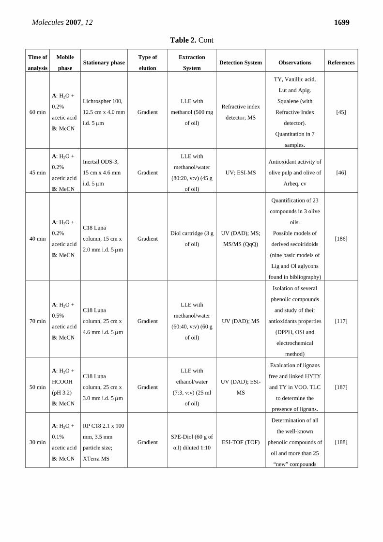
Table 2. Cont
Time of
analysis
Mobile
phase Stationary phase
Type of
elution
Extraction
System Detection System Observations References
60 min
A: H2O +
0.2%
acetic acid
B: MeCN
Lichrospher 100,
12.5 cm x 4.0 mm
i.d. 5 μm
Gradient
LLE with
methanol (500 mg
of oil)
Refractive index
detector; MS
TY, Vanillic acid,
Lut and Apig.
Squalene (with
Refractive Index
detector).
Quantitation in 7
samples.
[45]
45 min
A: H2O +
0.2%
acetic acid
B: MeCN
Inertsil ODS-3,
15 cm x 4.6 mm
i.d. 5 μm
Gradient
LLE with
methanol/water
(80:20, v:v) (45 g
of oil)
UV; ESI-MS
Antioxidant activity of
olive pulp and olive of
Arbeq. cv
[46]
40 min
A: H2O +
0.2%
acetic acid
B: MeCN
C18 Luna
column, 15 cm x
2.0 mm i.d. 5 μm
Gradient Diol cartridge (3 g
of oil)
UV (DAD); MS;
MS/MS (QqQ)
Quantification of 23
compounds in 3 olive
oils.
Possible models of
derived secoiridoids
(nine basic models of
Lig and Ol aglycons
found in bibliography)
[186]
70 min
A: H2O +
0.5%
acetic acid
B: MeCN
C18 Luna
column, 25 cm x
4.6 mm i.d. 5 μm
Gradient
LLE with
methanol/water
(60:40, v:v) (60 g
of oil)
UV (DAD); MS
Isolation of several
phenolic compounds
and study of their
antioxidants properties
(DPPH, OSI and
electrochemical
method)
[117]
50 min
A: H2O +
HCOOH
(pH 3.2)
B: MeCN
C18 Luna
column, 25 cm x
3.0 mm i.d. 5 μm
Gradient
LLE with
ethanol/water
(7:3, v:v) (25 ml
of oil)
UV (DAD); ESI-
MS
Evaluation of lignans
free and linked HYTY
and TY in VOO. TLC
to determine the
presence of lignans.
[187]
30 min
A: H2O +
0.1%
acetic acid
B: MeCN
RP C18 2.1 x 100
mm, 3.5 mm
particle size;
XTerra MS
Gradient SPE-Diol (60 g of
oil) diluted 1:10 ESI-TOF (TOF)
Determination of all
the well-known
phenolic compounds of
oil and more than 25
“new” compounds
[188]
Molecules 2007, 12 1700
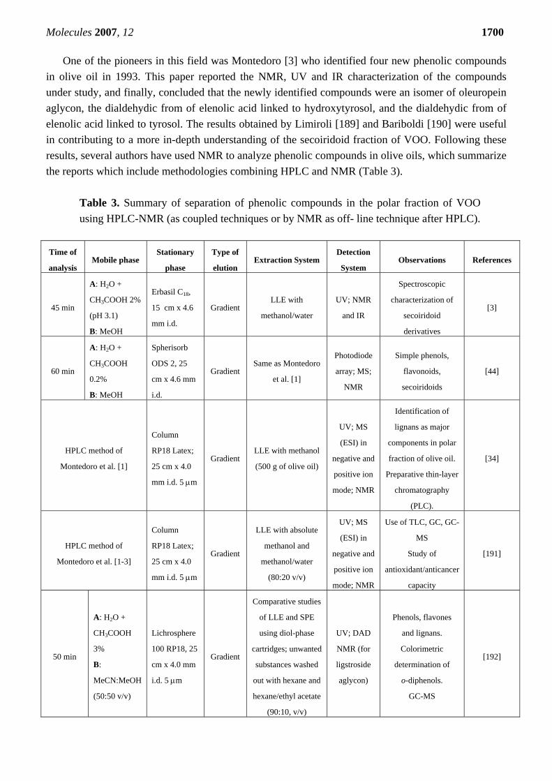
One of the pioneers in this field was Montedoro [3] who identified four new phenolic compounds in olive oil in 1993. This paper reported the NMR, UV and IR characterization of the compounds under study, and finally, concluded that the newly identified compounds were an isomer of oleuropein aglycon, the dialdehydic from of elenolic acid linked to hydroxytyrosol, and the dialdehydic from of elenolic acid linked to tyrosol. The results obtained by Limiroli [189] and Bariboldi [190] were useful in contributing to a more in-depth understanding of the secoiridoid fraction of VOO. Following these results, several authors have used NMR to analyze phenolic compounds in olive oils, which summarize the reports which include methodologies combining HPLC and NMR (Table 3).
Table 3. Summary of separation of phenolic compounds in the polar fraction of VOO using HPLC-NMR (as coupled techniques or by NMR as off- line technique after HPLC).
Time of
analysis Mobile phase
Stationary
phase
Type of
elution Extraction System
Detection
System Observations References
45 min
A: H2O +
CH3COOH 2%
(pH 3.1)
B: MeOH
Erbasil C18,
15 cm x 4.6
mm i.d.
Gradient LLE with
methanol/water
UV; NMR
and IR
Spectroscopic
characterization of
secoiridoid
derivatives
[3]
60 min
A: H2O +
CH3COOH
0.2%
B: MeOH
Spherisorb
ODS 2, 25
cm x 4.6 mm
i.d.
Gradient Same as Montedoro
et al. [1]
Photodiode
array; MS;
NMR
Simple phenols,
flavonoids,
secoiridoids
[44]
HPLC method of
Montedoro et al. [1]
Column
RP18 Latex;
25 cm x 4.0
mm i.d. 5 μm
Gradient LLE with methanol
(500 g of olive oil)
UV; MS
(ESI) in
negative and
positive ion
mode; NMR
Identification of
lignans as major
components in polar
fraction of olive oil.
Preparative thin-layer
chromatography
(PLC).
[34]
HPLC method of
Montedoro et al. [1-3]
Column
RP18 Latex;
25 cm x 4.0
mm i.d. 5 μm
Gradient
LLE with absolute
methanol and
methanol/water
(80:20 v/v)
UV; MS
(ESI) in
negative and
positive ion
mode; NMR
Use of TLC, GC, GC-
MS
Study of
antioxidant/anticancer
capacity
[191]
50 min
A: H2O +
CH3COOH
3%
B:
MeCN:MeOH
(50:50 v/v)
Lichrosphere
100 RP18, 25
cm x 4.0 mm
i.d. 5 μm
Gradient
Comparative studies
of LLE and SPE
using diol-phase
cartridges; unwanted
substances washed
out with hexane and
hexane/ethyl acetate
(90:10, v/v)
UV; DAD
NMR (for
ligstroside
aglycon)
Phenols, flavones
and lignans.
Colorimetric
determination of
o-diphenols.
GC-MS
[192]
Molecules 2007, 12 1701
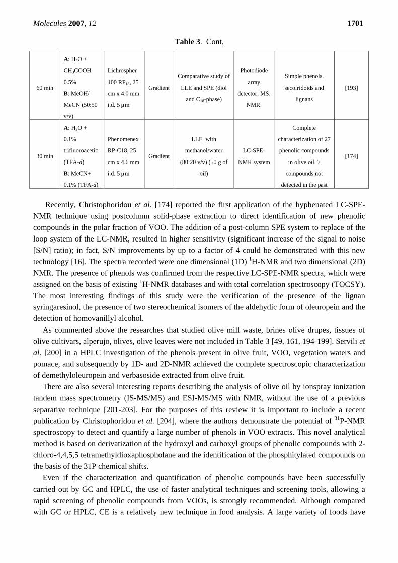
Table 3. Cont,
Recently, Christophoridou et al. [174] reported the first application of the hyphenated LC-SPE-
NMR technique using postcolumn solid-phase extraction to direct identification of new phenolic compounds in the polar fraction of VOO. The addition of a post-column SPE system to replace of the loop system of the LC-NMR, resulted in higher sensitivity (significant increase of the signal to noise [S/N] ratio); in fact, S/N improvements by up to a factor of 4 could be demonstrated with this new technology [16]. The spectra recorded were one dimensional (1D) 1H-NMR and two dimensional (2D) NMR. The presence of phenols was confirmed from the respective LC-SPE-NMR spectra, which were assigned on the basis of existing 1H-NMR databases and with total correlation spectroscopy (TOCSY). The most interesting findings of this study were the verification of the presence of the lignan syringaresinol, the presence of two stereochemical isomers of the aldehydic form of oleuropein and the detection of homovanillyl alcohol.
As commented above the researches that studied olive mill waste, brines olive drupes, tissues of olive cultivars, alperujo, olives, olive leaves were not included in Table 3 [49, 161, 194-199]. Servili et al. [200] in a HPLC investigation of the phenols present in olive fruit, VOO, vegetation waters and pomace, and subsequently by 1D- and 2D-NMR achieved the complete spectroscopic characterization of demethyloleuropein and verbasoside extracted from olive fruit.
There are also several interesting reports describing the analysis of olive oil by ionspray ionization tandem mass spectrometry (IS-MS/MS) and ESI-MS/MS with NMR, without the use of a previous separative technique [201-203]. For the purposes of this review it is important to include a recent publication by Christophoridou et al. [204], where the authors demonstrate the potential of 31P-NMR spectroscopy to detect and quantify a large number of phenols in VOO extracts. This novel analytical method is based on derivatization of the hydroxyl and carboxyl groups of phenolic compounds with 2-chloro-4,4,5,5 tetramethyldioxaphospholane and the identification of the phosphitylated compounds on the basis of the 31P chemical shifts.
Even if the characterization and quantification of phenolic compounds have been successfully carried out by GC and HPLC, the use of faster analytical techniques and screening tools, allowing a rapid screening of phenolic compounds from VOOs, is strongly recommended. Although compared with GC or HPLC, CE is a relatively new technique in food analysis. A large variety of foods have
60 min
A: H2O +
CH3COOH
0.5%
B: MeOH/
MeCN (50:50
v/v)
Lichrospher
100 RP18, 25
cm x 4.0 mm
i.d. 5 μm
Gradient
Comparative study of
LLE and SPE (diol
and C18-phase)
Photodiode
array
detector; MS,
NMR.
Simple phenols,
secoiridoids and
lignans
[193]
30 min
A: H2O +
0.1%
trifluoroacetic
(TFA-d)
B: MeCN+
0.1% (TFA-d)
Phenomenex
RP-C18, 25
cm x 4.6 mm
i.d. 5 μm
Gradient
LLE with
methanol/water
(80:20 v/v) (50 g of
oil)
LC-SPE-
NMR system
Complete
characterization of 27
phenolic compounds
in olive oil. 7
compounds not
detected in the past
[174]
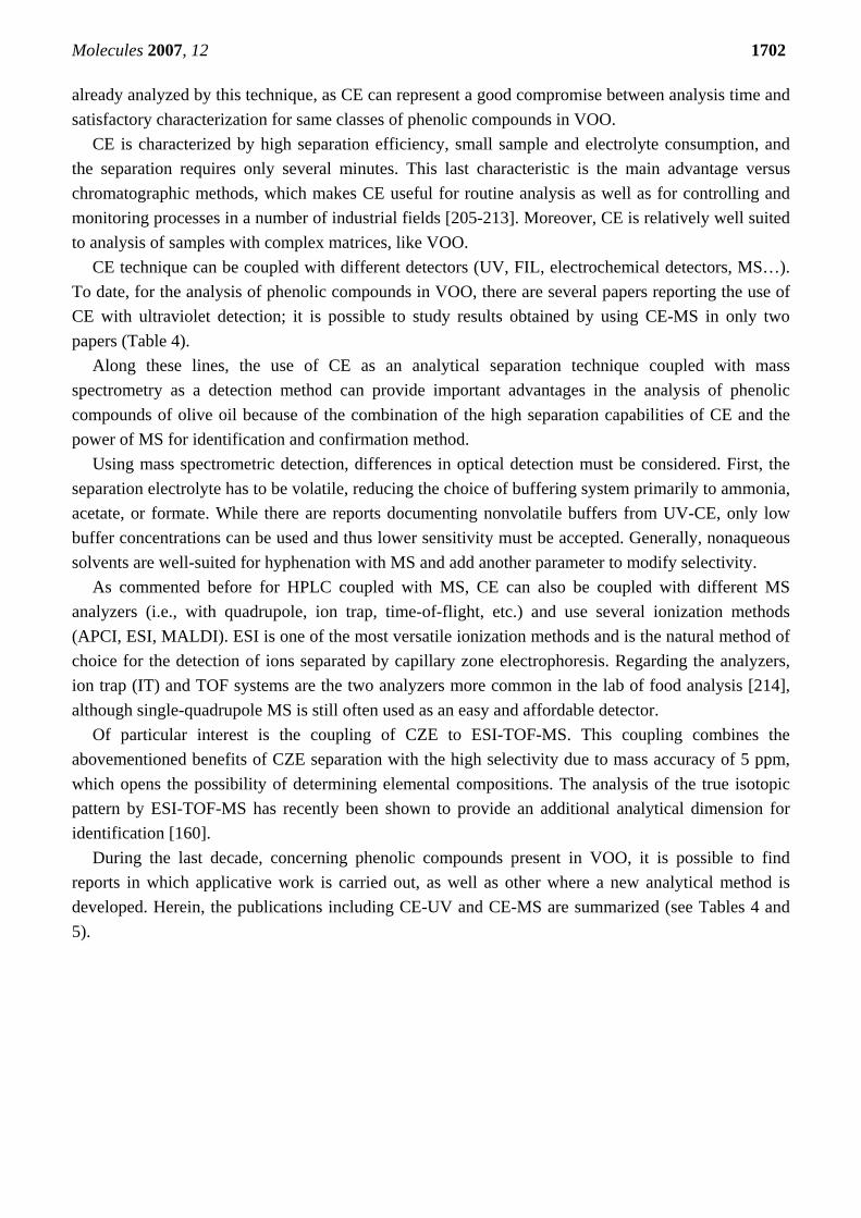
Molecules 2007, 12 1702 already analyzed by this technique, as CE can represent a good compromise between analysis time and satisfactory characterization for same classes of phenolic compounds in VOO.
CE is characterized by high separation efficiency, small sample and electrolyte consumption, and the separation requires only several minutes. This last characteristic is the main advantage versus chromatographic methods, which makes CE useful for routine analysis as well as for controlling and monitoring processes in a number of industrial fields [205-213]. Moreover, CE is relatively well suited to analysis of samples with complex matrices, like VOO.
CE technique can be coupled with different detectors (UV, FIL, electrochemical detectors, MS…). To date, for the analysis of phenolic compounds in VOO, there are several papers reporting the use of CE with ultraviolet detection; it is possible to study results obtained by using CE-MS in only two papers (Table 4).
Along these lines, the use of CE as an analytical separation technique coupled with mass spectrometry as a detection method can provide important advantages in the analysis of phenolic compounds of olive oil because of the combination of the high separation capabilities of CE and the power of MS for identification and confirmation method.
Using mass spectrometric detection, differences in optical detection must be considered. First, the separation electrolyte has to be volatile, reducing the choice of buffering system primarily to ammonia, acetate, or formate. While there are reports documenting nonvolatile buffers from UV-CE, only low buffer concentrations can be used and thus lower sensitivity must be accepted. Generally, nonaqueous solvents are well-suited for hyphenation with MS and add another parameter to modify selectivity.
As commented before for HPLC coupled with MS, CE can also be coupled with different MS analyzers (i.e., with quadrupole, ion trap, time-of-flight, etc.) and use several ionization methods (APCI, ESI, MALDI). ESI is one of the most versatile ionization methods and is the natural method of choice for the detection of ions separated by capillary zone electrophoresis. Regarding the analyzers, ion trap (IT) and TOF systems are the two analyzers more common in the lab of food analysis [214], although single-quadrupole MS is still often used as an easy and affordable detector.
Of particular interest is the coupling of CZE to ESI-TOF-MS. This coupling combines the abovementioned benefits of CZE separation with the high selectivity due to mass accuracy of 5 ppm, which opens the possibility of determining elemental compositions. The analysis of the true isotopic pattern by ESI-TOF-MS has recently been shown to provide an additional analytical dimension for identification [160].
During the last decade, concerning phenolic compounds present in VOO, it is possible to find reports in which applicative work is carried out, as well as other where a new analytical method is developed. Herein, the publications including CE-UV and CE-MS are summarized (see Tables 4 and 5).
Molecules 2007, 12 1703
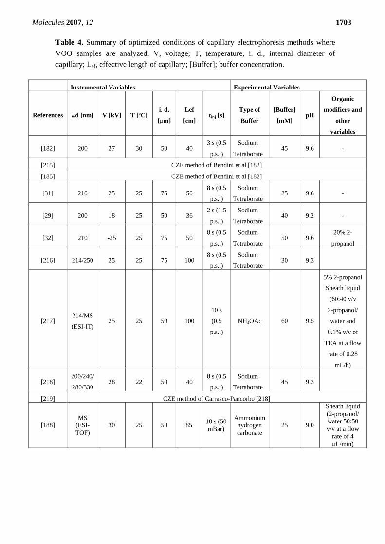
Table 4. Summary of optimized conditions of capillary electrophoresis methods where VOO samples are analyzed. V, voltage; T, temperature, i. d., internal diameter of capillary; Lef, effective length of capillary; [Buffer]; buffer concentration.
Instrumental Variables Experimental Variables
References λd [nm] V [kV] T [ºC] i. d.
[μm]
Lef
[cm] tinj [s]
Type of
Buffer
[Buffer]
[mM] pH
Organic
modifiers and
other
variables
[182] 200 27 30 50 40 3 s (0.5
p.s.i)
Sodium
Tetraborate 45 9.6 -
[215] CZE method of Bendini et al.[182]
[185] CZE method of Bendini et al.[182]
[31] 210 25 25 75 50 8 s (0.5
p.s.i)
Sodium
Tetraborate 25 9.6 -
[29] 200 18 25 50 36 2 s (1.5
p.s.i)
Sodium
Tetraborate 40 9.2 -
[32] 210 -25 25 75 50 8 s (0.5
p.s.i)
Sodium
Tetraborate 50 9.6
20% 2-
propanol
[216] 214/250 25 25 75 100 8 s (0.5
p.s.i)
Sodium
Tetraborate 30 9.3
[217] 214/MS
(ESI-IT) 25 25 50 100
10 s
(0.5
p.s.i)
NH4OAc 60 9.5
5% 2-propanol
Sheath liquid
(60:40 v/v
2-propanol/
water and
0.1% v/v of
TEA at a flow
rate of 0.28
mL/h)
[218] 200/240/
280/330 28 22 50 40
8 s (0.5
p.s.i)
Sodium
Tetraborate 45 9.3
[219] CZE method of Carrasco-Pancorbo [218]
[188] MS
(ESI-TOF)
30 25 50 85 10 s (50 mBar)
Ammonium hydrogen carbonate
25 9.0
Sheath liquid (2-propanol/ water 50:50 v/v at a flow
rate of 4 μL/min)
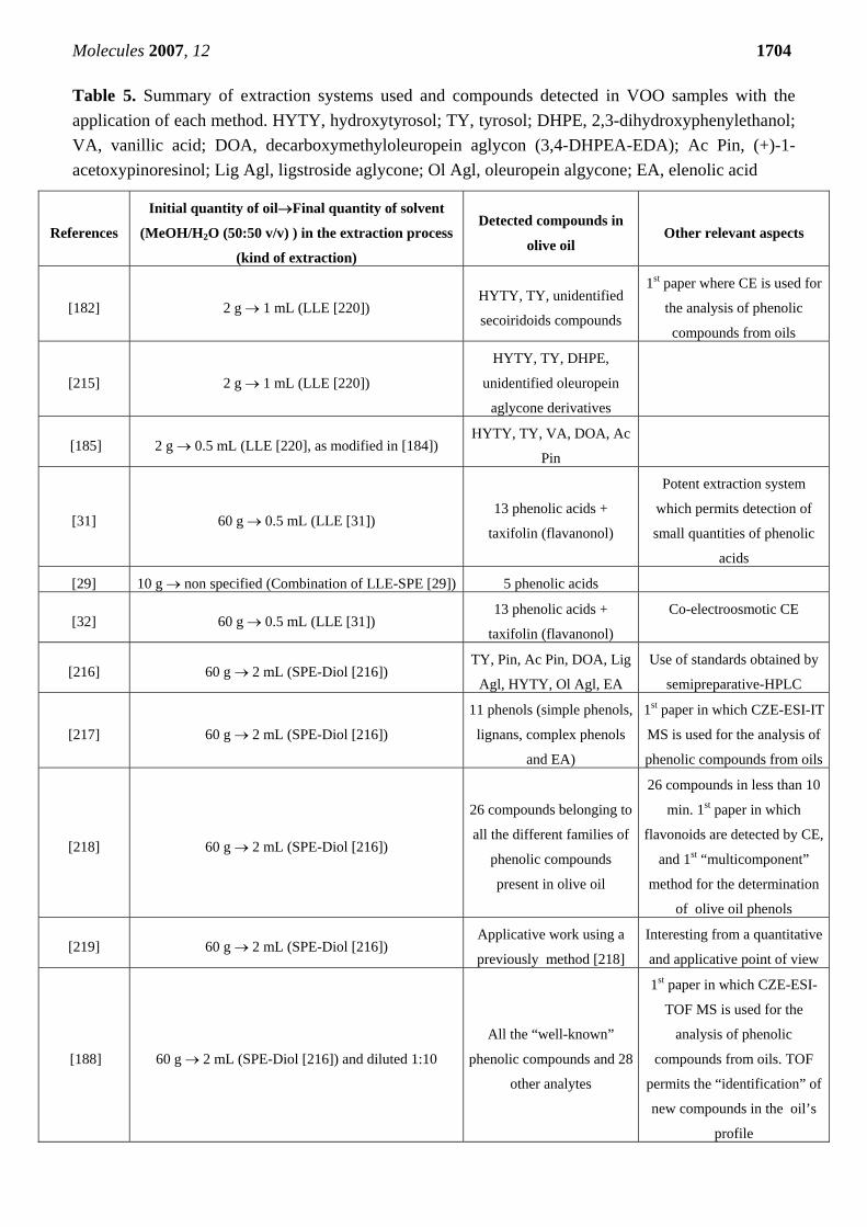
Molecules 2007, 12 1704 Table 5. Summary of extraction systems used and compounds detected in VOO samples with the application of each method. HYTY, hydroxytyrosol; TY, tyrosol; DHPE, 2,3-dihydroxyphenylethanol; VA, vanillic acid; DOA, decarboxymethyloleuropein aglycon (3,4-DHPEA-EDA); Ac Pin, (+)-1-acetoxypinoresinol; Lig Agl, ligstroside aglycone; Ol Agl, oleuropein algycone; EA, elenolic acid
References
Initial quantity of oil→Final quantity of solvent
(MeOH/H2O (50:50 v/v) ) in the extraction process
(kind of extraction)
Detected compounds in
olive oil Other relevant aspects
[182] 2 g → 1 mL (LLE [220]) HYTY, TY, unidentified
secoiridoids compounds
1st paper where CE is used for
the analysis of phenolic
compounds from oils
[215] 2 g → 1 mL (LLE [220])
HYTY, TY, DHPE,
unidentified oleuropein
aglycone derivatives
[185] 2 g → 0.5 mL (LLE [220], as modified in [184]) HYTY, TY, VA, DOA, Ac
Pin
[31] 60 g → 0.5 mL (LLE [31]) 13 phenolic acids +
taxifolin (flavanonol)
Potent extraction system
which permits detection of
small quantities of phenolic
acids
[29] 10 g → non specified (Combination of LLE-SPE [29]) 5 phenolic acids
[32] 60 g → 0.5 mL (LLE [31]) 13 phenolic acids +
taxifolin (flavanonol)
Co-electroosmotic CE
[216] 60 g → 2 mL (SPE-Diol [216]) TY, Pin, Ac Pin, DOA, Lig
Agl, HYTY, Ol Agl, EA
Use of standards obtained by
semipreparative-HPLC
[217] 60 g → 2 mL (SPE-Diol [216])
11 phenols (simple phenols,
lignans, complex phenols
and EA)
1st paper in which CZE-ESI-IT
MS is used for the analysis of
phenolic compounds from oils
[218] 60 g → 2 mL (SPE-Diol [216])
26 compounds belonging to
all the different families of
phenolic compounds
present in olive oil
26 compounds in less than 10
min. 1st paper in which
flavonoids are detected by CE,
and 1st “multicomponent”
method for the determination
of olive oil phenols
[219] 60 g → 2 mL (SPE-Diol [216]) Applicative work using a
previously method [218]
Interesting from a quantitative
and applicative point of view
[188] 60 g → 2 mL (SPE-Diol [216]) and diluted 1:10
All the “well-known”
phenolic compounds and 28
other analytes
1st paper in which CZE-ESI-
TOF MS is used for the
analysis of phenolic
compounds from oils. TOF
permits the “identification” of
new compounds in the oil’s
profile
Molecules 2007, 12 1705 Concluding remarks and future outlook
The amount of phenolic compounds is a very important parameter when evaluating the quality of VOOs. Phenols are closely related with both the resistance of the oil to oxidation and the typical bitter and pungent tastes. Furthermore, some studies have shown that the amount of phenols, particularly those with a catecholic structure, together with a favorable monounsaturated to polyunsaturated fatty acid ratio, is related to several healthy attributes. These different aspects make VOO a very valuable and appreciated dietary lipidic condiment, and add importance to the determination of its phenolic compounds, both qualitative and quantitatively. The most commonly methods used for phenolic determination in VOO are based on GC and HPLC, and more recently on CE, coupled with different detector systems (UV, FLD, amperometric or coulometric). If the literature regarding phenolic compounds of VOO is analyzed in detail, it is evident that this class of compounds has not been completely studied, because of the complexity of their chemical nature and the complexity of the matrix in which they are found. During the last ten years, MS and NMR have become indispensable to study the quali-quantitative profiles of phenols and their oxidative forms, and detectors with the power to identify compounds and provide the analyst with information about the molecular structure are essential.
Apart from the interest on knowing in composition of the polar fraction of VOO, the determination of these compounds also helps to understand their health benefits that include reduction of risk factors of coronary heart disease, prevention of several varieties of cancer and modification of immune and inflammatory responses. It is also of interest to distinguish what phenolic molecules are responsible for bitterness, pungency, astringency and metallic sensations and to evaluate the antioxidant activities of the polar fraction.
Although excellent progress has already been made, it is expected that the use of different methodologies of potent techniques coupled with rapid, reliable and sophisticated detectors will become more common in the near future; there are still many “unknown” compounds present in the polar fraction of olive oil and it is very important to carry out collaborative studies to join the efforts of the scientific community.
Abbreviations: 3,4-dihydroxyphenyl-ethanol or hydroxytyrosol (3,4-DHPEA); 3,4-dihydroxyphenyl-ethanol acetate or hydroxytyrosol acetate (3,4-DHPEA-AC); 3,4-dihydroxyphenyl-ethanol linked to elenolic acid (3,4-DHPEA-EA); 3,4-dihydroxyphenyl-ethanol linked to dialdehydic form of elenolic acid (3,4-DHPEA-EDA); 1-acetoxypinoresinol (Ac Pin); buthylated hydroxytoluene (BHT); Capillary Electrophoresis (CE); cyclooxygenase (COX); diphenylpicrylhydrazyl (DPPH); elenolic acid (EA); electrospray ionization mass spectrometry (ESI-MS); Flame Ionization Detector (FID); gas chromatography (GC); High Performance Liquid Chromatography (HPLC); HPLC-atmospheric pressure chemical ionization mass spectrometry (HPLC-APCI-MS); ion-trap mass spectrometry (IT-MS); low-density lipoprotein (LDL); Liquid-Liquid- extraction (LLE); limit of detection (LOD); mass spectrometry (MS); tandem mass spectrometry (MS/MS); multiple-stage mass spectrometry (MSn); nuclear magnetic resonance (NMR); Oxidative Stability Instrument (OSI); p-hydroxyphenyl-ethanol or tyrosol (p-HPEA); p-hydroxyphenyl-ethanol linked to elenolic acid (p-HPEA-EA); p-hydroxyphenyl-ethanol linked to dialdehydic form of elenolic acid (p-HPEA-EDA); pinoresinol (Pin); triple
Molecules 2007, 12 1706 quadrupole (QqQ); Solid-Phase Extraction (SPE); virgin olive oil (VOO); secoiridoid (SID); time of flight mass spectrometry (TOF-MS); Total Peak Area Ratio (TPAR); ultraviolet (UV). References and Notes 1. Montedoro, G.F.; Servili, M.; Baldioli, M.; Miniati, E. Simple and Hydrolyzable Phenolic
Compounds in Virgin Olive Oil. 1. Their Extraction, Separation, and Quantitative and Semiquantitative Evaluation by HPLC. J. Agric. Food. Chem. 1992, 40, 1571–1576.
2. Montedoro, G.F.; Servili, M.; Baldioli, M.; Miniati, E. Simple and Hydrolyzable Phenolic Compounds in Virgin Olive Oil. 2. Initial Characterization of the Hydrolyzable Fraction. J. Agric. Food. Chem. 1992, 40, 1577–1580.
3. Montedoro, G.F.; Servili, M.; Baldioli, M.; Selvaggini, R.; Miniati, E.; Macchioni, A. Simple and Hydrolyzable Compounds in Virgin Olive Oil. 3. Spectroscopic Characterizations of the Secoiridoid Derivatives. J. Agric. Food Chem. 1993, 41, 2228-2234.
4. Amiot, M.J.; Fleuriet, A.; Macheix, J.J. Importance and evolution of phenolic compounds in olive during growth and maturation. J. Agric. Food Chem., 1986, 34, 823-825.
5. Amiot, M.J.; Fleuriet, A.; Mecheix, J.J. Accumulation of oleuropein derivatives during olive maturation. Phytochemistry 1989, 28, 67-69.
6. Gutierrez Gonzales-Quijano, R.; Janer del Valle, C.; Janier del Valle, M.L.; Gutierrez Rosales, F.; Vazquez Roncero, A. Relacion entre los polifenoles y la calidad y estabilidad del aeite de oliva virgen. Grasas Aceites 1977, 28, 101-106.
7. Gutfinger, T. Polyphenols in olive oils. J. Am. Oil Chem. Soc. 1981, 58, 966-968. 8. Tsimidou, M. Polyphenols and quality of virgin olive oil in retrospective. Ital. J. Food Sci. 1998,
10, 99-115. 9. Montedoro, G.F.; Cantarelli, C. Indagini sulle sostanze fenoliche presenti negli oli d’oliva. Riv.
Ital. Sost. Grasse 1969, 46, 115-124. 10. Kuwajima, H.; Uemura, T.; Takaishi, K.; Inoue, K.; Inouye, H. A secoiridoid glucoside from
Olea europea. Phytochemistry 1988, 27, 1757-1759. 11. Cortesi, N.; Azzolini, M.; Rovellini, P.; Fedeli, E. I componenti minori polari degli oli vergini di
oliva: ipotesi di struttura mediante LC-MS. Riv. Ital. Sost. Grasse 1995, 72, 241-251. 12. Cortesi, N.; Azzolini, M.; Rovellini, P.; Fedeli, E. Dosaggio dei componenti minori polari
(CMP) in oli vergini di oliva. Riv. Ital. Sostanze Grasse 1995, 72, 333-337. 13. Angerosa, F.; D’Alessandro, N.; Konstantinou, P.; Di Giacinto, L. Characterization of phenolic
and secoiridoid aglycons present in virgin olive oil by gas chromatography-chemical ionization mass spectrometry. J. Chromatogr. A 1996, 736, 195–203.
14. Brenes, M.; Garcıa, A.; Garcıa, P.; Rios, J. J.; Garrido, A. Phenolic Compounds in Spanish Olive Oils. J. Agric. Food Chem. 1999, 47, 3535-3540.
16. Exarchou, V.; Godejohann, M.; van Beek, T. A.; Gerothanassis, I. P.; Vervoort, J. LC-UV-Solid-Phase Extraction-NMR-MS Combined with a Cryogenic Flow Probe and Its Application to the Identification of Compounds Present in Greek Oregano. Anal. Chem. 2003, 75, 6288-6294.
Molecules 2007, 12 1707 17. Andreasen, M.F.; Christensen, L.P.; Meyer, A.S.; Hansen, A. Content of phenolic acids and
ferulic acid dehydrodimers in 17 rye (Secale cereale L.) varieties J. Agric. Food Chem. 2000, 48, 2837-2842.
18. Barthelmebs, L.; Divies, C.; Cavin, J.F. Knockout of the p-coumarate decarboxylase gene from lactobacillus plantarum reveals the existence of two other inducible enzymatic activities involved in phenolic acid metabolism. Appl. Environ. Micro. 2000, 66, 3368-3375.
19. Hakkinen, S.; Heinonen, M.; Karenlampi, S.; Mykkanen, H.; Ruuskanen, J.; Torronen, R. Screening of selected flavonoids and phenolic acids in 19 berries. Food Res. Int. 1999, 32, 345-353.
20. Xu, L.; Diosady, L.L. Rapid method for total phenolic acid determination in rapeseed/canola meals. Food Res. Int. 1997, 30, 571-574.
21. Maga, J. A. Simple phenol and phenolic compounds in food flavor. Crit. Rev. Food Sci. Nutr. 1978, 10, 323-372.
22. Nergiz, C.; Unal, K. Determination of phenolic acids in virgin olive oil. Food Chem. 1991, 39, 237-240.
23. Harborne, J. B. The Flavonoids: Advances in Research since 1986; Chapman and Hall: London, U.K., 1994.
24. Peleg, H.; Naim, M.; Rouseff, R. L.; Zehavi, U. Distribution of bound and free phenolic acids in oranges (Citrus sinensis) and Grapefruits (Citrus paradisi). J. Sci. Food Agric. 1991, 57, 417-426.
25. Shahidi, F.; Nacsk, M. Food phenolics: Sources, Chemistry, Effects, and Applications; Technomic Publishing Company, Inc.: Lancaster, PA, 1995.
26. Robbins, R. J. Phenolic Acids in Foods: An Overview of Analytical Methodology. J. Agric. Food Chem. 2003, 51, 2866-2887.
27. Gomes, C. A.; Girao da Cruz, T.; Andrade, J. L.; Milhazes, N.; Borges, F.; Marques. M. P. M. Anticancer Activity of Phenolic Acids of Natural or Synthetic Origin: A Structure-Activity Study. J. Med. Chem. 2003, 46, 5395-5401.
28. Masaki, H.; Okamoto, N.; Sakaki, S.; Sakurai, H. Protective effects of hydroxybenzoic acids and their esters on cell damage induced by hydroxyl radicals and hydrogen peroxides. Biol. Pharm. Bull. 1997, 20, 304-308.
29. Buiarelli, F.; Di Berardino, S.; Coccioli, F.; Jasionowska, R.; Russo, M. V. Determination of phenolic acids in olive oil by capillary electrophoresis. Anal. Chim. 2004, 94, 699-705.
30. Cartoni, G. P; Coccioli, F; Janionowska, R; Ramirez, D. HPLC analysis of the benzoic and cinnamic acids in edible vegetable oils. Ital. J. Food Sci. 2000, 12, 163-173.
31. Carrasco Pancorbo, A.; Cruces-Blanco, C.; Segura Carretero, A.; Fernández Gutiérrez, A. Sensitive determination of phenolic acids in extra-virgin olive oil by capillary zone electrophoresis. J. Agric. Food. Chem. 2004, 52, 6687-6693.
32. Carrasco-Pancorbo, A.; Segura-Carretero, A.; Fernández-Gutiérrez, A. Co-electroosmotic capillary electrophoresis determination of phenolic acids in comercial olive oil. J. Sep. Sci. 2005, 28, 925-934.
33. Davin, B.D.; Bedgar, D.L.; Katayama, T.; Lewis, N.G. On the stereoselective synthesis of (1)-pinoresinol in Forsythia suspensa from its achiral precursor, coniferyl alcohol. Phytochemistry 1992, 31, 3869–3874.
Molecules 2007, 12 1708 34. Owen, R. W.; Mier, W.; Giacosa, A.; Hull, W. E.; Spiegelhalder, B.; Bartsch, H. Identification
of Lignans as Major Components in the Phenolic Fraction of Olive Oil, Clinical Chem. 2000, 46, 976–988.
35. Brenes, M.; Garcıa, A.; Rios, J. J.; Garcıa, P.; Garrido, A. Use of 1-acetoxypinoresinol to authenticate Picual olive oils. Int. J. Food Sci. Technol. 2002, 37, 615–625.
36. Bianco, A.; Coccioli, F.; Guiso, M.; Marra C. The occurrence in olive oil of a new class of phenolic compounds: hydroxy-isochromans, Food Chem. 2001, 77, 405–411.
37. Le March, L. Cancer preventive effects of flavonoids—a review. Biomed. Pharmacother. 2002, 56, 296–301.
38. Kanadaswami, C.; Lee, L. T.; Lee, P. P. H.; Hwang, J. J.; Ke, F. C.; Huanh Y. T.; Lee M.T. The antitumour activities of flavonoids. In vivo 2005, 19, 895-909.
39. Steinberg, F. M.; Bearden, M. M.; Keen, C. L. Cocoa and chocolate flavonoids: implications for cardiovascular health. J. Am. Diet Assoc. 2003, 103, 215– 223.
40. Nestel, P. Isoflavones: their effects on cardiovascular risk and functions. Curr. Opin. Lipidol. 2003, 14, 3–8.
41. Rovellini, P.; Cortesi, N.; Fedeli, E. Analysis of flavonoids from Olea europaea by HPLC-UV and HPLC-electrospray-MS. Riv. Ital. Sost. Grasse 1997, 74, 273-279.
42. Vázquez-Roncero, A.; Janer Del Valle, L., Janer Del Valle, C. Componentes fenòlicos de la aceituna. III. Polifenoles del aceite. Grasas Aceites 1976, 27, 185-191.
43. Carrasco-Pancorbo, A.; Gómez-Caravaca, A. M.; Cerretani, L.; Bendini, A.; Segura-Carretero, A.; Fernández-Gutiérrez, A. Rapid quantification of the phenolic fraction of spanish virgin olive oils by capillary electrophoresis with uv detection. J. Agric. Food Chem. 2006, 54, 7984-7991.
44. Brenes, M.; García, A.; García, P.; Ríos, J. J.; Garrido, A. Phenolic compounds in Spanish olive oils. J. Agric. Food Chem. 1999, 47, 3535-3540.
45. Murkovic, M.; Lechner, S.; Pietzka, A.; Bratacos, M.; Katzogiannos, E. Analysis of minor components in olive oil. J. Biochem. Methods 2004, 61, 155-160.
46. Morelló, J. R.; Vuorela, S.; Romero, M. P.; Motilva, M. J.; Heinonen, M. Antioxidant activity of olive pulp and olive oil phenolic compounds of the arbequina cultivar. J. Agric. Food Chem. 2005, 53, 2002-2008.
47. Bouaziz, M; Grayer, R. J; Simmonds, M. S. J; Damak, M; Sayadi, S. Identification and antioxidant potential of flavonoids and low molecular weight phenols in olive cultivar Chemlali growing in Tunisia. J. Agric. Food Chem. 2005, 53, 236-241.
48. Vlahov, G. Flavonoids in three olives (Olea europaea) fruit varieties during maturation. J. Sci. Food Agric. 1992, 58, 157-159.
49. Ryan, D.; Prenzler, P. D.; Lavee, S.; Antolovich, M.; Robards, K. Quantitative changes in phenolic content during physiological development of olive (Olea europaea) cultivar Hardy´s Mammoth. J. Agric. Food Chem. 2003, 51, 2532-2538.
50. Romani, A.; Pinelli, P.; Mulinacci, N.; Vincieri, F. F.; Gravano, E.; Tattini, M. HPLC analysis of flavonoids and secoiridoids in leaves of Ligustrum vulgare L. (Oleaceae). J. Agr. Food Chem. 2000, 48, 4091-4096.
51. Boskou, D. Sources of natural antioxidants. Trends Food Sci. Technol. 2006, 17, 505-512. 52. Stark, A.H.; Madar, Z. Olive oil as a functional food: Epidemiology and nutritional approaches,
Nutr. Rev. 2002, 60, 170-176.
Molecules 2007, 12 1709 53. Perez-Jimenez, F. (coordinator) International conference on the healthy effect of virgin olive oil.
Eur. J. Clinic. Invest. 2005, 35, 421–424. 54. Bisignano, G.; Tomaino, A.; Lo Cascio, R.; Crisafi, G.; Uccella, N.; Saija, A. On the in-vitro
antiomicrobial activity of oleuropein and hydroxytyrosol, J. Pharm. Pharmacol. 1999, 31, 971–974.
55. Glatzle, J.; Beckert, S.; Kasparek, M. S.; Mueller, M. H.; Mayer, P.; Meile, T.; Konigsrainer, A.; Steurer, W. Olive oil is more potent than fish oil to reduce septic pulmonary dysfunctions in rats. Langenbecks Arch Surg 2007, 392, 323–329.
56. Tuck, K.L.; Hayball, P.J. Major phenolic compounds in olive oil: metabolism and health effects. J. Nutr. Biochem. 2002, 13, 636-644.
57. Tuck, K.L.; Hayball, P.J.; Stupans, I. Structural characterisation of the metabolites of hydroxytyrosol, the principal phenolic component in olive oil, in rats. J. Agric. Food Chem. 2002, 50, 2404–2409.
58. Visioli, F.; Bellomo, G.; Galli, C. Free radical-scavenging of olive oil phenols, Biochem. Biophys. Res. Commun. 1998, 247, 60–64.
59. Visioli, F.; Galli, C.; Bornet, F.; Mattei, A.; Patelli, R.; Galli, G.; Caruso, D. Olive oil phenolics are dose-dependently absorbed in humans. FEBS Lett. 2000, 468, 159–160.
60. Tuck, K.L.; Freeman, M.P.; Hayball, P.J.; Stretch, G.L.; Stupans, I. The in vivo fate of hydroxytyrosol and tyrosol, antioxidant phenolic constituents of olive oil, following intravenous and oral dosing of labelled compounds to rats. J. Nutr. 2001, 131, 1993–1996.
61. Visioli, F.; Galli, C. Olive oil phenols and their potential effects on human health. J. Agric. Food Chem. 1998, 46, 4292-4296.
62. Coni, E.; Di Benedetto, R.; Di Pasquale, M.; Masella, R.; Modesti, D.; Attei, R.; Carlini, E.A. Protective effect of Oleuropein, and olive oil biophenol, on low density lipoprotein oxidizability in rabbits, Lipids 2000, 35, 45–54.
63. Menéndez, J. A.; Vázquez-Martín, A.; Colomer, R.; Carrasco-Pancorbo, A.; García-Villalba, R.; Fernández-Gutiérrez, A.; Segura-Carretero, A. Oleuropein aglycone, the bitter principle of olives and Olive oil, is a potent anti-HER2 oncogene agent capable to reverse breast cancer acquired autoresistance to trastuzumab (Herceptin). BMC Cancer 2007, 7, 80. DOI: 10.1186/1471-2407-7-80.
64. Beauchamp, G.K.; Keast, R. S. J.; Morel, D.; Lin, J.; Pika, J.; Han, Q.; Lee, C-H.; Smith, A. B.; Breslin, P. A. S. Ibuprofen-like activity in extra-virgin olive oil, Nature 2005, 437, 45-46.
65. Smith, A. B.; Han, Q.; Breslin, P. A. S.; Beauchamp, G. K. Synthesis and assignment of absolute configuration of (-)-oleocanthal: A potent, naturally occurring non-steroidal anti-inflammatory and anti-oxidant agent derived from extra virgin olive oils. Org. Lett. 2005, 7, 5075-5078.
66. Fogliano, V.; Sacchi, R. Oleocanthal in olive oil: Between myth and reality, Mol. Nutr. Food Res. 2006, 50, 5-6.
67. Visioli, F.; Bellomo, G.; Montedoro, G.; Galli, C. Low-density lipoprotein oxidation is inhibited in vitro by olive oil constituents. Atherosclerosis 1995, 117, 25-32.
68. Fito, M.; Covas, M. I.; Lamuela-Raventos, R. M.; Vila, J.; Torrents, J.; de la Torre, C.; Marrugat, J. Protective effect of olive oil and its phenolic compounds against low density lipoprotein oxidation. Lipids 2000, 35, 633-638.
Molecules 2007, 12 1710 69. Leenen, R.; Roodenburg, A. J. C.; Vissers, M. N.; Schuubiers, J. A. E.; van Putte, K. P. A. M.;
Wiseman, S. A.; van de Put, F.H. M. M. Supplementation of plasma with olive oil phenols and extracts: Influence on LDL oxidation. J. Agric. Food Chem. 2002, 50, 1290-1297.
70. Bogani, P.; Galli, C.; Villa, M.; Visioli, F. Postprandial anti-inflammatory and antioxidant effects of extra virgin olive oil. Atherosclerosis, 2007, 190, 181–186.
71. Serraino, M.; Thompson, L.U. The effect of flaxseed consumption on the initiation and promotional stages of mammary carcinogenesis. Nutr. Cancer. 1992, 17, 153–159.
72. Togna, G.I.; Togna, A.R.; Franconi, M.; Marra, C.; Guiso, M. Olive oil isochromans inhibit human platelet reactivity. J. Nutr. 2003, 133, 2532-2536.
73. Covas, M. I.; Ruiz-Gutiérrez, V.; de la Torre, R.; Kafatos, A.; Lamuela-Raventós, R. M.:, Osada, J; Owen, R. W.; Visioli, F. Minor components of olive oil: Evidence to date of health benefits in humans. Nutr. Rev. 2006, 64, S20-S30.
74. Fitó, M.; Covas, M.I.; Lamuela-Raventós, R.M.; Vila, J., Torrents, J.; De La Torre, C.; Marrugat, J. Protective effect of olive oil and its phenolic compounds against low density lipoprotein oxidation. Lipids 2000, 35, 633-638.
75. Masella, R.; Varì, R.; D'Archivio, M.; Di Benedetto, R.; Matarrese, P.; Malorni, W.; Scazzocchio, B.; Giovannini, C. Extra virgin olive oil biophenols inhibit cell-mediated oxidation of LDL by increasing the mRNA transcription of glutathione-related enzymes. J. Nutr. 2004, 134, 785-791.
76. De la Puerta, R.; Ruiz-Gutiérez, V.; Hoult, J. R. Inhibition of leukocyte 5-lipooxigenase by phenolics from virgin olive oil. Biochem. Pharmacol. 1999, 157, 445-449.
77. Owen, R. W.; Giacosa, A.; Hull, W. E.; Haubner, R.; Spigelhalder, B.; Bartsch, H. The antioxidant/anticancer potencial of phenolic compounds from olive oil. Eur. J. Cancer 2000, 36, 1235-1247.
78. Carluccio, M.A.; Siculella, L.; Ancora, M.A.; Massaro, M.; Scoditti, E.; Storelli, C.; Visioli, F.; Distante, A.; De Caterina, R. Olive oil and red wine antioxidant polyphenols inhibit endothelial activation: antiatherogenic properties of the Mediterranean diet phytochemicals. Arterioscler. Thromb. Vasc. Biol. 2003, 23, 622-629.
79. Moreno, J. J. Effect of olive oil minor components on oxidative stress and arachidonic acid mobilization and metabolism by macrophages RAW 264.7. Free Radic. Biol. Med. 2003, 35, 1073-1081.
80. Massaro, M.; Basta, G.; Lazzerini, G.; Carluccio, M.A.; Bosetti, F.; Solaini, G.; Visioli, F.; Paolicchi, A.; De Caterina, R. Quenching of intracellular ROS generation as a mechanism for oleate-induced reduction of endothelial activation in early atherogenesis. Thromb. Haemost. 2002, 88, 335-344.
81. Petroni, A.; Blasevich, M.; Salami, M.; Papini, N.; Montedoro, G. F.; Galli, C. Inhibition of platelet aggregation and eicosanoid production by phenolic components of olive oil. Thromb. Res. 1995, 78, 151-160.
82. Quiles, J. L.; Farquharson, A. J.; Simpson, D. K.; Grant, I.; Wahle, K. W. Olive oil phenolics: effects on DNA oxidation and redoenzyme RNA in prostate cells. Br. J. Nutr. 2002, 88, 225-234.
83. Hashim Y. Z. H. Y.; Eng M. E.; Gill C. I. R.; McGlynn, H.; Rowland. I. R. Components of olive oil and chemoprevention of colorectal cancer. Nutr. Rev. 2005, 63, 374-386.
84. Stavric, B. Role of chemopreventers in human diet. Clin. Biochem. 1994, 27, 319-332.
Molecules 2007, 12 1711 85. Yang, C.S.; Landau, J.M.; Huang, M.-T.; Newmark, H.L. Inhibition of carcinogenesis by dietary
polyphenolic compounds. Ann. Rev. Nutr. 2001, 21, 381-406. 86. Frankel, E. N. Volatile lipid oxidation products. Prog. Lipid Res. 1982, 22, 1-33. 87. Montedoro, G. Costituenti fenolici presenti negli oli vergini di oliva Nota I:Identificazione di
alcuni acidi fenolici e loro potere antiossidante. Sci. Tecnol. Aliment. 1972, 3, 177-186. 88. Frankel, E. N. Chemistry of autoxidation: mechanism, products and flavor significance. In:
Flavor Chemistry of Fats and Oils. Min, D. B.; Smouse, T. H., Eds.; AOCS Press: Champaign, IL (USA) 1985; pp. 1-37.
89. Porter, W.L.; Black, E.D.; Drolet, A.M. Use of polyamide oxidative fluorescence test on lipid emulsions: contrast in relative effectiveness of antioxidants in bulk versus dispersed systems. J. Agric. Food Chem. 1989, 37, 615-624.
90. Frankel, E. N. Antioxidants in lipid foods and their impact on food quality. Food Chem. 1996, 57, 51-55.
91. Paiva-Martins, F.; Santos, V.; Mangericao, H.; Gordon, M. H. Effects of Copper on the Antioxidant Activity of Olive Polyphenols in Bulk Oil and Oil-in-Water Emulsions. J. Agric. Food Chem. 2006, 54, 3738-3743.
92. Fregapane, G.; Lavelli, V.; Leon, S.; Kapuralin, J.; Salvador, M.D. Effect of filtration on virgin olive oil stability during storage. Eur. J. Lipid Sci. Technol. 2006, 108, 134-142.
93. Mendez, A.I.; Falque, E. Effect of storage time and container type on the quality of extra-virgin olive oil. Food Control 2007, 18, 521-529.
94. Lercker, G.; Bendini, A.; Cerretani, L. Quality composition and production process of virgin olive oils, Progr. Nutr. 2007, 9, 9-23.
95. Gómez-Caravaca, A.M.; Cerretani, L.; Bendini, A.; Segura-Carretero, A.; Fernández-Gutiérrez, A.; Lercker, G. Effect of filtration systems on the phenolic content in virgin olive oil by HPLC-DAD-MSD, Am. J. Food Technol. in press.
96. Tsimidou, M. Z.; Georgiou, A.; Koidis, A.; Boskou, D. Loss of stability of ‘‘veiled’’ (cloudy) virgin olive oils in storage, Food Chem. 2005, 93, 377-383.
97. Shahidi, F.; Wanasundra, P. D. Phenolic antioxidants. Crit. Rev. Food Sci. Nutr. 1992, 32, 67-103.
98. Roginsky, V.; Lissi, E. A. Review of methods to determine chainbreaking antioxidant activity in food. Food Chem. 2005, 92, 235-254.
99. Huang, D.; Ou, B; Prior, R. L. The chemistry behind antioxidant capacity assays. J. Agric. Food Chem. 2005, 53, 1841-1856.
100. Prior, R.; Wu, X.; Schich, K. Standardized methods for the determination of antioxidant capacity and phenolics in foods and dietary supplements. J. Agric. Food Chem. 2005, 53, 4290-4302.
102. Paiva-Martins, F.; Gordon, M. H. Effects of pH and ferric ions on the antioxidant activity of olive polyphenols in oil-in-water emulsions. J. Am. Oil Chem. Soc. 2002, 79, 571-576.
103. Paiva-Martins, F.; Gordon, M.H. Interactions of Ferric Ions with Olive Oil Phenolic Compounds. J. Agric. Food Chem. 2005, 53, 2704-2709.
Extra Virgin Olive Oil Phenolics on Oxidative Stability in the Presence or Absence of Copper Ions. J. Agric. Food Chem. 2006, 54, 4880-4887.
105. Pedrielli, P.; Skibsted, L. H. Antioxidant synergy and regeneation effect of quercetin, (-)-epicatechin, and (+)-catechin on R-tocopherol in homogeneous solutions of peroxidating methyl linoleate. J. Agric. Food Chem. 2002, 50, 7138-7144.
106. Cai, Y. J.; Ma, L. P.; Hou, L. F.; Zhou, B.; Yang, L.; Liu, Z. L. Antioxidant effects of green tea polyphenols on free radical initiated peroxidation of rat liver microsomes. Chem. Phys. Lipids 2002, 120, 109-117.
107. Pazos, M.; Andersen, M.L.; Medina, I.; Skibsted, L.H. Efficiency of Natural Phenolic Compounds Regenerating a-Tocopherol from a-Tocopheroxyl Radical. J. Agric. Food Chem. 2007, 55, 3661-3666.
108. Baldioli, M.; Servili, M.; Perretti, G.; Montedoro, G. Antioxidant Activity of Tocopherols and Phenolic Compounds in Virgin Olive Oil. J. Am. Oil Chem. Soc. 1996, 73, 1589-1593.
109. Trujillo, M.; Mateos, R.; Collantes De Teran, L.; Espartero, J. L.; Cert, R.; Jover, J.; Alcudia, F.; Bautista, J.; Cert, A.; Parrado, J. Lipophilic Hydroxytyrosyl Esters. Antioxidant Activity in Lipid Matrices and Biological Systems. J. Agric. Food Chem. 2006, 54, 3779-3785.
110. Fki, I.; Allouche, N.; Sayadi, S. The use of polyphenolic extract, purified hydroxytyrosol and 3,4-dihydroxyphenyl acetic acid from olive mill wastewater for the stabilization of refined oils: a potential alternative to synthetic antioxidants. Food Chem. 2005, 93, 197–204.
111. Perez-Bonilla, M.; Salido, S.; van Beek, T. A.; Linares-Palomino, P. J.; Altarejos, J.; Nogueras, M.; Sanchez, A. Isolation and identification of radical scavengers in olive tree (Olea europaea) wood. J. Chromatogr. A. 2006, 1112, 311–318.
112. Tsimidou, M.; Papadopoulos, G.; Boskou, D. Phenolic Compounds and Stability of Virgin Olive Oil - Part I. Food Chem. 1992, 45, 141-144.
113. Visioli, F.; Bellomo, G.; Galli, C. Free Radical-Scavenging Properties of Olive Oil Polyphenols. Biochem. Biophys.Res. Commun. 1998, 247, 60-64.
114. Chimi, H.; Cillard, J.; Cillard, P.; Rahmani, M. Peroxyl and Hydroxyl Radical Scavenging Activity of Some Natural Phenolic Antioxidants. J. Am. Oil Chem. Soc. 1991, 68, 307-312.
115. Gordon, M.H.; Paiva-Martins, F.; Almeida, M. Antioxidant Activity of Hydroxytyrosol Acetate Compared with That of Other Olive Oil Polyphenols. J. Agric. Food Chem. 2001, 49, 2480-2485.
116. Aparicio R.; Roda L.; Albi M. A.; Gutierrez F. Effect of Various Compounds on Virgin Olive Oil Stability Measured by Rancimat. J. Agric. Food Chem. 1999, 47, 4150-4155.
117. Carrasco-Pancorbo, A.; Cerretani, L.; Bendini, A.; Segura-Carretero, A.; Del Carlo, M.; Gallina-Toschi, T.; Lercker, G.; Compagnone, D.; Fernandez-Gutierrez, A. Evaluation of the Antioxidant Capacity of Individual Phenolic Compounds in Virgin Olive Oil. J. Agric. Food Chem. 2005, 53, 8918-8925.
118. Briante, R.; Febbraio, F.; Nucci, R. Antioxidant properties of low molecular weight phenols present in the Mediterranean diet. J. Agric. Food Chem. 2003, 51, 6975-6981.
119. Nenadis, N.; Wang, L. F.; Tsimidou, M. Z.; Zhang, H.Y. Radical Scavenging Potential of Phenolic Compounds Encountered in O. europaea Products as Indicated by Calculation of Bond Dissociation Enthalpy and Ionization Potential Values. J. Agric. Food Chem. 2005, 53, 295-299.
Molecules 2007, 12 1713 120. Lorenz, P.; Zeh, M.; Martens-Lobenhoffer, J.; Schmidt, H.; Wolf, G.; Horn, T.F.W. Natural and
newly synthesized hydroxy-1-aryl-isochromans: A class of potential antioxidants and radical scavengers. Free Rad. Res. 2005, 39, 535-545.
121. Franconi, F.; Coinu, R.; Carta, S.; Urgeghe, P. P.; Ieri, F.; Mulinacci, N.; Romani A. Antioxidant Effect of Two Virgin Olive Oils Depends on the Concentration and Composition of Minor Polar Compounds. J. Agric. Food Chem. 2006, 54, 3121-3125.
122. Rìos, J.J.; Gil, M.J.; Gutiérrez-Rosales, F. Solid-phase extraction gas chromatography-ion trap-mass spectrometry qualitative method for evaluation of phenolic compounds in virgin olive oil and structural confirmation of oleuropein and ligstroside aglycons and their oxidation products. J. Chromatogr. A 2005, 1093, 167-176.
123. Rovellini, P.; Cortesi, N. Liquid chromatography-mass spectrometry in the study of oleuropein and ligstroside aglycons in virgin olive oils: aldehydic, dialdehydic forms and their oxidized products. Riv. Ital. Sost. Grasse 2002, 79, 1-14.
124. Carrasco-Pancorbo, A.; Cerretani, L.; Bendini, A.; Segura-Carretero, A.; Lercker, G.; Fernández-Gutiérrez, A. Evaluation of the Influence of Thermal Oxidation in the Phenolic Composition and in Antioxidant Activity of Extra-Virgin Olive Oils. J. Agric. Food Chem. 2007, 55, 4771-4780.
125. Armaforte, E.; Mancebo-Campos, V.; Bendini, A.; Salvador, M. D.; Fregapane, G.; Cerretani, L. Retention effects of oxidized polyphenols during analytical extraction of phenolic compounds of virgin olive oil. J. Sep. Sci. 2007, 15, DOI: 10.1002/jssc.200700130.
126. Kilcast, D. Sensory science, Chem. Brit. 2003, 39, 62-62. 127. Kiritsakis, A. K. Flavor components of olive oil – a review. J. Am. Oil Chem. Soc. 1998, 75,
673-681. 128. Angerosa, F.; Mostallino, R.; Basti, C.; Vito, R. Virgin olive oil odour notes: their relationships
with volatile compounds from the lipoxygenase pathway and secoiridoid compounds. Food Chem. 2000, 68, 283-287.
129. Caponio, F.; Gomes, T.; Pasqualone, A. Phenolic compounds in virgin olive oils: influence of the degree of olive ripeness on organoleptic characteristics and shelf-life. Eur Food Res. Technol. 2001, 212, 329–333.
130. Rotondi, A.; Bendini, A.; Cerretani, L.; Mari, M.; Lercker, G.; Gallina Toschi, T. Effect of Olive Ripening Degree on the Oxidative Stability and Organoleptic Properties of Cv. Nostrana di Brisighella Extra Virgin Olive Oil. J. Agric. Food Chem. 2004, 52, 3649-3654.
131. Perez, A. G.; Luaces, P.; Rios, J. J.; Garcia, J. M.; Sanz, C. Modification of Volatile Compound Profile of Virgin Olive Oil Due to Hot-Water Treatment of Olive Fruit. J. Agric. Food Chem. 2003, 51, 6544-6549.
132. García, J. M.; Yousfi, K.; Mateos, R.; Olmo, M.; Cert, A. Reduction of oil bitterness by heating of olive (Olea europaea) fruits. J. Agric. Food Chem. 2001, 49, 4231-4235.
133. European Commission Regulation EEC/2568/91 of 11 July on the Characteristics of Olive and Olive Pomace Oils and on Their Analytical Methods, Off. J. Eur. Commun., 1991, L 248, 1–82.
134. Gutiérrez, F.; Perdiguero, S.; Gutiérrez, R.; Olías, J.M. Evaluation of the Bitter Taste in Virgin Olive Oil. J. Am. Oil Chem. Soc. 1992, 69, 394–395.
135. Mateos, R.; Cert, A.; Pérez-Camino, C. M.; García, J. M. Evaluation of Virgin Olive Oil Bitterness by Quantification of Secoiridoid Derivatives. J. Am. Oil Chem. Soc. 2004, 81, 71-75.
Molecules 2007, 12 1714 136. Frank, O.; Ottinger, H.; Hofmann, T. Characterization of an intense bitter-tasting 1H,4H-
quinolizinium-7-olate by application of the taste dilution analysis, a novel bioassay for the screening and identification of taste-active compounds in foods. J. Agric.Food Chem. 2001, 49, 231-238.
137. Andrewes, P.; Busch, J. L. H. C.; De Joode, T.; Groenewegen, A.; Alexandre, H. Sensory Properties of Virgin Olive Oil Polyphenols: Identification of Deacetoxy-ligstroside Aglycon as a Key Contributor to Pungency. J. Agric. Food Chem. 2003, 51, 1415-1420.
138. Gutierrez-Rosales, F.; Rios, J. J.; Gomez-Rey, MA. L. Main Polyphenols in the Bitter Taste of Virgin Olive Oil. Structural Confirmation by On-Line High-Performance Liquid Chromatography Electrospray Ionization Mass Spectrometry. J. Agric. Food Chem. 2003, 51, 6021-6025.
139. Sinesio, F.; Moneta, E.; Esti, M. The dynamic sensory evaluation of bitterness and pungency in virgin olive oil. Food Qual. Pref. 2005, 16, 557–564.
140. Gutierrez, F.; Albi, M.A.; Palma, R.; Rios, J.J.; Olias, J.M. Bitter taste of virgin olive oil: Correlation of sensory evaluation and instrumental HPLC analysis. J. Food Sci. 1989, 54, 68–70.
141. Busch, J. L. H. C.; Hrncirik, K.; Bulukin, E.; Boucon, C.; Mascini, M. Biosensor measurements of polar phenolics for the assessment of the bitterness and pungency of virgin olive oil. J Agric Food Chem. 2006, 54, 4371–4377.
142. Beltrán, G.; Ruano, M. T.; Jiménez, A.; Uceda, M.; Aguilera, M. P. Evaluation of virgin olive oil bitterness by total phenol content analysis. Eur. J. Lipid Sci. Technol. 2007, 108, 193-197.
143. Vazquez-Roncero, A.; Janer del Valle, C.; Janer del Valle M.L. Determinación de polifenoles totales en el aceite de oliva. Grasas Aceites. 1973, 24, 350–357.
144. Graciani Costante, E.; Vazquez Roncero, A. Estudio de los components polares del aceite de oliva por cromatografia liquida de alta eficacia (HPLC) II Cromatografia en fase inverse. Grasas y Aceites. 1980, 31, 237-243.
145. Solinas, M.; Cichelli, A. Sulla determinazione delle sostanze fenoliche dell'olio di oliva. Riv. Ital. Sost. Grasse. 1981, 58, 159-164.
146. Solinas, M.; Cichelli, A. GLC and HPLC quantitation of phenolic compounds in olive oil: the possible role of tyrosol in assessing virgin olive oil amount in mixture with refined oils. Riv. Ital. Soc. Aliment. 1982, 11, 4- 12.
147. Tsimidou, M.; Papadopoulos, G.; Boskou, D. Determination of phenolic compounds in virgin olive oil by reverse-phase HPLC with emphasis on UV detection. Food Chem. 1992, 45, 141-144.
148. Akasbi, M.; Shoeman, D.W.; Saari Csallany A. High Performance Liquid Chromatography of selected phenolic compounds in olive oils. J. Am. Oil Chem. Soc. 1993, 70, 367-370.
149. Mannino, S.; Cosio, M.S.; Bertuccioli, M. High performance liquid chromatography of phenolic compounds in virgin olive oils using amperometric detection. Ital. J. Food Sci., 1993, 4, 363-370.
150. Tsimidou, M.; Lytridou, M.; Boskou, D.; Pappa-Louis, A.; Kotsifaki, F.; Petrakis, C. On determination of minor phenolic acids of virgin olive oil by RP-HPLC. Grasas y Aceites. 1996, 47, 151-157.
151. Janer Del Valle, C.; Vazquez Roncero, A. A study of the polar compounds in olive oil by gas chromatography. Grasas y Aceites. 1980, 31, 309-316.
Molecules 2007, 12 1715 152. Solinas, M. HRGC analysis of phenolic components in virgin olive oils in relation to the
ripening and the variety of olives. Riv. Ital. Sost. Grasse. 1987, 64, 255-262. 153. Angerosa, F.; D’Alessandro, N.; Konstantinou, P.; Di Giacinto, L. GC-MS evaluation of
phenolic compounds in virgin olive oil. J. Agric. Food Chem. 1995, 43, 1802-1807. 154. Angerosa, F.; D’Alessandro, N.; Corana, F.; Mellerio, G. Characterisation of phenolic and
secoiridoid aglycons present in virgin olive oil by gas chromatography-chemical ionisation mass spectrometry. J. Chromatogr. A. 1996, 736, 195-203.
155. Ohnesorge, J.; Sänger-van de Griend, C.; Wätzig, H. Quantitation in capillary electrophoresis-mass spectrometry. Electrophoresis. 2005, 26, 2360–2375.
156. Ryan, D.; Robards, K.; Prenzler, P.; Jardine, D.; Herlt, T.; Antolovich. Liquid chromatography with electrospray ionisation mass spectrometric detection of phenolic compounds from Olea elropaea. J. Chromatogr. A. 1999, 855, 529-537.
157. Soler, C.; Hamilton, B.; Furey, A.; James, K. J.; Mañes, J.; Picó, Y. Comparison of tour mass analyzers for determining carbosulfan and its metabolites in citrus by liquid chromatography/mass spectrometry. Rapid Commun. Mass Spectrom. 2006, 20, 2151-2164.
158. Bristow, A. W. T.; Webb, K. S. Intercomparison study on accurate mass measurement of small molecules in mass spectrometry. J. Am. Soc. Mass Spectrom. 2003, 14, 1086–1098.
159. Pelzing, M.; Decker, J.; Neusüß, C.; Räther, O. talk A042670, presented on 52nd ASMS Conference on Mass Spectrometry and Allied Topics, Nashville, TN, May 23–27, 2004.
160. Bringmann, G.; Kajahn, I.; Neusüß, C.; Pelzing, M.; Laug, S.; Unger, M.; Holzgrabe, U. Analysis of the glucosinolate pattern of Arabidopsis thaliana seeds by capillary zone electrophoresis coupled to electrospray ionization-mass spectrometry. Electrophoresis. 2005, 26, 1513-1522.
161. Owen R.W.; Haubner, R.; Mier, W.; Giacosa, A.; Hull, W.E.; Spiegelhalder, B.; Bartsch, H. Isolation, structure elucidation and antioxidant potential of the major phenolic and flavonoid compounds in brined olive drupes. Food Chem. Toxicol. 2003, 41, 703-717.
162. Cardoso S.M.; Guyot, S.; Marnet, N.; Lopes-da-Silva, J.A.; Renard, C.M.G.C.; Coimbra, M.A. Characterisation of phenolic extracts from olive pulp and olive pomace by electrospray mass spectrometry. J. Sci. Food Agric. 2005, 85, 21-32.
163. Di Donna, L.; Mazzotti, F.; Napoli, A.; Salerno, R.; Sajjad, A.; Sindona, G. Secondary metabolism of olive secoiridoids. New microcomponents detected in drupes by electrospray ionization and high-resolution tandem mass spectrometry. Rapid Commun. Mass. Spectrom. 2007, 21, 273-278.
164. Silva, S.; Gomes, L.; Leitao, F.; Coelho, A.V.; Boas, L.V. Phenolic compounds and antioxidant activity of Olea europaea L. fruits and leaves. Food Sci. Technol. Intern. 2006, 12, 385-395.
165. Vinha, A. F.; Ferreres, F.; Silva, B. M.; Valentao, P.; Gonçalves, A.; Pereira, J. A.; Oliveira, M. B.; Seabra, R. M.; Andrade, P. B. Phenolic profiles of Portuguese olive fruits (Olea europaea L.): Influences of cultivar and geographical origin. Food Chem. 2005, 89, 561-568.
166. Mulinacci, N.; Innocenti, M.; La Marca, G.; Mercalli, E.; Giaccherini, C.; Romani, A.; Erica, S.; Vincieri, F.F. Solid olive residues: Insight into their phenolic composition. J. Agric. Food Chem. 2005, 53, 8963-8969.
Molecules 2007, 12 1716 167. Innocenti, M.; La Marca, G.; Malvagia, S.; Giaccherini, C.; Vincieri, F.F.; Mulinacci N.
Electrospray ionisation tandem mass spectrometric investigation of phenylpropanoids and secoiridoids from solid olive residue. Rapid Commun. Mass. Spectrom. 2006, 20, 2013-2022.
168. Mulinacci, N.; Romani, A.; Galardi, C.; Pinelli, P.; Giaccherini, C.; Vincieri, F. F. Polyphenolic contentt in olive oil waste waters and related olive samples. J. Agric. Food Chem. 2001, 49, 3509-3514.
169. De la Torre-Carbot, K.; Jauregui, O.; Castellote, A.I.; Lamuela-Raventos, R.M.; Covas, M.I.; Casals, I.; Lopez-Sabater, M.C. Rapid high-performance liquid chromatography-electro spray ionization tandem mass spectrometry method for qualitative and quantitative analysis of virgin olive oil phenolic metabolites in human low-density lipoproteins. J. Chromatogr. A 2006, 1116, 69-75.
170. De la Torre-Carbot, K.; Chavez-Servin, J.L.; Jauregui, O.; Castellote, A.I.; Lamuela-Raventos, R.M.; Fito, M.; Covas, M.I.; Munoz-Aguayo, D.; Lopez-Sabater M.C. Presence of virgin olive oil phenolic metabolites in human low density lipoprotein fraction: Determination by high-performance liquid chromatography-electrospray ionization tandem mass spectrometry. Anal. Chim. Acta 2007, 583, 402-410.
171. Sacchi, R.; Addeo, F.; Paolillo, L. 1H and 13C NMR of virgin olive oil. An overview. Magn. Reson. Chem. 1997, 35, S5133-S5145.
172. Vlahov, G. Application of NMR to the study of olive oils. Prog. Nucl. Magn. Reson. Spectrosc. 1999, 35, 341-357.
173. Gerothanassis, I. P.; Exarchou, V.; Lagouri, V.; Troganis, A.; Tsimidou, M.; Boskou, D. Methodology fro identification of phenolic acids in complex mixtures by high-resolution two-dimensional nuclear magnetic resonance. Application to methanolic extracts of two oregano species. J. Agric. Food Chem. 1998, 46, 4185-4192.
174. Christophoridou, S.; Dais, P.; Tseng, L. H.; Spraul, M. Separation and identification of phenolic compounds in olive oil by coupling High-performance liquid chromatography with postclumn solid-phase extraction to nuclear magnetic resonance spectroscopy (LC-SPE-NMR). J. Agric. Food Chem 2005, 53, 4667-4679.
175. Litridou, M.; Linssen, J.; Schols, H.; Bergmans, M.; Posthumus, M.; Tsimidou, M.; Boskou, D. Phenolic compounds in virgin olive oils: Fractionation by solid phase extraction and antioxidant activity assessment. J. Sci. Food Agric. 1997, 74, 169, 174.
176. Bianco, A.; Buiarelli, F.; Cartoni, G.; Coccioli, F.; Muzzalupo, I.; Polidori, A.; Uccella, N. Analysis by HPLC-MS/MS of biophenolic components of olive oil. Anal. Letters 2001, 34, 1033, 1051.
177. Cortesi, N.; Azzolini, M.; Rovellini, P.; Fedeli, E. Minor polar components of virgin olive oils: a hypothetical structure by LC-MS. Riv. Ital. Sost. Grasse 1995, 72, 241-251.
178. Caruso, D.; Colombo, R.; Patelli, R.; Giavarini, F.; Galli, G. Rapid evaluation of phenolic component profile and analysis of oleuropein aglycon in olive oil by atmospheric pressure chemical ionization-mass spectrometry (APCI-MS). J. Agric. Food Chem. 2000, 48, 1182-185.
179. Romani, A.; Mulinacci, N.; Pinelli, P.; Vincieri, F. F.; Cimato, A. Polyphenolic content in five Tuscany cultivars of Olea europaea L. J. Agric. Food Chem. 1999, 47, 964-967.
Molecules 2007, 12 1717 180. Romani, A.; Pinelli, P.; Mulinacci, N.; Galardi, C.; Vincieri, F. F.; Liberatore, L.; Cichelli, A.
HPLC and HRGC analyses of polyphenols and secoiridoid in olive oil. Chromatographia 2001, 53, 279-284.
181. Gutiérrez-Rosales, F.; Ríos, J. J.; Gómez-Rey, M. L. Main polyphenols in the bitter taste of virgin olive oil. Structural confirmation by on-line high-performance liquid chromatography electrospray ionization mass spectrometry. J. Agric. Food Chem. 2003, 51, 6021-6025.
182. Bendini, A.; Bonoli, M.; Cerretani, L.; Biguzzi, B.; Gallina-Toschi, T.; Lercker, G. Liquid-liquid and solid-phase extraction from phenols from virgin olive oil and their separation by chromatographic and electrophoretic methods. J. Chromatogr. A 2003, 985, 425-433.
183. Bianco, A.; Buiarelli, F.; Cartoni, G.; Coccioli, F.; Jasionowska, R.; Margherita, P. Analysis by liquid chromatography-tandem mass spectrometry of biophenolic compounds in virgin olive oil. Part II. J. Sep. Sci. 2003, 26, 417-424.
184. Rotondi, A.; Bendini, A.; Cerretani, L.; Mari, M.; Lercker, G.; Gallina Toschi, T. Effect of olive ripening degree on the oxidative stability and organoleptic properties of cv. Nostrana di Brisighella extra virgin olive oil. J. Agric. Food Chem. 2004, 52, 3649-3654.
185. Bonoli, M.; Bendini, A.; Cerretani, L.; Lercker, G.; Gallina-Toschi, T. Qualitative and semiquantitative analysis of phenolic compounds in extra virgin olive oils as a function of the ripening degree of olive fruits by different analytical techniques. J. Agric. Food Chem. 2004, 52, 7026-7032.
186. De la Torre-Carbot, K.; Jauregui, O.; Gimeno, E.; Castellote, A. I.; Lamuela-Raventós, R. M.; López-Sabater, M. C. Characterization and quantification of phenolic compounds in olive oils by solid-phase extraction, HPLC-DAD and HPLC-MS/MS. J. Agric. Food Chem. 2005, 53, 4331-4340.
187. Mulinaci, N.; Giaccherini, C.; Ieri, F.; Innoccenti, M.; Romani, A.; Vincieri, F. F. Evaluation of lignans and free and linked hydroxy-tyrosol and tyrosol in extra virgin olive oil after hydrolysis processes. J. Sci. Food Agric. 2006, 86, 757, 764.
188. Carrasco-Pancorbo, A.; Neusüß, C.; Pelzing, M.; Segura-Carretero, A.; Fernández-Gutiérrez, A. CE- and HPLC-TOF-MS for the characterization of phenolic compounds in olive oil. Electrophoresis 2007, 28, 806-821.
189. Limiroli, R.; Consonni, R.; Ottolina, G.; Marsilio, V.; Bianchi, G.; Zetta, L. H-1 and C-13 NMR characterization of new oleuropein aglycones. J. Chem. Soc-Perkin. 1995, 1, 1519-1523.
190. Gariboldi, P.; Jommi, G.; Verotta, L. Secoiridoids from olea-europaea. Phytochemistry 1986, 25, 865-869.
191. Owen, R. W.; Mier, W.; Giacosa, A.; Hull, W. E.; Spiegelhalder, B.; Bartsch, H. Phenolic compounds and squalene in olive oils: the concentration and antioxidant potential of total phenols, simple phenols, secoiridoids, lignans and squalene. Food Chem. Toxicol. 2000, 38, 647-659.
192. Mateos, R.; Espartero, J. L.; Trujillo, M.; Ríos, J. J.; León-Camacho, M.; Alcudia, F.; Cert, A. Determination of phenols, flavones, and lignans in virgin olive oils by solid-phase extraction and high-performance liquid chromatography with diode array ultraviolet detection. J. Agric. Food Chem. 2001, 49, 2185-2192.
Molecules 2007, 12 1718 193. Hrncirik, K.; Fritsche, S. Comparability and reliability of different techniques for the
determination of phenolic compounds in virgin olive. Eur. J. Lipid Sci. Technol. 2004, 106, 540-549.
194. Fernández-Bolanos, J.; Rodríguez, G.; Rodríguez, R.; Heredia, A.; Guillén, R.; Jiménez, A. Production in large quantities of highly purified hydroxytyrosol from liquid-solid waste of two-phase olive oil processing or “alperujo”. J. Agric. Food Chem. 2002, 50, 6804-6811.
195. Bastoni, L.; Bianco, A.; Piccioni, F.; Uccella, N. Biophenolic profile in olives by nuclear magnetic resonance. Food Chem. 2001, 73, 145-151.
196. Savournin, C.; Baghdikian, B.; Elías, R.; Dargouth-Kesraoui, F.; Boukef, K.; Balansard, G. Rapid high-performance liquid chromatography analysis for the quantitative determination of oleuropein in Olea europaea leaves. J. Agric. Food Chem. 2001, 49, 618-621.
197. Limiroli, R.; Consonni, R.; Ranalli, A.; Bianchi, G.; Zetta, L. H-1 NMR study of phenolics in the vegetation water of three cultivars of Olea europaea: Similarities and differences. J. Agric. Food Chem. 1996, 44, 2040-2048.
198. Bianco, A.; Uccella, N. Biophenolic components of olives. Food Res. Int. 2000, 33, 475-485. 199. Obied, H. K.; Karuso, P.; Prenzler, P. D.; Robards, K. Novel secoiridoids with antioxidant
activity from Australian olive mill waste. J. Agric. Food Chem. 2007, 55, 2848-2853. 200. Servili, M.; Baldioli, M.; Selvagginni, R.; Miniati, E.; Macchioti, A.; Montedoro, G. High-
performance liquid chromatography evaluation of phenols in olive fruit, virgin olive oil, vegetation waters, and pomace and 1D- and 2D-nuclear magnetic resonance. J. Am. Oil Chem. Soc. 1999, 76, 873-882.
201. Capasso, R. A review on the electron ionization and fast atom bombardment mass spectrometry of polyphenols naturally occurring in olive wastes and some of their synthetic derivatives. Phytochem. Anal. 1999, 10, 299-306.
202. De Nino, A.; Mazzotti, F.; Perri, E.; Procopio, A.; Raffaelli, A.; Sindona, G. Virtual freezing of hemiacetal-aldehyde equilibrium of the aglycones of oleuropein and ligstroside present in olive oils from Carolea and Coratina cultivars by ionspray ionization tandem mass spectrometry. J. Mass Spectrom. 2000, 35, 461-467.
203. Cardoso, S. M.; Guyot, S.; Marnet, N.; Lopes-da-Silva, J. A.; Silva, A. M. S.; Renard, C. M. G. C.; Coimbra, M. A. Identification of oleuropein oligomers in olive pulp and pomace. J. Sci. Food Agric. 2006, 86, 1495-1502.
204. Christophoridou, S.; Dais, P. Novel approach to the detection and quantification of phenolic compounds in olive oil based on P-31 nuclear magnetic resonance spectroscopy. J. Agric. Food Chem. 2006, 54, 656-664.
205. Frazier, R. A. Recent advances in capillary electrophoresis methods for food analysis. Electrophoresis 2001, 22, 4197-4206.
206. Ibáñez, E.; Cifuentes, A. New analytical techniques in food science. Crit. Rev. Food Sci. Nutr. 2001, 41, 413-450.
207. Senorans, F. J.; Ibañez, E.; Cifuentes, A. New trends in food processing. Crit. Rev. Food Sci. Nutr. 2003, 43, 507-526.
208. Escarpa, A.; González, M. C. An overview of analytical chemistry of phenolic compounds in foods. Crit. Rev. Food Sci. Nutr. 2001, 31, 57-139.
Molecules 2007, 12 1719 209. Frazier, R. A.; Ames, J. M.; Nursten, H. E. The development and application of capillary
electrophoresis methods for food analysis. Electrophoresis 1999, 20, 3156-3180. 210. Boyce, M. C. Determination of additives in food by capillary electrophoresis. Electrophoresis
2001, 22, 1447-1459. 211. Andrade, P.; Ferreres, F.; Gil, M. I.; Tomás Barberán, F. A. Determination of phenolic
compounds in honeys with different floral origin by capillary zone electrophoresis. Food Chem. 1997, 60, 79-84.
212. Issaq, H. J. Capillary electrophoresis of natural products-II. Electrophoresis 1999, 20, 3190-3202.
213. Pazourek, J.; González, G.; Revilla, A. L.; Havel, J. Separation of polyphenols in Canary Islands wine by capillary zone electrophoresis without preconcentration. J. Chromatogr. A. 2000, 874, 111-119.
214. Schmitt-Kopplin, P.; Englmann. Capillary electrophoresis-mass spectrometry: Survey on developments and applications 2003-2004. Electrophoresis 2005, 26, 1209-1220.
215. Bonoli, M.; Montanucci, M.; Gallina-Toschi, T.; Lercker, G. Fast separation and determination of tyrosol, hydroxytyrosol and other phenolic compounds in extra-virgin olive oil by capillary zone electrophoresis with ultraviolet-diode array detection. J. Chromatogr. A. 2003, 1011, 163-172.
216. Gómez-Caravaca, A. M.; Carrasco-Pancorbo, A.; Cañabate-Díaz, B.; Segura-Carretero, A.; Fernández-Gutiérrez, A. Electrophoretic identification and quantitation of compounds in the polyphenolic fraction of extra-virgin olive oil. Electrophoresis 2005, 26, 3538-3551.
217. Carrasco-Pancorbo, A.; Arráez-Román, D.; Segura-Carretero, A.; Fernández-Gutiérrez, A. Capillary electrophoresis-electrospray ionization-mass spectrometry method to determine the phenolic fraction of extra-virgin olive oil. Electrophoresis 2006, 27, 2182-2196.
218. Carrasco-Pancorbo, A.; Gómez-Caravaca, A. M.; Cerretani, L.; Bendini, A.; Segura-Carretero, Fernández-Gutiérrez. A simple and rapid electrophoretic method to characterize simple phenols, lignans, complex phenols, phenolic acids, and flavonoids in extravirgin olive oil. J. Sep. Sci. 2006, 29, 2221-2233.
219. Carrasco-Pancorbo, A.; Gómez-Caravaca, A. M.; Cerretani, L.; Bendini, A.; Segura-Carretero, Fernández-Gutiérrez. Rapid quantification of the phenolic fraction of Spanish virgin olive oils by capillary electrophoresis with UV detection. J. Agric. Food Chem. 2006, 54, 7984-7991.
220. Pirisi, F. M.; Cabras, P.; Falqui Cao, C.; Migliorini, M.; Muggelli, M. Phenolic compounds in virgin olive oil. 2. Reappraisal of the extraction, HPLC separation, and quantification procedures. J. Agric. Food Chem. 2000, 48, 1191-1196.