Phosphinositide binding in Kir channels 1 Energetics and location of phosphoinositide binding in human Kir2.1 channels Nazzareno D’Avanzo †* , Sun-Joo Lee † , Wayland W.L. Cheng, Colin G. Nichols From the Department of Cell Biology and Physiology and the Center for Investigation of Membrane Excitability Diseases, Washington University School of Medicine, St. Louis, Missouri 63110 Running title: Phosphinositide binding in Kir channels Keywords: Kir, lipid, gating, PIP2, potassium channel, inward rectifier, liposome, arrays Background: Kir2.1 channels are uniquely activated by PI(4,5)P 2 and can be inhibited by other PIPs. Results: A different subset of residues controls channel binding to each PIP. PIPs can encompass multiple orientations in two sites. Conclusions: Selective activation by PI(4,5)P 2 involves orientational specificity and other PIPs inhibit through direct competition. Significance: Our findings reveal unanticipated complexities of PIP interactions. SUMMARY Kir2.1 channels are uniquely activated by PI(4,5)P 2 and can be inhibited by other PIPs. Using biochemical and computational approaches, we assess PIP- channel interactions, and distinguish residues that are energetically critical for binding from those that alter PIP sensitivity by shifting the open-closed equilibrium. Intriguingly, binding of each PIP is disrupted by a different subset of mutations. In silico ligand-docking indicates that PIPs bind to two sites. . The second minor site may correspond to the secondary anionic phospholipid site required for channel activation. However, 96-99% of PIP binding localizes to the first cluster, which corresponds to the general PI(4,5)P 2 binding location in recent Kir crystal structures. PIPs can encompass multiple orientations; each di- and tri-phosphorylated species binds with comparable energies, and is favored over mono-phosphorylated PIPs. The data suggest that selective activation by PI(4,5)P 2 involves orientational specificity and that other PIPs inhibit this activation through direct competition. Inward rectifier potassium (Kir) channels are integral membrane proteins that selectively control the permeation of K + ions across cell membranes. Members of this family are directly regulated by phosphoinositides (PIPs) even in the absence of other proteins or downstream signaling pathways (1-4). Some members show variable specificity for the activating PIP, but all eukaryotic Kir channels are activated by PI(4,5)P 2 (2), and members of the Kir2 subfamily, including human Kir2.1 channels, are quite selectively activated by this ligand (2,5). To understand why Kir2.1 channels are selectively activated by PI(4,5)P 2 over other PIPs, it is necessary to identify the location and structure of the PIP binding site(s). Many previous studies have used mutagenesis combined with electrophysiology or biochemical assays on GST-fusions of isolated channel domains to identify molecular determinants of PI(4,5)P 2 regulation (6-11). Such studies have suggested that numerous positively charged residues in the N- and C-termini determine sensitivity of Kir2.1 channels to PI(4,5)P 2 activation (7,12-18). Recently, atomic structures of Kir2.2 (19) and Kir3.2 bound to PI(4,5)P 2 [41] have been solved. These reveal one specific site, formed at the interface of N- and C-terminal domains, just beyond the transmembrane segments and clearly involving some of the key residues previously identified as controlling PI(4,5)P 2 sensitivity. However, these structures provide no insight into the energetic contributions of the various residues to ligand binding, nor explain how multiple residues outside the binding http://www.jbc.org/cgi/doi/10.1074/jbc.M113.452540 The latest version is at JBC Papers in Press. Published on April 5, 2013 as Manuscript M113.452540 Copyright 2013 by The American Society for Biochemistry and Molecular Biology, Inc. by guest on April 8, 2018 http://www.jbc.org/ Downloaded from
Transcript
Phosphinositide binding in Kir channels
1
Energetics and location of phosphoinositide binding in human Kir2.1 channels
Nazzareno D’Avanzo†*, Sun-Joo Lee†, Wayland W.L. Cheng, Colin G. Nichols
From the Department of Cell Biology and Physiology and the Center for Investigation of Membrane Excitability Diseases, Washington University School of Medicine, St. Louis, Missouri 63110
Running title: Phosphinositide binding in Kir channels
Keywords: Kir, lipid, gating, PIP2, potassium channel, inward rectifier, liposome, arrays Background: Kir2.1 channels are uniquely activated by PI(4,5)P2 and can be inhibited by other PIPs. Results: A different subset of residues controls channel binding to each PIP. PIPs can encompass multiple orientations in two sites. Conclusions: Selective activation by PI(4,5)P2 involves orientational specificity and other PIPs inhibit through direct competition. Significance: Our findings reveal unanticipated complexities of PIP interactions. SUMMARY
Kir2.1 channels are uniquely activated by PI(4,5)P2 and can be inhibited by other PIPs. Using biochemical and computational approaches, we assess PIP-channel interactions, and distinguish residues that are energetically critical for binding from those that alter PIP sensitivity by shifting the open-closed equilibrium. Intriguingly, binding of each PIP is disrupted by a different subset of mutations. In silico ligand-docking indicates that PIPs bind to two sites. . The second minor site may correspond to the secondary anionic phospholipid site required for channel activation. However, 96-99% of PIP binding localizes to the first cluster, which corresponds to the general PI(4,5)P2 binding location in recent Kir crystal structures. PIPs can encompass multiple orientations; each di- and tri-phosphorylated species binds with comparable energies, and is favored over mono-phosphorylated PIPs. The data suggest that selective activation by PI(4,5)P2 involves orientational specificity and that other PIPs
inhibit this activation through direct competition.
Inward rectifier potassium (Kir) channels are integral membrane proteins that selectively control the permeation of K+ ions across cell membranes. Members of this family are directly regulated by phosphoinositides (PIPs) even in the absence of other proteins or downstream signaling pathways (1-4). Some members show variable specificity for the activating PIP, but all eukaryotic Kir channels are activated by PI(4,5)P2 (2), and members of the Kir2 subfamily, including human Kir2.1 channels, are quite selectively activated by this ligand (2,5). To understand why Kir2.1 channels are selectively activated by PI(4,5)P2 over other PIPs, it is necessary to identify the location and structure of the PIP binding site(s). Many previous studies have used mutagenesis combined with electrophysiology or biochemical assays on GST-fusions of isolated channel domains to identify molecular determinants of PI(4,5)P2 regulation (6-11). Such studies have suggested that numerous positively charged residues in the N- and C-termini determine sensitivity of Kir2.1 channels to PI(4,5)P2 activation (7,12-18). Recently, atomic structures of Kir2.2 (19) and Kir3.2 bound to PI(4,5)P2 [41] have been solved. These reveal one specific site, formed at the interface of N- and C-terminal domains, just beyond the transmembrane segments and clearly involving some of the key residues previously identified as controlling PI(4,5)P2 sensitivity.
However, these structures provide no insight into the energetic contributions of the various residues to ligand binding, nor explain how multiple residues outside the binding
http://www.jbc.org/cgi/doi/10.1074/jbc.M113.452540The latest version is at JBC Papers in Press. Published on April 5, 2013 as Manuscript M113.452540
Copyright 2013 by The American Society for Biochemistry and Molecular Biology, Inc.
pocket may affect activation. They also leave unexplained the unique sensitivity of Kir2.1 channel activity to PI(4,5)P2 over other PIPs. Using direct binding approaches on full-length Kir2.1 channels, we identify the energetic contribution of specific residues to binding of multiple PIP ligands, and for the first time distinguish them from residues that when mutated primarily act to alter channel gating. This analysis reveals that there is a different subset of residues that when mutated disrupts binding of each PIP. We developed homology models of human Kir2.1 channels based on the PI(4,5)P2-bound structure of chicken Kir2.2 channels, and employed ligand docking approaches to identify and compare putative binding sites and conformations for each phosphoinositide. These studies reveal that all PIPs bind within the same general pocket, but with different conformational orientations and rotational freedom, suggesting an explanation for why Kir2.1 channels are selectively activated by PI(4,5)P2, and the molecular basis of competitive inhibition of PI(4,5)P2 dependent Kir2.1 channel activity by other PIPs . EXPERIMENTAL PROCEDURES Human Kir2.1 Protein Purification WT and mutant Kir2.1-FLAG-His8 fusion proteins were expressed in and purified from the FGY217 strain of Saccharomyces cerevisiae as previously described(2,5,20,21). Mutagenesis was performed using QuickChange II site-directed mutagenesis kits (Stratagene Cloning Systems, CA) and verified by sequencing. All mutant channel proteins peaked in the same fractions as WT Kir2.1 channels on gel filtration profiles (Fig. 1A, dashed lines), and only protein in these three 0.5 mL fractions were used for functional and binding assays. 86Rb+ Uptake Assay Channel activity and lipid dependence were assessed by measuring 86Rb+ uptake into proteoliposomes containing reconstituted Kir2.1 protein as previously described(2,20,21). Valinomycin was used to measure maximal 86Rb+ uptake. Uptake counts measured after reaching a plateau (typically 60 mins after commencing the assay(2,20,21) were subtracted from uptake counts measured from
protein-free liposomes, and expressed relative to valinomycin-induced uptake counts. Counts were then renormalized to counts from proteoliposomes made with 0.01% PI(4,5)P2 in order to quantitatively determine the lipid-activity relationship.
Electrophysiology of Human Kir2.1 in Giant Liposomes Giant liposomes were prepared using a dehydration-rehydration method in a similar manner to that previously described(2,5). For patch-clamp, giant proteo-liposomes were pipetted onto a glass cover slip in an oil-gate chamber(22) and allowed to settle for ~5 min
before starting the solution exchange to wash away debris. Patch-clamp recordings were performed in symmetrical K-MOPS buffer (10 mM MOPS acid, 150mM KCl, pH 7.4 with KOH). Membrane patches were voltage-clamped using a CV-4 headstage, an Axopatch 1-D amplifier, and a Digidata 1322A digitizer board (MDS Analytical Technologies). Patch pipettes were pulled from soda lime glass microhematocrit tubes (Kimble) to a resistance
of ~1–3 MΩ and data were collected at a sampling rate of 10 kHz, with a 1 kHz low-pass analogue filter. Analysis was performed using
the pClamp 9.2 software suite (MDS Analytical Technologies) and Origin7.0 (Microcal). Phosphoinositide Binding Assay Binding of channel proteins to various PIPs was assessed using PIP-Arrays (Echelon Biosciences Inc.) similar to methods previously described(23). Briefly, PIP-Arrays were blocked for 1 hr at room temperature in TBK-T (10 mM Tris HCl pH = 7.4, 150 mM KCl, 0.06% Tween-20) supplemented with 1% BSA. Blocking buffer was replaced and PIP-Arrays were incubated with purified Kir2.1 protein (10 µg/mL) overnight at 4 ºC. Following 3 washes in TBK-T for 15 mins each, PIP-Arrays were probed using anti-His Probe (Santa Cruz Technologies) at 1:1000 in blocking buffer for 1 hr at room temperature. Following 3 more washes in TBK-T, blots were developed on film and scanned images analyzed by densitometry using ImageJ software. Background was subtracted for each PIP (using the lowest value spot for each PIP as an estimate) and densities measured within a PIP
Array were normalized to the 100 pmol/spot for PI(5)P as an internal control. To minimize over-interpretation of data, densities were not compared from different arrays without this normalization. Experiments were performed at least twice for each protein with similar results, however, for the sake of clarity, densitometry data are shown for one set of these experiments. Only when a mutation caused a reduction of binding to a spot on the array with intensity <50% of the average for the remaining spots, was the mutation considered to have a meaningful effect on binding. Homology Modeling of Human Kir2.1 Channels Homology models of human Kir2.1 were built based on tetrameric template structures including the chicken Kir2.2 (PDB 3JYC, 3SPC, and 3SPI) crystal structures and mouse Kir3.2 (PDB 3SYQ) open model. Sequence alignment was performed using the ClustalW. One hundred homology models were generated through random seeding using MODELLER 9.10 program (24,25) for each template structures. Due to high sequence homology (76 % and 56% sequence identity or 90% and 78% positive substitutions to chicken Kir2.2 and mouse Kir3.2 respectively), each model was structurally highly similar (1.68 – 3.07 Å all atom rmsd, 0.21- 0.37 Å Cα rmsd) to the template structures in regions where electron density was observed in the crystal structures. However, two missed loops, one that connects the N-terminal beta-strand and the sliding helix, and one that connects the two transmembrane alpha-helices, varied between models. A few derived models that had unphysical bond length and/or bond angle, particularly in Gln residues, were detected and discarded. PDBQT structure files, containing charge and atom type were generated from the PDB file of every homology model beginning with protonation, assigning Gasteiger charge, merging of non-polar hydrogen to the bonded carbon atom, and assigning atom type using Autodock Tool (ADT) 1.5.4(26), and then the protein was aligned to the initial crystal structure using MacPyMOL programs (PyMOL) so that the docking search area was consistent between models. Homology models of mutant Kir2.1 channels were
generated in the same way, with R80, R82, K182, K185, K187, and K188 mutated to glutamine individually. Docking simulations (i) Ligand Preparation: Previous studies have experimentally shown that the head groups are critical determinants for PIP activations with the Kir protein (Fig. 1B). In order to facilitate simulations, only the head groups for each of the 7 PIP molecules were used in our docking simulations, with the PIPs truncated at the first carbon atom of glycerol moiety (Fig. 3B). This results in an inositol phosphate singly methylated at the C1 position, which reduces the charge at the phosphate group and better mimics the PIPs than the inositol phosphate itself. An initial PI(4,5)P2 structure and PDB file were obtained using the PRODRG server, and the structure was constrained such that the hydroxyl group was axial at the second carbon position of the inositol ring. Hydrogen atoms on the inositol ring were merged to the carbon atoms. Six other PIPs were generated by modifying the PI(4,5)P2 molecule using Maestro (Schrödinger, Portand, OR). Atom charges for the PIPs were adopted from those reported(27), with PI(4,5)P2 charges shown in Fig. 3B. ADT 1.5.4 was used to assign atom type and torsion tree for each ligand(26). PI(3)P, PI(4)P, PI(5)P had 9, PI(3,4)P2, PI(3,5)P2, PI(4,5)P2 had 10, and PI(3,4,5)P3 had 11 rotatable bonds. (ii) Search space and grid map generation: The grid space for docking simulation was limited to the region into which lipid head groups can reach with the lipid tail still embedded in the membrane. The membrane was not present in the docking simulations but its potential location with respect to the protein was approximated based on the slide helix position. The searched region fully covered the sliding helix, the lower end of the TM1 helix, the N-terminal beta-strand of subunit A and the adjacent part of neighboring subunits B and D, which was necessary to identify potential interactions at the subunit interface as shown in Fig. 4A. The resulting grid space differed slightly between individual models based on different crystal structures. In particular 3JYC, which exhibits a greater displacement of the cytoplasmic domain
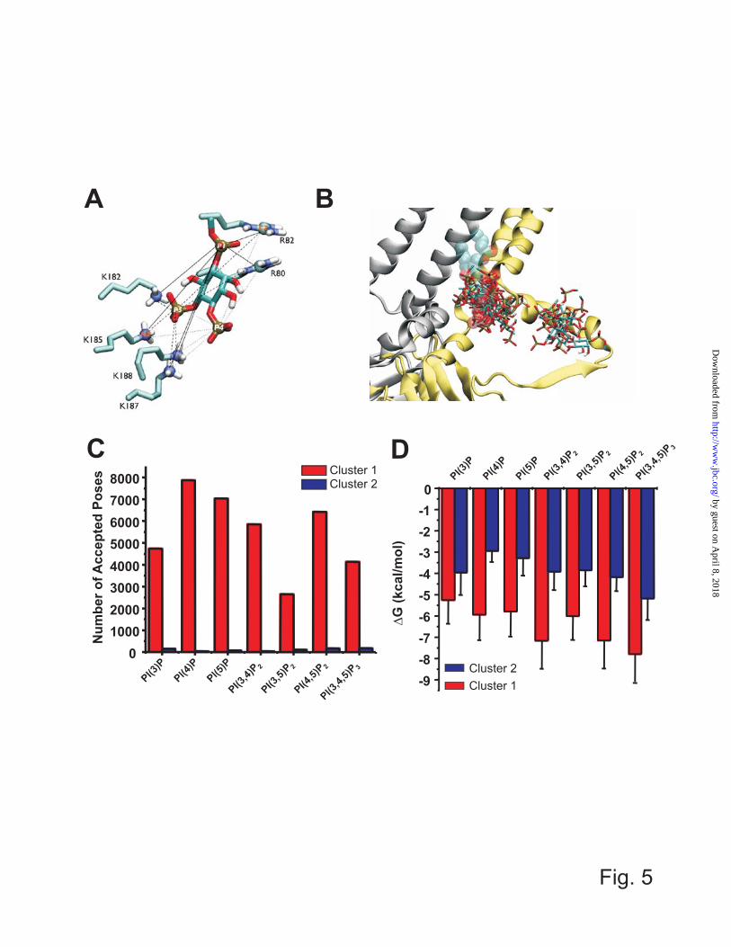
from the TM domain than other structures, resulted in a wider search space although the structural parts included for the search were almost equivalent. With grid space of ~0.34 Å, the grid points were 126x102x100 for 3SPI and 3SYQ, 126x104x100 for 3SPC, and 126x112x100 for 3JYC. Grid parameter files were generated using ADT 1.5.4. and grid maps were generated by AutoGrid 4.2. program. (iii) Docking parameters and simulations: For each model, 100 independent docking simulations were performed on the 100 homology models for 4 templates by Autodock 4.2 program using a Lamarkcian genetic algorithm(28). All default parameters were used except that the maximum number of evaluations was raised from 2,500,000 to 25,000,000. This ensured that a stable minimum was found in each case. Each docking simulation was run for approximately 8 hours on a single processor of IBM x3650-m2 nodes for PI(3)P, PI(4)P, PI(5)P, 10 hours for PI(3,4)P2, PI(3,5)P2, PI(4,5)P2, and 11 hours for PI(3,4,5)P3. (iv) Acceptance: For each template, 10,000 poses were respectively generated for seven ligands. Poses were accepted based on their orientation. A pose was accepted if the angle between the molecular vector ( ) and bilayer normal ( ) (Fig. 2B) was greater than 90o, which occurred when the head group was either pointing away from the membrane or less than parallel to the membrane surface. Current and following analyses were performed by in-house programs running on MatLab (MathWorks, Natick, MA) (v) Clustering: To elucidate representative binding sites the accepted poses were clustered into sub-groups. A K-mean clustering algorithm was adopted using as a metric the RMSD of distances of the phosphorus atom to an atom (CZ for Arg and NZ for Lys) of putative binding residues (80 (R), 82 (R), 182 (K), 185(K), 187(K), 188(K)) (Fig. 5A) was used to cluster the accepted poses.
In order to cluster all accepted poses (N), relative location of a ligand to the protein was used as a metric. The relative position was determined by the distance from each phosphate atom in the ligand to the 6 putative binding residues. Distances were combined into a vector
lir1, r2, …., rn where ri is the distance between one phosphorus atom and one sidechain atom and n is 6 x p (phosphorus atom number in the ligand). Matrix M (n x N) consists of distance vector li of all the accepted poses, after clustering M is sub-grouped into
(1) where MK is a sub-matrix and K is the number of clusters. K-mean clustering is performed according to the following: i) K poses are randomly assigned as initial centroids w1, w2,…, wK. ii) The distance of a pose from these centroids is computed and the pose assigned to the closest centroid. iii) After assignment is made for all poses, new centroids are computed from the members belonging to each centroid and iv) tested for change from the previous centroids. v) If centroid is different, steps 2 through 5 are repeated until centroids do not change. vi) If the new centroids and previous centroids are the same, the clustering is accomplished and the cluster indices of each pose are stored and performance index is computed according to the following equation(29)
(2)
where δi and si are relative weights and a mean intra-cluster variance of the ith sub-group respectively, and di is the minimum distance of ith sub-group to one of all other clusters that accounts for inter-cluster variance. δi, di, and si are computed as following.
(3)
where ki is the number of poses and wi is the centroid of the ith cluster.
where dij is the shortest distance among all the possible pairs between the members of Mi and Mj and the smallest dij is finally determines di of ith cluster.
(5)
where ki is the number of poses in the ith sub-group and N is the number of accepted poses. The best clustering was determined by the minimum Q value. The number of clusters for each ligand was determined to minimize the performance index (Q). The smaller the Q value, the greater the inter-sub-group variance and the smaller the intra-sub-group variance, which indicates better clustering performance. Five clustering trials on the accepted poses for each ligand were performed, and the trace of a trial with the minimum Q value at any cluster number was chosen and shown in Fig 6A. Two clusters (cluster 1 for the larger and cluster 2 for the smaller cluster) were identified. The poses in cluster 1 were sub-grouped for each ligand to identify major conformations of bound ligands. (vi) Contact analysis: To visualize the clustering results based on the multi-dimensional variables, contact patterns between the ligand and frequently contacting residues were determined. Hydrogen bonds were defined by two criteria: (1) the distance between the donor and acceptor atoms being shorter than or equal to 3.4 Å, and the angular orientation being smaller than or equal to 30º between the two unit vectors. One joins hydrogen bond donor atom and hydrogen atom and the other joins hydrogen bond donor and acceptor atoms. Statistical Analysis: Statistical significance was analyzed using an unpaired T-test or one-way ANOVA with Tukey post-hoc analysis as
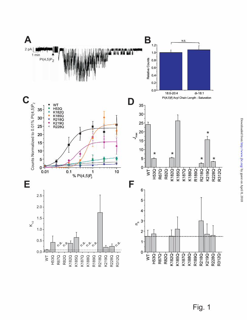
appropriate, and statistical significance (P < 0.05) is indicated by an asterisk. RESULTS AND DISCUSSION Identification of Kir2.1 residues that control PIP binding Kir2.1 channels show an absolute requirement for the phosphoinositide PI(4,5)P2 for channel activity(2). PI(4,5)P2 can be superfused onto the inner leaflet of an excised patch of membrane containing purified Kir2.1 channels and re-stimulate channel activity following rundown, in the absence of any other proteins or intracellular signaling pathways (Fig. 1A). Maximal reconstituted Kir2.1 channel activities measured by 86Rb+ uptake into proteoliposomes containing di-18:1 (dioleoyl) or 18:0 - 24:4 (stearoyl – arachidonoyl) PI(4,5)P2 were indistinguishable (Fig. 1B), indicating that the length and degree of saturation in the acyl-chain is not a major determinant of channel activity, and reflects similar conclusions drawn from patching channels in mammalian cells (13). We also showed previously that Kir2.1 are very specifically activated by PI(4,5)P2, with only ~10% of maximal activity by PI(3,4,5)P3 and little or no activation by the remaining PIPs (2). Furthermore, in the presence of high concentrations of anionic phospholipids such as phosphatidylglycerol (PG), other PIPs can competitively inhibit PI(4,5)P2-dependent channel activity(5). This suggests that the various PIPs may bind to the same general location in Kir2.1 channels, but does not explain why they do not equivalently trigger channel activation. To address this, we have examined the effects of mutations of positively charged residues on the cytoplasmic side of Kir2.1 channels on PIP-driven channel activation in the absence of other proteins or intracellular pathways, and then the effects of these mutations on PIP binding directly.
Twelve positively charged residues on the cytoplasmic side of the channel have been previously shown to alter Kir2.1 channel sensitivity to activation by PI(4,5)P2 (7,12-16). Similar mutational effects have been identified in other Kir channels (10,12,15,17,30-39). We quantified the PI(4,5)P2 dependence of wild type and mutant channel activation directly and independently of other proteins or downstream
signaling pathways in liposomes of defined composition using the 86Rb+ uptake assay (Fig. 1C). K185Q and K219Q mutations primarily shifted the K1/2 of activation (Fig. 1E), while H53Q, K182Q, R218Q, and R228Q mutations reduced the maximal flux (Fig. 1D) as well as the K1/2 of PI(4,5)P2 (Fig. 1E). There was no discernable activity of R67Q, R82Q, K187Q, K188Q, R189Q and R312Q mutant channels, even in liposomes containing 30% PI(4,5)P2. Interestingly, none of the mutations caused any obvious change in the Hill co-efficient compared to WT (nh ≈ 1.5; Fig. 1F) suggesting that stoichiometry or co-operativity of PI(4,5)P2 activation is unaffected by these mutations. The results are generally consistent with the effects of these mutations on Kir2.1 currents in recombinant cells (12). Thus, each of these mutations directly affects the sensitivity of Kir2.1 channels to activation by PI(4,5)P2., although the above analyses cannot determine whether any particular mutation does so through disruption of ligand binding or by affecting transduction to channel opening.
To assess channel-PIP binding affinity directly, we made use of a biochemical lipid binding assay (PIP-Arrays), as has previously been employed to assess PIP binding sites in the C-terminus of KCNQ1 channels (40). WT Kir2.1 channels, detected with anti-His anti-body, bound to PIP-Arrays with a characteristic pattern (Fig. 2). Kir2.1 showed the highest binding to PI(4)P and PI(5)P with only modest binding to PI(4,5)P2, PI(3)P, PI(3,4)P2, PI(3,4,5)P3 and with no observable affinity to PI and PI(3,5)P2. This characteristic binding pattern suggests that higher binding affinity is not the determinant of PI(4,5)P2 specificity.
To ensure that the binding pattern on PIP-Arrays was not due to protein aggregation in the buffer used, two experiments were performed. Firstly, purified protein from the peak fractions that corresponded to the size of Kir2.1 tetramers, and used for both functional studies and the binding assay (Supplementary Fig. S1A), was incubated overnight in Tween-20 detergent and re-run on a gel filtration column in TBK-T buffer. Kir2.1 protein maintained its tetrameric assembly in TBK-T (Supplementary Fig. S1B). Additionally, purified Kir2.1 that was
purposely aggregated by heating to 95 ºC showed no evidence of binding to the PIP-Array (Supplementary Fig. S2), suggesting that (a) any binding of aggregated protein to the array will not be detected by the anti-His probe, and (b) there is no non-specific binding of the anti-His probe to the array. Together, these data indicate the pattern we observed resulted from binding of correctly folded tetrameric protein, and that changes in the binding pattern observed in the following experiments reflect disrupted binding due to the specific mutation.
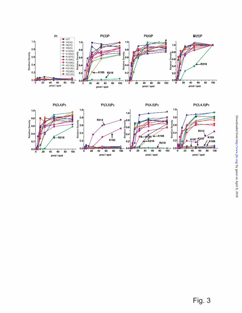
H53Q, R67Q, R82Q, K182Q, and K228Q mutations did not markedly affect the binding of any of the PIPs (although PI(3,5)P2 binding was slightly enhanced with the K182Q mutation) (Fig. 3 and Supplementary Fig. S2 for the raw images of PIP-Array). Thus, while these residues may interact with bound PIP, their role in regulating channel activity appears to be primarily in transduction through the down-stream gating mechanism. On the other hand, K185, K187, K188, R189, R218, R219 and R312 all markedly affected the binding of various PIPs (Fig. 3) and, interestingly, binding of each PIP isoform appears to be regulated by a different subset of key residues (Table 1). For example, PI(4,5)P2 binding appears to be controlled by K185, R189, R218 and K219, with R218Q abolishing binding in the range of the array. On the other hand, PI(3,4,5)P3 binding was disrupted by mutations of K185, K187, K188, K219 and R312, while PI(5)P and PI(3,4)P2 binding was only disrupted when R218 was mutated to glutamine. Estimates of ΔΔG of PIP binding between WT and these various mutants are shown in Table 1. These data imply that the key interactions which govern binding of each particular PIP are different, which may help explain why PIPs other than PI(4,5)P2 fail to stimulate channel activation. Notably, none of the N-terminal residues tested (H53, R67, and R82) disrupted PIP binding when mutated, suggesting that the N-terminus primarily plays a role in regulating channel gating transitions rather than controlling PIP binding itself. R189 is a particularly notable residue, since mutation to glutamine only slightly reduced channel binding to PI(4,5)P2 and had no effect on binding of other PIPs (Fig. 3), even
though it completely abolished channel activity (Fig. 2). This residue may thus serve as a lynch-pin, coupling PI(4,5)P2 binding to a transduction mechanism that leads to channel activation. Ligand Docking of Various PIPs to Human Kir2.1 As noted, Kir2.1 channels are uniquely activated by PI(4,5)P2, with only weak activation by PI(3,4,5)P3 and little or no activation by the remaining PIPs (2). Biochemical assessment of binding (Fig. 2, 3) suggests that this selectivity does not arise from the specificity of PIP binding, since PI(4)P and PI(5)P bind at least as well as PI(4,5)P2, and suggests instead that differences in the nature of the binding, or of the coupling to channel opening, are key. To gain further insight to how the various PIPs interact with Kir2.1 channels, and why these channels cannot be activated by all PIP isoforms, we turned to computational ligand docking experiments. We built homology models of human Kir2.1 (Fig. 4A) based on the recently solved chicken Kir2.2 (PDB: 3JYC, 3SPC, 3SPI)(9,19) and mouse Kir3.2 (PDB: 3SYQ) structures (41), and docked different PIPs to the model structures thus generated. Loops that were not resolved in the structure were added through a loop sampling module implemented in Modeller. To overcome the limited number of rotatable bonds allowed in Autodock 4.2, side-chain flexibility was accounted for by generating 100 models for each of these templates, thus enabling “pseudo-flexible” docking simulations. Since the length and saturation of the acyl-chain was not critical for channel activation (Fig. 1B) we used only the headgroups of the PIP ligands (Fig. 4B) in our computational docking experiments. However, to minimize the bias that may be introduced by using just the headgroups, the acyl-chains of the PIP ligands were removed from the P1 phosphate at C1 (C1_tail) (Fig. 4B, see Methods). Docking poses were analyzed for correct orientation, with poses in which the “tails” were oriented facing away from the expected plane of the membrane removed. In our docking simulations, the membrane was not present. The relative affinity for the various PIPs (assessed by the number of correctly oriented poses in our computational experiments) were compared to the relative binding affinities
determined from the PIP Arrays, for each group of models (Fig. 4C). The closest agreement between model and experiment suggests that, when bound to the PIP Arrays, the WT protein resides in a similar conformation to the closed PI(4,5)P2 bound Kir2.2 structure (PDB: 3SPI). Additional docking simulations were therefore performed on models of Kir2.1 derived from this structure (hereby denoted as Kir2.1-3SPI models). As an internal control, we first ensured that docking simulations in the Kir2.1 models could recapitulate PI(4,5)P2 binding in a similar position and orientation to that observed in the PI(4,5)P2 bound Kir2.2 structure (Fig. 4D). Accepted poses were clustered using the relative distance of all phosphorus atoms in the ligand to the 6 putative binding residues (Fig. 5A), which demonstrated 2 regions near the cytoplasmic domain-membrane interface to which all of the 7 PIP ligands bound (Fig. 5B). A summary of the number of accepted poses in each region (clusters 1 and 2) for the various PIPs is presented in Fig. 5C. Cluster 1 reflects the location of the bound PI(4,5)P2 in the Kir channel crystal structures, and 96-99% of accepted poses reside in this cluster (Fig. 5C). Furthermore, the average binding energy determined by Autodock4.2 is significantly lower in cluster 1 than in cluster 2, for all PIPs (Fig. 4D). We speculate that cluster 2 may reflect the secondary anionic phospholipid site that we described previously (5). A recent simulation also predicted the end of slide helix as a putative anionic lipids binding site (42). However, further in-depth computational analysis and direct experimentation are required to test this idea. We examined the interaction of the PIP ligands within cluster 1 in further detail. Poses in cluster 1 were sub-clustered with the number of sub-clusters (2 to 5) for each ligand being determined by the minimized performance index value (Q) (Figs. 6A and Supplementary Fig. S3). The number of poses in each sub-cluster is shown in Fig. 6B. The free energy of binding (ΔGbinding) estimated from the docking simulations (Fig. 6C), is significantly higher for all mono-phosphorylated PIPs than for di- or tri-phosphorylated PIPs. This appears contrary to the observation of stronger binding of mono-
phosphorylated, particularly PI(4)P and PI(5)P in the PIP Arrays. However, sub-clustering indicates that both PI(4)P and PI(5)P dock with a wider range of orientations than PI(4,5)P2 (Fig. 6), suggesting that preferential binding to these mono-phosphorylated PIPs observed in the array may be entropically driven. Additionally, it is noticeable that the phosphoinositides that contain a 3′ phosphate show significantly fewer acceptable poses than other PIPs (Figs. 5C, 6B). This appears to result from a steric clash that arises between the 2′ hydroxyl group that is axial to the inositol ring and residue W81 side-chain if these ligands were to dock in similar positions to PI(4,5)P2. As a result, these 3′ phosphorylated ligands bind more shallowly within the pocket, with fewer stabilizing interactions with channel side-chains and therefore with weaker binding affinity (Figs. 5D, 6C). Assuming that the orientation of PI(4,5)P2 in the recent crystal structure of Kir2.2 (19) is the one that underlies the activation of the channel, then the present docking experiments provide a clear explanation why the other PIPs do not activate the channel, since they cannot coordinate the same group of side-chains with the same bonding interactions. Interestingly, PI(3,4,5)P3, the only other ligand that marginally activates the channel(2,13), can co-ordinate with the appropriate side-chains in a similar manner to PI(4,5)P2 (PI(3,4,5)P3 cluster 1b; Supplementary Fig. S3), and with similar energies (Fig. 6C), although this pose is observed with much lower frequency (Fig. 6C) than it is with PI(4,5)P2. Ligand Docking of Various PIPs to mutant Human Kir2.1 To assess the effect of individual residues on the binding of various PIPs to the Kir2.1 proteins, docking simulations were repeated on mutant channels. Mutations of residues located away from cluster 1 (namely R189, R218, K219, and R312) were excluded due to the inability to capture conformational changes that might result from mutation of these residues. The 6 remaining basic residues (R80, R82, K182, K185, K187, and K188) that form cluster 1 were mutated into Gln individually, with the results of these docking simulations summarized in Fig 7.
The number of accepted poses of each PIP in cluster 1 is shown as a ratio of the accepted poses in the previous simulations to the wild type protein. Except for PI(3,5)P2, docking to R80Q and R82Q mutant proteins, the number of accepted poses was lower for all mutants than for wild type protein. All the mutations increased the binding energy for all PIPs. Among the 6 mutations, K185Q resulted in the greatest attenuating effect on PIP binding to cluster 1. Polyphosphorylated PIPs were more susceptible to mutations than monophosphorylated PIPs. These docking results qualitatively match the PIP-Array data in the two following aspects: no single mutation in the binding pocket abolishes binding and the K185Q mutation had the greatest effect on PIP binding. Mechanism of PIP activation of Kir2.1 Both our biochemical and computational analyses indicate that PI(4,5)P2 specific activation arises not from a uniquely low free binding energy for this ligand. Instead, multiple PIP ligands bind with varying energies in the same overall location (cluster 1), but non-identical conformations, implying that the appropriate structural changes that lead to channel activation are specific to the preferred PI(4,5)P2 conformation. Only PI(3,4,5)P3, which binds to this site with similar energy to PI(4,5)P2, occasionally samples a very similar conformation. Mono-phosphorylated PIPs interact in the same binding pocket with lower affinity, but greater conformational freedom, which may lead to entropically driven competitive inhibition.
Kir2 channels are constitutively closed in the absence of PI(4,5)P2, and increased PI(4,5)P2 reduces Kir2.1 channel closed times(14,43), suggesting that the predominant pathway to channel activation may be PI(4,5)P2 binding to a closed state, followed by channel opening (Fig. 8). Multiple mutations affect the process of PI(4,5)P2 activation (12,44), and using a combination of biochemical and computational approaches, we can identify mutations that affect binding of phoshoinositides separately from mutations that primarily act to alter channel gating by shifting the relative stability of the open state. Only a relatively
small subset of those mutations that significantly alter channel activation actually interfere with ligand binding (Fig. 8). Of these, K185Q identifies a PI(4,5)P2-interacting residue in the Kir2.2 crystal structure. Thus, residues R189, R218 and K219, which are located some distance from the crystal structure binding site – and from the identified sites in the docking analysis - may be influencing binding indirectly, potentially by disruption of the extensive hydrogen bonding network that links the cytoplasmic domain of one subunit to the slide helix of the neighbouring subunit(19,41). Disruption of such a network could transition to a structure resembling the 3JYC Kir2.2 structure (PI(4,5)P2-unbound closed 1; Fig. 8), thereby disrupting PIP binding due to a loss of the three-dimensional conformation needed to co-ordinate these ligands. This is supported by the fact that the binding energy of all ligands to 3JYC models was increased by ~3 kcal/mol. Recent FRET studies indicate that extensive co-ordinated re-arrangements of the Kir cytoplasmic domain occur during PI(4,5)P2 gating (45). Residues R82, K182, K187 and K188 all interact with the PI(4,5)P2 in the 3SPI structure, yet mutation of these residues (together with multiple other mutations that inhibit activation, including H53Q, R67Q, R228Q, and R312Q) do not significantly disrupt PI(4,5)P2 binding, indicating that loss of any one interacting residue does not abolish binding. Instead, we suggest that the primary effect of these mutations may be disruption of the necessary gating transitions that lead to channel opening following PI(4,5)P2 binding (Fig. 8).
Mutations in Kir2.1 channels cause cardiac arrhythmias, periodic paralysis and developmental phenotypes of Andersen-Tawil syndrome (16,44). Many disease mutations, including R67W, R189I, R218W/Q, R312C, have been suggested to disrupt PI(4,5)P2 binding
(16), although this has not been confirmed by biochemical binding assays. Our data indicate that mutations of residues R189 and R218 do indeed disrupt PI(4,5)P2 binding, although mutations of R67 and R312 do not. Pegan et al. [8] observed stable tetramers for WT cytoplasmic domain-only constructs, but not for 218Q mutants. They thus suggested that this mutation might lead to disruption of the channel assembly. However, full-length R218Q constructs remain tetrameric in solution (Supplementary Fig. S1A) and retain very low but still observable activity (Fig. 1C). Thus, it is unlikely that the disease mechanism of mutation R218Q is through a mis-folded cytoplasmic domain, but rather through disruption of PI(4,5)P2 binding. Distinct modes of action of various Andersen-Tawil mutations should be considered during efforts to develop therapeutic treatments for diseases involving Kir2 channels. Conclusions Using a combination of biochemical and computational approaches, we have determined the role of charged residues in the cytoplasmic domain in PIP activation of Kir2.1 channels. We determine residues that are critical for binding of phoshoinositides and residues that primarily act to alter channel gating. The data indicate that PI(4,5)P2 specific activation arises not from uniquely low free binding energy for this ligand and instead suggest that interactions in a specific conformation trigger appropriate changes that lead to channel activation. Only PI(3,4,5)P3, which binds to this site with similar energy, may occasionally sample a similar conformation. Mono-phosphorylated PIPs may interact in the same binding pocket with greater conformational freedom, thereby leading to entropically driven competitive inhibition.
REFERENCES 1. Cheng, W. W., Enkvetchakul, D., and Nichols, C. G. (2009) KirBac1.1: it's an inward rectifying
potassium channel. J Gen Physiol 133, 295-305 2. D'Avanzo, N., Cheng, W. W., Doyle, D. A., and Nichols, C. G. (2010) Direct and specific
activation of human inward rectifier K+ channels by membrane phosphatidylinositol 4,5-bisphosphate. J Biol Chem 285, 37129-37132
3. Enkvetchakul, D., Jeliazkova, I., and Nichols, C. G. (2005) Direct modulation of Kir channel gating by membrane phosphatidylinositol 4,5-bisphosphate. J Biol Chem 280, 35785-35788
4. Leal-Pinto, E., Gomez-Llorente, Y., Sundaram, S., Tang, Q. Y., Ivanova-Nikolova, T., Mahajan, R., Baki, L., Zhang, Z., Chavez, J., Ubarretxena-Belandia, I., and Logothetis, D. E. (2010) Gating of a G protein-sensitive mammalian Kir3.1 prokaryotic Kir channel chimera in planar lipid bilayers. J Biol Chem 285, 39790-39800
5. Cheng, W. W., D'Avanzo, N., Doyle, D. A., and Nichols, C. G. (2011) Dual-Mode Phospholipid Regulation of Human Inward Rectifying Potassium Channels. Biophys J 100, 620-628
6. Nishida, M., Cadene, M., Chait, B. T., and MacKinnon, R. (2007) Crystal structure of a Kir3.1-prokaryotic Kir channel chimera. EMBO J 26, 4005-4015
7. Pegan, S., Arrabit, C., Slesinger, P. A., and Choe, S. (2006) Andersen's syndrome mutation effects on the structure and assembly of the cytoplasmic domains of Kir2.1. Biochemistry 45, 8599-8606
8. Pegan, S., Arrabit, C., Zhou, W., Kwiatkowski, W., Collins, A., Slesinger, P. A., and Choe, S. (2005) Cytoplasmic domain structures of Kir2.1 and Kir3.1 show sites for modulating gating and rectification. Nat Neurosci 8, 279-287
9. Tao, X., Avalos, J. L., Chen, J., and MacKinnon, R. (2009) Crystal structure of the eukaryotic strong inward-rectifier K+ channel Kir2.2 at 3.1 A resolution. Science 326, 1668-1674
10. Haider, S., Tarasov, A. I., Craig, T. J., Sansom, M. S., and Ashcroft, F. M. (2007) Identification of the PIP2-binding site on Kir6.2 by molecular modelling and functional analysis. Embo J 26, 3749-3759
11. Rohacs, T., Lopes, C. M., Jin, T., Ramdya, P. P., Molnar, Z., and Logothetis, D. E. (2003) Specificity of activation by phosphoinositides determines lipid regulation of Kir channels. Proc Natl Acad Sci U S A 100, 745-750
12. Lopes, C. M., Zhang, H., Rohacs, T., Jin, T., Yang, J., and Logothetis, D. E. (2002) Alterations in conserved Kir channel-PIP2 interactions underlie channelopathies. Neuron 34, 933-944
13. Rohacs, T., Chen, J., Prestwich, G. D., and Logothetis, D. E. (1999) Distinct specificities of inwardly rectifying K(+) channels for phosphoinositides. J Biol Chem 274, 36065-36072
14. Xie, L. H., John, S. A., Ribalet, B., and Weiss, J. N. (2008) Phosphatidylinositol-4,5-bisphosphate (PIP2) regulation of strong inward rectifier Kir2.1 channels: multilevel positive cooperativity. J Physiol 586, 1833-1848
15. Zhang, H., He, C., Yan, X., Mirshahi, T., and Logothetis, D. E. (1999) Activation of inwardly rectifying K+ channels by distinct PtdIns(4,5)P2 interactions. Nat Cell Biol 1, 183-188
16. Donaldson, M. R., Jensen, J. L., Tristani-Firouzi, M., Tawil, R., Bendahhou, S., Suarez, W. A., Cobo, A. M., Poza, J. J., Behr, E., Wagstaff, J., Szepetowski, P., Pereira, S., Mozaffar, T., Escolar, D. M., Fu, Y. H., and Ptacek, L. J. (2003) PIP2 binding residues of Kir2.1 are common targets of mutations causing Andersen syndrome. Neurology 60, 1811-1816
17. Huang, C. L., Feng, S., and Hilgemann, D. W. (1998) Direct activation of inward rectifier potassium channels by PIP2 and its stabilization by Gbetagamma. Nature 391, 803-806
18. Soom, M., Schonherr, R., Kubo, Y., Kirsch, C., Klinger, R., and Heinemann, S. H. (2001) Multiple PIP2 binding sites in Kir2.1 inwardly rectifying potassium channels. FEBS Lett 490, 49-53
19. Hansen, S. B., Tao, X., and MacKinnon, R. (2011) Structural basis of PIP2 activation of the classical inward rectifier K+ channel Kir2.2. Nature 477, 495-498
20. D'Avanzo, N., Cheng, W. W., Xia, X., Dong, L., Savitsky, P., Nichols, C. G., and Doyle, D. A. (2010) Expression and purification of recombinant human inward rectifier K+ (KCNJ) channels in Saccharomyces cerevisiae. Protein Expr Purif 71, 115-121
21. D'Avanzo, N., Hyrc, K., Enkvetchakul, D., Covey, D. F., and Nichols, C. G. (2011) Enantioselective Protein-Sterol Interactions Mediate Regulation Of Both Prokaryotic And Eukaryotic Inward Rectifier K+ Channels By Cholesterol. In Submission
22. Lederer, W. J., and Nichols, C. G. (1989) Nucleotide modulation of the activity of rat heart ATP-sensitive K+ channels in isolated membrane patches. J Physiol 419, 193-211
23. Thomas, A. M., and Tinker, A. (2008) Determination of phosphoinositide binding to K(+) channel subunits using a protein-lipid overlay assay. Methods Mol Biol 491, 103-111
24. Eswar, N., Marti-Renom, M. A., Webb, B., Madhusudhan, M. S., Eramian, D., Shen, M., Pieper, U., and Sali, A. (2006) Comparative Protein Structure Modeling with MODELLER. in Current Protocols in Bioinformatics, John Wiley & Sons, Inc. pp 5.6.1-5.6.3
25. Marti-Renom, M. A., Stuart, A. C., Fiser, A., Sanchez, R., Melo, F., and Sali, A. (2000) Comparative protein structure modeling of genes and genomes. Annu Rev Biophys Biomol Struct 29, 291-325
26. Sanner, M. F. (1999) Python: a programming language for software integration and development. J Mol Graph Model 17, 57-61
27. Lupyan, D., Mezei, M., Logothetis, D. E., and Osman, R. (2010) A molecular dynamics investigation of lipid bilayer perturbation by PIP2. Biophys J 98, 240-247
28. Morris, G. M., Goodsell, D. S., Halliday, R. S., Huey, R., Hart, W. E., Belew, R. K., and Olson, A. J. (1998) Automated Docking Using a Lamarckian Genetic Algorithm and Empirical Binding Free Energy Function. J. Computational Chemistry 19, 1639-1662
29. Savaresi, S. M., Boley, D. L., Bittanti, S., and Gazzaniga, G. (2002) Cluster selection in divisive clustering algorithms. in Second SIAM International Conference on Data Mining (SDM 2002)
30. Fan, Z., and Makielski, J. C. (1997) Anionic phospholipids activate ATP-sensitive potassium channels. J Biol Chem 272, 5388-5395
31. Inanobe, A., Nakagawa, A., Matsuura, T., and Kurachi, Y. (2010) A structural determinant for the control of PIP2 sensitivity in G protein-gated inward rectifier K+ channels. J Biol Chem 285, 38517-38523
32. Leung, Y. M., Zeng, W. Z., Liou, H. H., Solaro, C. R., and Huang, C. L. (2000) Phosphatidylinositol 4,5-bisphosphate and intracellular pH regulate the ROMK1 potassium channel via separate but interrelated mechanisms. J Biol Chem 275, 10182-10189
33. Liou, H. H., Zhou, S. S., and Huang, C. L. (1999) Regulation of ROMK1 channel by protein kinase A via a phosphatidylinositol 4,5-bisphosphate-dependent mechanism. Proc Natl Acad Sci U S A 96, 5820-5825
34. Schulze, D., Krauter, T., Fritzenschaft, H., Soom, M., and Baukrowitz, T. (2003) Phosphatidylinositol 4,5-bisphosphate (PIP2) modulation of ATP and pH sensitivity in Kir channels. A tale of an active and a silent PIP2 site in the N terminus. J Biol Chem 278, 10500-10505
35. Shyng, S. L., Cukras, C. A., Harwood, J., and Nichols, C. G. (2000) Structural determinants of PIP(2) regulation of inward rectifier K(ATP) channels. J Gen Physiol 116, 599-608
36. Shyng, S. L., and Nichols, C. G. (1998) Membrane phospholipid control of nucleotide sensitivity of KATP channels. Science 282, 1138-1141
37. Stansfeld, P. J., Hopkinson, R., Ashcroft, F. M., and Sansom, M. S. (2009) PIP(2)-binding site in Kir channels: definition by multiscale biomolecular simulations. Biochemistry 48, 10926-10933
38. Zeng, W. Z., Liou, H. H., Krishna, U. M., Falck, J. R., and Huang, C. L. (2002) Structural determinants and specificities for ROMK1-phosphoinositide interaction. Am J Physiol Renal Physiol 282, F826-834
39. Cukras, C. A., Jeliazkova, I., and Nichols, C. G. (2002) The role of NH2-terminal positive charges in the activity of inward rectifier KATP channels. J Gen Physiol 120, 437-446
40. Thomas, A. M., Harmer, S. C., Khambra, T., and Tinker, A. (2011) Characterization of a binding site for anionic phospholipids on KCNQ1. J Biol Chem 286, 2088-2100
41. Whorton, M. R., and MacKinnon, R. (2011) Crystal structure of the mammalian GIRK2 K+ channel and gating regulation by G proteins, PIP2, and sodium. Cell 147, 199-208
42. Schmidt, M. R., Stansfeld, P. J., Tucker, S. J., and Sansom, M. S. (2013) Simulation-based prediction of phosphatidylinositol 4,5-bisphosphate binding to an ion channel. Biochemistry 52, 279-281
43. Jin, T., Sui, J. L., Rosenhouse-Dantsker, A., Chan, K. W., Jan, L. Y., and Logothetis, D. E. (2008) Stoichiometry of Kir channels with phosphatidylinositol bisphosphate. Channels (Austin) 2, 19-33
44. Plaster, N. M., Tawil, R., Tristani-Firouzi, M., Canun, S., Bendahhou, S., Tsunoda, A., Donaldson, M. R., Iannaccone, S. T., Brunt, E., Barohn, R., Clark, J., Deymeer, F., George, A. L., Jr., Fish, F. A., Hahn, A., Nitu, A., Ozdemir, C., Serdaroglu, P., Subramony, S. H., Wolfe, G., Fu, Y. H., and Ptacek, L. J. (2001) Mutations in Kir2.1 cause the developmental and episodic electrical phenotypes of Andersen's syndrome. Cell 105, 511-519
45. Wang, S., Lee, S. J., Heyman, S., Enkvetchakul, D., and Nichols, C. G. (2011) Structural rearrangements underlying ligand-gating in Kir channels. Nat Commun 3, 617
Acknowledgements The authors would like to thank Dr. Oscar Harari for useful discussion regarding clustering analysis. FOOTNOTES This work was supported by grants from the National Institutes of Health (HL54171 to CGN; K99 HL112300-01 to ND) and an American Heart Association Fellowship (13POST14660069 to SJL). The funders had no role in study design, data collection and analysis, decision to publish, or preparation of the manuscript. †Contributed equally to the work *Current and permanent address: GÉPROM, Université de Montréal, Montréal, Québec, Canada, H3T1J4 For correspondence: Ph: (314) 362-6630, FAX: (314) 362-7463, email: [email protected] or [email protected]
TABLE 1. ΔΔG (in kcal/mol) of PIP binding between WT and Gln mutant Kir2.1 channels from PIP Arrays.
FIGURE LEGENDS Figure 1. (A) Superfusion of PI(4,5)P2 onto a patch of membrane containing Kir2.1 channels can recover channel activity after rundown. (B) Activation of Kir2.1 channels by PI(4,5)P2 is not sensitive to the acyl-chain length or saturation as determined by comparing maximal activation between di-18:1 (dioleoyl) and 18:0 - 24:4 (stearoyl – arachidonoyl) PI(4,5)P2. (C) PI(4,5)P2 concentration – activity relationship for WT and mutant Kir2.1 channels. For clarity, only mutants in which some activity could be observed are shown. Solid lines are fit curves using the Hill equation. No discernable activity was detected for R67Q, R82Q, K187Q, K188Q, R189Q and R312Q Kir2.1 channels even in liposomes containing 30% PI(4,5)P2. (D – F) Parameters of fits to the Hill equation indicate that K185Q and K219Q mutants primarily shift the K1/2 of PI(4,5)P2, while H53Q, K182Q, R218Q, and R228Q mutations reduce the maximal flux as well as increase the K1/2 of PI(4,5)P2. No mutation significantly altered the Hill co-efficient (nh ≈ 1.5) which has been suggested to be a measure of co-operativity between ligands. (n.a. denotes no detectable activity). Figure 2. The interaction of wild type Kir2.1 to PIP-arrays. A schematic diagram of PIP-Arrays (left) shows increasing amount of lipids from left to right. PIP-Arrays were incubated with WT Kir2.1 and bound proteins were probed with an anti-His antibody (center). Densities in each array were internally normalized to density measured for the 100 pmol spot of PI(5)P (right). Figure 3. The interaction of individual mutant Kir 2.1 to the PIP-arrays. Mutant Kir2.1 protein bound to the PIP-arrays were probed with an anti-His antibody. The raw images are shown in Supplementary Fig. S2. Densitometry measurements from PIP-Arrays were internally normalized to density measured for the 100 pmol spot of PI(5)P and are plotted for lipids individually. The mutations that caused more than 50 % reduction in binding compared to wt Kir2.1 are designated by an arrow and residue name. These data indicate that for each PIP, it is a different subset of residues that when mutated to Gln (Q) disrupts channel binding. This suggests these ligands orient differently in the binding pocket, thereby interacting with different subset of residues, which may explain why they do not equivalently trigger activation in Kir2.1 channels. Figure 4. Docking of various PIPs to wild type Kir2.1 protein models (A) Cα trace of representative homology models of human Kir2.1 based on the chicken Kir2.2 structures (PDB: 3JYC (Blue); 3SPC (Cyan), or 3SPI (Green)) and mouse Kir3.2 (PDB: 3SYQ (Red)). The search region for autodocking is designated by a black box. (B) Surrogate structure of the PI(4,5)P2 ligand with atom names and charges. The surrogates are the mimics of the PIPs with acyl chains cut off at the C1 position. Other PIPs were generated through substituting with either a hydroxyl or phosphate group at position 3, 4, and 5 on the
inositol ring. Atom charges are transferable to other PIPs. The vector for molecular axis ( ) and bilayer normal ( ) are used for docking simulations and analysis. (C) Relative affinities (assessed by number of correctly oriented poses) for each ligand from our computational experiments were compared to the relative binding affinities determined from the PIP Arrays. R2 values for the linear fit were 0.5514, 0.4669, 0.4403, and 0.4211 for 3SPI, 3JYC, 3SPC, and 3SYQ derived Kir2.1 models respectively. This analysis suggests that the protein bound to the PIP Arrays most likely resides in a similar conformation to that observed in the closed PI(4,5)P2 bound structure (PDB: 3SPI), with weak binding in both cases to PI(3,5)P2, and so the remainder of our analysis was performed on docking simulations to models derived from this structure. (D) A docked pose of PI(4,5)P2 bound to Kir2.1-3SPI in darker color compared to the crystallographic PI(4,5)P2 bound to Kir2.2 in light color. Chicken Kir2.2 subunit A is shown in cartoon image, and 6 basic residues in the binding pocket are shown in sticks. Figure 5. (A) Distances between a ligand and the six putative binding residues. The pairs between the phosphorus atoms (P1, P4, and P5) of PI(4,5)P2 and the sidechain atoms (CZ of R80, R82 and NZ of K182, K185, K187, and K188) are visualized by solid, dashed, and dotted lines. The distances of these pairs were used to cluster accepted poses. (B) Kir 2.1-3SPI subunit A and D are shown in yellow and silver ribbon respectively with the crystallographic PI(4,5)P2 bound to the subunit A shown in sphere translucently. An overlay of a single representative pose from each cluster of 7 different C1-PIP ligands stacked on the Kir2.1-3SPI surface clearly indicates these ligand bind primarily within 2 clusters. (C) The number of accepted poses within cluster 1 (red) and cluster 2 (blue) for each PIP are indicated. For each PIP, 96 – 99 % of poses reside in cluster 1. (D) Average binding energy for poses in cluster 1 (red) and cluster 2 (blue) for each PIP docked to Kir2.1-3SPI. Figure 6. (A) The minimum value determined from the performance index (Q) versus cluster number was taken to determine the optimal number of sub-clusters for PIP binding in cluster 1. (B) The number of accepted poses in each sub-cluster for each of the 7 PIP ligands docked to Kir2.1-3SPI. (C) A box-whisker plot of binding free energy (ΔGbinding in kcal/mol) within a sub-cluster for each PIP ligand. Mono-phosphorylated PIPs and PI(3,5)P2 bind with higher average energies than PI(4,5)P2, however, selective activation of Kir2.1 channels by PI(4,5)P2 cannot be solely accounted for based on this, since PI(3,4)P2 and PI(3,4,5)P3 bind with similar energies. Figure 7. Docking of various PIPs to Kir2.1 mutant protein models. The same docking simulations were carried out on the Kir2.1 model proteins with a single mutation as listed in this figure. (A) The number of accepted poses in cluster 1 relative to that of wild type protein is shown. (B) The difference of binding free energy in the cluster 1 between the wild type and each mutant protein is shown in kcal/mol unit. Error bars represents standard errors. Figure 8. A proposed model for the predominant pathway to channel activation. Kir2.1 channels may undergo a conformational change in which the cytoplasmic domain moves from a 3JYC type closed conformation (1) toward the plasma membrane and interacts through a hydrogen bond network with the slide helix, leading to a PI(4,5)P2 unbound structure (similar to what was observed for the Kir3.2 apo structure). We suggest this transition may be less favoured by mutations R189Q, R218Q, and K219Q, leading to reduced binding of PIP ligands. However, once the transition occurs, this state generates a three-dimensional binding pocket that strongly co-ordinates PI(4,5)P2, binding of which is disrupted by K185Q mutation. Further conformational changes lead to channel opening. Multiple mutations disrupt this transition, without affecting earlier steps of binding.