Phosphorylation of MAP kinase-like proteins mediate the response of the halotolerant alga Dunaliella viridis to hypertonic shock Carlos Jime ´nez a , Tomas Berl b , Christopher J. Rivard b , Charles L. Edelstein b , Juan M. Capasso b, * a Department of Ecology, Faculty of Science, University of Ma ´laga, Ma ´laga 29071, Spain b Division of Nephrology, Department of Medicine, University of Colorado, School of Medicine, 4200 East 9th Avenue, Denver, CO 80262, USA Received 24 February 2003; received in revised form 15 September 2003; accepted 31 October 2003 Abstract The microalga Dunaliella viridis has the ability to adapt to a variety of environmental stresses including osmotic and thermal shocks, UV irradiation and nitrogen starvation. Lacking a rigid cell wall, Dunaliella provides an excellent model to study stress signaling in eukaryotic unicellular organisms. When exposed to hyperosmotic stress, UV irradiation or high temperature, a 57-kDa protein is recognized by antibodies specific to mammalian p38, to its yeast homologue Hog1, and to the phospho-p38 MAP kinase motif. This 57-kDa protein appears to be both up-regulated and phosphorylated. Three other proteins (50, 45, 43 kDa) were transiently phosphorylated under stress conditions as detected with an antibody specific to the mammalian phospho c-Jun N-terminal kinase (JNK) motif. Treatment with specific inhibitors of p38 MAP kinase (SB203580) and JNK (SP600125) activities markedly impaired the adaptation of Dunaliella to osmotic stress. From an evolutionary standpoint, these data strongly suggest that MAP kinase signaling pathways, other than ERK, were already operating in the common ancestor of plant and animal kingdoms, probably as early as 1400 million years ago. D 2004 Elsevier B.V. All rights reserved. Keywords: Microalgae; Hypertonicity; MAP kinase; Cell survival; Adaptation 1. Introduction Mitogen-activated protein (MAP) kinases are highly conserved serine/threonine kinases found in all eukaryotic cells in combination with their upstream activators. These kinases have been extensively studied in organisms from yeast to humans as transducers of extracellular signals in a variety of cytoplasmic and nuclear events [1]. In contrast, the characterization of MAP kinases in plants is much more limited but also of increasing interest [1–4]. Several studies have established that plants express proteins homologous to some of the components of the MAP kinase cascade de- scribed in mammals [4]. Specifically, tobacco leaves stressed by cutting activate a 46-kDa protein designated as PMSAP [5]. Likewise, in this plant two MAP kinases, one induced by salicylate/salcylic (SIPK) and a wound-induced protein ki- nase (WIPK), have been described [6]. Also in tobacco, the bacterial protein harpin induces activation of a 49-kDa kinase [7] and the fungal elicitor cryptogein induces activa- tion of both a 46- and a 50-kDa MAP kinase [8]. In tomato leaves, wounds turn on a 48-kDa protein [9] and in alfalfa a kinase designated as MKK4 is activated by cold stress and drought [10]. Signal pathways have been explored in Arabi- dopsis thaliana leaf cells exposed to cold, touch [11], bacterial products [12,13] and oxidative stress [14]. Osmotic stress homologues to the yeast osmoreceptor [15] have been sought in plants [12]. In A. thaliana a two- component system was cloned [16] including the salt stress- induced transcription of a gene and the expression of a protein (ATMPK3) that is structurally related to MAP kinases [11]. The aforementioned alfalfa MKK4 activated by drought is not altered by salinity [10], but rather a 46-kDa MAPK is salt induced (SIMK), which is localized in the nucleus [17]. In tobacco plants placed under osmotic stress conditions, three different kinases (44, 46 and 50 kDa) were activated by tonicity [18], the latter two being identified as the aforementioned tobacco SIPK and WIPK, respectively. The 44-kDa protein does not appear to belong to the MAP kinase family as it failed to be recognized by anti-human extracellular signal-regulated kinase (ERK 1 /ERK 2 ) antibody. 0167-4889/$ - see front matter D 2004 Elsevier B.V. All rights reserved. doi:10.1016/j.bbamcr.2003.10.009 * Corresponding author. Tel.: +1-303-315-6723; fax: +1-303-315- 4852. E-mail address: [email protected] (J.M. Capasso). www.bba-direct.com Biochimica et Biophysica Acta 1644 (2004) 61 – 69
Transcript
www.bba-direct.com
Biochimica et Biophysica Acta 1644 (2004) 61–69
Phosphorylation of MAP kinase-like proteins mediate the response of
the halotolerant alga Dunaliella viridis to hypertonic shock
Carlos Jimeneza, Tomas Berlb, Christopher J. Rivardb, Charles L. Edelsteinb, Juan M. Capassob,*
aDepartment of Ecology, Faculty of Science, University of Malaga, Malaga 29071, SpainbDivision of Nephrology, Department of Medicine, University of Colorado, School of Medicine, 4200 East 9th Avenue, Denver, CO 80262, USA
Received 24 February 2003; received in revised form 15 September 2003; accepted 31 October 2003
Abstract
The microalga Dunaliella viridis has the ability to adapt to a variety of environmental stresses including osmotic and thermal shocks, UV
irradiation and nitrogen starvation. Lacking a rigid cell wall, Dunaliella provides an excellent model to study stress signaling in eukaryotic
unicellular organisms. When exposed to hyperosmotic stress, UV irradiation or high temperature, a 57-kDa protein is recognized by
antibodies specific to mammalian p38, to its yeast homologue Hog1, and to the phospho-p38 MAP kinase motif. This 57-kDa protein appears
to be both up-regulated and phosphorylated. Three other proteins (50, 45, 43 kDa) were transiently phosphorylated under stress conditions as
detected with an antibody specific to the mammalian phospho c-Jun N-terminal kinase (JNK) motif. Treatment with specific inhibitors of p38
MAP kinase (SB203580) and JNK (SP600125) activities markedly impaired the adaptation of Dunaliella to osmotic stress. From an
evolutionary standpoint, these data strongly suggest that MAP kinase signaling pathways, other than ERK, were already operating in the
common ancestor of plant and animal kingdoms, probably as early as 1400 million years ago.
amounts of protein were loaded per lane (50 Ag/lane) forSDS-PAGE. To ensure adequate data analysis several experi-
ments were run in duplicate with one of the gels stained with
Coomassie blue to confirm uniform protein loading. Electro-
phoresis conditions, electroblotting to PVDF membranes,
and immunodetection were performed as previously de-
scribed [27,29]. Antibodies were purchased from Cell Sig-
naling Technology (Beverly, MA) and Santa Cruz
Biotechnology (Santa Cruz, CA).
Antibody specificity: Both p38 and Hog1 antibodies detect
the presence of the corresponding kinases, regardless of their
phosphorylation state. These antibodies are specific for p38/
Hog1 and do not cross-react with JNK and ERK. The
reactivity represents a total concentration of all forms of the
protein. Hog1 from yeast has been previously demonstrated
to be functionally and structurally homologous to the mam-
malian p38 [30]. The phospho-p38 antibody detects only the
phosphorylated form of this kinase and is specific for the
antigen sequence T*GY* and does not cross-react with
similar sequences such as TPY (JNK) or TEY (ERK). This
phospho-p38 antibody does not react with either the non-
phosphorylated or single phosphorylated form of the protein.
C. Jimenez et al. / Biochimica et Biophysica Acta 1644 (2004) 61–69 63
At this time an antibody specific to the non-phosphorylated
form of p38 MAPK is not available although the non-
phosphorylated p38 pools may be inferred with respect to
the difference in signal kinetics from Western blots using the
two antibodies described above.
The phospho-JNK antibody used is specific for the T*PY*
sequence and only when double-phosphorylated. This anti-
body does not recognize either the non-phosphorylated or
single phosphorylated form of the protein. This antibody does
not cross-react with p38 or ERK. This antibody recognizes all
isoforms of the JNK protein. This information has been
provided by the manufacturers as well as corroborated by
the authors.
Band analysis was performed as described [26]. Analysis
of the same blot with different antibodies was done by
striping with Western Re-Probe (Geno Technology, St.
Louis, MO) as directed by the manufacturer.
2.5. Metabolic labeling of Dunaliella with radioactive
phosphate
To detect protein phosphorylation in Dunaliella in res-
ponse to stress, mid-log cultures growing at 2 M NaCl were
concentrated by centrifugation to 10% of the initial volume,
and incubated overnight in the presence of 2 mCi of 32P
(K2HPO4, SpAc 1 Ci/mM, NEX055, Perkin Elmer Life
Sciences, Boston, MA). Cells were osmotically challenged
by adding NaCl to a 4 M final concentration. Cells were
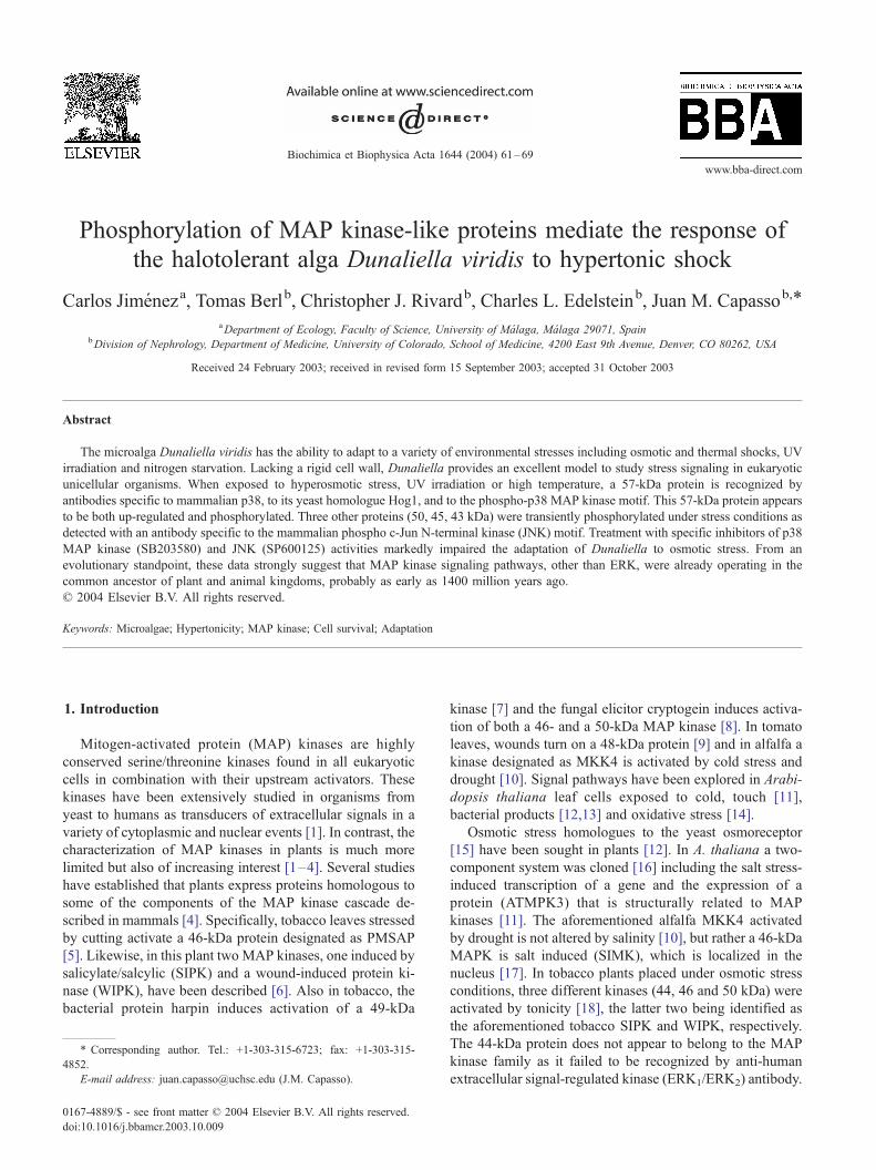
Fig. 1. Aliquots of mid log phase Dunaliella cultures were challenged as described
hypoosmotic conditions) for increasing time periods. Cell protein extracts were an
form of mammalian p38 MAP kinase. The graph depicts the MeanF S.E. of the 5
blot is also shown.
harvested by centrifugation after 4 h, and washed three
times in 4 M NaCl medium. Samples were prepared for
electrophoresis as above. Gels were stained with Coomassie
blue, digitized, dried and autoradiographed using Kodak
BioMax ML film with one intensifying screen for different
times.
2.6. Cell viability measurement
Algal cell viability was measured using CellTiter 96AQ
(Promega, Madison, WI), as described by Capasso et al.
[31].
2.7. Statistics
Results were analyzed by using the INSTAT software
package (GraphPad, San Diego CA). A value of P < 0.05
was considered significant.
3. Results
3.1. Effects of alterations in NaCl concentration on
proteins probed with a phosphorylated p38 MAP kinase
antibody
Cultures of D. viridis, growing under basal conditions (2
M NaCl), were exposed to either a hypertonic stress
in Section 2 with three different NaCl treatments (control, hyperosmotic and
alyzed by Western blot using an antibody raised against the phosphorylated
7-kDa band intensity from three independent experiments. A representative
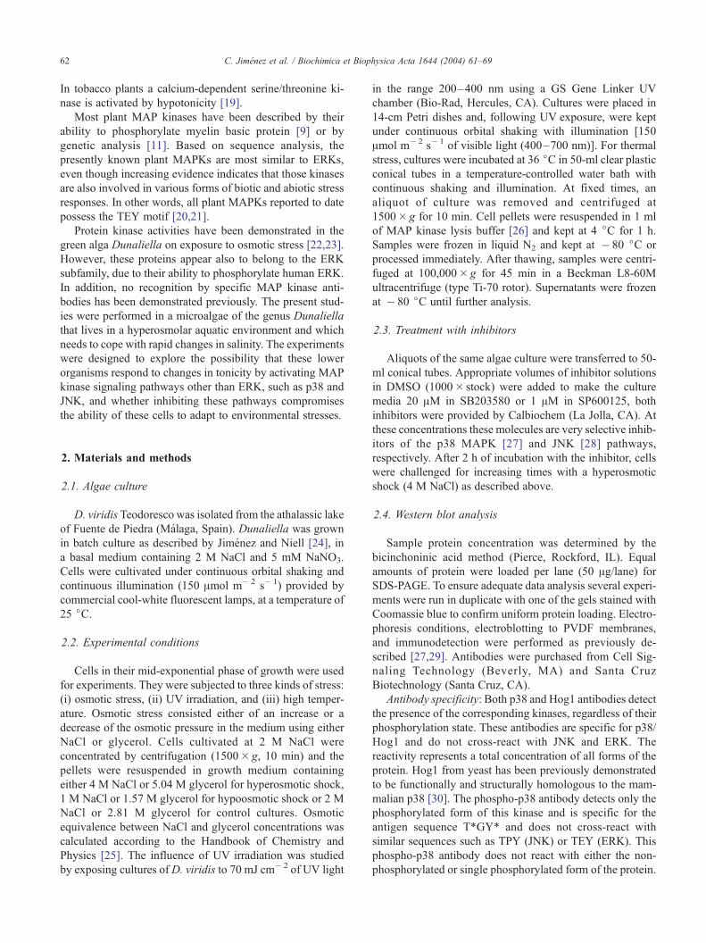
Fig. 2. Cell cultures were subjected to thermal and UV stresses. Protein
extracts were analyzed by Western blot using antibodies specific for
phosphorylated p38 MAP kinase (as in Fig. 1). (A) Cells were exposed to
thermal shock by incubation at 36 jC, quantitative analysis of band intensity(n= 4) and one representative blot is shown. (B) Cells were exposed to
irradiation with UV light (70 mJ cm� 2/200–400 nm); the graph depicts the
MeanF S.E. of the 57-kDa band intensity (n= 3); a representative blot is
also shown. The difference between 0 and 15 min is highly significant
( P < 0.01).
C. Jimenez et al. / Biochimica et Biophysica Acta 1644 (2004) 61–6964
(medium supplemented to 4 M NaCl) or to a hypotonic
stress (medium containing 1 M NaCl) for varying times
ranging from 0 to 4 h. Exposure to 4 M NaCl was
associated with a time-dependent increase in the level of a
protein band with an estimated molecular weight of 57 kDa
(Fig. 1). This band cross-reacts with a highly specific
antibody raised against the phosphorylated form of the
mammalian p38 MAP kinase (P-Thr/Gly/P-Tyr). This phos-
phorylated protein was not detected in control cells main-
tained at 2 M NaCl or cells exposed to a hypotonic stress
for as long as 4 h. The same result was obtained using
equivalent concentrations of glycerol instead of NaCl in
order to increase or diminish the medium tonicity (data not
shown).
3.2. Effect of UV-irradiation and thermal shock on the
phosphorylation of the 57-kDa protein
To assess whether alternative environmental stresses also
caused the phosphorylation of the 57-kDa protein, algal
cells were exposed to heat shock or to UV irradiation for
increasing times. Exposure of algal cells to elevated tem-
perature (36 jC) induced rapid appearance of the 57-kDa
phosphoprotein using the phosphorylated p38 MAP kinase
antibody (Fig. 2A). However, the time course of phosphor-
ylation was very different from that obtained during os-
motic shock. After only 30 min of incubation at 36 jCmore than 50% of the maximum level had been reached
and after 60 min the level had essentially reached saturation
(Fig. 1 vs. Fig. 2A). The effect of UV irradiation (70 mJ/
cm2) on algal cell cultures is shown in Fig. 2B. Similar to
the effect of thermal shock stress, a very rapid increase in
the 57-kDa phosphoprotein was detected upon irradiation
of the cells; the kinetics of the phosphorylation was slightly
different.
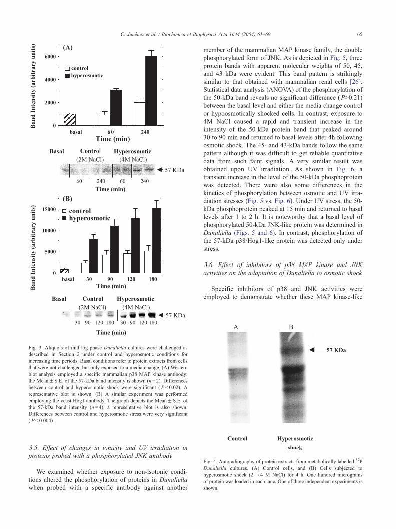
3.3. Effect of hypertonicity on the expression of a protein
that cross-reacts with non-phosphorylated p38 MAP kinase
and Hog1 antibodies
In view of the above results, we examined whether the
57-kDa protein could also be detected in algal cells using
antibodies specific for the mammalian p38 MAP kinase and/
or its yeast homologue, Hog1 kinase, and if so, whether its
expression is altered by exposure to hypertonicity. As
described in Section 2, these antibodies recognize the
presence of both phosphorylated and unphosphorylated
states of the kinase. Fig. 3 shows that a protein band of
57 kDa is detected using both the p38 MAP kinase antibody
(Fig. 3A) and the Hog1 antibody (Fig. 3B). These data
indicate that the protein band cross-reacts more strongly
with the yeast antibody than with the mammalian one. In
contrast to the observation in mammalian cells that p38
MAP kinase is present at a relatively constant level [26], the
57-kDa protein is present at a low level under basal
conditions and is up-regulated in a time-dependent manner
following osmotic stress (Fig. 3A). This indicates that in
Dunaliella, stress induces both phosphorylation and synthe-
sis of the p57 protein, suggesting that it is part of the stress-
induced response mechanism. Interestingly, changing the
culture medium by itself (involving centrifugation and
resuspension) caused a small time-dependent increase in
the protein level (Fig. 3A and B), but no phosphorylation of
this protein occurred (see control data in Fig. 1).
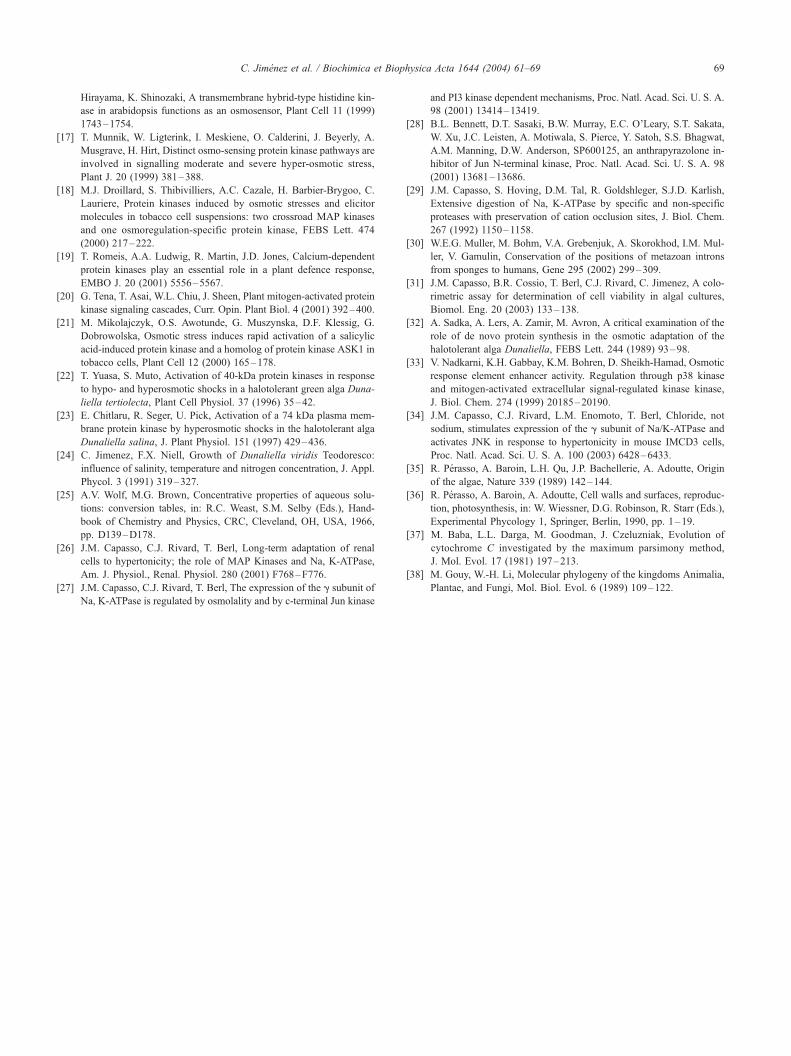
3.4. 32P metabolic labeling of Dunaliella cultures under
osmotic stress
Cell cultures of Dunaliella were metabolically labelled
with 32P, as described in Section 2, to confirm the phos-
phorylation of the p57 protein in response to osmotic stress.
Fig. 4 clearly depicts incorporation of radioactivity into
several protein bands in response to hyperosmotic stress.
The main ones had apparent molecular weight of 54, 51 and
48 kDa, and of special interest for this work is the band at
57 kDa.
Fig. 3. Aliquots of mid log phase Dunaliella cultures were challenged as
described in Section 2 under control and hyperosmotic conditions for
increasing time periods. Basal conditions refer to protein extracts from cells
that were not challenged but only exposed to a media change. (A) Western
blot analysis employed a specific mammalian p38 MAP kinase antibody;
the MeanF S.E. of the 57-kDa band intensity is shown (n= 2). Differences
between control and hyperosmotic shock were significant ( P < 0.02). A
representative blot is shown. (B) A similar experiment was performed
employing the yeast Hog1 antibody. The graph depicts the MeanF S.E. of
the 57-kDa band intensity (n= 4); a representative blot is also shown.
Differences between control and hyperosmotic stress were very significant
( P< 0.004).
Fig. 4. Autoradiography of protein extracts from metabolically labelled 32P
Dunaliella cultures. (A) Control cells, and (B) Cells subjected to
hyperosmotic shock (2! 4 M NaCl) for 4 h. One hundred micrograms
of protein was loaded in each lane. One of three independent experiments is
shown.
C. Jimenez et al. / Biochimica et Biophysica Acta 1644 (2004) 61–69 65
3.5. Effect of changes in tonicity and UV irradiation in
proteins probed with a phosphorylated JNK antibody
We examined whether exposure to non-isotonic condi-
tions altered the phosphorylation of proteins in Dunaliella
when probed with a specific antibody against another
member of the mammalian MAP kinase family, the double
phosphorylated form of JNK. As is depicted in Fig. 5, three
protein bands with apparent molecular weights of 50, 45,
and 43 kDa were evident. This band pattern is strikingly
similar to that obtained with mammalian renal cells [26].
Statistical data analysis (ANOVA) of the phosphorylation of
the 50-kDa band reveals no significant difference (P>0.21)
between the basal level and either the media change control
or hypoosmotically shocked cells. In contrast, exposure to
4M NaCl caused a rapid and transient increase in the
intensity of the 50-kDa protein band that peaked around
30 to 90 min and returned to basal levels after 4h following
osmotic shock. The 45- and 43-kDa bands follow the same
pattern although it was difficult to get reliable quantitative
data from such faint signals. A very similar result was
obtained upon UV irradiation. As shown in Fig. 6, a
transient increase in the level of the 50-kDa phosphoprotein
was detected. There were also some differences in the
kinetics of phosphorylation between osmotic and UV irra-
diation stresses (Fig. 5 vs. Fig. 6). Under UV stress, the 50-
kDa phosphoprotein peaked at 15 min and returned to basal
levels after 1 to 2 h. It is noteworthy that a basal level of
phosphorylated 50-kDa JNK-like protein was determined in
Dunaliella (Figs. 5 and 6). In contrast, phosphorylation of
the 57-kDa p38/Hog1-like protein was detected only under
stress.
3.6. Effect of inhibitors of p38 MAP kinase and JNK
activities on the adaptation of Dunaliella to osmotic shock
Specific inhibitors of p38 and JNK activities were
employed to demonstrate whether these MAP kinase-like
Fig. 5. Aliquots of mid log phase Dunaliella cultures were challenged as described in Section 2 with three different NaCl treatments (control, hyperosmotic and
hypoosmotic conditions) for increasing time periods. Cell protein extracts were analyzed by Western blot using an antibody raised against the phosphorylated
form of mammalian JNK. The graph depicts the MeanF S.E. of the 50-kDa band intensity from three independent experiments; a representative blot is also
shown. Basal refers to cells that were not manipulated. Differences in control and hyperosmotic stress conditions were significant between 30 and 90 min
( P< 0.02).
Fig. 6. Dunaliella cells were irradiated with UV light (70 mJ cm� 2 at 200–
400 nm), and protein extracts were obtained at increasing time periods after
shock and analyzed by Western blot using an antibody specific for
phosphorylated JNK as described in Section 2. The graph shows the band
intensity of the 50-kDa phosphoprotein (MeanF S.E., n= 3); a typical blot
is also shown. Differences in band intensity between 0 and 15 min were
very significant ( P< 0.01).
C. Jimenez et al. / Biochimica et Biophysica Acta 1644 (2004) 61–6966
signaling pathways are present and physiologically re-
levant for adaptation of Dunaliella cells. For this purpose
hyperosmotic shock experiments were performed using
the specific p38 MAP kinase inhibitor SB203580 and
JNK pathway inhibitor SP600125. Cell cultures were
incubated for 2 h with 20 AM SB203580 (standard
concentration for mammalian cell experiments [27]) prior
to a 4 M NaCl shock. The change in osmolality pro-
duces a well-known and profound decrease in the
physiological function of the cells with recovery occur-
ring over the ensuing 48 h [32]. However, this recovery
was markedly impaired in cells exposed to the p38 MAP
kinase inhibitor (Fig. 7A). This effect was neither a
consequence of the solvent (dimethyl sulfoxide) (Fig.
7A) nor a nonspecific toxic effect of SB203580 per se,
since it had no effect on cells maintained at 2 M NaCl
(Fig. 7B). As shown in Fig. 7A, a more profound effect
was noted when Dunaliella cells were challenged with
hyperosmotic stress after preincubation for 2 h with 1
AM of the JNK pathway inhibitor SP600125. The
recovery of algal cells after hyperosmotic shock was
completely abolished by the JNK inhibitor, although
Fig. 7. Cell cultures were preincubated for 2 h with MAP kinase
inhibitors or with the same volume of solvent (DMSO) before being
subjected to (A) hyperosmotic stress (4 M NaCl) or (B) allowed to
remain at 2M NaCl. Culture samples were removed at increasing times to
assess cell viability by the CellTiter 96 assay as described in Section 2.
(A) Depicts the effect of the p38 MAP kinase inhibitor SB203580 (20
AM) on cell viability after osmotic shock. Data points are the
MeanF S.E. of five independent experiments. Differences between
control and inhibitor treatment were significant at 30 h ( P < 0.014) and
very significant at 48 h ( P< 0.001). Preincubation with the JNK inhibitor,
SP600125, demonstrated more profound effects. The carrier DMSO alone
had no effect on Dunaliella response to hypertonic conditions. (B) shows
the effect of the specific MAP kinase inhibitors on non-stressed cell
cultures.
C. Jimenez et al. / Biochimica et Biophysica Acta 1644 (2004) 61–69 67
the presence of inhibitor did not adversely affect control
cells (Fig. 7B).
4. Discussion
Recent studies indicate that MAP kinase signaling cas-
cades which have been extensively described in organisms
from yeast to mammals [1] are also present in vascular
plants such as Arabidopsis, tobacco, and alfalfa [20].
However, while three MAPK subfamilies (ERK, JNK, and
p38 MAP kinase) are present in animal cells, plant kinase
genes appear to belong solely to the ERK subfamily [20].
The presence of MAP kinase pathways in aquatic cellular
plants such as green algae (from which higher plants
evolved in the transition to terrestrial life) has not been
previously described. The use of Dunaliella as a model to
study signaling systems in unicellular algae is based on the
lack of a cell wall and its ability to adapt to environmental
stresses. The lack of a cell wall allows for the use of
common protocols used for mammalian cells to study
genetic and protein systems. This study reveals that the
microalgae Dunaliella possess a 57-kDa protein that is
induced in response to increasing tonicity, but not by
hypotonic stress. This 57-kDa protein was detected by
cross-reactivity with mammalian p38 MAP kinase antibody
and even more robustly with the antibody against the yeast
homologue, Hog1, an osmo-sensitive kinase. Furthermore,
exposure of algal cells to hypertonicity is rapidly followed
by phosphorylation of the protein, which is detected by a
highly specific antibody raised against the phospho p38
MAP kinase motif (T*GY*). This up-regulation and phos-
phorylation of the 57-kDa protein is not a specific NaCl
effect but rather an osmotic effect, since equivalent
increases in osmolality using glycerol had an almost iden-
tical effect. In addition, this protein is also up-regulated and
phosphorylated by non-osmotic stresses such as UV irradi-
ation and thermal shock. It is also of interest that the
phosphoprotein was not detected under basal conditions
(i.e., 2 M NaCl). Confirmation of the presence of the p57
kDa phosphoprotein was validated through metabolic label-
ing experiments, in which clear phosphorylation of a 57-
kDa protein band appeared in response to hyperosmotic
stress.
We also explored the possibility that another member of
the MAPK family, the c-Jun N-terminal kinases (JNKs), was
activated in Dunaliella by the same stresses that activate
them in mammalian cells. In fact, we were able to detect a
pattern of three protein bands similar to that found in
IMCD3 (inner medullary collecting duct) cells using a
highly specific antibody raised against the phospho JNK
motif (T*PY*). These bands, as occurs in mammalian cells,
were present at basal conditions in the algae, and their
intensity was also greatly and transiently increased under
hyperosmotic shock and UV irradiation.
The most compelling evidence for the presence and the
physiological role of p38-like and JNK-like pathways in
Dunaliella is provided by using highly specific inhibitors of
the p38 and JNK signaling pathways at standard concen-
trations. The relevance of the activation of these pathways in
the adaptation and survival of mammalian cells in response
to stress has been well established. For instance, in MDCK
osmotically stressed cells, inhibition of the p38 MAP kinase
with SB203580 was associated with loss of induction of
aldose reductase, an enzyme that generates sorbitol, which
in turn is an osmolyte involved in hypertonic adaptation
[33]. Also, inhibition of the JNK pathway with SP600125
C. Jimenez et al. / Biochimica et Biophysica Acta 1644 (2004) 61–6968
results in a significant decrease in cell survival after hyper-
osmotic shock in IMCD3 cells [34]. To date, despite the
increasing evidence of protein phosphorylation in response
to a great variety of stresses in plants, no indisputable direct
evidence for its physiological role has been provided. The
present work gives strong evidence for the relationship
between activation of the signaling pathways and cell
adaptation and survival upon hyperosmotic stress. We
propose that the p38-like kinase signaling cascade is in-
volved in the late response to environmental stress in
Dunaliella as protein synthesis is involved. It is well known
that the early response (glycerol synthesis) is independent of
protein synthesis. This de novo protein synthesis of the
signaling kinase is dissimilar to that in mammalian cells
[26]. However, Dunaliella’s decrease in adaptability and,
eventually, in cell survival in the presence of the specific
inhibitors of the p38 and JNK signaling pathways strongly
suggests the existence of signaling mechanisms in these
unicellular microalgae similar to those found in mammalian
cells.
Cloning of some of the components of these putative
signaling pathways will have very interesting evolutionary
implications. The fact that we have found p38-like and c-
Jun-like MAP kinases in an unicellular green microalga
suggests that these pathways were already present in the
common ancestor of the three main eukaryotic lines (i.e.
green plants, including both the green algae and the higher
plants, eukaryotic fungi of the phylum Ascomycota which
includes yeasts and molds, and the metazoan animals).
According to these results, we hypothesize that higher plants
are expected to have p38-like and c-Jun-like MAP kinases.
At present, none of them have yet been described. On the
other hand, the fact that no c-Jun-like MAP kinase has been
found in yeast, even though at least 12 complete yeast
genomes are known, would indicate an evolutionary loss of
this signaling pathway. Analysis of the 18S and 28S rRNA
nucleotide sequences shows that the ancestors of all the
main eukaryotic evolutionary lines were probably unicellu-
lar flagellates, represented in the fossil record by acritarchs
during Neoproterozoic times, some 1400 million years ago
[35,36]. This close phylogenetic relationship between the
green algae and the higher animals has also been confirmed
by comparisons of several gene and amino acid sequences
[37,38].
In summary, these data identify, for the first time, the
presence of p38- and c-Jun-like MAP kinase proteins in
algae and their phosphorylation in response to increments in
osmolality and other stress conditions. Also, these data
reveal that specific chemical agents that inhibit the activity
of these enzymes have markedly deleterious effects on the
ability of the algae to overcome hyperosmotic stress, there-
by revealing their critical role in adaptation and survival of
Dunaliella under such environmental conditions. Finally,
the data suggest that the origin of these MAP kinase
signaling pathways predates the divergence of the plant
and the fungi/animal lineages.
Acknowledgements
We thank Zafie Craft for excellent secretarial assistance.
This work was supported by a grant from the National
Institute of Health, DK-19928 to TB. C.J. was supported by
Research Project REN2002-00340/MAR of the Spanish
Ministry of Science and Technology, and by a Research
Grant from the University of Malaga. We thank Professors
Lynn Heasley and Uri Pick for critical reading of the
manuscript.
References
[1] C. Widmann, S. Gibson, M.B. Jarpe, G.L. Johnson, Mitogen-acti-
vated protein kinase: conservation of a three-kinase module from
yeast to human, Physiol. Rev. 79 (1999) 143–180.
[2] K. Zwerger, H. Hirt, Recent advances in plant MAP kinase signalling,
Biol. Chem. 382 (2001) 1123–1131.
[3] T. Romeis, Protein kinases in the plant defence response, Curr. Opin.
Plant Biol. 4 (2001) 407–414.
[4] C. Jonak, W. Ligterink, H. Hirt, MAP kinases in plant signal trans-
duction, Cell. Mol. Life Sci. 55 (1999) 203–213.
[5] S. Usami, H. Banno, Y. Ito, R. Nishihama, Y. Machida, Cutting
activates a 46-kilodalton protein kinase in plants, Proc. Natl. Acad.
Sci. U. S. A. 92 (1995) 8660–8664.
[6] S. Seo, M. Okamoto, H. Seto, K. Ishizuka, H. Sano, Y. Ohashi,
Tobacco MAP kinase: a possible mediator in wound signal transduc-
tion pathways, Science 270 (1995) 1988–1992.
[7] A. Adam, S. Pike, E. Hoyos, J.M. Stone, J.C. Walker, A. Novacky,
Rapid and transient activation of a myelin basic protein kinase in
Tobacco leaves treated with harpin from Erwinia amylovora, Plant
Physiol. 115 (1997) 853–861.
[8] A. Lebrun-Garcıa, F. Ouaked, A. Chiltz, A. Pugin, Activation of
MAPK homologues by elicitors in Tobacco cells, Plant J. 15 (1998)
773–781.
[9] J.W. Stratmann, C.A. Ryan, Myelin basic protein kinase activity in
tomato leaves is induced systemically by wounding and increases in
response to systemin and oligosaccharide elicitors, Proc. Natl. Acad.
Sci. U. S. A. 94 (1997) 11085–11089.
[10] C. Jonak, S. Kiegerl,W. Ligterink, P.J. Barker, N.S. Huskisson, H. Hirt,
Stress signaling in plants: a mitogen-activated protein kinase pathway
is activated by cold and drought, Proc. Natl. Acad. Sci. U. S. A. 93
(1996) 11274–11279.
[11] T. Mizoguchi, K. Irie, T. Hirayama, N. Hayashida, K. Yamaguchi-
Shinozaki, K. Matsumoto, K. Shinozaki, A gene encoding a mitogen-
activated protein kinase kinase kinase is induced simultaneously with
genes for a mitogen-activated protein kinase and an S6 ribosomal
protein kinase by touch, cold, and water stress in Arabidopsis thali-
ana, Proc. Natl. Acad. Sci. U. S. A. 93 (1996) 765–769.
[12] T.S. Nuhse, S.C. Peck, H. Hirt, T. Boller, Microbial elicitors induce
activation and dual phosphorylation of the Arabidopsis thaliana
MAPK 6, J. Biol. Chem. 275 (2000) 7521–7526.
[13] R. Desikan, J.T. Hancock, K. Ichimura, K. Shinozaki, S.J. Neill,
Harpin induces activation of the Arabidopsis mitogen-activated pro-
tein kinases AtMPK4 and AtMPK6, Plant Physiol. 126 (2001)
1579–1587.
[14] Y. Kovtun, W.L. Chiu, G. Tena, J. Sheen, Functional analysis of
oxidative stress-activated mitogen-activated protein kinase cascade
in plants, Proc. Natl. Acad. Sci. U. S. A. 97 (2000) 2940–2945.
[15] F. Posas, H. Saito, Osmotic activation of the HOG MAPK pathway
via Ste11p MAPKKK: scaffold role of Pbs2p MAPKK, Science 276
(1997) 1702–1705.
[16] T. Urao, B. Yabukov, R. Satoh, K. Yamaguchi-Shinozaki, M. Seki, T.
C. Jimenez et al. / Biochimica et Biophysica Acta 1644 (2004) 61–69 69
Hirayama, K. Shinozaki, A transmembrane hybrid-type histidine kin-
ase in arabidopsis functions as an osmosensor, Plant Cell 11 (1999)
1743–1754.
[17] T. Munnik, W. Ligterink, I. Meskiene, O. Calderini, J. Beyerly, A.
Musgrave, H. Hirt, Distinct osmo-sensing protein kinase pathways are
involved in signalling moderate and severe hyper-osmotic stress,
Plant J. 20 (1999) 381–388.
[18] M.J. Droillard, S. Thibivilliers, A.C. Cazale, H. Barbier-Brygoo, C.
Lauriere, Protein kinases induced by osmotic stresses and elicitor
molecules in tobacco cell suspensions: two crossroad MAP kinases
and one osmoregulation-specific protein kinase, FEBS Lett. 474
(2000) 217–222.
[19] T. Romeis, A.A. Ludwig, R. Martin, J.D. Jones, Calcium-dependent
protein kinases play an essential role in a plant defence response,
EMBO J. 20 (2001) 5556–5567.
[20] G. Tena, T. Asai, W.L. Chiu, J. Sheen, Plant mitogen-activated protein