Photocurrent Generation from Porphyrin/Fullerene Complexes Assembled
in a Tethered Lipid Bilayer
Wei Zhan,*,† Kai Jiang,† Matthew D. Smith,§ Heidi E. Bostic,§ Michael D. Best,*,§
Maria L. Auad,‡ Joshua V. Ruppel,^ Chungsik Kim,^ and X. Peter Zhang^
†Department of Chemistry and Biochemistry, and ‡Department of Polymer and Fiber Engineering, AuburnUniversity, Auburn, Alabama 36849, §Department of Chemistry, The University of Tennessee, Knoxville,Tennessee 37996, and ^Department of Chemistry, The University of South Florida, Tampa, Florida 33620
Received July 20, 2010. Revised Manuscript Received August 30, 2010
A modular photocurrent generation system, based on amphiphilic porphyrin and fullerene species assembled in atethered lipid bilayer matrix, is reported here. The key findings are (1) the amount of photoactive species can bequantitatively controlled in each leaflet of the bilayer and (2) the sequential formation of the bilayer allows a directionalorganization of these agents on electrodes. Photocurrent generation from seven differently configured photoactivebilayers is studied, which reveals several critical factors in achieving efficient photoinduced electron transfer across lipidmembranes. Detailed fluorescence characterization is performed on porphyrin samples either in liposomes or surface-tethered bilayers; and the observed fluorescence quenching is correlated with photocurrents generated from theelectrode-immobilized lipid films. The potential usefulness of this lipid-based approach is discussed in connection toseveral existing molecular photovoltaic systems.
Introduction
Wewish to report a newmodular photocurrent generation systemthat is based on amphiphilic porphyrin and fullerene speciesassembled in a tethered lipid bilayer matrix. The interesting newfindings are that the amount of photoactive species can be quantita-tively controlled in each leaflet of the bilayer, and a directional orga-nizationof these agents on electrodes affordedby the tetheredbilayercan significantlymodify the vectorial photoinduced electron transferand thus the obtained photocurrents. This approach introduces aninteresting alternative method of organizing multiple photoactivespecies on electrodes formolecular photovoltaic studies,1-4 of whichmost preexisting systems rely on organic synthesis to achieve mole-cular organization and surface immobilization.
Photoactive complexes based on porphyrins (P) and fullerenes(F) have been extensively used in buildingmolecular photovoltaicsystems owing to efficient electron transfer from porphyrin tofullerene upon photoexcitation. While ground-state porphyrinsabsorb light very strongly (i.e., ε of∼105M-1 cm-1) at∼400 nm,their excited-state counterparts are normally long-lived and canoften exchange electrons readily with redox species in the surro-undings.5,6 More important, the redox potential of excited-state
porphyrins generally matches well with that of fullerenes, a classof species with strong electron-accepting capability.7,8 Time-resolved spectroscopic measurements9,10 of covalently linkedP-F dyads, for example, have recorded photoinduced electrontransfer (ET) rate constants on the order of 107-1010 s-1. In thepresence of additional electron-donating group, such as ferro-cene10-12 and carotene,9,13 this efficient ET step can be furtherstabilized, yielding prolonged charge-separation (CS) states withlifetimes in the microsecond to millisecond range. When theseP-F-based dyads/triads are further affixed on electrodes for pho-tocurrent generation, a general correlation of the CS lifetime vsconversion efficiency has been observed.High photon-to-electronconversion efficiencies,3,10,14,15 for example, ∼15-20%, can be
*To whom correspondence should be addressed. E-mail: [email protected] (W.Z.); [email protected] (M.D.B.). Tel.: 334-844-6984 (W.Z.);865-974-8658 (M.D.B.). Fax.: 334-844-6959 (W.Z.).(1) Imahori, H.; Sakata, Y. Donor-Linked Fullerenes: Photoinduced Electron
Transfer and Its Potential Application. Adv. Mater. 1997, 9, 537–546.(2) Imahori, H.; Mori, Y.; Matano, Y. Nanostructured Artificial Photosynth-
esis. J. Photochem. Photobiol. C 2003, 4, 51–83.(3) Cho, Y. J.; Ahn, T. K.; Song, H.; Kim, K. S.; Lee, C. Y.; Seo, W. S.; Lee, K.;
Kim, S. K.; Kim, D.; Park, J. T. Unusually High Performance Photovoltaic CellBased on a [60]Fullerene Metal Cluster-Porphyrin Dyad SAM on an ITOElectrode. J. Am. Chem. Soc. 2005, 127, 2380–2381.(4) Matsuo, Y.; Kanaizuka, K.; Matsuo, K.; Zhong, Y.-W.; Nakae, T.;
Nakamura, E. Photocurrent-Generating Properties of Organometallic FullereneMolecules on an Electrode. J. Am. Chem. Soc. 2008, 130, 5016–5017.(5) Kalyanasundaram, K. Photochemistry of Polypyridine and Porphyrin Com-
plexes; Academic Press Ltd.; London, 1992.(6) D’Souza, F.; Ito, O. Photoinduced Electron Transfer in Supramolecular
Systems of Fullerenes Functionalized with Ligands Capable of Binding to ZincPorphyrins and Zinc Phthalocyanines. Coord. Chem. Rev. 2005, 249, 1410–1422.
(7) Guldi, D. M.; Kamat, P. V. In Fullerenes: Chemistry, Physics, and Technol-ogy; Kadish, K. M., Ruoff, R. S. (Eds.); John Wiley & Sons, Inc.: New York, 2000; pp225-281.
(8) Guldi, D. M.; Prato, M. Excited-State Properties of C60 Fullerene Deriva-tives. Acc. Chem. Res. 2000, 33, 695–703.
(9) Kuciauskas, D.; Liddell, P. A.; Lin, S.; Stone, S. G.; Moore, A. L.; Moore,T. A.; Gust, D. Photoinduced Electron Transfer in Carotenoporphyrin-FullereneTriads: Temperature and Solvent Effects. J. Phys. Chem. B 2000, 104, 4307–4321.
(10) Imahori, H.; Tamaki, K.; Guldi, D. M.; Luo, G.; Fujitsuka, M.; Ito, O.;Sakata, Y.; Fukuzumi, S.Modulating Charge Separation and Charge Recombina-tion Dynamics in Porphyrin-Fullerene Linked Dyads and Triads: Marcus-Normalversus Inverted Region. J. Am. Chem. Soc. 2001, 123, 2607–2617.
(11) Luo, C.; Guldi, D. M.; Imahori, H.; Tamaki, K.; Sakata, Y. SequentialEnergy and Electron Transfer in an Artificial Reaction Center: Formation of aLong-Lived Charge-Separated State. J. Am. Chem. Soc. 2000, 122, 6535–6551.
(12) Imahori, H.; Guldi, D. M.; Tamaki, K.; Yoshida, Y.; Luo, C.; Sakata, Y.;Fukuzumi, S. Charge Separation in a Novel Artificial Photosynthetic ReactionCenter Lives 380 ms. J. Am. Chem. Soc. 2001, 123, 6617–6528.
(13) Liddell, P. A.; Kuciauskas, D.; Sumida, J. P.; Nash, B.; Nguyen, D.;Moore,A. L.; Moore, T. A.; Gust, D. Photoinduced Charge Separation and ChargeRecombination to a Triplet State in a Carotene-Porphyrin-Fullerene Triad. J. Am.Chem. Soc. 1997, 119, 1400–1405.
(14) Imahori, H.; Yamada, H.; Nishimura, Y.; Yamazaki, I.; Sakata, Y.Vectorial Multistep Electron Transfer at the Gold Electrodes Modified withSelf-Assembled Monolayers of Ferrocene-Porphyrin-Fullerene Triads. J. Phys.Chem. B 2000, 104, 2099–2108.
(15) Imahori, H.; Norieda, H.; Yamada, H.; Nishimura, Y.; Yamazaki, I.;Sakata, Y.; Fukuzumi, S. Light-Harvesting and Photocurrent Generation byGoldElectrodes Modified with Mixed Self-Assembled Monolayers of Boron-Dipyrrinand Ferrocene-Porphyrin-Fullerene Triad. J. Am. Chem. Soc. 2001, 123, 100–110.
realized, once the molecular orientation, ET directionality, andenergetics in such systems are optimized.
On the other hand, there has been a long-term research interestin mimicking natural photosynthetic processes with syntheticconstructs.16-19 Through these simplified but well controlledanalogs, researchers are able to bypass the intrinsic complexityof natural photosynthesis and examine directly the molecularorganization and energetics essential for photoconversion. Usinglipid vesicles and black lipid membranes as experimental mod-els,20,21 people have studied photoelectrochemical behaviors of ahost of photoactive species including porphyrins and fullerenes ina lipid-bilayer environment. These studies have revealed severalcritical characteristics of lipid bilayers as the host of photosyn-thetic complexes, in particular, in scaffolding and compartmen-talization of different agents within close distances, facilitatingdirectional ET and suppressing charge recombination.
We have been interested in building lipid-based photoconver-sion systems using electrode-supported lipid bilayers.22,23 Whilemost previous lipid-based artificial photosynthetic systems adoptto follow the involved photoelectrochemical events spectrosco-pically,24-26 our effort is focused on organizing/immobilizingphotoactive species directly on electrodes for a terminal conver-sion of light to photocurrents. Findings from these studies mayeventually put us at a better position evaluating the potentialusefulness of lipid-based materials in organic solar devices.Recently,we have shown that single-component photoconversionsystems, based on either fullerene22 or ruthenium tris(bipyridyl)complexes,23 can be constructed on lipid-bilayer based structures.Stable photocurrents have been obtained, manifesting that theselipid-fixed photoagents can reliably communicate with the sacri-ficial electron donor species in solution, as well as with electrodeson which they are immobilized. The results reported here repre-sent our first step toward lipid-based, multicomponent photo-conversion systems. Specifically, we show that a series of P/Fassembled bilayer structures can be configured on electrodes viatethered lipid bilayers and the corresponding photocurrentsgenerated from these structures.
Results
System Design and Fabrication. Scheme 1 depicts the orga-nization, as well as materials, used in building the photoconverting
assemblies. To prepare the tethered lipid bilayer (TLB),27,28 athiolated lipid, 1,2-dipalmitoyl-sn-glycero-3-phosphothioethanol(DPPT, 3), was first used to form a self-assembled lipid monolayeron a gold surface, which was then covered by a lipid monolayer of1-palmitoyl-2-oleoyl-sn-glycero-3-phosphocholine (POPC, 1) vialiposome fusion.27 The two photoactive components employed,the zinc porphyrin conjugated with 1,2-dioleoyl-sn-glycero-3-phos-phoethanolamine, ZnP-DOPE (2), and malonic fullerene (C63, 4)were sequentially assembled into the architecture during the forma-tion of the lipid bilayer (see below).
The formation of gold-tethered DPPT/POPC bilayers wascharacterized by cyclic voltammetry and atomic force micro-scopy. Figure 1 shows the electrochemical reduction/oxidation ofa probe species, ferricyanide, by gold substrates with/withoutdeposited lipid layers.Aquasi-reversible electrochemical responsewas obtained from the naked gold electrode, whereas the presenceof a DPPT layer largely blocked the probe molecules from ex-changing electrons with the gold surface underneath. When theelectrode was further covered with a POPC top layer, its directcommunication with ferricyanide was nearly completely shut offin the probed potential window. These distinctive voltammetricfeatures indicate controlled deposition of thiols and lipids onelectrodes and are in good agreementwith previous studies29,30 onlipid based bilayers formed under similar conditions.
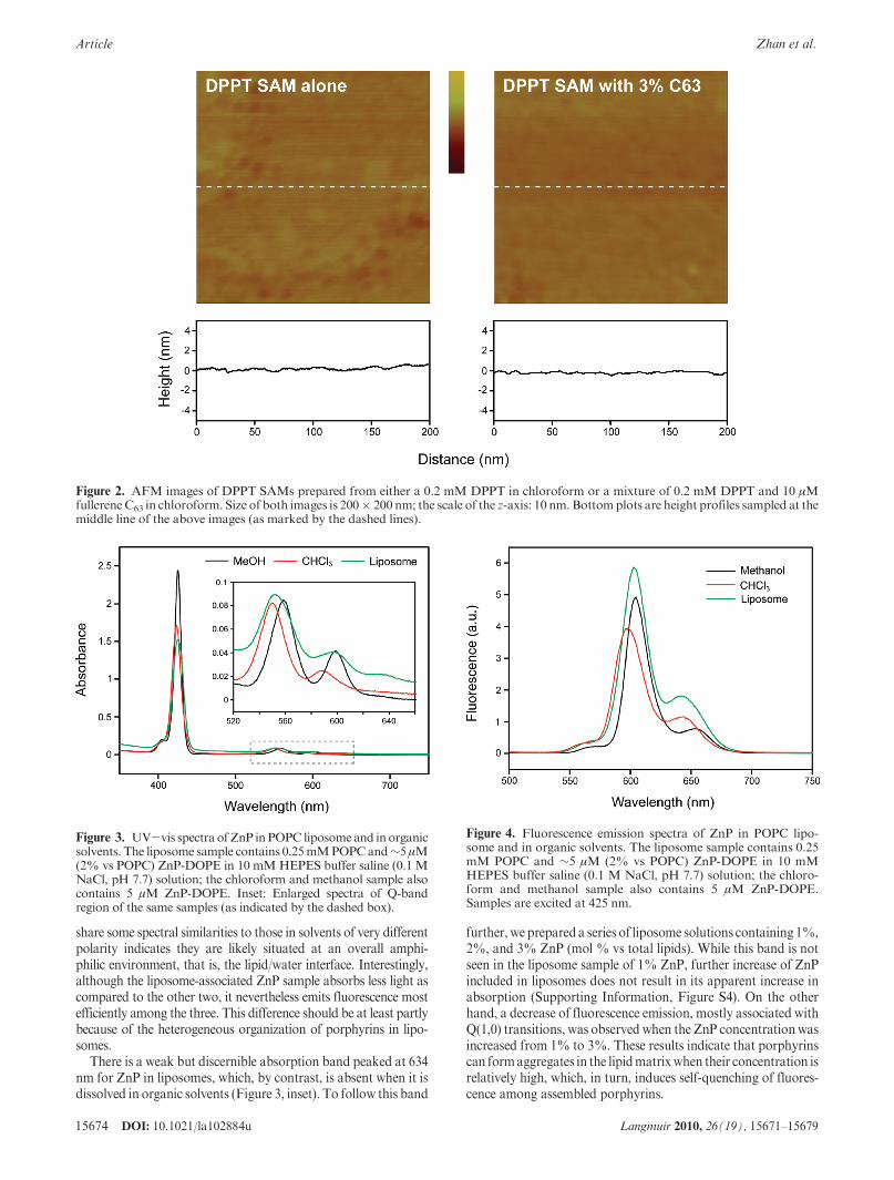
In search of an assembly-based method for fullerene surfaceimmobilization, we have identified a thiolated lipid, DPPT, as asuitable candidate. We chose a lipid-based thiol over conventionalalkanethiols for the consideration that the diacyl chain of lipidsallows formation of relatively loosely packed SAMs,31 which inturn should provide more room for inclusion of fullerenes withinthe structure. As shown in Figure 2, the SAM formed from amixture of DPPT and 3% fullerene C63 (mol % vs that of DPPT)displays a surface smoothness comparable to that formedbyDPPTalone. This result indicates that thus deposited fullerenes, at least atthe low dosages used here, aremost likely well dispersed in the lipidmatrix, rather than forming aggregates on the SAM surface.
On the other hand, the low fullerene coverage on an opaquesubstrate prevented us fromdirectly quantifying the exact amountof deposited material with conventional techniques based onelectrochemical and absorption spectroscopic methods. To cir-cumvent this limitation, we opted for an indirect method bycomparing photocurrents generated from these fullerene-contain-ing monolayers. The assumption behind this experiment is thatthe obtained photocurrent is proportional to the number offullerenes included in a monolayer, which is expected to be thecase for low-coverage,monolayer-based photocurrent-generatingsystems.21,23 The photocurrent measured from DPPT SAMscontaining 1, 2, and 3% fullerenes is 3.5, 7 ( 1 (from 3 parallelmeasurements), and 17 nA, respectively. These results imply thatthis assembly based method does not immobilize fullerenes ongold precisely proportional to its mixing ratio in the lipidprecursor. This method, however, does enable us to reproduciblyprepare electrodes comprising a fixed amount of fullerenes, which
(16) Calvin, M. Simulating Photosynthetic Quantum Conversion. Acc. Chem.Res. 1978, 10, 369–374.(17) Gust, D.; Moore, T. A.; Moore, A. L. Molecular Mimicry of Photosyn-
thetic Energy and Electron Transfer. Acc. Chem. Res. 1993, 26, 198–205.(18) Gust, D.; Moore, T. A.; Moore, A. L. Mimicking Photosynthetic Solar
Energy Transduction. Acc. Chem. Res. 2001, 34, 40–48.(19) Fukuzumi, S. Development of Bioinspired Artificial Photosynthetic Sys-
tems. Phys. Chem. Chem. Phys. 2008, 10, 2283–2297.(20) Fendler, J. H.Membrane Mimetic Chemistry; John Wiley & Sons, Inc.: New
York, 1982; Chapter 13.(21) Lymar, S. V.; Parmon, V. N.; Zamaraev, K. I. Photoinduced Electron
Transfer across Membranes. Top. Curr. Chem. 1991, 159, 1–65.(22) Zhan,W.; Jiang, K. AModular Photocurrent Generation System Based on
Phospholipid-Assembled Fullerenes. Langmuir 2008, 24, 13258–13261.(23) Jiang, K.; Xie, H.; Zhan, W. Photocurrent Generation from Ru(bpy)3
2þ
Immobilized on Phospholipid/Alkanethiol Hybrid Bilayers. Langmuir 2009, 25,11129–11136.(24) Ford, W. E.; Otvos, J. W.; Calvin, M. Photosensitized Electron Transport
across Lipid Vesicle Walls - Quantum Yield Dependence on Sensitizer Concentra-tion. Proc. Natl. Acad. Sci. U.S.A. 1979, 76, 3590–3593.(25) Armitage, B.; O’Brien, D. F. Vectorial Photoinduced Electron Transfer
between Phospholipid Membrane-Bound Donors and Acceptors. J. Am. Chem.Soc. 1992, 114, 7396–7403.(26) Hammarstrom, L.; Norrby, T.; Stenhagen, G.; Martensson, J.; Akermark,
B.; Almgren, M. Two-Dimensional Emission Quenching and Charge SeparationUsing a Ru(II)-Photosensitizer Assembled with Membrane-Bound Acceptors. J.Phys. Chem. B 1997, 101, 7494–7504.(27) Sinner, E.-K.; Knoll, W. Functional Tethered Membranes. Curr. Opin.
Chem. Biol. 2001, 5, 705–711.
(28) Dorvel, B. R.; Keizer, H. M.; Fine, D.; Vuorinen, J.; Dodabalapur, A.;Duran, R. S. Formation of Tethered Bilayer Lipid Membranes on Gold Surfaces:QCM-Z and AFM Study. Langmuir 2007, 23, 7344–7355.
(29) Plant, A. L. Self-Assembled Phospholipid/Alkanethiol Biomimetic Bilayerson Gold. Langmuir 1993, 9, 2764–2767.
(30) Twardowski, M.; Nuzzo, R. G. Molecular Recognition at Model OrganicInterfaces: Electrochemical Discrimination Using Self-Assembled Monolayers(SAMs) Modified via the Fusion of Phospholipid Vesicles. Langmuir 2003, 19,9781–9791.
(31) Kunze, J.; Leitch, J.; Schwan, A. L.; Faragher, R. J.; Naumann, R.; Schiller,S.; Knoll, W.; Dutcher, J. R.; Lipkowski, J. New Method to Measure PackingDensities of Self-Assembled Thiolipid Monolayers. Langmuir 2006, 22, 5509–5519.
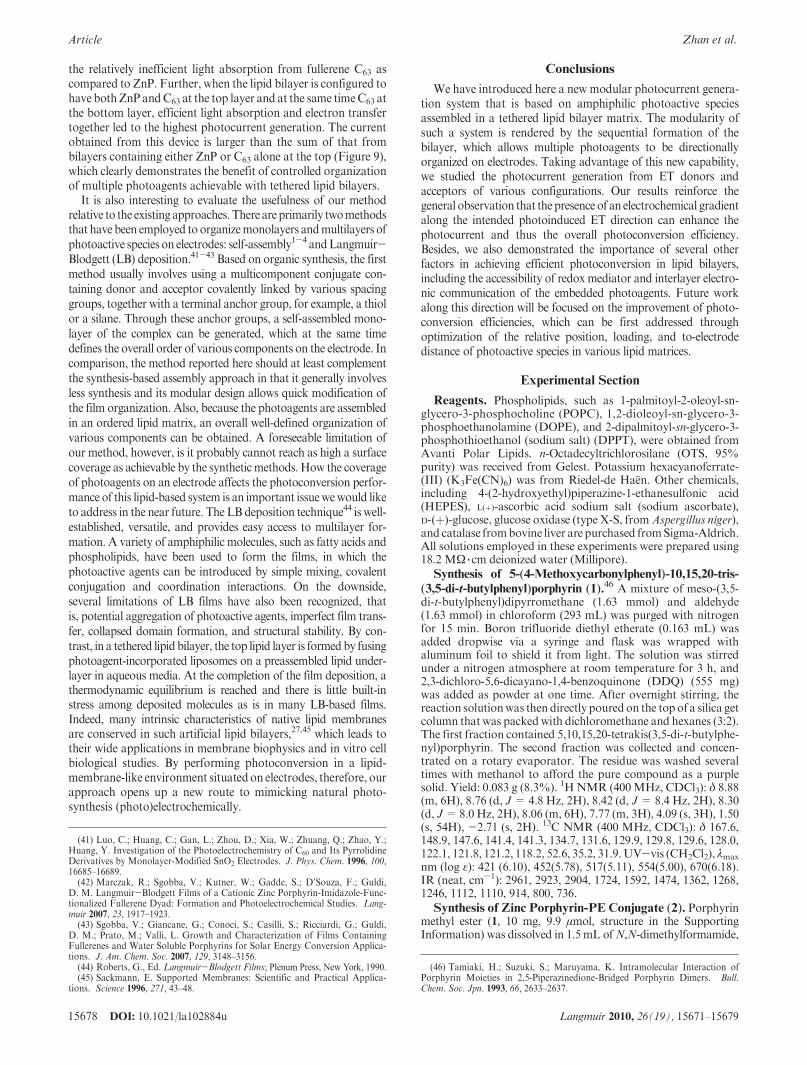
is sufficient for making quantitative comparisons from device todevice and for devices with different configurations (see below).Spectroscopic Characterization of the System. UV-vis
absorption spectroscopy of the ZnP-DOPE-incorporated lipo-some solution reveals a Soret band at 425 nm, together with threebroader and less intenseQbands at 552, 596, and634nm(Figure 3).In comparison, the corresponding bands obtained from ZnPcomplexes dissolved in organic solvents are considerably shifted:423, 550, and 589 nm in chloroform and 425, 558, and 598 nm inmethanol. These spectral shifts, as well as the difference in absor-bance, suggest the ZnP moieties reside in a distinctive microenvir-onment of different local dielectrics and polarity in liposomes ascompared to chloroform (ε=4.8) andmethanol (ε=33). This ob-servation is further revealed by the fluorescence emission spectra ofthe same samples. As shown in Figure 4, two emission bands at 604and 643 nm with an intensity ratio of 3.2:1 was obtained for ZnP-DOPE in liposome, which can be assigned to Q(1,0) and Q(0,0)transitions,32 respectively.ForZnP-DOPEdissolved in chloroform,the corresponding bands are located at 598 and 644nm,with a simi-lar intensity ratio (3.5:1) but lower intensity; in methanol, the twobands appear at 605 and 655 nmwith an fluorescence intensity ratioof 6.5:1. Together, the fact that liposome-associated ZnPmolecules
Scheme 1. Cartoon Depiction of the Multi-Component Photocurrent Generation System Based on Fullerene/Porphyrin Assembled
in a Tethered Lipid Bilayera
a Structures of species employed in forming the assemblies (from 1 to 4): 1-palmitoyl-2-oleoyl-sn-glycero-3-phosphocholine (POPC), zinc por-phyrin conjugated to 1,2-dioleoyl-sn-glycero-3-phosphoethanolamine (ZnP-DOPE), 1,2-dipalmitoyl-sn-glycero-3-phosphothioethanol (DPPT),and monomalonic fullerene (C63). This illustrative schematic does not imply the actual organization or stoichiometry of involved species in thebilayers.
Figure 1. Formation of tethered lipid bilayers on gold sub-strates as characterized by cyclic voltammetry. The probedsolution contains 1.0 mM Fe(CN)6
3- in 1 M KCl in DI waterand the gold substrates are covered with a DPPT SAM or aDPPT/POPC bilayer. Three-electrode setup with a Ag/AgClreference electrode and a Pt counter electrode was used. Scanrate: 100 mV/s.
(32) Gouterman, M. In The Porphyrins, Vol. III.; Dolphin, D., Ed.; AcademicPress: New York, 1978; pp 1-165.
share some spectral similarities to those in solvents of very differentpolarity indicates they are likely situated at an overall amphi-philic environment, that is, the lipid/water interface. Interestingly,although the liposome-associated ZnP sample absorbs less light ascompared to the other two, it nevertheless emits fluorescence mostefficiently among the three. This difference should be at least partlybecause of the heterogeneous organization of porphyrins in lipo-somes.
There is a weak but discernible absorption band peaked at 634nm for ZnP in liposomes, which, by contrast, is absent when it isdissolved in organic solvents (Figure 3, inset). To follow this band
further,we prepareda series of liposome solutions containing 1%,2%, and 3% ZnP (mol % vs total lipids). While this band is notseen in the liposome sample of 1% ZnP, further increase of ZnPincluded in liposomes does not result in its apparent increase inabsorption (Supporting Information, Figure S4). On the otherhand, a decrease of fluorescence emission, mostly associated withQ(1,0) transitions, was observed when the ZnP concentrationwasincreased from 1% to 3%. These results indicate that porphyrinscan formaggregates in the lipidmatrixwhen their concentration isrelatively high, which, in turn, induces self-quenching of fluores-cence among assembled porphyrins.
Figure 2. AFM images of DPPT SAMs prepared from either a 0.2 mM DPPT in chloroform or a mixture of 0.2 mM DPPT and 10 μMfullereneC63 in chloroform. Size of both images is 200� 200 nm; the scale of the z-axis: 10 nm. Bottomplots are height profiles sampled at themiddle line of the above images (as marked by the dashed lines).
Figure 3. UV-vis spectra ofZnP inPOPC liposome and inorganicsolvents. The liposome sample contains 0.25mMPOPCand∼5μM(2% vs POPC) ZnP-DOPE in 10 mMHEPES buffer saline (0.1 MNaCl, pH 7.7) solution; the chloroform and methanol sample alsocontains 5 μM ZnP-DOPE. Inset: Enlarged spectra of Q-bandregion of the same samples (as indicated by the dashed box).
Figure 4. Fluorescence emission spectra of ZnP in POPC lipo-some and in organic solvents. The liposome sample contains 0.25mM POPC and ∼5 μM (2% vs POPC) ZnP-DOPE in 10 mMHEPES buffer saline (0.1 M NaCl, pH 7.7) solution; the chloro-form and methanol sample also contains 5 μM ZnP-DOPE.Samples are excited at 425 nm.
Liposomes coassembledwith ZnP-DOPE and fullerene C63 weresimilarly prepared by including the two in the lipid precursorsolution. As shown in Figure 5, the UV-vis absorption spectrumof the resulting liposome solution is essentially a superimposition ofabsorption from liposomes containing either ZnP or C63 alone.Compared to the ZnP-loaded liposome sample, the fluorescence ofZnP in 1:1 fullerene C63-co-assembled liposomes was significantlyquenched, showing a 48% decrease in emission intensity at 603 nmand a 70% decrease at 640 nm. This “uneven” quenching is pro-bably resulted from the fact that the two photoagents are situated ina highly anisotropic environment. Complete to nearly completequenching of porphyrin fluorescence by fullerene is commonly seenin covalently33 and noncovalently34 linked donor/acceptor systems;in polar media, such quenching predominantly follows the photo-induced electron-transfer mechanism.11,33 The less quenchingobtained from the present system is likely due to the low concentra-tion (2%of both species) of ZnP/C63 in the liposome,which rendersa large lateral distance between the two that disfavors efficientelectron transfer. Indeed, by assuming a homogeneous distributionfor both ZnP and C63 in the lipid bilayer and a POPC packingdensity of 0.7 nm2 per molecule, we can estimate that there areapproximately only three of eachmolecules in any 10� 10nm2 lipidmonolayer patch.
Further fluorescence characterization of ZnP-DOPE assembledin varioushybridbilayers reveals distinctive emissionandquenchingfeatures as compared to liposome-associated samples. First, for 2%ZnP in POPC monolayer formed on the DPPT SAM, the peakemissions appear at 610 and650nm.Accompanying these shifts, theQ(0,0) band now emits more intensely than that of Q(1,0). Thesechanges may be due to the reorganization and aggregation ofZnP-DOPEmolecules that occurred during the lipid deposition. Torule out the potential interference of excitation/emission by the goldsubstrate, we also prepared ZnP-containing hybrid bilayers usingoctadecyltrichlorosilane (OTS) SAMon glass as a reference (see theExperimental Section). Besides that the fluorescence intensity from
ZnP assembled on gold is significantly quenched relative to that onglass, the two spectra share similar profile. Similar spectral variationfrom surface-bound thin films of Zn-complexed porphyrins hasbeen reported and is attributed to the formation of face-to-faceaggregation of these ring-shaped fluorophores.35 Second, for sur-face-bound bilayers containing both ZnP and C63, the fluorescencesignals are found to be more completely quenched as compared totheir liposome counterparts (Table 1). This difference in quenchingefficiency may be caused by the structural rearrangement of lipidsduring deposition: when the bilayer changes from a spherical to aplanar geometry, the orientation and relative position of photoa-gents assembled therein can be altered, which would then modifythe efficiencyof electron transfer.Third, betweenbilayers formedonglass and gold, a higher quenching is always observed for the latter.This can be understood from the fact that the gold surface pro-vides an additional energy-dissipation channel for the excited ZnPthrough dipole-dipole interaction.36,37 Interestingly, when bothSAM and lipid monolayers are doped with fullerene C63, a >90%quenching of fluorescence was resulted (Table 1). Presumably, thefullerenes immobilized in the POPC layer can serve as an additionalenergy-transfer conduit that would effectively shorten the dielectricdistance between porphyrins and the gold substrate underneath. Inthis case, the presence of fullerenes in both layers can photoelec-trochemically connect the fluorophores all thewaydown to the goldsurface, thus affording a more efficient energy-dissipation pathwaythat produces a maximal quenching effect. As shown in the nextsection, a correlation between the level of fluorescence quenchingand the generated photocurrents can be drawn for some of thesedifferently configured bilayers.Photocurrent Generation and Enhancement. It is thus
established above that amphiphilic fullerenes and porphyrinscan be quantitatively assembled in liposomes and in electrode-supported lipid bilayers. Because of their bulky structure, thesecomplexes are expected to be primarily associated with theirrespective lipid layers during the bilayer formation. As theliposomes containing these photoagents encounter the hydro-phobic DPPT SAM, they rupture and fuse onto the latter surfaceto form the tethered lipid bilayer (TLB), resulting in a finitetransfer of porphyrins and fullerenes onto the electrode. Impor-tantly, because the upper- and under-leaflet of a TLB are placedon the electrode surface sequentially, it becomes possible to
Figure 5. UV-vis spectra of POPC liposome samples assembledwith ZnP and fullerene C63. The liposome sample contains0.25 mM POPC and ∼5 μM (2% vs POPC) ZnP-DOPE and∼5 μM fullerene C63 in 10 mMHEPES buffer saline (0.1MNaCl,pH 7.7) solution. Inset: Corresponding fluorescence emissionspectra of the same samples; λex = 425 nm.
Table 1. Summary of Fluorescence Emission of 2% ZnP-DOPE
(mol % vs. POPC) and its Quenching by 2% Fullerene C63
a Spectra of liposome samples were acquired on a standard fluoros-pectrometer. b Spectra of surface-bound samples were obtained from aspectrometer attached to an epifluorescencemicroscope. See the Experi-mental Section for details. cQuenching ratios are obtainedby comparingfluorescence intensity of samples containing 2% ZnP and C63 togetherwith that of 2% ZnP alone at Q(1,0) and Q(0,0) peak wavelengths.
(33) Imahori, H.; El-Khouly, M. E.; Fujitsuka, M.; Ito, O.; Sakata, Y.;Fukuzumi, S. Solvent Dependence of Charge Separation and Charge Recombina-tion Rates in Porphyrin-Fullerene Dyad. J. Phys. Chem. A 2001, 105, 325–332.(34) Balbinot, D.; Atalick, S.; Guldi, D.M.; Hatzimarinaki, M.; Hirsch, A.; Jux,
N. Electrostatic Assemblies of Fullerene-Porphyrin Hybrids: Toward Long-LivedCharge Separation. J. Phys. Chem. B 2003, 107, 13273–13279.
(35) Rochford, J.; Galoppini, E. Zinc(II) Tetraarylporphyrins Anchored toTiO2, ZnO, and ZrO2 Nanoparticle Films through Rigid-Rod Linkers. Langmuir2008, 24, 5366–5374.
(36) Barnes, W. L. Fluorescence near Interfaces: The Role of Photonic ModeDensity. J. Modern Optics 1998, 45, 661–699.
(37) Imahori, H.; Norieda, H.; Ozawa, S.; Ushida, K.; Yamada, H.; Azuma, T.;Tamaki, K.; Sakata, Y. Chain Length Effect on Photocurrent from Polymethy-lene-Linked Porphyrins in Self-AssembledMonolayers. Langmuir 1998, 14, 5335–5338.
directionally organize multiple photoagents (e.g., a donor/accep-tor pair) on the electrode surface.
Shown in Figure 7 are the photocurrent responses obtained froma series of fullerene(/porphyrin) structures organized in gold-tethered lipid bilayers. Relatively low photocurrents, that is, <10nA/cm2, were obtained from the following two architectures: 2%(mol%) porphyrins alone in the top layer and2%fullerenes38 aloneassembled in the bottom layer. For the TLB containing 2%porphyrins and fullerenes coassembled in the top lipid layer, thephotocurrent quickly decayed to <30 nA/cm2 (see below). Bycontrast, when the two agents are aligned vertically, that is,porphyrins in the top and fullerenes in the bottom layer, respec-tively, a stable photocurrent of ∼140 nA/cm2 was generated.Quantitative fluorescencemeasurements give relatively comparablefluorescence intensities for ZnP-DOPE deposited on the DPPTSAMs with/without fullerenes coassembled (Figure 6), thus rulingout the possibility that this increase of photocurrent is caused by alarger amount of porphyrins accumulated on the fullerene-contain-ing lipid base layer.
A peculiar case in the photocurrent generation arose frombilayers comprising ZnP and C63 coassembled in the top lipidlayer and DPPT SAM alone at the bottom layer. As shown inFigure 8, the photoelectrochemical cell comprising this bilayerinitially produced a large current of about 370 nA/cm2, whichthen decayed sharply to less than 10% of the initial magnitudeafter several minutes of light irradiation. The probable causes ofsuch a drastic decay are discussed in the next section.
The sequential formation of tethered lipid bilayers gave us theflexibility to explore other porphyrin/fullerene configurations in thebilayer and two such cases are presented in Figure 9. Here, a photo-current of 230 nA/cm2 could be generated from the cell containing
fullerene C63 agents vertically organized in the bilayer, which wasprepared by fusing 2%-C63-loaded POPC liposomes on the DPPTSAM immobilized with 2% C63. Similarly, it is also possible to laybothZnPandC63 on aC63-incorporatedDPPTSAMbyemployingliposomes containing both species. The obtained photocurrent,∼550 nA/cm2, is the largest among the seven configurations exa-mined, under otherwise identical experimental conditions (e.g., theexcitation light intensity and concentration of sacrificial electrondonor).
Discussions
Several general characteristics about lipid-bilayer based photo-current generation systems can bededuced from the above results.
Figure 7. Anodic photocurrents generated from differently con-figured fullerene(/porphyrin) in the POPC/DPPT bilayers. Thedrawings on the right show the primary association of ZnP (P) andfullerene C63 (F) with the two leaflets after the bilayer formation.Ascorbate of 50mMwas used as the sacrificial electrondonor in 10mMHEPES buffer saline (0.1MNaCl, pH 7.7) solution, in whichthe oxygen was depleted by adding 50 mM glucose, 50 units/mLglucoseoxidase, and200units/mLcatalase.All photocurrentswererecorded at the cell open-circuit potential measured in the dark.The cell was irradiated with light from a Hg lamp filtered at 417(30 nm (average intensity 53.5 mW/cm2).
Figure 6. Fluorescence emission spectra from 2% ZnP in variouslipid-based bilayers, with/without fullerene C63 assembled in eitherthe top (same as ZnP) or the bottom leaflet of the bilayer. Two toplipid layers (POPC containing 2% ZnP and POPC containing 2%(ZnP þ C63)) are used to form bilayers on three underlayerstructures (DPPT SAM, DPPT SAM assembled with C63, andOTS SAM on glass). The spectrum of 2% ZnP immobilized onOTS/glass (solid green line) is normalized to that formedonDPPT/Au (solid black line) at 654 nm; the spectrum of 2% (ZnP þ C63)formed on OTS/glass (dashed green line) is adjusted according toits fluorescence intensity relative to the sample containing 2%ZnPalone on glass (solid green line). The preparation of these bilayerstructures are detailed in the Experimental Section.
Figure 8. Decay of photocurrent from the photoelectrochemicalcell containing 2% (ZnP þ C63) or 2% fullerene C63 alone at thePOPC top layer formed on a DPPT SAM. Other experimentalconditions are identical to those described in Figure 7.
(38) As discussed above, 2% (mol% vs that of DPPT) here only reflects thestarting concentration of fullerenes in the precursor solutions used to form C63-incorporated DPPT SAMs.
First, with proper functionalization, it is possible to confinephotoactive species at a relatively fixed position in the lipidbilayer and thus achieve some control on the distance betweenthese agents versus the underlying electrode. A particularlyeffective strategy, as exploited here, is to conjugate the photoagentdirectly to the headgroup of a lipidmolecule, which can then placethe photoactive moiety at the top of the lipid layer. Second, thesequential formation of the bilayer lends great flexibilities incontrolling the organization and type of photoagents assembledin the final structure. This in turn will allow us to study the effectof distance and energetics of electron donor/acceptor pair on thephotoconversion efficiency in a lipid-bilayer-like environment.Third, while a lipid bilayer itself generally imposes a barrier forcross-membrane electron transfer processes, decorating it withphotoactive species, even at relatively low concentrations, canturn the bilayer into a reasonably good photoconducting matrix.Our results clearly point to the necessity of decorating bothleaflets of the bilayer for robust cross-membrane ET to occur.At the water/lipid interface, the lipid presents a mass-transferbarrier that slows down electron exchange between sacrificialelectron donors (i.e., ascorbate) and the photoactive speciesembedded in the lipids. This explains the low current obtainedfrom the bilayer containing fullerenes at the bottom layer only(Figure 7). On the other hand, the bottom lipid layer acts as anelectron transfer barrier simply because its presence adds extradistance between the redoxmolecules at the top lipid layer and theelectrode, which discourages efficient electron tunneling. Thisexplains the low currents obtained from the bilayer having ZnPonly at the top layer (Figure 7).
The initial large current produced by the bilayer containingboth ZnP and C63 at the upper layer (Figure 8) indicates thatefficient photoinduced ET can be established between the twowhen they are situated in the same lipid layer, as also manifestedby the>80% quenching of ZnP fluorescence seen from the samestructure. However, without a layer of fullerenes underneath toquickly relay the electrons to the electrode, extra charges willaccumulate on fullerenes.Note that, the typical energydissipationchannel, that is, charge recombination, is significantly suppressedin this case, because, upon electron transfer to fullerenes, the ZnPspecies are reduced directly by ascorbate (which is in large supply
from the surrounding solution) and thus cannot take electronsback from fullerenes. If these charges are not immediatelycompensated by the supporting electrolytes (which is likely thecase for NaCl), the lipid bilayer will be polarized and across it alocal electric field will build up. This transient electric field, inturn, can drive charged fullerenes further down into the bilayer.As the photoagents are being pushed away from lipid/waterinterface, their effective electrochemical communication withthe sacrificial donor is cut off, resulting in a sharp decrease inthe obtainable photocurrents. Particularly, our results identifyfullerene C63 as the species penetrating the lipid bilayer uponpolarization: for the bilayer contains fullerenes alone at the toplayer, a continuous photocurrent decay, instead of a stable out-put, was observed (red trace, Figure 8). Moreover, it is evidentthat fullerene’s penetration can be significantly accelerated whenZnP is coassembled in the same top layer, which is likely resultedfrom the efficient electron transfer between porphyrins and full-erenes assembled in the same lipid leaflet. Trans-membranemigration of lipid-tagged photoagents has been previously sug-gested to at least partially account for photoinduced electrontransfer across lipid membranes; however, experimental evidencecollected on viologens39 and ruthenium tri(bipyridyl)40 com-plexes, conjugated to lipids of length similar to DOPE used here,all favors an alternative, electron-exchange based mechanism.For the tethered lipid bilayer systemdiscussed here, the fact that astable photocurrent can be generated from the cell with ZnP aloneat the top layer supports these early observations.
For the photoelectrochemical cell having ZnP and C63 as-sembled in the top andbottom lipid layer, respectively, the verticalalignment of the two establishes a redox gradient that allowsdirectional electron flow from the top of the lipid bilayer to theelectrode. Through this gradient, the photogenerated charges onZnP moieties can be promptly taken away by the underlyingfullerenes, minimizing the polarization of the lipid bilayer andthus translocation of photoagents. As a result, stable photocur-rent generation is observed (top trace, Figure 7). However, thecurrent achieved here, 140 nA/cm2, is significantly smaller thanthe initial response obtained from the cellwith both agents fixed inthe top layer, 370 nA/cm2, indicating the distance between thusorganized ZnP and C63 is still not optimal for their electroniccommunication. This ismainly because of the lackof photoagentsin the hydrocarbon region of the upper leaflet, where the ZnPmoiety is expected to be positioned by the DOPE anchor slightlyabove the water/lipid interface. Photocurrents generated fromtwo additional configurations support this analysis (Figure 9).The seemingly surprisingly large current obtained from the cellwith C63 assembled in both layers underscores the importance ofestablishing the chain of electron transfer throughout the entirelipid bilayer. Previously, we have proposed22 that the overallamphiphilic characteristic of fullerene C63 enables it to be posi-tioned at a relatively fixed position in the lipids. While thehydrophilic malonic group should reside close to the lipid/waterinterface, the bulk of the buckyball should be buried within thehydrocarbon region of the lipid layer. Such an inclusion, in turn,would place the molecule close to the fullerene C63 at the bottomlayer once the bilayer is formed, thus giving a viable pathway forelectron transfer between the two lipid layers. This pathwayapparently can compensate more than the difference caused by
Figure 9. Enhanced photocurrent generation from differentlyconfigured fullerene(/porphyrin) in the POPC/DPPT bilayers.The drawings on the right show the primary association of ZnP(P) and fullerene C63 (F) with the two leaflets after the bilayerformation. Other experimental conditions are identical to thosedescribed in Figure 7.
(39) Patterson, B. C.; Hurst, J. K. Pathways of Viologen-Mediated Oxidation-Reduction Reactions across Dihexadecyl Phosphate Bilayer Membranes. J. Phys.Chem. 1993, 97, 454–465.
(40) Ford, W. E.; Otvos, J. W.; Calvin, M. Photosensitized Electron Transportacross Lipid Vesicle Walls - Quantum Yield Dependence on Sensitizer Concentra-tion. Proc. Natl. Acad. Sci. U.S.A. 1979, 76, 3590–3593.
the relatively inefficient light absorption from fullerene C63 ascompared to ZnP. Further, when the lipid bilayer is configured tohave bothZnP andC63 at the top layer and at the same timeC63 atthe bottom layer, efficient light absorption and electron transfertogether led to the highest photocurrent generation. The currentobtained from this device is larger than the sum of that frombilayers containing either ZnP or C63 alone at the top (Figure 9),which clearly demonstrates the benefit of controlled organizationof multiple photoagents achievable with tethered lipid bilayers.
It is also interesting to evaluate the usefulness of our methodrelative to the existingapproaches.There areprimarily twomethodsthat have been employed to organizemonolayers andmultilayers ofphotoactive species on electrodes: self-assembly1-4 andLangmuir-Blodgett (LB) deposition.41-43 Based on organic synthesis, the firstmethod usually involves using a multicomponent conjugate con-taining donor and acceptor covalently linked by various spacinggroups, together with a terminal anchor group, for example, a thiolor a silane. Through these anchor groups, a self-assembled mono-layer of the complex can be generated, which at the same timedefines the overall order of various components on the electrode. Incomparison, the method reported here should at least complementthe synthesis-based assembly approach in that it generally involvesless synthesis and its modular design allows quick modification ofthe film organization. Also, because the photoagents are assembledin an ordered lipid matrix, an overall well-defined organization ofvarious components can be obtained. A foreseeable limitation ofour method, however, is it probably cannot reach as high a surfacecoverage as achievable by the syntheticmethods.How the coverageof photoagents on an electrode affects the photoconversion perfor-mance of this lipid-based system is an important issuewewould liketo address in the near future. The LB deposition technique44 is well-established, versatile, and provides easy access to multilayer for-mation. A variety of amphiphilic molecules, such as fatty acids andphospholipids, have been used to form the films, in which thephotoactive agents can be introduced by simple mixing, covalentconjugation and coordination interactions. On the downside,several limitations of LB films have also been recognized, thatis, potential aggregation of photoactive agents, imperfect film trans-fer, collapsed domain formation, and structural stability. By con-trast, in a tethered lipid bilayer, the top lipid layer is formedby fusingphotoagent-incorporated liposomes on a preassembled lipid under-layer in aqueous media. At the completion of the film deposition, athermodynamic equilibrium is reached and there is little built-instress among deposited molecules as is in many LB-based films.Indeed, many intrinsic characteristics of native lipid membranesare conserved in such artificial lipid bilayers,27,45 which leads totheir wide applications in membrane biophysics and in vitro cellbiological studies. By performing photoconversion in a lipid-membrane-like environment situatedon electrodes, therefore, ourapproach opens up a new route to mimicking natural photo-synthesis (photo)electrochemically.
Conclusions
We have introduced here a new modular photocurrent genera-tion system that is based on amphiphilic photoactive speciesassembled in a tethered lipid bilayer matrix. The modularity ofsuch a system is rendered by the sequential formation of thebilayer, which allows multiple photoagents to be directionallyorganized on electrodes. Taking advantage of this new capability,we studied the photocurrent generation from ET donors andacceptors of various configurations. Our results reinforce thegeneral observation that thepresenceof an electrochemical gradientalong the intended photoinduced ET direction can enhance thephotocurrent and thus the overall photoconversion efficiency.Besides, we also demonstrated the importance of several otherfactors in achieving efficient photoconversion in lipid bilayers,including the accessibility of redox mediator and interlayer electro-nic communication of the embedded photoagents. Future workalong this direction will be focused on the improvement of photo-conversion efficiencies, which can be first addressed throughoptimization of the relative position, loading, and to-electrodedistance of photoactive species in various lipid matrices.
Experimental Section
Reagents. Phospholipids, such as 1-palmitoyl-2-oleoyl-sn-glycero-3-phosphocholine (POPC), 1,2-dioleoyl-sn-glycero-3-phosphoethanolamine (DOPE), and 2-dipalmitoyl-sn-glycero-3-phosphothioethanol (sodium salt) (DPPT), were obtained fromAvanti Polar Lipids. n-Octadecyltrichlorosilane (OTS, 95%purity) was received from Gelest. Potassium hexacyanoferrate-(III) (K3Fe(CN)6) was from Riedel-de Ha
::en. Other chemicals,
including 4-(2-hydroxyethyl)piperazine-1-ethanesulfonic acid(HEPES), L(þ)-ascorbic acid sodium salt (sodium ascorbate),D-(þ)-glucose, glucose oxidase (type X-S, fromAspergillus niger),and catalase frombovine liver are purchased fromSigma-Aldrich.All solutions employed in these experiments were prepared using18.2 MΩ 3 cm deionized water (Millipore).
Synthesis of 5-(4-Methoxycarbonylphenyl)-10,15,20-tris-(3,5-di-t-butylphenyl)porphyrin (1).46 A mixture of meso-(3,5-di-t-butylphenyl)dipyrromethane (1.63 mmol) and aldehyde(1.63 mmol) in chloroform (293 mL) was purged with nitrogenfor 15 min. Boron trifluoride diethyl etherate (0.163 mL) wasadded dropwise via a syringe and flask was wrapped withaluminum foil to shield it from light. The solution was stirredunder a nitrogen atmosphere at room temperature for 3 h, and2,3-dichloro-5,6-dicayano-1,4-benzoquinone (DDQ) (555 mg)was added as powder at one time. After overnight stirring, thereaction solutionwas then directly pouredon the top of a silica getcolumn that was packedwith dichloromethane and hexanes (3:2).The first fraction contained 5,10,15,20-tetrakis(3,5-di-t-butylphe-nyl)porphyrin. The second fraction was collected and concen-trated on a rotary evaporator. The residue was washed severaltimes with methanol to afford the pure compound as a purplesolid. Yield: 0.083 g (8.3%). 1H NMR (400MHz, CDCl3): δ 8.88(m, 6H), 8.76 (d, J= 4.8 Hz, 2H), 8.42 (d, J= 8.4 Hz, 2H), 8.30(d, J=8.0 Hz, 2H), 8.06 (m, 6H), 7.77 (m, 3H), 4.09 (s, 3H), 1.50(s, 54H), -2.71 (s, 2H). 13C NMR (400 MHz, CDCl3): δ 167.6,148.9, 147.6, 141.4, 141.3, 134.7, 131.6, 129.9, 129.8, 129.6, 128.0,122.1, 121.8, 121.2, 118.2, 52.6, 35.2, 31.9.UV-vis (CH2Cl2), λmax
Synthesis of Zinc Porphyrin-PE Conjugate (2). Porphyrinmethyl ester (1, 10 mg, 9.9 μmol, structure in the SupportingInformation) was dissolved in 1.5 mL ofN,N-dimethylformamide,
(41) Luo, C.; Huang, C.; Gan, L.; Zhou, D.; Xia, W.; Zhuang, Q.; Zhao, Y.;Huang, Y. Investigation of the Photoelectrochemistry of C60 and Its PyrrolidineDerivatives by Monolayer-Modified SnO2 Electrodes. J. Phys. Chem. 1996, 100,16685–16689.(42) Marczak, R.; Sgobba, V.; Kutner, W.; Gadde, S.; D’Souza, F.; Guldi,
D. M. Langmuir-Blodgett Films of a Cationic Zinc Porphyrin-Imidazole-Func-tionalized Fullerene Dyad: Formation and Photoelectrochemical Studies. Lang-muir 2007, 23, 1917–1923.(43) Sgobba, V.; Giancane, G.; Conoci, S.; Casilli, S.; Ricciardi, G.; Guldi,
D. M.; Prato, M.; Valli, L. Growth and Characterization of Films ContainingFullerenes and Water Soluble Porphyrins for Solar Energy Conversion Applica-tions. J. Am. Chem. Soc. 2007, 129, 3148–3156.(44) Roberts, G., Ed. Langmuir-Blodgett Films; Plenum Press, New York, 1990.(45) Sackmann, E. Supported Membranes: Scientific and Practical Applica-
and zinc acetate (13 mg, 792 μmol) was added. The solution washeated to 60 �Cand allowed to stir overnight. The solvent was thenremoved under reduced pressure, followed by extraction of theresulting crude with methylene chloride (3 � 25 mL) from water.The resulting organic layers were combined, dried withmagnesiumsulfate, and filtered, and the solvent was removed by rotaryevaporation. The residue was next dissolved in 5 mL of a 1:1mixture of tetrahydrofuran/ethanol.With stirring, 0.5 mL of a 2Npotassium hydroxide solution was added, and the reactionmixturewas heated to reflux overnight. Next, 5mL each of chloroform andwater were added, and the solution was neutralized to pH 4 byadding 2 N hydrochloric acid. The reaction mixture was thenextracted with chloroform (3� 25 mL), and the combined organiclayers were dried with magnesium sulfate, filtered and the solventwas removed by rotary evaporation. Column chromatography onsilica gel using 10%methanol/methylene chloride was then used toobtain the zinc-porphyrin-carboxylic acid derivative of 2. Theresulting carboxylic acid (9.8 mg, 9.3 μmol) was dissolved in2 mL of a 1:1 N,N-dimethylformamide/chloroform mixture anddiisopropylethylamine (DIEA, 3.3 μL, 20.1 μmol) and O-benzo-triazole-N,N,N0,N0-tetramethyluronium hexafluorophosphate(HBTU, 3.8 mg, 10.1 μmol) were added. After it was stirred for20 min, a solution of DOPE (5 mg, 6.7 μmol) in chloroform wasadded, and the reaction mixture was allowed to stir at roomtemperature overnight. The solvent was then removed via rotaryevaporation and column chromatography on silica gel using 10%methanol/chloroform as eluant yielded the final zinc porphyrin-PEconjugate (4.4 mg, 36% over 3 steps). 1H NMR (300 MHz,CDCl3): δ 8.88 (s, 6H), 8.72-8.78 (m, 2H), 8.22-8.30 (m, 2H),8.14-8.22 (m, 2H), 8.02 (s, 6H), 7.72-7.75 (m, 1H), 7.69-7.72 (s,3H), 5.13-5.28 (m, 4H), 4.29-4.40 (m, 1H), 4.04-4.16 (m, 2H),3.93-4.04 (m, 2H), 3.70-3.81 (m, 2H), 3.57-3.61 (m, 2H), 3.33 (s,4H), 2.17-2.28 (m, 4H), 1.80-1.92 (m, 8H), 1.45 (s, 40H),1.13-1.22 (m, 54H), 0.74-0.86 (m, 6H). MALDI-HRMS: [M þH]þ calculated 1784.0639, found 1784.0593.
Assembly of Tethered Lipid Bilayers. Gold-coated sub-strates were fabricated by sputtering gold on the chromium-coated silicon wafers. The thickness of gold layer was about 100nm. Alternatively, different gold-coated substrates were used forfluorescence (10 nm gold-coated glass slides, Sigma-Aldrich) andAFM (150 nm gold-coated mica substrates, SPI Supplies) mea-surements. Prior to the self-assembling of alkanethiol mono-layers, the gold-coated substrates were cleaned in piranhasolution (3:1, concentrated H2SO4 to 30% H2O2 solution, v/v)for 15min, and thoroughly rinsed bywater and ethanol and driedby an argon stream. Thus cleaned gold electrodes were incubatedin 0.2 mMDPPT in chloroform at room temperature for at least12 h. To assemble the malonic fullerene22 (C63) into DPPT SAM,proper amounts of fullerenes were added into DPPT precursorsolution in chloroform. The mixture was sonicated (Bransonic,3510R-DTH) for 30 min and then incubated with cleaned goldsubstrates to form monolayers for at least 12 h. Following that,the SAM-modified gold substrates were gently rinsed with co-pious chloroform, methanol, and DI water (in that order), driedunder argon, and then assembled in Teflon photoelectrochemicalcells for further use.
The preparation of liposomes using an extrusion-basedmethodhas been described previously.22 To form the POPC monolayerson either DPPT or OTS SAMs, appropriate volumes of liposomesolution, with a total lipid concentration of∼2.5mM,were addedinto the Teflon cell containing the corresponding SAMs andincubated for 2 h. The unbound liposome solution was thenremoved completely from the cell by a thorough buffer exchange(10 mM HEPES, 100 mMNaCl, pH 7.7).
Preparation of OTS SAMs on Glass. Prior to silanization,glass slides (precleaned micro slides, VWR) were first cleaned bysonication in dilute detergent, DI water, acetone, and DI wateragain, each for 30 min. Thus treated slides were then boiled in amixture of DI water/H2O2 (30%)/NH4OH (30%) (5:1:1, v/v) for
1 h. The slides were then rinsed with copious amount of DI waterand thoroughly dried by an argon stream. Following this step,these slides were immediately transferred into a glovebox, inwhich the water vapor level is controlled typically under 3 ppm.To form the OTS SAM, the cleaned slides were immersed in a2.5 mMOTS in anhydrous toluene for 45min. After silanization,the slides were further rinsed by toluene and methanol to removethe unbound silane and finally, annealed at 120 �C for 1 h. Theseslides are normally used to form lipid/SAM bilayers within 24 h.Fluorescencemicroscopy results confirm the successful formationof lipid monolayers on thus prepared OTS SAM.47
UV-vis Absorption and Fluorescence Spectroscopy.UV-vis spectroscopy was carried out using a UV-visible spec-trophotometer (Cary 50 Bio, Varian). The fluorescence emission/excitation spectra of ZnP-DOPE in liposome and chloroformsolutions were collected on a Shimadzu RF-5301 fluorospectro-meter.
The fluorescence emission spectra of ZnP-containing hybridbilayers on semitransparent gold-coated glass slides (Sigma-Aldrich) and on glass slides (precleaned micro slides, VWR) wereacquired using a PI Acton spectrometer (SpectraPro SP 2356,Acton, NJ) that is connected to the side port of an epifluorescencemicroscope (Nikon TE-2000U, Japan). The emission signal wasrecorded by a back-illuminated digital CCD camera (PI ActonPIXIS:400B,Acton,NJ) operatedbyaPC.The excitationwasgene-rated by a mercury lamp (X-Cite 120, EXFO, Ontario, Canada)filtered by a band-pass filter at 430( 5 nm. The emission signal wasfiltered by a long-pass filter with a cutoff wavelength of 515 nm.
AFM Measurements. Gold-coated mica substrates (thick-ness: 150 nm, SPI Supplies) were used directly in the AFMmeasurements without additional treatment. The AFM scanningwas operated in tapping mode on a Veeco atomic force micro-scope (Dimension 3000). The etched Si tips (FM-20, Nanoworld)have a force constant of 2.8 N/m and resonance frequency of75 kHz. The tip scanning was operated at 2 Hz. All images arepresented without graphical enhancement.
Electrochemical and Photoelectrochemical Measure-
ments. The electrochemical and photoelectrochemical measure-ments were carried out in a three-electrode Teflon photoelec-trochemical cell. The three-electrode setup contains the goldsubstrate (with/without lipid SAMs or lipid bilayers) as theworking electrode, Pt and Ag/AgCl (KCl saturated) as counterand reference electrode. The cyclic voltammetry experiments wereconducted by a PC-controlled potentiostat (CHI 910B, CHInstruments) in 1.0 mM potassium hexacyanoferrate (III) in 1M KCl and the scan rate was 100 mV/s. In the photoelectro-chemical measurements, the electrolytes contain 0.1 M NaCl inHEPES buffer. The cell was irradiated with light from a Hg lamp(X-Cite, Series 120 PC, EXFO) filtered at 417 ( 30 nm (averageintensity: 53.5 mW/cm2). Oxygen in the cell was removed by anenzyme cocktail containing 50 mM glucose, 50 units/mL glucoseoxidase, and 200 units/mL catalase. The resulting photocurrentwas recorded on the same potentiostat.
Acknowledgment. W.Z. is grateful for support from the NSF(CHE-0951743),USDA(throughAUDFS),andAuburnUniversity.M.D.B. acknowledges support from the NSF (CHE-0954297)and theUniversity ofTennessee, andX.P.Z. acknowledges supportfrom the University of South Florida. The authors thank one ofthe reviewers for his valuable comments and suggestions.
Supporting Information Available: NMR spectra of theporphyrin methyl ester and ZnP-DOPE and additional fluores-cence excitation/emission spectra of ZnP in liposomes andorganic solvents. This material is available free of charge viathe Internet at http://pubs.acs.org.