Diagnostic and Therapeutic Endoscopy, 1996, Vol. 2, pp. 203-206 Reprints available directly from the publisher Photocopying permitted by license only (C) 1996 OPA (Overseas Publishers Association) Amsterdam B. V. Published in The Netherlands by Harwood Academic Publishers GmbH Printed in Singapore Photodynamic Therapy of Lung Cancer with Bronchial Artery Infusion of Photofrin TETSUYA OKUNAKA, HARUBUMI KATO, CHIMORI KONAKA, KINYA FURUKAWA, MASAHIKO HARADA, and YUTAKA YAMAMOTO Department of Surgery, Tokyo Medical College, Tokyo, Japan (Received March 15, 1995; in finalform December 25, 1995) Photodynamic therapy (PDT) utilizing Photofrin is proving to be effective for the treatment of early stage lung cancer. However, wider clinical applications of Photofrin as a photosensitizer for various cancers are hampered by potentially serious and prolonged skin photosensitivity. To prevent these side effects and reduce the hospitalization period, we recently gave reduced doses of Photofrin by bronchial arterial infusion. Five patients with endoscopically evaluated minimally invasive carci- noma of the lung were given 0.7 mg/kg of Photofrin by bronchial arterial infusion 48 hr before PDT. Complete remission was obtained in all 5 cases and no case showed skin photosensitivity when ex- posed to sunlight under careful surveillance at one week after PDT. KEY WORDS: Photodynamic therapy, lung cancer, bronchial arterial infusion INTRODUCTION Photodynamic therapy (PDT) using hematoporphyrin de- rivative as an effective modality in the management of can- cer is currently receiving considerable attention (1-3). The photosensitizer preparation most commonly used is a complex mixture of porphyfins termed hematoporphyrin derivative (HpD) or Photofrin, a partially purified prepa- ration of HpD, which received government approval on October 1994 in Japan. In spite of excellent results in clin- ical studies, especially in early-stage lung cancer, wider clinical applications of PDT using HpD are hampered by the skin photosensitivity it induces (4,5). Photofrin has routinely been given intravenously at a dosage of 2.0 mg/kg body weight since the report of Dougherty et al. (6); however, patients receiving Photofrin must avoid sun- light for a minimum of 30 days to avoid sunburn (4). On the other hand, regional infusion of anticancer agents into the malignant lesion via a transarterial route could minimize fast deactivation while increasing the an- ticancer effect (7). Bronchial artery infusion (BAI) is known to achieve high concentrations of anticancer drug Address for correspondence: Dr. T. Okunaka, Department ofSurgery, Tokyo Medical College, 6-7-1, Nishishinjuku, Shinjuku-ku, Tokyo 160, Japan. Tel. 813-3342-6111 (ext. 5071); Fax: 813-3349-0326. 203 in lung cancer tissue (8). To reduce skin photosensitivity and to increase local photodynamic effects, a clinical study of low-dose Photofrin given by BAI was performed. MATERIALS AND METHODS Patient Selection The patients and their histories are outlined in Table 1. Five patients with endoscopically evaluated early-stage carcinoma of the lung and two with advanced carcinoma were treated. Histologically, all cases were squamous cell carcinoma. All 7 patients were men with a mean age of 73 years (range 60 to 81 years). Procedure The photosensitizer used was porfimer sodium (Photofrin, Ledede Japan Inc., Tokyo, Japan). One or more arteries feeding the tumor were identified angiographically before infusion of Photofrin. After catheterization of the target artery, a single dose of Photofrin (0.7 mg/kg body weight) was slowly infused into the bronchial artery followed by endoscopic PDT performed with topical anesthesia ap- proximately 48 hr later. The new laser light delivery sys- tem consists of an excimer laser, which emits a pulse laser beam coupled to a dye laser (Hamamatsu Photonics Ltd,
Transcript
Diagnostic and Therapeutic Endoscopy, 1996, Vol. 2, pp. 203-206Reprints available directly from the publisherPhotocopying permitted by license only
(C) 1996 OPA (Overseas Publishers Association)Amsterdam B. V. Published in The Netherlands
by Harwood Academic Publishers GmbHPrinted in Singapore
Photodynamic Therapy of Lung Cancerwith Bronchial Artery Infusion of Photofrin
Department ofSurgery, Tokyo Medical College, Tokyo, Japan
(Received March 15, 1995; infinalform December 25, 1995)
Photodynamic therapy (PDT) utilizing Photofrin is proving to be effective for the treatment of earlystage lung cancer. However, wider clinical applications of Photofrin as a photosensitizer for variouscancers are hampered by potentially serious and prolonged skin photosensitivity. To prevent theseside effects and reduce the hospitalization period, we recently gave reduced doses of Photofrin bybronchial arterial infusion. Five patients with endoscopically evaluated minimally invasive carci-noma of the lung were given 0.7 mg/kg of Photofrin by bronchial arterial infusion 48 hr before PDT.Complete remission was obtained in all 5 cases and no case showed skin photosensitivity when ex-posed to sunlight under careful surveillance at one week after PDT.
Photodynamic therapy (PDT) using hematoporphyrin de-rivative as aneffective modality in the managementofcan-ceris currentlyreceiving considerable attention (1-3). Thephotosensitizer preparation most commonly used is acomplex mixture of porphyfins termed hematoporphyrinderivative (HpD) or Photofrin, a partially purified prepa-ration of HpD, which received government approval onOctober 1994 in Japan. In spite ofexcellent results in clin-ical studies, especially in early-stage lung cancer, widerclinical applications ofPDT using HpD are hampered bythe skin photosensitivity it induces (4,5). Photofrin hasroutinely been given intravenously at a dosage of 2.0mg/kg body weight since the report of Dougherty et al.(6); however, patients receiving Photofrin must avoid sun-light for a minimum of 30 days to avoid sunburn (4).On the other hand, regional infusion of anticancer
agents into the malignant lesion via a transarterial routecould minimize fast deactivation while increasing the an-ticancer effect (7). Bronchial artery infusion (BAI) isknown to achieve high concentrations of anticancer drug
Addressforcorrespondence: Dr. T. Okunaka,DepartmentofSurgery,Tokyo Medical College, 6-7-1, Nishishinjuku, Shinjuku-ku, Tokyo 160,Japan. Tel. 813-3342-6111 (ext. 5071); Fax: 813-3349-0326.
203
in lung cancer tissue (8). To reduce skin photosensitivityand to increase local photodynamic effects, a clinicalstudy oflow-dose Photofrin given by BAI was performed.
MATERIALS AND METHODS
Patient Selection
The patients and their histories are outlined in Table 1.Five patients with endoscopically evaluated early-stagecarcinoma of the lung and two with advanced carcinomawere treated. Histologically, all cases were squamous cellcarcinoma. All 7 patients were men with a mean age of73 years (range 60 to 81 years).
Procedure
The photosensitizerusedwas porfimer sodium (Photofrin,Ledede Japan Inc., Tokyo, Japan). One or more arteriesfeeding the tumorwere identified angiographically beforeinfusion of Photofrin. After catheterization of the targetartery, a single dose ofPhotofrin (0.7 mg/kg body weight)was slowly infused into the bronchial artery followed byendoscopic PDT performed with topical anesthesia ap-proximately 48 hr later. The new laser light delivery sys-tem consists ofan excimer laser, which emits a pulse laserbeam coupled to a dye laser (Hamamatsu Photonics Ltd,
204 T. OKUNAKA et al.
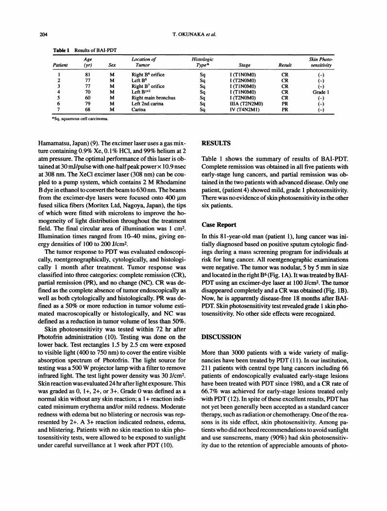
Table I Results of BAI-PDT
Age Location ofPatient (yr) Sex Tumor
Histologic Skin Photo-Type* Stage Result sensitivity
81 M Right B orifice2 77 M Left B3 77 M Right B orifice4 70 M Left B5 60 M Right main bronchus6 79 M Left 2nd cadna7 68 M Carina
Hamamatsu, Japan) (9). The excimer laser uses a gas mix-ture containing 0.9% Xe, 0.1% HC1, and 99% helium at 2atm pressure. The optimal performance of this laser is ob-tained at 30 rnJ/pulse with one-halfpeakpowerx 10.9 nsecat 308 nm. The XeC1 excimer laser (308 nm) can be cou-pled to a pump system, which contains 2 M RhodamineB dye in ethanol to convert the beam to 630nm. The beamsfrom the excimer-dye lasers were focused onto 400 I.tmfused silica fibers (Moritex Ltd, Nagoya, Japan), the tipsof which were fitted with microlens to improve the ho-mogeneity of light distribution throughout the treatmentfield. The final circular area of illumination was 1 cm2.Illumination times ranged from 10-40 mins, giving en-ergy densities of 100 to 200 J/cm2.The tumor response to PDT was evaluated endoscopi-
cally, roentgenographically, cytologically, and histologi-cally 1 month after treatment. Tumor response wasclassified into three categories: complete remission (CR),partial remission (PR), and no change (NC). CR was de-fined as the complete absence of tumor endoscopically aswell as both cytologically and histologically. PR was de-fined as a 50% or more reduction in tumor volume esti-mated macroscopically or histologically, and NC wasdefined as a reduction in tumor volume of less than 50%.
Skin photosensitivity was tested within 72 hr afterPhotofrin administration (10). Testing was done on thelower back. Test rectangles 1.5 by 2.5 cm were exposedto visible light (400 to 750 nm) to cover the entire visibleabsorption spectrum of Photofrin. The light source fortesting was a 500W projector lamp with a filter to removeinfrared light. The test light power density was 30 J/cm2.Skin reaction was evaluated 24hr after light exposure. Thiswas graded as 0, 1+, 2+, or 3+. Grade 0 was defined as anormal skin without any skin reaction; a 1+ reaction indi-cated minimum erythema and/or mild redness. Moderateredness with edema but no blistering or necrosis was rep-resented by 2+. A 3+ reaction indicated redness, edema,and blistering. Patients with no skin reaction to skin pho-tosensitivity tests, were allowed to be exposed to sunlightunder careful surveillance at 1 week after PDT (10).
RESULTS
Table 1 shows the summary of results of BAI-PDT.Complete remission was obtained in all five patients withearly-stage lung cancers, and partial remission was ob-tained in the two patients with advanced disease. Only onepatient, (patient 4) showed mild, grade 1 photosensitivity.There was no evidence ofskin photosensitivity in the othersix patients.
Case Report
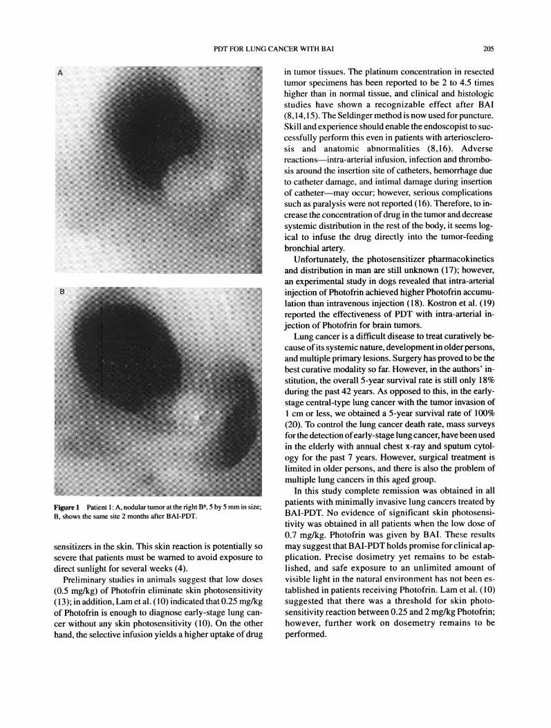
In this 81-year-old man (patient 1), lung cancer was ini-tially diagnosed based on positive sputum cytologic find-ings during a mass screening program for individuals atrisk for lung cancer. All roentgenographic examinationswere negative. The tumor was nodular, 5 by 5 mm in sizeand located in the right B6 (Fig. 1A). It was treatedby BAI-PDT using an excimer-dye laser at 100 J/cm2. The tumordisappeared completely and a CR was obtained (Fig. 1B).Now, he is apparently disease-free 18 months after BAI-PDT. Skin photosensitivity test revealed grade 1 skin pho-tosensitivity. No other side effects were recognized.
DISCUSSION
More than 3000 patients with a wide variety of malig-nancies have been treated by PDT (11). In our institution,211 patients with central type lung cancers including 66patients of endoscopically evaluated early-stage lesionshave been treated with PDT since 1980, and a CR rate of66.7% was achieved for early-stage lesions treated onlywith PDT (12). In spite ofthese excellent results, PDT hasnot yet been generally been accepted as a standard cancertherapy, such as radiation or chemotherapy. One ofthe rea-sons is its side effect, skin photosensitivity. Among pa-tients who did notheedrecommendations to avoid sunlightand use sunscreens, many (90%) had skin photosensitiv-ity due to the retention of appreciable amounts of photo-
PDT FOR LUNG CANCER WITH BAI 205
Figure 1 Patient 1" A, nodular tumor at the right B6, 5 by 5 mm in size;B, shows the same site 2 months after BAI-PDT.
sensitizers in the skin. This skin reaction is potentially sosevere that patients must be warned to avoid exposure to
direct sunlight for several weeks (4).Preliminary studies in animals suggest that low doses
(0.5 mg/kg) of Photofrin eliminate skin photosensitivity(13); in addition, Lam et al. (10) indicated that 0.25 mg/kgof Photofrin is enough to diagnose early-stage lung can-cer without any skin photosensitivity (10). On the otherhand, the selective infusion yields a higher uptake of drug
in tumor tissues. The platinum concentration in resectedtumor specimens has been reported to be 2 to 4.5 timeshigher than in normal tissue, and clinical and histologicstudies have shown a recognizable effect after BAI(8,14,15). The Seldinger method is now used for puncture.Skill and experience should enable the endoscopist to suc-cessfully perform this even in patients with arteriosclero-sis and anatomic abnormalities (8,16). Adversereactions--intra-arterial infusion, infection and thrombo-sis around the insertion site of catheters, hemorrhage dueto catheter damage, and intimal damage during insertionof cathetermmay occur; however, serious complicationssuch as paralysis were not reported (16). Therefore, to in-crease the concentration ofdrug in the tumor and decreasesystemic distribution in the rest of the body, it seems log-ical to infuse the drug directly into the tumor-feedingbronchial artery.
Unfortunately, the photosensitizer pharmacokineticsand distribution in man are still unknown (17); however,an experimental study in dogs revealed that intra-arterial
injection of Photofrin achieved higher Photofrin accumu-lation than intravenous injection (18). Kostron et al. (19)reported the effectiveness of PDT with intra-arterial in-jection of Photofrin for brain tumors.
Lung cancer is a difficult disease to treat curatively be-cause ofits systemic nature, development in older persons,and multiple primary lesions. Surgery has proved to be thebest curative modality so far. However, in the authors’ in-stitution, the overall 5-year survival rate is still only 18%during the past 42 years. As opposed to this, in the early-stage central-type lung cancer with the tumor invasion ofcm or less, we obtained a 5-year survival rate of 100%
(20). To control the lung cancer death rate, mass surveysfor the detection ofearly-stage lung cancer, have been usedin the elderly with annual chest x-ray and sputum cytol-ogy for the past 7 years. However, surgical treatment islimited in older persons, and there is also the problem ofmultiple lung cancers in this aged group.
In this study complete remission was obtained in allpatients with minimally invasive lung cancers treated byBAI-PDT. No evidence of significant skin photosensi-tivity was obtained in all patients when the low dose of0.7 mg/kg. Photofrin was given by BAI. These resultsmay suggest that BAI-PDT holds promise for clinical ap-plication. Precise dosimetry yet remains to be estab-lished, and safe exposure to an unlimited amount ofvisible light in the natural environment has not been es-
tablished in patients receiving Photofrin. Lam et al. (10)suggested that there was a threshold for skin photo-sensitivity reaction between 0.25 and 2 mg/kg Photofrin;however, further work on dosemetry remains to beperformed.
206 T. OKUNAKA et al.
ACKNOWLEDGMENT
This work was supported in part by grants from theMinistry of Health and Welfare and the Ministry ofIndustry and Trade, Japan. The authors wish to thankProfessor J. P. Barron of Tokyo Medical College for hisreview of the manuscript.
REFERENCES
1. Hayata Y, Kato H, KonakaC, et al. Hematoporphyrin derivative andlaser photoradiation in the treatment of lung cancer. Chest1982;81:269-277.
3. Hayata Y, Dougherty TY, eds. Lasers and hematoporphydn deriv-ative in cancer. Tokyo: Igaku-Shoin, 1983.
4. Razime N, Balchum OJ, Profio AF, et al. Skin photosensitivity; du-ration and intensity following intravenous HpD and DHE.Photochem Photobiol 1987;46:925-928.
5. Wooten RS, Smith KS, AhlquestDA, et al. Prospective study ofcu-taneous phototoxicity after systemic hematoporphydn derivative.Lasers Surg Med 1988;8:294-300.
6. Dougherty TJ, Laurence G, Kaufman JH, et al. Photoradiation in thetreatment of recurrent breast carcinoma. J Natl Cancer Inst1979;62:231-237.
7. Kahn PC, Paul RE, Rheinlander HF. Selective bronchial arteriog-raphy and intra-arterial chemotherapy in carcinoma of the lung. JThorac Cardiovasc Surg 1965;50:640-645.
8. Masuda H, Ogata T, Kikuchi K, et al. Bronchial arterial infusion ofcisplatin for lung cancer and platinum concentration in the tumor.Lung Cancer 1988;28:297-302.
9. Yamamoto H, Kato H, OkunakaT, et al. Photodynamic therapy withthe excimer dye laser in the treatment of respiratory tract malig-nancies. Lasers Life Sci 1991;4:125-133.
10. Lam S, Palcic B, McLean D, et al. Detection of early lung cancerusing low dose Photofrin II. Chest 1990;97:333-337.
11. Marcus SL, Dugan M. Global status of clinical phntodynamic ther-apy: the registration process for a new therapy. Lasers Surg Med1992; 12:318-324.
12. Okunaka T, KonakaC, Tsutsui H, et al. Present status ofendoscopictreatment of lung cancer: the role of photodynemic therapy. J JpnSoc Bronchol 1994;16:712-722.
13. Mang TS, Potter WR, Dougherty TJ. Fluorescence detection andHPLC characterization of intratumoral porphyrin in occult metas-tasis of the lymph nodes following injection of low levels of the pu-dried component of hematoporphyrin derivative. Lasers Surg Med1988;8:145-146.
14. Ten GJ, Chai XL, Gao GR, et al. Intraarterial digital subtraction an-giography in bronchogenic carcinoma treated with bronchial arteryinfusion. Eur J Radiol 1991; 12:91-94.
15. UchiyamaM, Kobayashi H, Nakajo M, et al. Treatment oflung can-cer with bronchial artery infusion of cisplatin and intravenoussodium thiosulfate rescue. Acta Oncol 1988;27:57-61.
16. Ogata T, Suemasu K, Yoneyama T, et al. Study of regional arterialinfusion ofanticancer agents as an adjuvant to surgery in carcinomaof the lung. Jpn J Cancer Clin 1977;23:1085-1089.
17. Henderson BW, Bellnier DA. Tissue localization of photosensitiz-ers and the mechanism ofphotodynamic tissue destruction. In: BoekG, Harnett S, eds. Photosensitizing compounds: their chemistry bi-ology and clinical use. Chichester: John Wiley and Sons,1989:112-130.
18. Andou T, Sekimoto I, Hoshiyama N, et al. Study of photoradiationtherapy by intra-arterial injection of HpD. J Jpn Laser Med1987;7:143-144.
19. Kostron H, Plangger C, Fritsch E, et al. Photodynamie treatment ofmalignant brain tumor. Wein Klin Wochenschr 1990; 102:531-535.
20. Kato H, Horai T, Furuse K, et al. Photodynamic therapy for can-cers: a clinical trial of porfimer sodium in Japan. Jpn J Cancer Res1993;84:1209-1214.