34
Physical-chemical properties of proteins; methods of its determination, precipitation reactions
| Date post: | 30-Dec-2015 |
| Category: |
Documents |
| Upload: | leilani-hopkins |
| View: | 47 times |
| Download: | 3 times |
Physical-chemical properties of proteins; methods of its determination, precipitation reactions

Separation of Amino Acids and Proteins
1. Chromatography – the method of separating amino acids on the basis of differences in absorption, ionic charges, size and solubility of molecules
2. Electrophoresis – effects separation in an electric field on the basis of differences in charges carried by amino acids and proteins under specific condition
3. Ultracentrifugation – effects separation on the basis of molecular weight when large gravitational forces are applied in the ultracentrifuge.
4. Precipitation Methods – salts as sodium sulfate, ammonium sulfate, cadmium nitrate, silver nitrate and mercuric chloride at specific conc. precipitate some proteins while others remain in solution
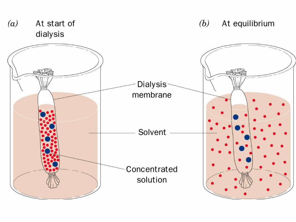
5. Dialysis – is for the removal of small, crystalloidal molecules from protein solution.


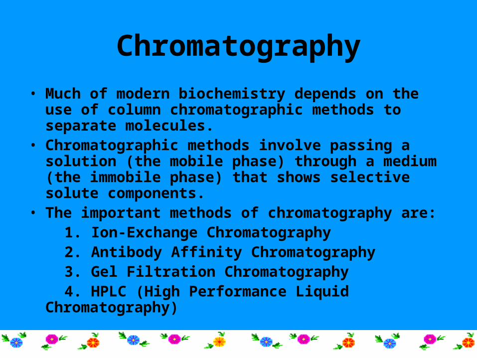
Chromatography
• Much of modern biochemistry depends on the use of column chromatographic methods to separate molecules.
• Chromatographic methods involve passing a solution (the mobile phase) through a medium (the immobile phase) that shows selective solute components.
• The important methods of chromatography are: 1. Ion-Exchange Chromatography 2. Antibody Affinity Chromatography 3. Gel Filtration Chromatography 4. HPLC (High Performance Liquid Chromatography)
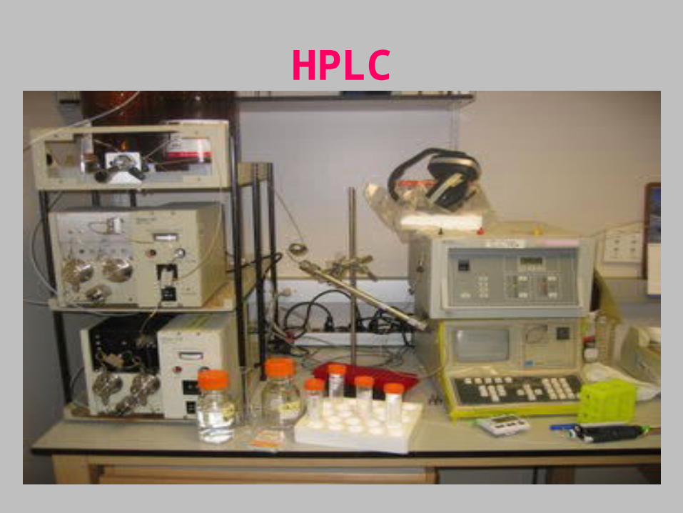
HPLC
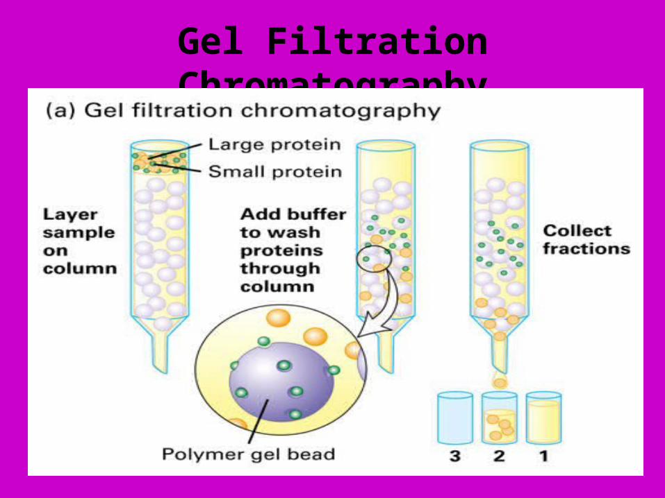
Gel Filtration Chromatography


Precipitation of proteins

Proteomics
• Proteomics is the science of protein expression of all the proteins made by a cell
• Proteome pertain to all proteins being made according to the transcriptome (RNA profile). It is often visualized by a system interaction map as seen in the proteogram.
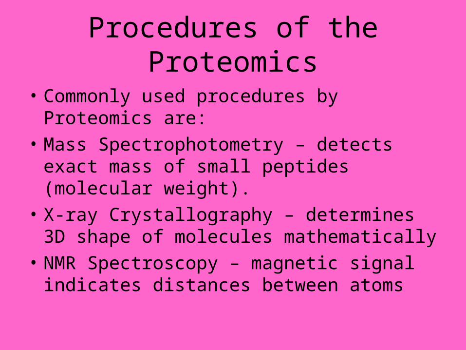
Procedures of the Proteomics
• Commonly used procedures by Proteomics are:
• Mass Spectrophotometry – detects exact mass of small peptides (molecular weight).
• X-ray Crystallography – determines 3D shape of molecules mathematically
• NMR Spectroscopy – magnetic signal indicates distances between atoms

Qualitative analysis of ProteinsPrecipitation reactionsColour Reactions of Proteins
Precipitation reactions
Precipitation by salts
Protein exist in colloidal solution due to solution due to hydration of hydration of polar groups (-COO, NH3
+, -OH)They can be precipitated by dehydration or by dehydration or neutralization of polar groups.
To 2 ml of protein solution add equal volume of saturated (NH4)2SO4 solution
White precipitation is formed

Precipitation by heavy metal saltsTo 2 ml of protein solution, add few drops of Heavy Metals (lead acetate or mercuric nitrate) solution, results in white precipitation
Precipitation by alkaloidal reagent
To a few ml of sample solution add 1-2 ml of picric acid solution. Formation of precipitation indicates the presence of proteins

Precipitation by organic solvents
To a few ml of sample solution, add 1 ml of alcohol. Mix and keep aside for 2 min. Formation of white precipitation indicates the presence of protein
Precipitation by heatTake few ml of protein solution in a test tube and heat over a flame. Cloudy white precipitation is observed
Precipitation by acidsTo 1 ml of protein solution in test tube, add few drops of 1% acetic acid, white precipitation is formed 15

CCoolloouur r Reactions of ProteinsReactions of ProteinsProteins give a number of colour reactions with different chemical reagents due to the presence of amino acid
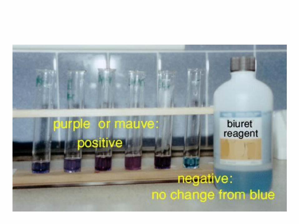
Biuret test
The Biuret test is a chemical test used for detecting the presence of peptide bonds In the presence of peptides, a copper (II) ion forms violet-colored coordination in an alkaline solution
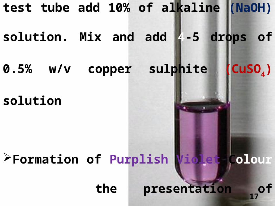
To 2 ml of protein solution in a test tube add 10%
of alkaline (NaOH) solution. Mix and add 4-5
drops of 0.5% w/v copper sulphite (CuSO4)
solution
Formation of Purplish Violet Colour indicates
the presentation of proteins 17
Biuret Test
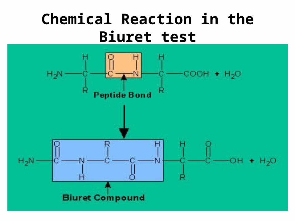
Chemical Reaction in the Biuret test
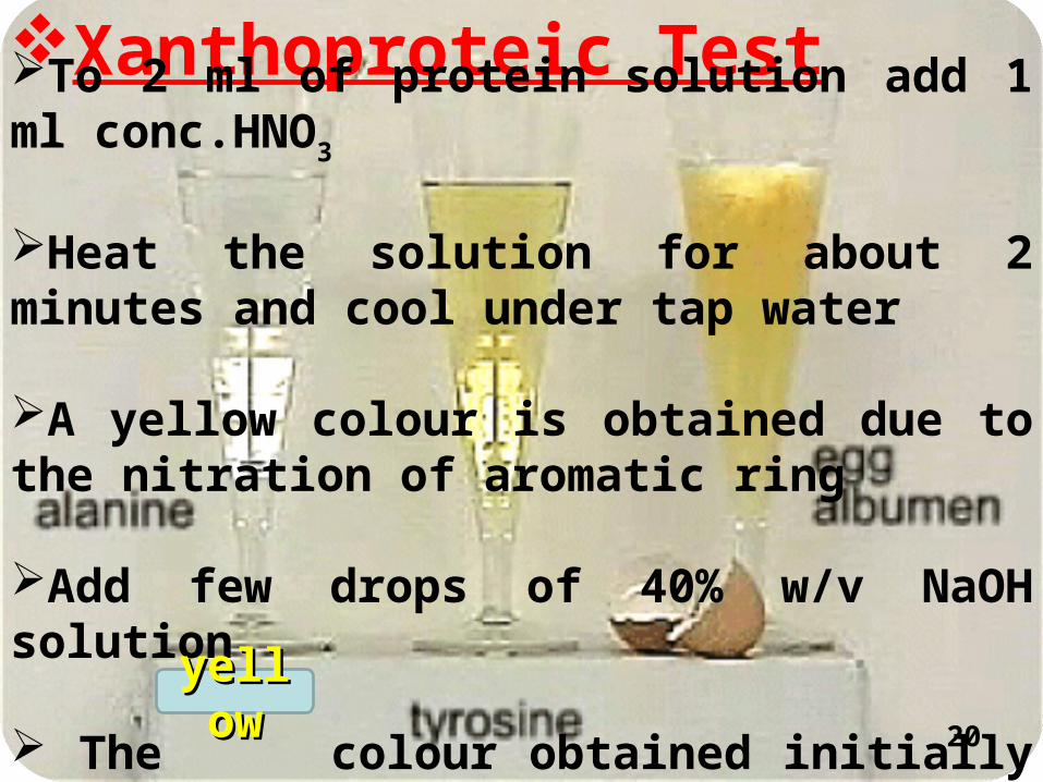
Xanthoproteic TestTo 2 ml of protein solution add 1 ml conc.HNO3
Heat the solution for about 2 minutes and cool under tap water
A yellow colour is obtained due to the nitration of aromatic ring
Add few drops of 40% w/v NaOH solution
The colour obtained initially changes to orange 20
yellowyellow
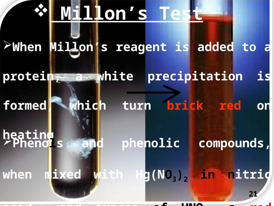
Millon’s Test
When Millon’s reagent is added to a protein,
a white precipitation is formed, which turn
brick red on heating
Phenols and phenolic compounds, when
mixed with Hg(NO3)2 in nitric acid and traces
of HNO2, a red colour is produced21
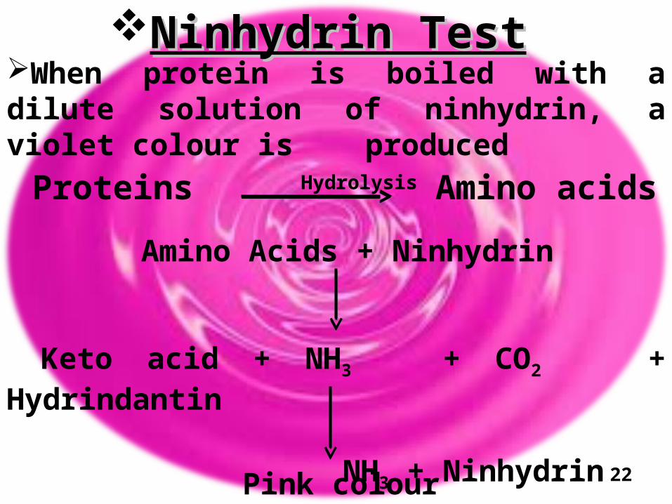
Ninhydrin TestNinhydrin TestWhen protein is boiled with a dilute solution of ninhydrin, a violet colour is produced
Proteins Hydrolysis Amino acids
Amino Acids + Ninhydrin
Keto acid + NH3 + CO2 + Hydrindantin
NH3 + Ninhydrin
Pink colour 22
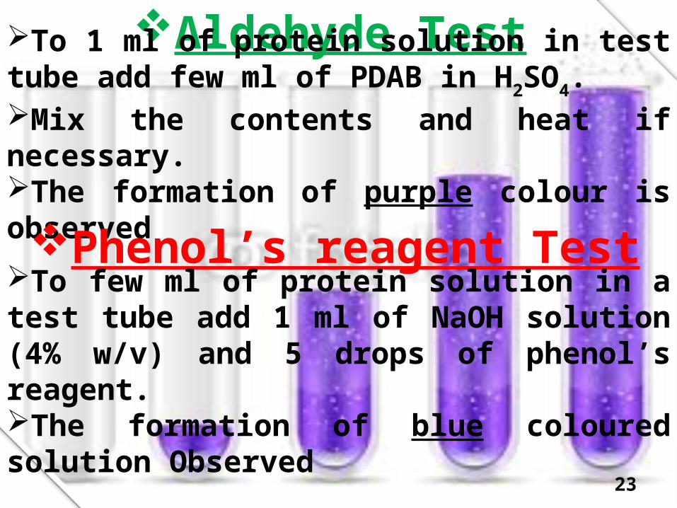
Aldehyde TestTo 1 ml of protein solution in test tube add few ml of PDAB in H2SO4. Mix the contents and heat if necessary.The formation of purple colour is observed
Phenol’s reagent TestTo few ml of protein solution in a test tube add 1 ml of NaOH solution (4% w/v) and 5 drops of phenol’s reagent.The formation of blue coloured solution Observed
23
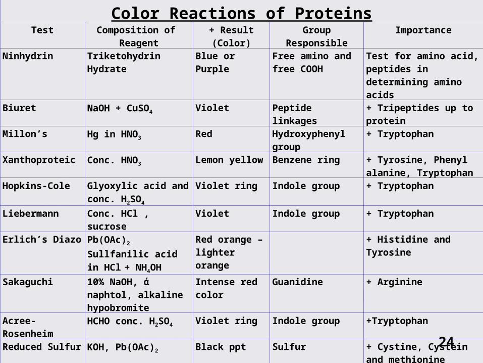
Color Reactions of ProteinsTest Composition of
Reagent+ Result (Color) Group Responsible Importance
Ninhydrin Triketohydrin Hydrate Blue or Purple Free amino and free COOH
Test for amino acid, peptides in determining amino acids
Biuret NaOH + CuSO4 Violet Peptide linkages + Tripeptides up to protein
Millon’s Hg in HNO3 Red Hydroxyphenyl group
+ Tryptophan
Xanthoproteic Conc. HNO3 Lemon yellow Benzene ring + Tyrosine, Phenyl alanine, Tryptophan
Hopkins-Cole Glyoxylic acid and
conc. H2SO4
Violet ring Indole group + Tryptophan
Liebermann Conc. HCl , sucrose Violet Indole group + TryptophanErlich’s Diazo Pb(OAc)2 Sullfanilic
acid in HCl + NH4OH
Red orange – lighter orange
+ Histidine and Tyrosine
Sakaguchi 10% NaOH, ά naphtol, alkaline hypobromite
Intense red color
Guanidine + Arginine
Acree-Rosenheim HCHO conc. H2SO4 Violet ring Indole group +Tryptophan
Reduced Sulfur KOH, Pb(OAc)2 Black ppt Sulfur + Cystine, Cystein and methionine
Br water Br.H2O, amyl alcohol Pink Indole group + Tryptophan
Molisch ά naphtol in alcoholic
H2SO4
Violet ring Carbohydrates Glycoprotein
Adamskiewez Glacial Acetic acid and
conc. H2SO4
Reddish violet ring at the junction
Indole group + Tryptophan24
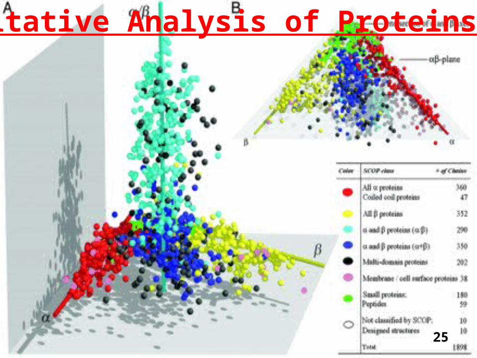
Quantitative Analysis of Proteins
25
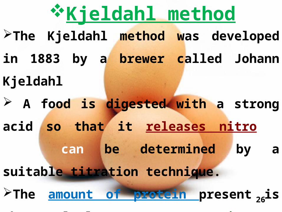
Kjeldahl methodThe Kjeldahl method was developed in 1883
by a brewer called Johann Kjeldahl
A food is digested with a strong acid so
that it releases nitrogen which can be
determined by a suitable titration technique.
The amount of protein present is then
calculated from the nitrogen concentration of
the food 26
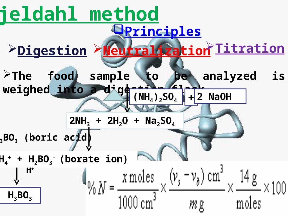
Kjeldahl methodPrinciples
Digestion Neutralization
The food sample to be analyzed is weighed into a digestion flask
(NH4)2SO4 + 2 NaOH
2NH3 + 2H2O + Na2SO4
H3BO3 (boric acid)
H+
H3BO3
Titration
NH4+ + H2BO3
- (borate ion)

Enhanced Dumas methodA sample of known mass
Combustion (900 oC) CO2, H2O and N2
Nitrogen
Thermal conductivity detector
The nitrogen content is then measured

Methods using UV-visible spectroscopyThese methods use either the natural ability of proteins to absorb (or scatter) light in the UV-visible region of the electromagnetic spectrum, or they chemically or physically modify proteins to make them absorb (or scatter) light in this region
PrinciplesDirect measurement at 280nmBiuret MethodLowry MethodDye binding methodsTurbimetric method 29
Direct measurement at 280nmTryptophan and tyrosine absorb ultraviolet light strongly at 280 nmThe tryptophan and tyrosine content of many proteins remains fairly constant, and so the absorbance of protein solutions at 280nm can be used to determine their concentration
Biuret MethodBiuret MethodA violet-purplish color is produced when cupric ions (Cu2+) interact with peptide bonds under alkaline conditions
The absorbance is read at 540 nm
31
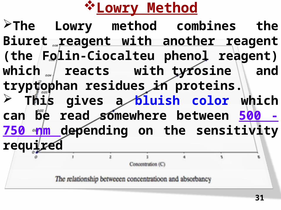
Lowry MethodThe Lowry method combines the Biuret reagent with another reagent (the Folin-Ciocalteu phenol reagent) which reacts with tyrosine and tryptophan residues in proteins. This gives a bluish color which can be read somewhere between 500 - 750 nm depending on the sensitivity required
32
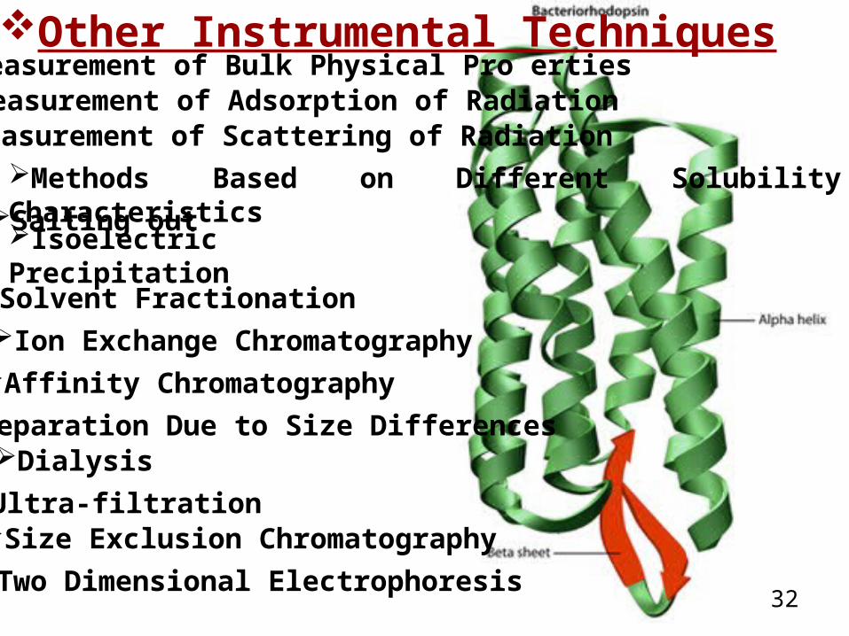
Other Instrumental TechniquesMeasurement of Bulk Physical PropertiesMeasurement of Adsorption of RadiationMeasurement of Scattering of RadiationMethods Based on Different Solubility CharacteristicsSalting outIsoelectric PrecipitationSolvent FractionationIon Exchange ChromatographyAffinity ChromatographySeparation Due to Size DifferencesDialysisUltra-filtrationSize Exclusion ChromatographyTwo Dimensional Electrophoresis

33
Amino Acid AnalysisAmino acid analysis is used to determine
the amino acid composition of proteins.
A protein sample is first hydrolyzed
(e.g. using a strong acid) to release the amino
acids, which are then separated using
chromatography, e.g., ion exchange, affinity
or absorption chromatography.
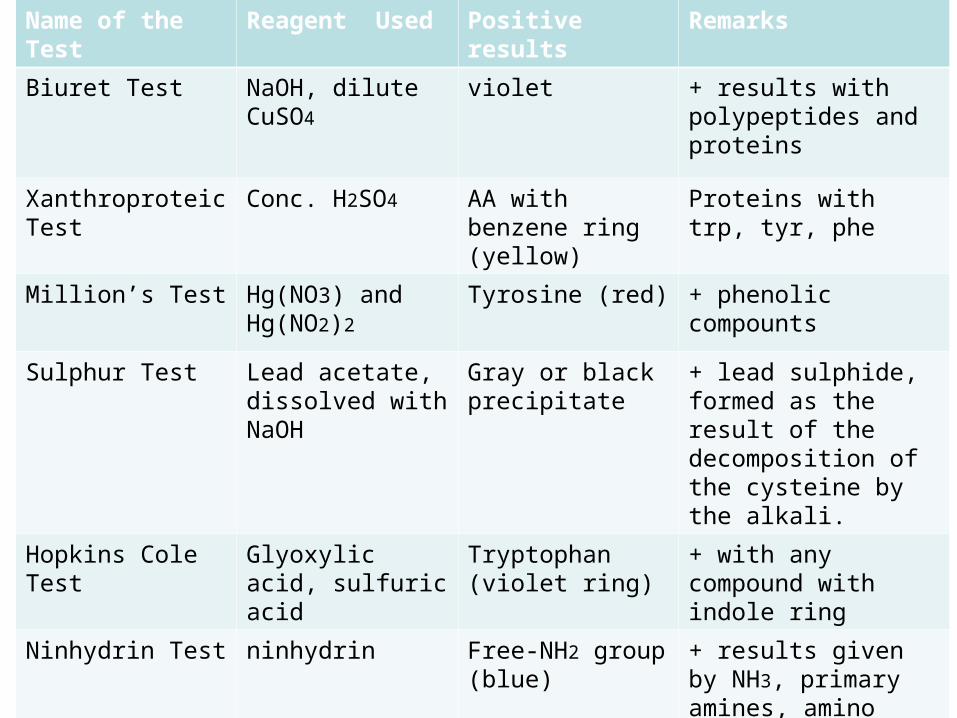
Name of the Test Reagent Used Positive results Remarks
Biuret Test NaOH, dilute CuSO4
violet + results with polypeptides and proteins
Xanthroproteic Test
Conc. H2SO4 AA with benzene ring (yellow)
Proteins with trp, tyr, phe
Million’s Test Hg(NO3) and Hg(NO2)2
Tyrosine (red) + phenolic compounts
Sulphur Test Lead acetate, dissolved withNaOH
Gray or black precipitate
+ lead sulphide, formed as the result of the decomposition of the cysteine by the alkali.
Hopkins Cole Test Glyoxylic acid, sulfuric acid
Tryptophan (violet ring)
+ with any compound with indole ring
Ninhydrin Test ninhydrin Free-NH2 group (blue)
+ results given by NH3, primary amines, amino acids, peptides, and proteins


