Physical exercise neuroprotects ovariectomized 3xTg-AD mice through BDNF mechanisms Yoelvis Garcı ´a-Mesa a,1,2 , Helios Pareja-Galeano b,1 , Vicent Bonet-Costa b , Susana Revilla a , M. Carmen Go ´mez-Cabrera b , Juan Gambini b , Lydia Gime´nez-Llort c , Rosa Cristo `fol a , Jose´Vin˜a b, ** , Coral Sanfeliu a, * a Institut d’Investigacions Biome`diques de Barcelona (IIBB), CSIC, IDIBAPS, Barcelona, Spain b Department of Physiology, University of Valencia, Fundacio´n Investigacio´n Hospital Clı ´nico Universitario/ INCLIVA, Valencia, Spain c Institute of Neuroscience and Medical Psychology Unit, Department of Psychiatry and Forensic Medicine, Universitat Auto `noma de Barcelona, Bellaterra, Barcelona, Spain Received 11 November 2013; received in revised form 29 March 2014; accepted 30 March 2014 Psychoneuroendocrinology (2014) 45, 154—166 KEYWORDS Alzheimer’s disease; Ovariectomy; 3xTg-AD mice; Physical exercise; Frailty; Behavioral tests; Cognition; Catalase; p-CREB; BDNF Summary Postmenopausal women may be more vulnerable to cognitive loss and Alzheimer’s disease (AD) than premenopausal women because of their deficiency in estrogens, in addition to their usually older age. Aerobic physical exercise has been proposed as a therapeutic approach for maintaining health and well-being in postmenopausal women, and for improving brain health and plasticity in populations at high risk for AD. To study the neuroprotective mechanisms of physical exercise in a postmenopausal animal model, we submitted previously ovariectomized, six-month old non-transgenic and 3xTg-AD mice to three months of voluntary exercise in a running wheel. At nine months of age, we observed lower grip strength and some exacerbation of the behavioral and psychological symptoms of dementia (BPSD)-like involving active exploratory activities. A similar major cognitive impairment was observed of ovariectomized 3xTg-AD mice in comparison with sham-operated 3xTg-AD mice. A reduction of bodily fitness and lack of retention of memory were observed in the ovariectomized non-transgenic mice. Physical exercise protected against all * Corresponding author: IIBB-CSIC, IDIBAPS, c/Rossello ´ 161, 6th Floor, 08036 Barcelona, Spain. Tel.: +34 93 3638338; fax: +34 93 3638301. ** Corresponding author: Department of Physiology, School of Medicine, University of Valencia, Av. Blasco Iba ´n ˜ez 15, 46010 Valencia, Spain. Tel.: +34 96 3864650. E-mail addresses: [email protected], [email protected](C. Sanfeliu), [email protected](J. Vin ˜a). 1 These authors equally contributed to this work. 2 Present address: Department of Molecular Biology and Microbiology, School of Medicine, Case Western Reserve University, Cleveland, OH, United States. Available online at www.sciencedirect.com ScienceDirect jou rn a l home pag e : ww w. el sev ier. com/ loca te /psyn eu en http://dx.doi.org/10.1016/j.psyneuen.2014.03.021 0306-4530/# 2014 Elsevier Ltd. All rights reserved.
Transcript
Physical exercise neuroprotectsovariectomized 3xTg-AD mice through BDNFmechanisms
Yoelvis Garcıa-Mesa a,1,2, Helios Pareja-Galeano b,1,Vicent Bonet-Costa b, Susana Revilla a,M. Carmen Gomez-Cabrera b, Juan Gambini b,Lydia Gimenez-Llort c, Rosa Cristofol a,Jose Vina b,**, Coral Sanfeliu a,*
a Institut d’Investigacions Biomediques de Barcelona (IIBB), CSIC, IDIBAPS, Barcelona, SpainbDepartment of Physiology, University of Valencia, Fundacion Investigacion Hospital Clınico Universitario/INCLIVA, Valencia, Spainc Institute of Neuroscience and Medical Psychology Unit, Department of Psychiatry and Forensic Medicine,Universitat Autonoma de Barcelona, Bellaterra, Barcelona, Spain
Received 11 November 2013; received in revised form 29 March 2014; accepted 30 March 2014
Summary Postmenopausal women may be more vulnerable to cognitive loss and Alzheimer’sdisease (AD) than premenopausal women because of their deficiency in estrogens, in addition totheir usually older age. Aerobic physical exercise has been proposed as a therapeutic approach formaintaining health and well-being in postmenopausal women, and for improving brain health andplasticity in populations at high risk for AD. To study the neuroprotective mechanisms of physicalexercise in a postmenopausal animal model, we submitted previously ovariectomized, six-monthold non-transgenic and 3xTg-AD mice to three months of voluntary exercise in a running wheel. Atnine months of age, we observed lower grip strength and some exacerbation of the behavioral andpsychological symptoms of dementia (BPSD)-like involving active exploratory activities. A similarmajor cognitive impairment was observed of ovariectomized 3xTg-AD mice in comparison withsham-operated 3xTg-AD mice. A reduction of bodily fitness and lack of retention of memory wereobserved in the ovariectomized non-transgenic mice. Physical exercise protected against all
* Corresponding author: IIBB-CSIC, IDIBAPS, c/Rossello 161, 6th Floor, 08036 Barcelona, Spain. Tel.: +34 93 3638338; fax: +34 93 3638301.** Corresponding author: Department of Physiology, School of Medicine, University of Valencia, Av. Blasco Ibanez 15, 46010 Valencia, Spain.Tel.: +34 96 3864650.
E-mail addresses: [email protected], [email protected] (C. Sanfeliu), [email protected] (J. Vina).1 These authors equally contributed to this work.2 Present address: Department of Molecular Biology and Microbiology, School of Medicine, Case Western Reserve University, Cleveland, OH,
United States.
Available online at www.sciencedirect.com
ScienceDirect
jou rn a l home pag e : ww w. el sev ie r. com/ loca te /psyn eu en
http://dx.doi.org/10.1016/j.psyneuen.2014.03.0210306-4530/# 2014 Elsevier Ltd. All rights reserved.
deleterious behaviors and normalized learning and memory. It also protected against body frailty,as expected. Analyses of hippocampal key markers of antioxidant and neuroplasticity signalingpathways, showed that ovariectomy impairs the activation of CREB through physical exercise.Furthermore, molecular and behavioral correlates suggested a central role of BDNF in theneuroprotection mediated by physical exercise therapy against apathy and memory loss inducedby ovariectomy and the AD-genotype.# 2014 Elsevier Ltd. All rights reserved.
Wheel-running in ovariectomized 3xTg-AD 155
1. Introduction
Alzheimer’s disease (AD) is the most common cause ofdementia in the elderly and the estimated number for ADpatients worldwide in years to come is staggering. As womenhave a higher life expectancy than men, the higher propor-tion of female patients could be due to the higher proportionof women in the upper age bracket. Despite the suggestedhigher risk of AD at menopause, several cohort studies havedemonstrated no gender effect in the risk of developing ADnor in the incidence rates (Rocca et al., 1998; Hebert et al.,2001; Barnes et al., 2003). However, the fact that there aremany more women than men suffering from AD has led to theresearch into gender-specific therapies. Hormone reposi-tion therapy, diet and physical exercise have been recom-mended as strategies to prevent and reverse the bodilyeffects of menopause in women. While more effectivetreatments are being developed, these general strategiesmay lead to better health and higher resistance to ADdevelopment in post-menopausal women. Treatment withestrogens has been shown to be neuroprotective in manyexperimental systems (Vina et al., 2011). However, clinicaltrials of hormone therapy yielded inconsistent results,including some negative outcomes in cognition and demen-tia, thus warranting further research before hormone repla-cement could be recommended as a treatment against AD(for review, see Maki and Henderson, 2012). Some nutrients,mainly those with antioxidant properties, yielded positiveresults in preclinical studies, although clinical trials held sofar have not produced evidence in support of such cognitivebenefits for AD patients (Kamat et al., 2008; Aisen et al.,2012). Aerobic exercise was demonstrated as improvingcognitive processes in animal models of AD (Intlekoferand Cotman, 2013) and in studies involving upper-agedhuman subjects with or without a known cognitive impair-ment (Heyn et al., 2004; Angevaren et al., 2008). Physicalexercise also impeded the cognitive deficits induced inovariectomized rodents (Ben et al., 2010), an animal modelof menopause. Therefore, regular physical exercise couldbe a preventive and/or protective strategy to decrease theAD burden in women and more research into its benefits andmechanisms will help to improve the lives of elderly femalesat risk of frailty and AD.
In this study we analyzed the presence of neuroprotectionas a result of voluntary running exercise, on the effectsinduced by ovariectomy in the triple transgenic mouse modelof AD (3xTg-AD) (Oddo et al., 2003) and in non-transgenicmice. In this mouse model, ovariectomy has been reported toinduce an increase of AD-like pathology (Carroll et al., 2010)including exacerbated mitochondrial dysfunction (Yao et al.,
2012). Physical exercise has been proved to be neuroprotec-tive at different stages of pathological severity in the 3xTg-ADmice (Garcıa-Mesa et al., 2011, 2012), although it has not beenassayed in a context mimicking ovarian hormone deficiency inAD Tg models. We aimed to investigate the benefits andmechanisms of a physical exercise therapy along a hypothe-sized progressive neurodegeneration from ovariectomizedwild type mice, to control AD mice and to ovariectomizedAD mice. For this purpose, we studied the mechanisms under-lying physical exercise-mediated neuroprotection in ovariec-tomized and sham-operated wild type mice and AD mice. Thiswas done by way of characterizing behavioral and cognitivechanges, brain pathology, and the levels of selected markers ofneuroprotection and plasticity in the hippocampus. Namely,we analyzed the antioxidant enzymes catalase, glutathioneperoxidase (GPx) and Mn superoxide dismutase (SOD); themodulator of mitochondrial function and biogenesis, peroxi-some proliferator-activated receptor-g coactivator-1a (PGC-1a); the mediators of synaptic plasticity and long-term mem-ory, phosphorylated cAMP response element-binding protein(p-CREB) and brain derived neurotrophic factor (BDNF); andthe receptor tyrosine kinase B (TrkB) that binds BDNF with highaffinity.
2. Materials and methods
2.1. Animals and experimental design
For this study we used 3xTg-AD (Tg) mice, which weregenetically engineered at the University of California Irvineto express familial AD mutations of amyloid precursor protein(APPSwe), presenilin-1 (PS1M146V), and tau (tauP301L)(Oddo et al., 2003). These mice presented a progressiveAD-like brain pathology and deterioration in multiple aspectsof brain physiology and behavior (Oddo et al., 2003; Gime-nez-Llort et al., 2007; Garcıa-Mesa et al., 2011), suggestingclose adherence to AD disease progression in patients. Weused Tg female mice and non-transgenic (NTg) mice with thesame genetic background hybrid (129 � C57BL/6) as the Tgmice. Both strains were bred in homozygosis from progenitorsobtained from Dr. Frank M. LaFerla (University of CaliforniaIrvine, CA). The animals were maintained in Macrolon cagesunder standard laboratory conditions of food and water adlibitum, 22 � 2 8C and 12 h light:dark. The genotypes wereconfirmed by PCR analysis of DNA obtained from tail biopsies.Animal handling and procedures were approved by the localanimal ethics committee (Ref: DMAH-5150, CEEA, UB), inaccordance with the EU Directive 2010/63/EU for animalexperiments.
156 Y. Garcıa-Mesa et al.
Mice were submitted to bilateral ovariectomy or shamoperation at 4 months of age. Two months later, at 6 monthsof age, half of the animals were subjected to a chronictreatment of voluntary physical exercise. Four to five animalsfrom different litters were housed together to avoid socialisolation stress, grouped by genotype, surgery type andexercise treatment. For those submitted to exercise, a read-ily available running wheel connected to a computerizedsystem (Activity Wheel Cage System for mice, Techniplast,Buguggiate, Italy) was added to each of the correspondingcages, as described previously (Garcıa-Mesa et al., 2011).The system allowed a continuous recording of the wheel turnnumber and therefore calculation of the average mouserunning activity per cage. The study was terminated at 9months of age, after 3 months of physical exercise. Subse-quently, grip strength, behavior and cognitive patterns wereevaluated along 13 days. Three days later, the animals wereeuthanized. Mice submitted to physical exercise treatmenthad access to the running wheel until sacrificed. All animalprocedures were performed between 9:00 h and 15:00 h in abalanced manner between mice of the different experimen-tal groups.
The experimental groups (n = 8—9 per group) were asfollows: (i) NTg, sham-operated NTg mice; (ii) NTg + Ex,sham-operated NTg mice subjected to exercise; (iii) NTgOv,ovariectomized NTg mice; (iv) NTgOv + Ex, ovariectomizedNTg mice subjected to exercise; (v) Tg, sham-operated Tgmice; (vi) Tg + Ex, sham-operated Tg mice subjected toexercise; (vii) TgOv, ovariectomized Tg mice; and (viii) TgO-v + Ex, ovariectomized Tg mice subjected to exercise.
2.2. Fitness, and behavioral and cognitive tests
Body weight of the mice was recorded at the beginning andend of the study. The running activity of those mice sub-mitted to exercise was recorded throughout the study. Theaverage distance covered (Km) per mouse was calculated in aweekly basis for each cage.
Fore limb muscle strength was measured by a gripstrength test. The mouse was held at the base of the tailand allowed to place its forepaws on the pull bar connectedto the force gauge of an electronic grip strength meter(Harvard Apparatus, Panlab, Cornella, Barcelona, Spain).The animal was slowly pulled away from the pull bar at arate of 2—3 cm/s until it released the bar. The force gaugeindicated the peak tension exerted by the animal. Eachmouse was tested in three sequential trials and the highestgrip strength reading was recorded as the total peak force(gram force).
Non-cognitive behavioral responses were evaluated as pre-viously described (Garcıa-Mesa et al., 2011). Each mouse wassubmitted to sequential testing of the following four tests, onetest per day. The corner test was executed to evaluate neo-phobia. Mice were placed into a home cage with fresh beddingand examined for 30 s for signs of adaptation to the newenvironment. Latency of rearing, number of rearings andnumber of corners were recorded. The open field test wasused to evaluate vertical and horizontal locomotor activity fora single trial period of 5 min. The mouse was positioned in thecenter of the apparatus (home-made, wooden, white,55 cm � 55 cm � 25 cm high) and measurements were taken
of latency of rearing, number of rearings, grooming activity,latencies and number of defecation and urine spots. Dis-tance covered was measured as the number of crossingsalong a virtual grid on the floor of the apparatus. Thedark—light box test was used to evaluate anxiety-likebehavior for 5 min. The dark—light box consisted oftwo compartments (black, 27 cm � 18 cm � 27 cm; white,27 cm � 27 cm � 27 cm; lit with a 20 W white bulb) con-nected by an opening (7 cm � 7 cm) (Panlab, Barcelona,Spain). In this test the mouse was placed in the dark com-partment of the apparatus and measures were taken forlatency of entry into the illuminated compartment as well asthe number of entries and the time spent in the illuminatedcompartment once there. Boissier’s 4 hole-board test wasused to assess exploratory behavior by measuring the num-ber of head-dips, latency and the time spent head-dipping ateach of the four holes during 5 min (3 cm diameter holes in ahome-made 32 cm � 32 cm, white, wooden board, withwalls 32 cm high).
Cognitive testing was then initiated on the fifth day ofbehavior evaluation. Spatial learning and memory weremeasured using the Morris water maze (MWM) test as pre-viously described (Garcıa-Mesa et al., 2011). The test con-sisted of a preliminary day for the cue learning of a visualplatform and six days of place task learning for spatialreference memory (days 1—6, four trial sessions per day).Mice were trained to locate a platform (7 cm diameter,1.5 cm below the water surface) in a circular pool (91 cmdiameter, 40 cm height, 25 8C opaque water) surrounded byblack curtains, by relying on visual cues. Within the blackenclosure, four different landmarks hung from a false ceilingat 908 to each other. The platform was placed between two ofthese landmarks. On day 7, after one trial of place learning,the platform was removed from the maze and the miceperformed a probe trial. A computerized tracking system(SMART, Panlab) enabled swimming distance to be measuredduring the learning tasks, in addition to the time spent ineach quadrant of the pool following the removal of theplatform in the probe trial.
2.3. Tissue analyses
The animals were decapitated under a light CO2 anesthesia.Each mouse brain was immediately dissected on ice to isolatethe hippocampus and cerebral cortex. The samples werestored at �80 8C for further analysis. The weight of theuterus and the intra-abdominal white adipose tissue (WAT)was obtained upon termination and measured as a percen-tage of body weight. WAT was measured as indicative ofobesity and fitness related changes. The uterus was measuredto check the efficacy of ovariectomy surgery.
RNA expression of antioxidant enzymes in the hippocam-pus was determined by real-time quantitative reverse tran-scriptase-polymerase chain reaction (qPCR). Total RNA wasisolated from hippocampal tissue using RNeasy1 Mini Kit(Qiagen Distributors, Valencia, CA) in accordance with themanufacturer’s instructions. TaqMan FAM labeled specificprobes (Applied Biosystems, Foster City, CA) were used forPCR quantitation of catalase (Mm00437992_m1), GPx(Mm00656767_g1) and MnSOD (Mm01313000_m1) mRNA inan iCycler iQ detection system (Bio-Rad, Hercules, CA). Gene
Wheel-running in ovariectomized 3xTg-AD 157
expression was normalized to that of glyceraldehyde-3P-dehydrogenase (GAPDH, Mm99999915_g1) and 18S(Mm03928990_g1) gene expression.
Protein levels of BDNF were quantified in the hippocampusby ELISA (CYT306 Millipore, Bedford, MA), as stipulated bythe manufacturer.
Western immunoblotting studies of cerebral cortical andhippocampal tissues were performed according to standardprocedures. The following primary antibodies were used:anti-CREB (1:800, Cell Signaling, Danvers, MA), anti-p-CREB(1:800, Cell Signaling), anti-TrkB (1:1000, Cell Signaling),anti-PGC-1a (1:1000, Cayman, Ann Arbor, MI), anti-C-termi-nus of the b-amyloid precursor protein (APP-CT) (1:1000,Covance, Princeton, NJ), anti-p-tau clone AT8, specific fordouble phosphorylated tau at Ser202/Thr205 (Pierce, Rock-ford, IL), anti-pan-actin (1:2000, Sigma—Aldrich, St. Louis,MO), and anti-a-tubulin (1:10000 Cell Signaling). The sec-ondary antibody was a horseradish peroxidase-conjugatedantibody (BD Amersham, Arlington Heights, IL).
2.4. Statistics
Data are expressed as the mean � SEM. Normal distributionwas checked with Kolmogorov—Smirnov test whereas theShapiro—Wilks test was used for small samples (C99/APPand p-tau protein levels). Skewed variables (i.e., latencyto enter into the lit area in the dark—light box test; andlatency to enter into the four holes in the Boissier’s 4 hole-board test) were analyzed with Kruskal—Wallis H test. Posthoc, non-parametric comparisons were performed withDunn’s test. Normally distributed variables were analyzedfor variance equality between groups with Levene’s test.Those variables that did not meet this assumption (i.e.,uterus weight, open field test parameters, and p-tau levels)were transformed to their natural logarithm. Next, all datawere submitted to ANOVA. Fisher’s LSD post hoc tests wereperformed where factor interactions were detected. Bonfer-roni test was used where indicated. Correlation betweenvariables was analyzed with Pearson’s test. Descriptive sta-tistics and analyses were performed using GraphPad Prism4.02 and IBM SPSS Statistics 21.0 software.
3. Results
3.1. Physical exercise protected ovariectomy-increased body frailty
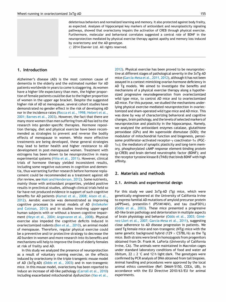
Sham-operated NTg and Tg mice showed a pattern of runningactivity similar to that seen in previous studies with youngerfemale mice of the same strains (Garcıa-Mesa et al., 2011),with Tg mice being more active than NTg ones (Fig. 1A).Ovariectomy induced a general decrease in running activitythat was greater in NTg than Tg mice, with respectivedecreases of 63% and 28% compared to the activity of corre-sponding sham-operated group. There were statisticaleffects of the genotype [F1,32 = 88.24, p < 0.001] and ovar-iectomy [F1,32 = 142.2, p < 0.001] factors, and an interactiongenotype � ovariectomy [F1,32 = 11.89, p = 0.002] on therunning distance.
The measure of grip strength of NTg mice was44.0 � 4.6 gf. Tg mice showed 113.2 � 7.8 gf, in agreement
with a higher muscular strength reported for this Tg mousecolony compared to NTg (Garcıa-Mesa et al., 2011). Results ofgrip strength of the diverse treatment groups are expressedas the percentage of their corresponding NTg or Tg values inFig. 1B. Ovariectomy reduced grip strength in sedentary Tgmice but not in NTg mice, whereas physical exercise gen-erally improved grip strength. There were effects of thegenotype [F1,67 = 51.217, p < 0.001], ovariectomy [F1,67 =4.295, p = 0.030] and exercise [F1,67 = 16.546, p < 0.001]factors, and an interaction genotype � ovariectomy [F1,67
= 8.623, p = 0.005].All experimental groups had similar body weights at the
beginning of the study, with group averages of 20—22 g.Body weights at termination and WAT values are shown inFig. 1C and D, respectively. Ovariectomy induced a generaldecrease in body weight [ovariectomy factor, F1,67 = 9.956,p = 0.003]. WAT values were increased in ovariectomizedNTg mice, whereas physical exercise induced a generaldecrease of WAT. There were effects of ovariectomy[F1,67 = 11.424, p = 0.001] and exercise [F1,67 = 9.837,p = 0.003] and an interaction genotype � ovariectomy[F1,67 = 4.159, p = 0.046] on WAT. Mouse uterus weight valuesare shown in Fig. 1E. Uterus weight decreased significantly inall ovariectomized mice [F1,67 = 5.732, p < 0.001], a clearindication of the efficacy of ovariectomy and consequentlack of ovarian hormones.
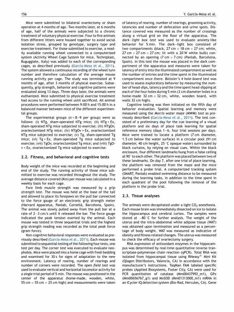
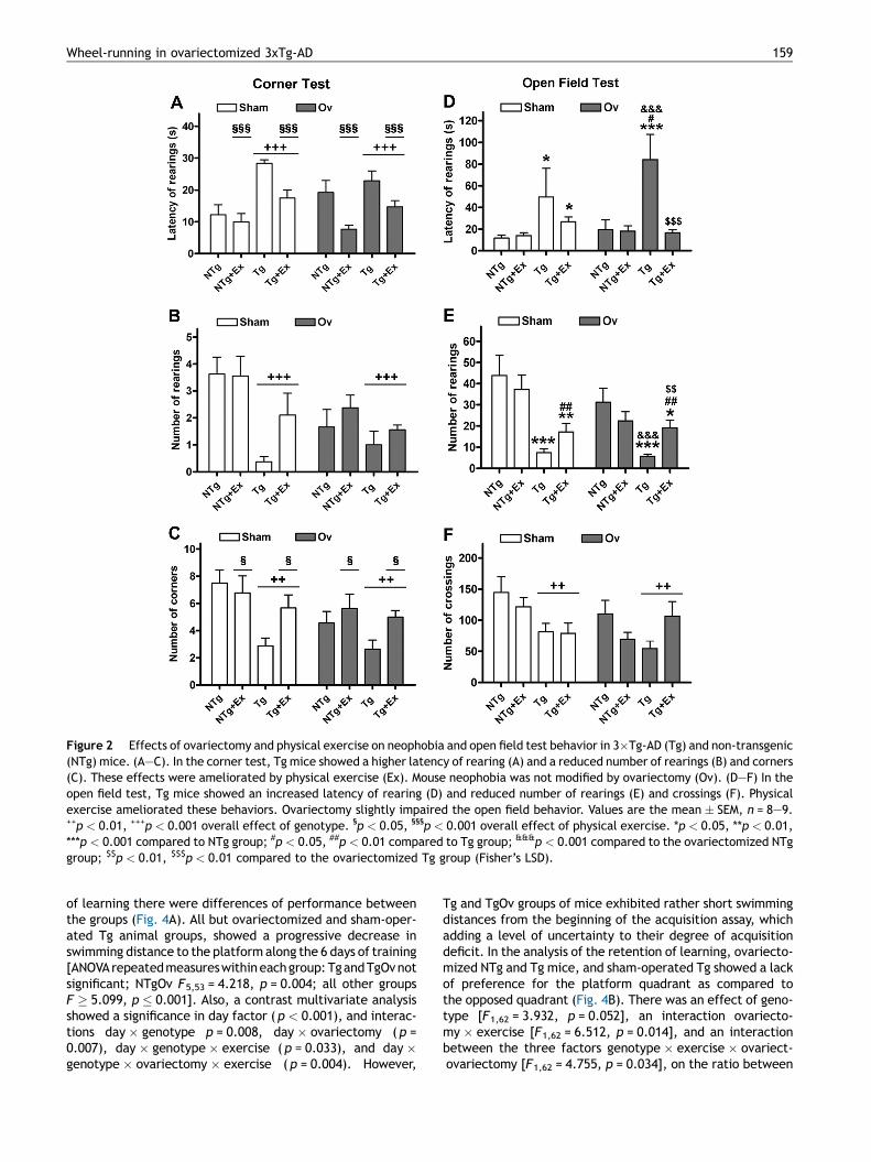
The behavioral tests confirmed the phenotype of neophobia,anxiety and reduced exploratory activity of these Tg miceand their protection by way of a therapy of physical exercise(Garcıa-Mesa et al., 2011, 2012). Selected results are shownin Figs. 2 and 3. Tg mice showed higher latencies of rearing(Fig. 2A), decreased number of rearings (Fig. 2B), andreduced number of corners (Fig. 2C) in the corned test[genotype factor, all F’s � 8.620, p � 0.007]. Ovariectomydid not induce significant changes in neophobia behavior.However, it induced a marginal decrease on the number ofrearings ( p = 0.065) and the number of corners ( p = 0.053).Physical exercise improved the latency of rearing and thenumber of corners of the Tg mouse groups [exercise factor,F’s � 4.706, p � 0.034].
In the open field test, Tg mouse groups showed higherlatency of rearing (Fig. 2D), a decreased number of rearings(Fig. 2E) and a decreased number of crossings (Fig. 2F)[genotype factor, all F’s � 6.580, p � 0.013]. Ovariecto-mized Tg mice showed more significant changes thansham-operated Tg mice (Fig. 2D), but ovariectomy factordid not reach significance. Physical exercise improved theresponse of Tg groups in the latency of rearing and thenumber of rearings [interaction genotype � exercise for bothparameters, F’s � 6.950, p � 0.011; effect of exercise on thenumber of rearings F1,67 = 6.756, p = 0.012]. Other changessuch as those of grooming activity and emotionality (numberof urine spots and defecation boli) did not reach a level ofsignificance (not shown).
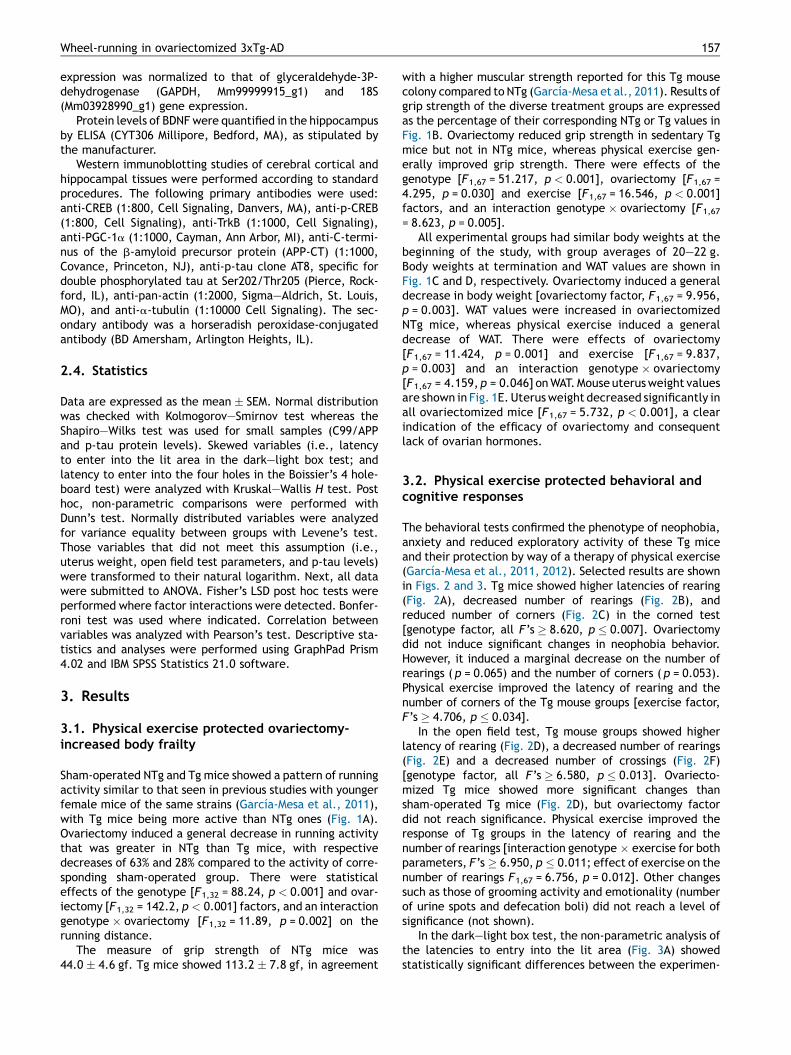
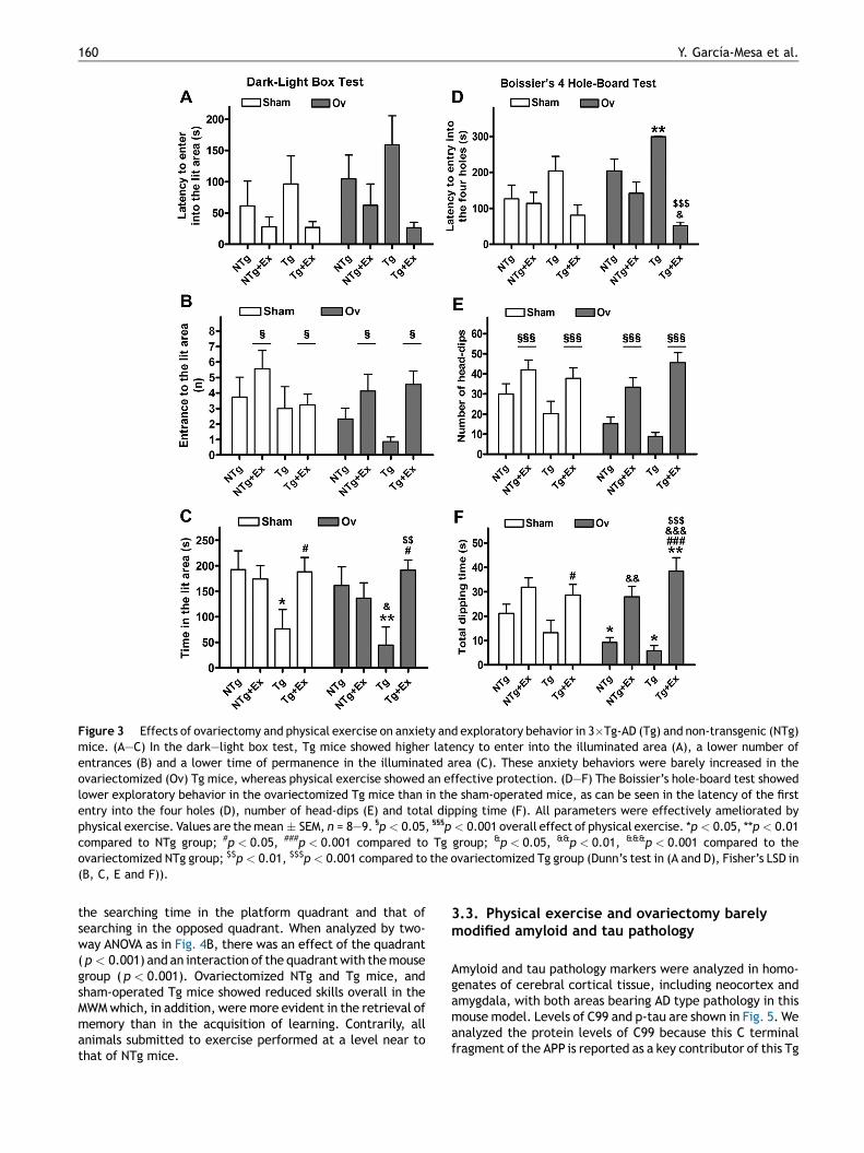
In the dark—light box test, the non-parametric analysis ofthe latencies to entry into the lit area (Fig. 3A) showedstatistically significant differences between the experimen-
Figure 1 Effects of ovariectomy on running activity (A) and ovariectomy and physical exercise on frailty status (B—D) in 3�Tg-AD (Tg)and non-transgenic (NTg) mice. (A) Tg mice submitted to physical exercise (Ex) through free access to a running wheel were moreactive than NTg mice, whereas ovariectomized (Ov) mice showed lower running activity than those of the same strain in sedentaryconditions. (B) Physical exercise increased grip strength in NTg mice, both control and ovariectomized mice. Exercise protected againsta decrease in grip strength seen in TgOv mice. (C) Ovariectomized mice showed decreased body weight. (D) White abdominal fat (WAT)was higher in ovariectomized NTg mice, whereas exercise showed a tendency to decrease WAT in most groups of mice. (E) Uterus weightis included to monitor the efficacy of ovariectomy. Values are the mean � SEM, n = 8—9. DDp < 0.01, DDDp < 0.001 overall effect ofovariectomy in (C and E). **p < 0.01, ***p < 0.001 compared to NTg + Ex group, and ###p < 0.001 compared to Tg + Ex group in (A).**p < 0.01 compared to NTg group, ##p < 0.01 compared to Tg group, &&p < 0.01 compared to the ovariectomized NTg group, $p < 0.05compared to the ovariectomized Tg group in (B and D) (Fisher’s LSD post hoc test).
158 Y. Garcıa-Mesa et al.
tal groups [H7 = 17.891, p = 0.012]. Post hoc comparisonsindicated higher latencies in the dark—light box for the Tggroup and the ovariectomized NTg and Tg, as well as protec-tion through physical exercise. Tg mice showed a decliningtrend in the number of entrances (Fig. 3B) and the time spentin the lit area (Fig. 3C), but the genotype factor not reachingsignificance. Physical exercise induced an increase in bothparameters [all F’s � 5.740, p � 0.020], although effects onthe time spent in the lit area were higher in Tg mice thanthe NTg [interaction genotype � exercise, F1,67 = 11.258,p = 0.001].
Ovariectomized Tg mice showed lower performance thansham-operated Tg mice in the hole-board test. Seven out of 8mice of the former group did not explore the four different
holes during the 5 min test, reaching the highest latency ofthe different experimental groups (Fig. 3D) [H7 = 28.919,p < 0.001]. Ovariectomy also had a marginal effect on thenumber on head-dips ( p = 0.051). Physical exercise totallyprotected from this apathetic behavior. Genotype factor didnot reach significance with respect to the number of head-dips (Fig. 3E) or the dipping time (Fig. 3F), whereas exerciseincreased both parameters [all F’s � 39.549, p < 0.001].Furthermore, the time of dipping was increased more notablyby exercise in the ovariectomized than in the sham-operatedmice [interaction ovariectomy � exercise, F1,67 = 4.645,p = 0.035].
The MWM test confirmed the known cognitive deficits ofthese Tg mice. Results are shown in Fig. 4. In the acquisition
Figure 2 Effects of ovariectomy and physical exercise on neophobia and open field test behavior in 3�Tg-AD (Tg) and non-transgenic(NTg) mice. (A—C). In the corner test, Tg mice showed a higher latency of rearing (A) and a reduced number of rearings (B) and corners(C). These effects were ameliorated by physical exercise (Ex). Mouse neophobia was not modified by ovariectomy (Ov). (D—F) In theopen field test, Tg mice showed an increased latency of rearing (D) and reduced number of rearings (E) and crossings (F). Physicalexercise ameliorated these behaviors. Ovariectomy slightly impaired the open field behavior. Values are the mean � SEM, n = 8—9.++p < 0.01, +++p < 0.001 overall effect of genotype. §p < 0.05, §§§p < 0.001 overall effect of physical exercise. *p < 0.05, **p < 0.01,***p < 0.001 compared to NTg group; #p < 0.05, ##p < 0.01 compared to Tg group; &&&p < 0.001 compared to the ovariectomized NTggroup; $$p < 0.01, $$$p < 0.01 compared to the ovariectomized Tg group (Fisher’s LSD).
Wheel-running in ovariectomized 3xTg-AD 159
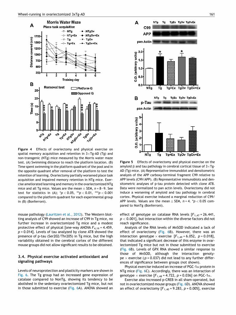
of learning there were differences of performance betweenthe groups (Fig. 4A). All but ovariectomized and sham-oper-ated Tg animal groups, showed a progressive decrease inswimming distance to the platform along the 6 days of training[ANOVA repeated measures within each group: Tg and TgOv notsignificant; NTgOv F5,53 = 4.218, p = 0.004; all other groupsF � 5.099, p � 0.001]. Also, a contrast multivariate analysisshowed a significance in day factor ( p < 0.001), and interac-tions day � genotype p = 0.008, day � ovariectomy ( p =0.007), day � genotype � exercise ( p = 0.033), and day �genotype � ovariectomy � exercise ( p = 0.004). However,
Tg and TgOv groups of mice exhibited rather short swimmingdistances from the beginning of the acquisition assay, whichadding a level of uncertainty to their degree of acquisitiondeficit. In the analysis of the retention of learning, ovariecto-mized NTg and Tg mice, and sham-operated Tg showed a lackof preference for the platform quadrant as compared tothe opposed quadrant (Fig. 4B). There was an effect of geno-type [F1,62 = 3.932, p = 0.052], an interaction ovariecto-my � exercise [F1,62 = 6.512, p = 0.014], and an interactionbetween the three factors genotype � exercise � ovariect-ovariectomy [F1,62 = 4.755, p = 0.034], on the ratio between
Figure 3 Effects of ovariectomy and physical exercise on anxiety and exploratory behavior in 3�Tg-AD (Tg) and non-transgenic (NTg)mice. (A—C) In the dark—light box test, Tg mice showed higher latency to enter into the illuminated area (A), a lower number ofentrances (B) and a lower time of permanence in the illuminated area (C). These anxiety behaviors were barely increased in theovariectomized (Ov) Tg mice, whereas physical exercise showed an effective protection. (D—F) The Boissier’s hole-board test showedlower exploratory behavior in the ovariectomized Tg mice than in the sham-operated mice, as can be seen in the latency of the firstentry into the four holes (D), number of head-dips (E) and total dipping time (F). All parameters were effectively ameliorated byphysical exercise. Values are the mean � SEM, n = 8—9. §p < 0.05, §§§p < 0.001 overall effect of physical exercise. *p < 0.05, **p < 0.01compared to NTg group; #p < 0.05, ###p < 0.001 compared to Tg group; &p < 0.05, &&p < 0.01, &&&p < 0.001 compared to theovariectomized NTg group; $$p < 0.01, $$$p < 0.001 compared to the ovariectomized Tg group (Dunn’s test in (A and D), Fisher’s LSD in(B, C, E and F)).
160 Y. Garcıa-Mesa et al.
the searching time in the platform quadrant and that ofsearching in the opposed quadrant. When analyzed by two-way ANOVA as in Fig. 4B, there was an effect of the quadrant( p < 0.001) and an interaction of the quadrant with the mousegroup ( p < 0.001). Ovariectomized NTg and Tg mice, andsham-operated Tg mice showed reduced skills overall in theMWM which, in addition, were more evident in the retrieval ofmemory than in the acquisition of learning. Contrarily, allanimals submitted to exercise performed at a level near tothat of NTg mice.
3.3. Physical exercise and ovariectomy barelymodified amyloid and tau pathology
Amyloid and tau pathology markers were analyzed in homo-genates of cerebral cortical tissue, including neocortex andamygdala, with both areas bearing AD type pathology in thismouse model. Levels of C99 and p-tau are shown in Fig. 5. Weanalyzed the protein levels of C99 because this C terminalfragment of the APP is reported as a key contributor of this Tg
Figure 5 Effects of ovariectomy and physical exercise on theamyloid b and tau pathology in cerebral cortical tissue of 3�Tg-AD (Tg) mice. (A) Representative immunoblot and densitometricanalysis of the APP carboxy-terminal fragment C99 relative toAPP levels (C99/APP). (B) Representative immunoblots and den-sitometric analyses of p-tau protein detected with clone AT8.Data were normalized to pan actin levels. Ovariectomy did notinduce a worsening of amyloid and tau pathology in cerebralcortex. Physical exercise induced a marginal reduction of C99/APP levels. Values are the mean � SEM, n = 4. *p < 0.05 com-pared to NonTg (Bonferroni).
Figure 4 Effects of ovariectomy and physical exercise onspatial memory acquisition and retention in 3�Tg-AD (Tg) andnon-transgenic (NTg) mice measured by the Morris water mazetest. (A) Swimming distance to reach the platform location. (B)Time spent swimming in the platform quadrant of the pool and inthe opposite quadrant after removal of the platform to test theretention of learning. Ovariectomy partially worsened place taskacquisition and impaired memory retention in NTg mice. Exer-cise ameliorated learning and memory in the ovariectomized NTgmice and all Tg mice. Values are the mean � SEM, n = 8—9. Seetext for statistics in (A); *p < 0.05, **p < 0.01, ***p < 0.001compared to the platform quadrant for each experimental groupin (B) (Bonferroni).
Wheel-running in ovariectomized 3xTg-AD 161
mouse pathology (Lauritzen et al., 2012). The Western blot-ting analysis of C99 showed an increase of C99 in Tg mice, nofurther increase in ovariectomized Tg mice and a modestprotective effect of physical [one-way ANOVA F4,19 = 4.459,p = 0.014]. Levels of tau analyzed by clone AT8 showed thepresence of p-tau (Ser202/Thr205) in Tg mice, but the highvariability obtained in the cerebral cortex of the differentmouse groups did not allow significant results to be obtained.
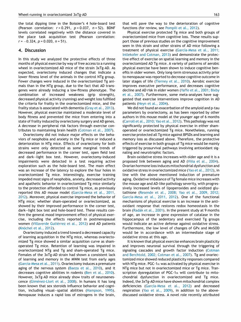
Levels of neuroprotection and plasticity markers are shown inFig. 6. The Tg group had an increased gene expression ofcatalase compared to NonTg, showing its tendency to beabolished in the sedentary ovariectomized Tg mice, but notin those submitted to exercise (Fig. 6A). ANOVA showed an
effect of genotype on catalase RNA levels [F1,47 = 26.441,p < 0.001], but interaction within the diverse factors did notreach significance.
Analysis of the RNA levels of MnSOD indicated a lack ofeffect of ovariectomy (Fig. 6B). However, there was aninteraction genotype � exercise [F1,49 = 6.052, p = 0.018],that indicated a significant decrease of this enzyme in ovar-iectomized Tg mice but not in those submitted to exercise(Fig. 6B). Levels of GPX RNA showed a similar response tothose of MnSOD, although the interaction genoty-pe � exercise ( p = 0.037) did not lead to any further differ-ences of significance between groups (not shown).
Physical exercise induced an increase of PGC-1a protein inNTg mice (Fig. 6C). Accordingly, there was an interaction ofgenotype � exercise [F1,48 = 4.722, p = 0.036] on PGC-1a.
Exercise also increased p-CREB in all sham-operated, butnot in ovariectomized mouse groups (Fig. 6D). ANOVA showedan effect of ovariectomy [F1,40 = 9.283, p = 0.005], exercise
Figure 6 Effects of ovariectomy and physical exercise on the levels of antioxidant and neuroplasticity signaling molecules in thehippocampus of 3�Tg-AD (Tg) and non-transgenic (NTg) mice. (A) Catalase, RNA levels. (B) MnSOD, RNA levels. (C) PGC-1a, and (D) p-CREB/CREB, protein levels by Western blot (representative immunoblots and densitometric analysis; data were normalized to a-tubulin levels). (E) BDNF, protein levels by ELISA. (F) TrkB, protein levels by Western blot. Catalase was increased in Tg mice.Ovariectomy inhibited the induction of p-CREB through physical exercise but did not significantly interact with the induction of PGC-1a
and BDNF. Values are the mean � SEM, n = 5—8. +p < 0.05 overall effect of genotype; §§§p < 0.001 overall effect of physical exercise.*p < 0.05, **p < 0.01 compared to NTg group; ##p < 0.01 compared to Tg group (Fisher’s LSD).
162 Y. Garcıa-Mesa et al.
[F1,40 = 9.880, p = 0.004], and an interaction between bothfactors [F1,40 = 8.095, p = 0.008] in p-CREB protein levels.
BDNF levels were generally increased by exercise, includ-ing Tg and all ovariectomized mice (Fig. 6E) [exercise factor,F1,51 = 11.867, p = 0.001]. Interaction genotype � exercisedid not reach significance ( p = 0.064), although sham-oper-ated NTg mice showed an unexpected lack of response. Asregards to BDNF receptor, the protein levels of TrkB were
generally decreased in Tg mice [genotype factor,F1,51 = 5.547, p = 0.023], without changes induced throughexercise or ovariectomy (Fig. 6F).
Correlation studies showed that PGC-1a, p-CREB and TrkBwere inter-correlated (all Pearson correlations: r � 0.549,p � 0.001). Levels of BDNF showed a positive correlation withthe time in the illuminated area in the dark—light box test(Pearson correlation: r = 0.325, p = 0.019, n = 52), and with
Wheel-running in ovariectomized 3xTg-AD 163
the total dipping time in the Boissier’s 4 hole-board test(Pearson correlation: r = 0.291, p = 0.037, n = 52). BDNFlevels correlated negatively with the distance covered inthe place task acquisition test (Pearson correlation:r = �0.324, p = 0.020, n = 51).
4. Discussion
In this study we analyzed the protective effects of threemonths of physical exercise by way of free access to a runningwheel in ovariectomized AD mice of the strain 3xTg-AD. Asexpected, ovariectomy induced changes that indicate alower fitness level of the animals in the control NTg group.Fewer changes were induced in the ovariectomized Tg ani-mals than in the NTg group, due to the fact that AD trans-genes were already inducing a low-fitness phenotype. Thecombination of increased weakness (grip strength),decreased physical activity (running) and weight loss fulfilledthe criteria for frailty in the ovariectomized mice, and thefrailty status is associated with dementia (Gray et al., 2013).However, physical exercise maintained a moderate level ofbody fitness and prevented the mice from entering into astate of frailty induced by ovariectomy surgery and AD genes.A decrease in peripheral risk factors through exercise con-tributes to maintaining brain health (Cotman et al., 2007).
Ovariectomy did not induce major effects on the beha-viors of neophobia and anxiety in the Tg mice or induced adeterioration in NTg mice. Effects of ovariectomy for bothstrains were only detected as some marginal trends ofdecreased performance in the corner test, open field testand dark—light box test. However, ovariectomy-inducedimpairments were detected in a test requiring activeexploration such as the hole-board test. The main effectwas an increase of the latency to explore the four holes inovariectomized Tg mice. Interestingly, exercise trainingimpeded most signs of neophobia, anxiety, decreased activityand apathetic behavior in ovariectomized Tg mice similarlyto the protection afforded to control Tg mice, as previouslyreported this AD mouse model (Garcıa-Mesa et al., 2011,2012). Moreover, physical exercise improved the behavior ofNTg mice; whether sham-operated or ovariectomized, asshowed by their improved performance in the corner test,dark—light box test and hole-board test. These results con-firm the general mood improvement effect of physical exer-cise, including the effects reported in postmenopausalwomen (Villaverde Gutierrez et al., 2012) and AD patients(Knochel et al., 2012).
Ovariectomy induced a trend toward a decreased capacityof learning acquisition in the NTg mice, whereas ovariecto-mized Tg mice showed a similar acquisition curve as sham-operated Tg mice. Retention of learning was impaired inovariectomized NTg and Tg and sham-operated Tg mice.Females of the 3xTg-AD strain had shown a consistent lackof learning and memory in the MWM test from early ages(Garcıa-Mesa et al., 2011). Ovariectomy induces a prematureaging of the nervous system (Baeza et al., 2010), and itdecreases cognitive abilities in rodents (Ben et al., 2010).However, 3xTg-AD mice already show traits of neurosenes-cence (Gimenez-Llort et al., 2008). In humans it has longbeen known that sex steroids influence behavior and cogni-tion, including visual—spatial abilities (Hampson, 1995).Menopause induces a rapid loss of estrogens in the brain,
that will pave the way to the deterioration of cognitivefunctions (for review, see Pompili et al., 2012).
Physical exercise protected Tg mice and both groups ofovariectomized mice from cognitive loss. These results sup-port those of previous studies on the cognitive improvementseen in this strain and other strains of AD mice following atreatment of physical exercise (Garcıa-Mesa et al., 2011;Intlekofer and Cotman, 2013) and demonstrate the protec-tive effect of exercise on spatial learning and memory in theovariectomized AD Tg mice. A variety of patterns of aerobicphysical exercise have been shown to induce cognitive ben-efits in older women. Only long-term strenuous activity priorto menopause was reported to decrease cognitive outcome inlater stages of life (Tierney et al., 2010). Aerobic exerciseimproves executive performance, and decreases cognitivedecline and AD risk in elder women (Yaffe et al., 2001; Bixbyet al., 2007). Furthermore, some meta-analyses have sug-gested that exercise interventions improve cognition in ADpatients (Heyn et al., 2004).
We did not found an exacerbation of the amyloid and p-tauparameters by ovariectomy, as has been reported by otherauthors in this mouse model at the younger age of 6 months(Carroll et al., 2010; Yao et al., 2012). This pathology was notsignificantly protected by physical exercise in either sham-operated or ovariectomized Tg mice. Nonetheless, runningexercise protected all Tg mice against BPSDs and learning andmemory loss as discussed above. Therefore, the protectiveeffects of exercise in both groups of Tg mice would be mainlytriggered by prosurvival pathways involving antioxidant sig-naling and neurotrophic factors.
Brain oxidative stress increases with older age and it is aproposed link between aging and AD (Vina et al., 2004).Ovarian hormone loss induces mitochondrial dysfunction andoxidative stress in ovariectomized mice (Yao et al., 2012), inline with the above mentioned induction of prematureaging. Oxidative imbalance of Tg female mice increases withthe mouse age and AD-like pathology severity, with progres-sively increased levels of lipoperoxides and oxidized glu-tathione (Resende et al., 2008; Yao et al., 2009, 2012;Garcıa-Mesa et al., 2011, 2012). One of the functionalmechanisms of physical exercise is an increase in the anti-oxidant response that restores redox homeostasis in thebrain (Radak et al., 2001). In the present study at 9 monthsof age, an increase in gene expression of catalase in thehippocampus of the sedentary and exercised Tg groupswould indicate an active defense against oxidative stress.Furthermore, the low level of changes of GPx and MnSODwould be in accordance with an intermediate stage ofoxidative stress at this age.
It is known that physical exercise enhances brain plasticityand improves neuronal survival through the triggering ofsignaling cascades and growth factor induction (Cotmanand Berchtold, 2002; Cotman et al., 2007). Tg and ovariec-tomized mice showed reduced plasticity responses comparedwith NTg mice. PGC-1a was activated by physical exercise inNTg mice but not in ovariectomized mice or Tg mice. Tran-scription dysregulation of PGC-1a will contribute to mito-chondrial dysfunction in ovariectomized and Tg mice.Indeed, the 3xTg-AD mice have shown mitochondrial complexdeficiencies (Garcıa-Mesa et al., 2012) and decreasedrespiration (Yao et al., 2009), in addition to the above-discussed oxidative stress. A novel role recently attributed
164 Y. Garcıa-Mesa et al.
to PGC-1a is and the formation and maintenance of dendriticspines and maturation of synapses in hippocampal neurons(Cheng et al., 2012). As regards CREB, impaired CREB signal-ing has been implicated in memory deficits associated withaging and AD (Saura and Valero, 2011). As expected, chronicwheel-running activity increased p-CREB in the sham-oper-ated NTg mice. Furthermore, Tg mice showed a similarresponse of CREB activation. Nonetheless we did not observebasal changes of p-CREB, whereas other authors havereported either a decrease (Caccamo et al., 2010) or anearly increase in this mouse model (Muller et al., 2011).The impairment of this plasticity pathway following ovar-iectomy supported previously reported decreases of hippo-campal p-CREB in ovariectomized rats (Sharma et al., 2007).Thus, deficient signaling of this transduction factor is prob-ably implicated in the cognitive deficits of Tg and ovariecto-mized mice.
In this study the exercise-induced increase of BDNFstrongly correlated with behavioral responses of decreasedanxiety, increased active exploration, and improved learn-ing. Therefore, hippocampal levels of BDNF increased coor-dinately with the behavioral and cognitive improvement inovariectomized and Tg groups, thus reinforcing the media-tion of BDNF in the neuroprotective changes induced byphysical exercise (Cotman and Berchtold, 2002; Adlardet al., 2005; Cotman et al., 2007). In humans, increasingBDNF through aerobic exercise was reported to ameliorateage-related hippocampal atrophy and memory dysfunctionand reduce depression (Erickson et al., 2012). We observed adecrease of TrkB expression in the hippocampus of the Tgmice, which would contribute to the derangements of BDNFsignaling in this mouse model. However we did not find asignificant decrease of BDNF in the hippocampus of thismouse model, as previously reported (Caccamo et al.,2010). Activation of TrkB receptors by BDNF induces severalsignaling pathways (Santos et al., 2010) that would convergein the synaptic plasticity and neurotransmission improve-ments required for the mood-related and memory improve-ments discussed previously. An effect of increased levels ofBDNF by physical exercise was noticeable in Tg mice andovariectomized NTg and Tg mice, but not in sham-operatedNTg. Therefore, it appears that BDNF was increased throughexercise when the hippocampal signaling was disturbed.Alternatively, middle-aged NTg mice submitted to chronicexercise training exhibited a lesser degree of BDNF increasethan expected for young mice (Adlard et al., 2005). Further-more, differential induction of BDNF could be influenced bythe complex interactions that occur between ovarian ster-oids and BDNF in the hippocampus (Franklin and Perrot-Sinal,2006; Scharfman and Maclusky, 2014). Studies in humans havefound either positive or negative correlations between phy-sical exercise and BNDF levels in blood (Vega et al., 2011; Leeet al., 2014), thus highlighting the complexity of BDNFregulation.
In summary, ovariectomy induced a deterioration of fit-ness and cognition in NTg mice and a worsening of apathyresponses in the Tg mice. However, ovariectomy did notinduce a significant exacerbation of AD pathology. Nonethe-less, three months of physical exercise in a freely availablerunning wheel protected 9-month old Tg and NTg femalemice submitted to ovariectomy (at two month before startingthe exercise training) against brain alterations and lost of
brain plasticity. Physical exercise induced a recovery of BPSD-like changes in Tg groups, and cognitive loss as seen in Tgmice and in ovariectomized Tg and NTg mice. Neuroprotec-tion in Tg mice did not appear mediated through a reductionof amyloid or tau pathology, but rather through the enhance-ment of mechanisms of neuroprotection and plasticity, suchas those involving catalase, p-CREB and BDNF. Remarkably,there was a positive correlation between BDNF levels andamelioration of behavior and cognition. These results confirmthe value of moderate aerobic exercise training as a lifestylethat effectively induces neuroprotection in postmenopausalwomen and reinforce the role of BDNF as a molecular effectorof physical exercise neuroprotection.
Role of the funding source
The authors declare that the founding sources cited in theacknowledgements section had no further role in this study.
Conflict of interest
The authors declare that there are no actual or potentialconflicts of interest.
Acknowledgements
This study was supported by grants: SAF2009-13093-C02-02,SAF2010-19498, SAF2012-39852-C02-02 and CSD2010-00045from the Spanish MINECO; 2009/SGR/214 from the General-itat and 062931 from the Fundacio La Marato de TV3, ofCatalonia; and 35NEURO GentxGent. Yoelvis Garcıa-Mesaacknowledges support received from the Fundacio La Maratode TV3. We thank Jofre Serret for his skillful technicalassistance. We are indebted to Dr Silvia Busquets from theUniversitat de Barcelona, for letting us use her grip strengthmeasuring apparatus and her advice with the measurements.
References
Aisen, P.S., Cummings, J., Schneider, L.S., 2012. Symptomatic andnonamyloid/tau based pharmacologic treatment for Alzheimerdisease. Cold Spring Harb. Perspect. Med. 2, a006395.
Adlard, P.A., Perreau, V.M., Cotman, C.W., 2005. The exercise-induced expression of BDNF within the hippocampus varies acrosslife-span. Neurobiol. Aging 26, 511—520.
Angevaren, M., Aufdemkampe, G., Verhaar, H.J., Aleman, A., Van-hees, L., 2008. Physical activity and enhanced fitness to improvecognitive function in older people without known cognitive im-pairment. Cochrane Database Syst. Rev. CD005381.
Baeza, I., De Castro, N.M., Gimenez-Llort, L., De la Fuente, M., 2010.Ovariectomy, a model of menopause in rodents, causes a prema-ture aging of the nervous and immune systems. J. Neuroimmunol.219, 90—99.
Barnes, L.L., Wilson, R.S., Schneider, J.A., Bienias, J.L., Evans, D.A.,Bennett, D.A., 2003. Gender, cognitive decline, and risk of AD inolder persons. Neurology 60, 1777—1781.
Ben, J., Soares, F.M., Scherer, E.B., Cechetti, F., Netto, C.A., Wyse,A.T., 2010. Running exercise effects on spatial and avoidancetasks in ovariectomized rats. Neurobiol. Learn. Mem. 94, 312—317.
Bixby, W.R., Spalding, T.W., Haufler, A.J., Deeny, S.P., Mahlow, P.T.,Zimmerman, J.B., Hatfield, B.D., 2007. The unique relation of
physical activity to executive function in older men and women.Med. Sci. Sports Exerc. 39, 1408—1416.
Caccamo, A., Maldonado, M.A., Bokov, A.F., Majumder, S., Oddo, S.,2010. CBP gene transfer increases BDNF levels and ameliorateslearning and memory deficits in a mouse model of Alzheimer’sdisease. Proc. Natl. Acad. Sci. U. S. A. 107, 22687—22692.
Carroll, J.C., Rosario, E.R., Villamagna, A., Pike, C.J., 2010. Contin-uous and cyclic progesterone differentially interact with estradiolin the regulation of Alzheimer-like pathology in female3�Transgenic-Alzheimer’s disease mice. Endocrinology 151,2713—2722.
Cheng, A., Wan, R., Yang, J.L., Kamimura, N., Son, T.G., Ouyang, X.,Luo, Y., Okun, E., Mattson, M.P., 2012. Involvement of PGC-1a inthe formation and maintenance of neuronal dendritic spines. Nat.Commun. 3, 1250.
Cotman, C.W., Berchtold, N.C., 2002. Exercise: a behavioral inter-vention to enhance brain health and plasticity. Trends Neurosci.25, 295—301.
Erickson, K.I., Miller, D.L., Roecklein, K.A., 2012. The aging hippo-campus: interactions between exercise, depression, and BDNF.Neuroscientist 18, 82—97.
Franklin, T.B., Perrot-Sinal, T.S., 2006. Sex and ovarian steroidsmodulate brain-derived neurotrophic factor (BDNF) protein levelsin rat hippocampus under stressful and non-stressful conditions.Psychoneuroendocrinology 31, 38—48.
Garcıa-Mesa, Y., Gimenez-Llort, L., Lopez, L.C., Venegas, C., Cris-tofol, R., Escames, G., Acuna-Castroviejo, D., Sanfeliu, C., 2012.Melatonin plus physical exercise are highly neuroprotective in the3�Tg-AD mouse. Neurobiol. Aging 33 1124.e13—29.
Garcıa-Mesa, Y., Lopez-Ramos, J.C., Gimenez-Llort, L., Revilla, S.,Guerra, R., Gruart, A., LaFerla, F.M., Cristofol, R., Delgado-Garcıa, J.M., Sanfeliu, C., 2011. Physical exercise protectsagainst Alzheimer’s disease in 3�Tg-AD mice. J. AlzheimersDis. 24, 421—454.
Gimenez-Llort, L., Arranz, L., Mate, I., De la Fuente, M., 2008.Gender-specific neuroimmunoendocrine aging in a triple-trans-genic 3�Tg-AD mouse model for Alzheimer’s disease and itsrelation with longevity. Neuroimmunomodulation 15, 331—343.
Gimenez-Llort, L., Blazquez, G., Canete, T., Johansson, B., Oddo, S.,Tobena, A., LaFerla, F.M., Fernandez-Teruel, A., 2007. Modelingbehavioral and neuronal symptoms of Alzheimer’s disease inmice: a role for intraneuronal amyloid. Neurosci. Biobehav.Rev. 31, 125—147.
Gray, S.L., Anderson, M.L., Hubbard, R.A., Lacroix, A., Crane, P.K.,McCormick, W., Bowen, J.D., McCurry, S.M., Larson, E.B., 2013.Frailty and incident dementia. J. Gerontol. A: Biol. Sci. Med. Sci.68, 1083—1090.
Hampson, E., 1995. Spatial cognition in humans: possible modulationby androgens and estrogens. J. Psychiatry Neurosci. 20, 397—404.
Hebert, L.E., Scherr, P.A., McCann, J.J., Beckett, L.A., Evans, D.A.,2001. Is the risk of developing Alzheimer’s disease greater forwomen than for men? Am. J. Epidemiol. 153, 132—136.
Heyn, P., Abreu, B.C., Ottenbacher, K.J., 2004. The effects ofexercise training on elderly persons with cognitive impairmentand dementia: a meta-analysis. Arch. Phys. Med. Rehabil. 85,1694—1704.
Intlekofer, K.A., Cotman, C.W., 2013. Exercise counteracts declininghippocampal function in aging and Alzheimer’s disease. Neuro-biol. Dis. 57, 47—55.
Kamat, C.D., Gadal, S., Mhatre, M., Williamson, K.S., Pye, Q.N.,Hensley, K., 2008. Antioxidants in central nervous system dis-eases: preclinical promise and translational challenges. J. Alz-heimers Dis. 15, 473—493.
Knochel, C., Oertel-Knochel, V., O‘Dwyer, L., Prvulovic, D., Alves, G.,Kollmann, B., Hampel, H., 2012. Cognitive and behavioural
effects of physical exercise in psychiatric patients. Prog. Neuro-biol. 96, 46—68.
Lauritzen, I., Pardossi-Piquard, R., Bauer, C., Brigham, E., Abraham,J.D., Ranaldi, S., Fraser, P., St-George-Hyslop, P., Le Thuc, O.,Espin, V., Chami, L., Dunys, J., Checler, F., 2012. The b-secretase-derived C-terminal fragment of bAPP, C99, but not Ab, is a keycontributor to early intraneuronal lesions in triple-transgenicmouse hippocampus. J. Neurosci. 32, 16243—21655.
Lee, T.M., Wong, M.L., Lau, B.W., Lee, J.C., Yau, S.Y., So, K.F., 2014.Aerobic exercise interacts with neurotrophic factors to predictcognitive functioning in adolescents. Psychoneuroendocrinology39, 214—224.
Maki, P.M., Henderson, V.W., 2012. Hormone therapy, dementia, andcognition: the Women’s Health Initiative 10 years on. Climacteric15, 256—262.
Muller, M., Cardenas, C., Mei, L., Cheung, K.H., Foskett, J.K., 2011.Constitutive cAMP response element binding protein (CREB) acti-vation by Alzheimer’s disease presenilin-driven inositol trispho-sphate receptor (InsP3R) Ca2+ signaling. Proc. Natl. Acad. Sci. U.S. A. 108, 13293—13298.
Oddo, S., Caccamo, A., Shepherd, J.D., Murphy, M.P., Golde, T.E.,Kayed, R., Metherate, R., Mattson, M.P., Akbari, Y., LaFerla, F.M.,2003. Triple-transgenic model of Alzheimer’s disease with plaquesand tangles: intracellular Ab and synaptic dysfunction. Neuron39, 409—421.
Pompili, A., Arnone, B., Gasbarri, A., 2012. Estrogens and memory inphysiological and neuropathological conditions. Psychoneuroen-docrinology 37, 1379—1396.
Radak, Z., Kaneko, T., Tahara, S., Nakamoto, H., Pucsok, J., Sasvari,M., Nyakas, C., Goto, S., 2001. Regular exercise improves cogni-tive function and decreases oxidative damage in rat brain. Neu-rochem. Int. 38, 17—23.
Resende, R., Moreira, P.I., Proenca, T., Deshpande, A., Busciglio, J.,Pereira, C., Oliveira, C.R., 2008. Brain oxidative stress in a triple-transgenic mouse model of Alzheimer disease. Free Radic. Biol.Med. 44, 2051—2057.
Rocca, W.A., Cha, R.H., Waring, S.C., Kokmen, E., 1998. Incidence ofdementia and Alzheimer’s disease: a reanalysis of data fromRochester, Minnesota, 1975—1984. Am. J. Epidemiol. 148, 51—62.
Santos, A.R., Comprido, D., Duarte, C.B., 2010. Regulation of localtranslation at the synapse by BDNF. Prog. Neurobiol. 92, 505—516.
Saura, C.A., Valero, J., 2011. The role of CREB signaling in Alzhei-mer’s disease and other cognitive disorders. Rev. Neurosci. 22,153—169.
Scharfman, H.E., Maclusky, N.J., 2014. Differential regulation ofBDNF, synaptic plasticity and sprouting in the hippocampal mossyfiber pathway of male and female rats. Neuropharmacology 76,696—708.
Sharma, K., Mehra, R.D., Dhar, P., Vij, U., 2007. Chronic exposure toestrogen and tamoxifen regulates synaptophysin and phosphory-lated cAMP response element-binding (CREB) protein expressionin CA1 of ovariectomized rat hippocampus. Brain Res. 1132,10—19.
Tierney, M.C., Moineddin, R., Morra, A., Manson, J., Blake, J., 2010.Intensity of recreational physical activity throughout life andlater life cognitive functioning in women. J. Alzheimers Dis.22, 1331—1338.
Vega, S.R., Kleinert, J., Sulprizio, M., Hollmann, W., Bloch, W.,Struder, H.K., 2011. Responses of serum neurotrophic factorsto exercise in pregnant and postpartum women. Psychoneuroen-docrinology 36, 220—227.
Villaverde Gutierrez, C., Torres Luque, G., Abalos Medina, G.M.,Argente del Castillo, M.J., Guisado, I.M., Guisado Barrilao, R.,Ramırez Rodrigo, J., 2012. Influence of exercise on mood inpostmenopausal women. J. Clin. Nurs. 21, 923—928.
Vina, J., Gambini, J., Lopez-Grueso, R., Abdelaziz, K.M., Jove, M.,Borras, C., 2011. Females live longer than males: role of oxidativestress. Curr. Pharm. Des. 17, 3959—3965.
Vina, J., Lloret, A., Ortı, R., Alonso, D., 2004. Molecular bases of thetreatment of Alzheimer’s disease with antioxidants: prevention ofoxidative stress. Mol. Aspects Med. 25, 117—123.
Yaffe, K., Barnes, D., Nevitt, M., Lui, L.Y., Covinsky, K., 2001. Aprospective study of physical activity and cognitive decline inelderly women: women who walk. Arch. Intern. Med. 161, 1703—1708.
Yao, J., Irwin, R.W., Zhao, L., Nilsen, J., Hamilton, R.T., Brinton,R.D., 2009. Mitochondrial bioenergetic deficit precedes Alzhei-mer’s pathology in female mouse model of Alzheimer’s disease.Proc. Natl. Acad. Sci. U. S. A. 106, 14670—14675.
Yao, J., Irwin, R., Chen, S., Hamilton, R., Cadenas, E., Brinton, R.D.,2012. Ovarian hormone loss induces bioenergetic deficits andmitochondrial b-amyloid. Neurobiol. Aging 33, 1507—1521.