Membrane-mediated interaction of intercellular cylindrical nanoparticles
Zeming Wu and Xin Yi *
Department of Mechanics and Engineering Science, College of Engineering, Peking University, Beijing 100871, P. R. China
(Received 3 May 2021; accepted 20 August 2021; published 7 September 2021)
Nanoparticles in intercellular gaps, junctions, or seals could have close contact with neighboring cellssimultaneously. Understanding the interaction between intercellular nanoparticles and confining cell membranesis of fundamental importance, not only to the unravelling of endocytic mechanisms but also to implicationssuch as controlled drug delivery in tumor tissues. Here we theoretically examine the mechanical behaviorsof adhesive cylindrical nanoparticles confined between two lipid membrane patches of finite size. As the sizeof the particle-membrane contact region or wrapping degree increases, neighboring cylindrical nanoparticlesbecome separated and the nanoparticle distance increases first and then decreases until the particles are fullytrapped by adjacent membrane patches. Depending on the nanoparticle size, adhesion energy, membrane bendingrigidity and tension, and intermembrane distance, three characteristic particle-membrane interaction phases aredetermined as no wrapping, partial trapping, and full trapping, and the corresponding interaction phase diagramis established. Further energy comparison indicates that multiple nanoparticles undergoing the two-membranetrapping process do not exhibit cooperative effects. Analytical estimations on the system energy and configura-tions at equilibrium are performed based on the force balance of the membranes at small deformation and matchwell with numerical solutions. The results shed light on the mechanical behaviors of multiple nanoparticles incell junctions or gaps and may have implications for drug delivery in tumor tissues.
DOI: 10.1103/PhysRevE.104.034403
I. INTRODUCTION
Cellular interaction with nanoparticles has been the sub-ject of renewed focus for decades owing to its fundamentalnecessity in many fields of physical and molecular biology ofcells, nanomedicine, and safe applications of nanotechnology,such as endocytosis, drug delivery, and nanomaterial safety[1–3]. Once the nanoparticles come into contact with a cell,the overall adhesive force arising from either specific bindingor nonspecific interaction between the nanoparticle and cellmembrane, or both, lowers the free energy of interaction,and leads the membrane curving and wrapping around thenanoparticle at the cost of elastic deformation energy frommembrane bending and tension as well as possible particledeformation. The free-energy reduction from the energeticcompetition mentioned above drives the wrapping processuntil the nanoparticle is fully wrapped by the cell mem-brane. Nanoparticles attaching on a cell surface or in a dilutecell cluster are interacting with a single cell membrane, andit has become well-known that the single-membrane wrap-ping around nanoparticles exhibits dependence of particlesize, shape, elasticity and surface functionalization [1–4]. Forexample, the cell uptake of nanoparticles is regulated notonly by the particle stiffness [5] but also the substrate stiff-ness [6]. In cases of two identical spherical nanoparticlesbinding to a membrane, the membrane-mediated interactionbetween particles on the same membrane side could be at-tractive or repulsive, depending on the wrapping degree [7];
while the interaction between spherical particles on the oppo-site membrane sides is always attractive [8]. Two-dimensionaltheoretical studies on the interaction between two rigidcylindrical nanoparticles on a membrane indicate repulsive(attractive) interaction for particles on the same (opposite)membrane side [8–11].
Other intriguing theoretical works on the membrane-mediated interaction between anisotropic nanoparticles at-taching on the membrane [12] or inclusions such as membraneproteins embedded in the membrane [13–17] have also beenperformed. For nanoparticles attaching on the membrane, themembrane surface is intact as a simply connected regionand the membrane-particle contact zone evolves as the mem-brane deforms and wraps around the nanoparticles, whilein the case of membrane inclusion interaction the mem-brane is multiply connected and it could be adopted thatthe membrane-inclusion interface geometry and associatedcontact angles are fixed by the inclusion shape and centralhydrophobic region.
In contrast to the single-membrane wrapping, nanoparti-cles confined between intercellular gaps, junctions, or sealscould form tight contact with neighboring cell membranessimultaneously [18,19]. Understanding mechanical behaviorsof confined nanoparticles and their interaction with confiningmembranes is of importance to deep drug delivery in tumortissues, whose microenvironment is structurally heteroge-neous and contains dense interstitial structures with separationdistances as short as a few or tens of nanometers [20]. So farlittle work has been done in exploring the interaction betweenintercellular nanoparticles and confining cell membranes, par-ticularly from a theoretical viewpoint [21,22]. A recent work
ZEMING WU AND XIN YI PHYSICAL REVIEW E 104, 034403 (2021)
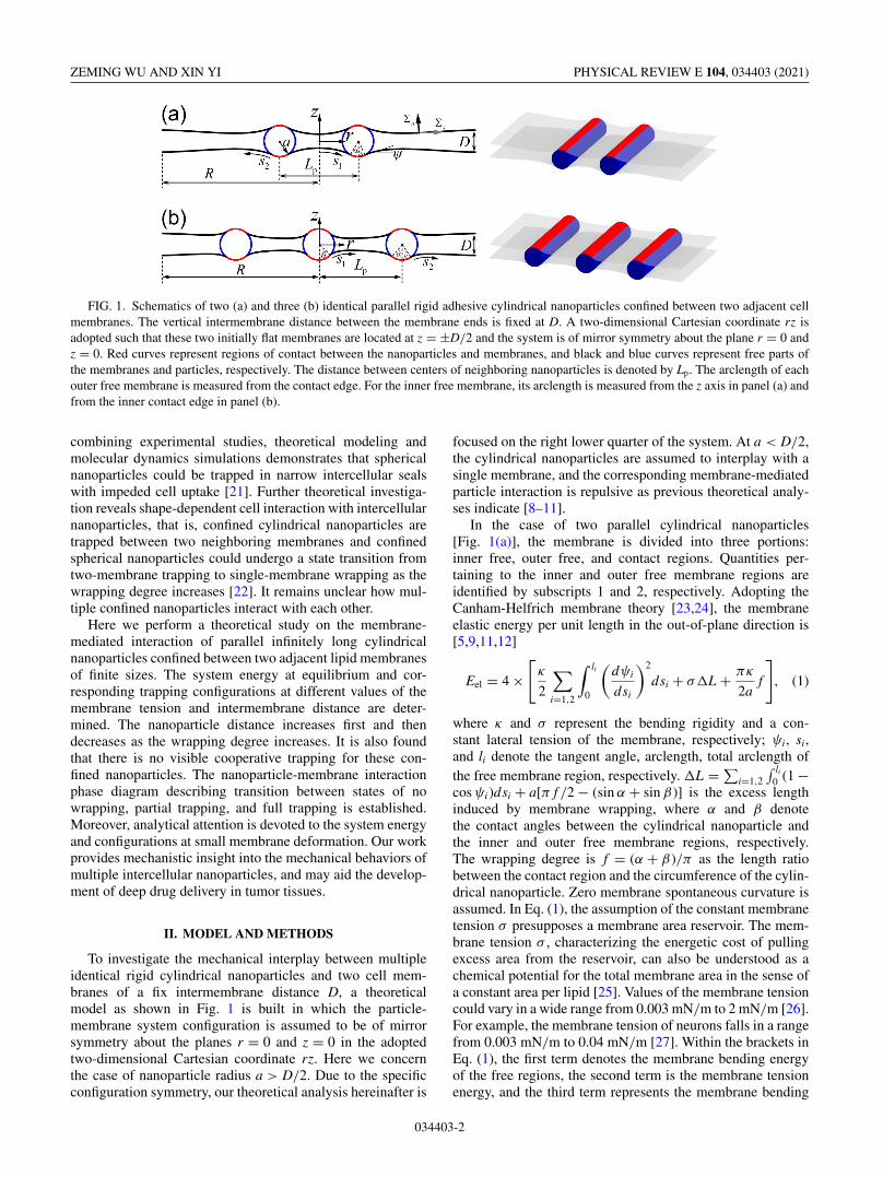
FIG. 1. Schematics of two (a) and three (b) identical parallel rigid adhesive cylindrical nanoparticles confined between two adjacent cellmembranes. The vertical intermembrane distance between the membrane ends is fixed at D. A two-dimensional Cartesian coordinate rz isadopted such that these two initially flat membranes are located at z = ±D/2 and the system is of mirror symmetry about the plane r = 0 andz = 0. Red curves represent regions of contact between the nanoparticles and membranes, and black and blue curves represent free parts ofthe membranes and particles, respectively. The distance between centers of neighboring nanoparticles is denoted by Lp. The arclength of eachouter free membrane is measured from the contact edge. For the inner free membrane, its arclength is measured from the z axis in panel (a) andfrom the inner contact edge in panel (b).
combining experimental studies, theoretical modeling andmolecular dynamics simulations demonstrates that sphericalnanoparticles could be trapped in narrow intercellular sealswith impeded cell uptake [21]. Further theoretical investiga-tion reveals shape-dependent cell interaction with intercellularnanoparticles, that is, confined cylindrical nanoparticles aretrapped between two neighboring membranes and confinedspherical nanoparticles could undergo a state transition fromtwo-membrane trapping to single-membrane wrapping as thewrapping degree increases [22]. It remains unclear how mul-tiple confined nanoparticles interact with each other.
Here we perform a theoretical study on the membrane-mediated interaction of parallel infinitely long cylindricalnanoparticles confined between two adjacent lipid membranesof finite sizes. The system energy at equilibrium and cor-responding trapping configurations at different values of themembrane tension and intermembrane distance are deter-mined. The nanoparticle distance increases first and thendecreases as the wrapping degree increases. It is also foundthat there is no visible cooperative trapping for these con-fined nanoparticles. The nanoparticle-membrane interactionphase diagram describing transition between states of nowrapping, partial trapping, and full trapping is established.Moreover, analytical attention is devoted to the system energyand configurations at small membrane deformation. Our workprovides mechanistic insight into the mechanical behaviors ofmultiple intercellular nanoparticles, and may aid the develop-ment of deep drug delivery in tumor tissues.
II. MODEL AND METHODS
To investigate the mechanical interplay between multipleidentical rigid cylindrical nanoparticles and two cell mem-branes of a fix intermembrane distance D, a theoreticalmodel as shown in Fig. 1 is built in which the particle-membrane system configuration is assumed to be of mirrorsymmetry about the planes r = 0 and z = 0 in the adoptedtwo-dimensional Cartesian coordinate rz. Here we concernthe case of nanoparticle radius a > D/2. Due to the specificconfiguration symmetry, our theoretical analysis hereinafter is
focused on the right lower quarter of the system. At a < D/2,the cylindrical nanoparticles are assumed to interplay with asingle membrane, and the corresponding membrane-mediatedparticle interaction is repulsive as previous theoretical analy-ses indicate [8–11].
In the case of two parallel cylindrical nanoparticles[Fig. 1(a)], the membrane is divided into three portions:inner free, outer free, and contact regions. Quantities per-taining to the inner and outer free membrane regions areidentified by subscripts 1 and 2, respectively. Adopting theCanham-Helfrich membrane theory [23,24], the membraneelastic energy per unit length in the out-of-plane direction is[5,9,11,12]
Eel = 4 ×[
κ
2
∑i=1,2
∫ li
0
(dψi
dsi
)2
dsi + σ�L + πκ
2af
], (1)
where κ and σ represent the bending rigidity and a con-stant lateral tension of the membrane, respectively; ψi, si,and li denote the tangent angle, arclength, total arclength ofthe free membrane region, respectively. �L = ∑
i=1,2
∫ li0 (1 −
cos ψi )dsi + a[π f /2 − (sin α + sin β )] is the excess lengthinduced by membrane wrapping, where α and β denotethe contact angles between the cylindrical nanoparticle andthe inner and outer free membrane regions, respectively.The wrapping degree is f = (α + β )/π as the length ratiobetween the contact region and the circumference of the cylin-drical nanoparticle. Zero membrane spontaneous curvature isassumed. In Eq. (1), the assumption of the constant membranetension σ presupposes a membrane area reservoir. The mem-brane tension σ , characterizing the energetic cost of pullingexcess area from the reservoir, can also be understood as achemical potential for the total membrane area in the sense ofa constant area per lipid [25]. Values of the membrane tensioncould vary in a wide range from 0.003 mN/m to 2 mN/m [26].For example, the membrane tension of neurons falls in a rangefrom 0.003 mN/m to 0.04 mN/m [27]. Within the brackets inEq. (1), the first term denotes the membrane bending energyof the free regions, the second term is the membrane tensionenergy, and the third term represents the membrane bending
034403-2
INTERACTION OF INTERCELLULAR CYLINDERS PHYSICAL REVIEW E 104, 034403 (2021)
energy of the contact region. The prefactor 4 arises from equalenergy contribution from four quarters of the system.
By introducing a new variable ti ≡ si/li and converting theintegral interval [0, li] for si to a unit interval [0,1] for ti,Eq. (1) can be expressed as
Eel
4κ/a=
∑i=1,2
a
2li
∫ 1
0
(dψi
dti
)2
dti + 2σa2
κ× �L
2a+ π
2f .
The total system energy is Etot = Eel − γ × 4πa f , whereγ (>0) is the nanoparticle-membrane adhesion energy. In adimensionless form, one has
Etot (σ̄ , γ̄ , ψi, dψi/dti, li/a)
κ/a= Eel
κ/a− 2π f γ̄ ,
where σ̄ = 2σa2/κ and γ̄ = 2γ a2/κ .In the case of three nanoparticles [Fig. 1(b)], the membrane
is divided into four portions: inner and outer free membraneregions, and inner and outer contact regions. Subscripts 1 and2 are used to identify quantities associated with the inner andouter free membrane regions, respectively, unless stated oth-erwise. Therefore, the membrane elastic energy of the systemis
Eel = 4 ×
⎡⎢⎢⎢⎣
κ
2
∑i=1,2
∫ li
0
(dψi
dsi
)2
dsi
+ σ�L + πκ
4af1 + πκ
2af2
⎤⎥⎥⎥⎦, (2)
or
Eel
4κ/a=
∑i=1,2
a
2li
∫ 1
0
(dψi
dti
)2
dti
+ 2σa2
κ× �L
2a+ π
4( f1 + 2 f2),
where f1 = 2θ/π and f2 = (α + β )/π represent thewrapping degrees of the inner and outer nanoparticles,respectively, and the wrapping-induced excess length is�L = ∑
i=1,2 li∫ 1
0 (1 − cos ψi )dti + a[π ( f1 + 2 f2)/4 −(sin α + sin β + sin θ )]. The total system energy is
Etot = Eel − γ × 2πa( f1 + 2 f2)
or in a dimensionless form as
Etot (σ̄ , γ̄ , ψi, dψi/dti, li/a)
κ/a= Eel
κ/a− π ( f1 + 2 f2)γ̄ .
With Eqs. (1) and (2) and geometric relations dri/dti =li cos ψi and dzi/dti = li sin ψi, the system energy and con-figurations can be characterized as functions of ψi = ψi(ti )with ti ∈ [0, 1] (i = 1, 2). In our numerical calculations, thetangent angle ψi is approximated by a cubic B-spline curveψi = ∑n
j=0 c(i)j N (i)
j (ti ), where c(i)j are coefficients of the basis
functions N (i)j defined recursively on a knot vector of ti. A
typical choice of the knot vector of ti in a cubic B-spline fittingis {t (0)
i , t (1)i , t (2)
i , . . . , t (n+4)i } with t (k)
i = 0 (k = 0, 1, 2, 3) andt (k)i = 1 (k = n + 1, . . . , n + 4).
At a given intermembrane distance D and wrapping de-gree f (or fi), the minimum energy state of the system isdetermined using the interior-point approach in constrained
nonlinear optimization. During energy minimization the fol-lowing boundary and constraint conditions provide eitherinput parameters or equality constraints for the determinationof variables c(i)
j and li.In the case of two cylindrical nanoparticles, the center
of the right nanoparticle is located at (Lp/2, 0) with Lp
as the prescribed distance between centers of neighboringnanoparticles. The tangent angles ψ1 = 0 at the mirror planer = 0 (s1 = 0 or t1 = 0) and ψ2 = 0 at the remote boundarys2 = l2 (or t2 = 1) are required to enforce the membrane flat-ness there. At the contact edges, the continuities of r andz coordinates and tangent angles of the membrane are re-quired. With the relations r2(l2) = r2(0) + l2
∫ 10 cos ψ2dt2 and
z2(l2) = z2(0) + l2∫ 1
0 sin ψ2dt2, the r and z coordinates of theend of the right lower membrane is constrained at r2(l2) = Rand z2(l2) = −D/2. We take R = 20a in our calculations,unless stated otherwise. The prescribed total wrapping degreef with a partition of f = (α + β )/π with positive α and β
acts as an equality constraint. For three nanoparticles with theprescribed particle distance Lp, the boundary and constraintconditions are quite similar to the case of two particles.
III. NUMERICAL RESULTS AND DISCUSSION
A. Membrane interaction with two confinedcylindrical nanoparticles
We first probe how two cylindrical nanoparticles mechan-ically interact with confining membranes. As an example, weperform case studies of different values of f and σ̄ at D/a =1.4 (Fig. 2). It is found that the two cylindrical nanoparti-cles are energetically favorable to stay in contact with bothmembranes and be trapped between them, similar to the caseof one intercellular cylindrical nanoparticle at D/a < 2 [22].At f = 0 (no-wrapping state), the energy minimum state is atLp = 2a with two nanoparticles staying in contact with eachother. Similar to this phenomenon that cylindrical nanoparti-cles are in tight contact at the no-wrapping state with smallintermembrane distance D < 2a, confined spherical nanopar-ticles at the no-wrapping state in a membrane nanotube of adiameter D < 2a stay in contact [28–30]. As f increases, twoneighboring nanoparticles gradually become separated andthe nanoparticle distance Lp increases first and then decreasesuntil the nanoparticles are fully trapped by the adjacent mem-branes with both inner and outer regions staying in touch. Inthe full-trapping state [marked by the symbols in Fig. 2(c)],the nanoparticles have no exposure to external environments.For a given intermembrane distance D, the maximum valueof Lp decreases as σ̄ increases. During the transition fromthe partial-trapping state to full-trapping state, the inner mem-branes form contact first and then outer membranes come intocontact [Figs. 2(d) and 2(e)] with a further decrease in Lp
[Fig. 2(c)]. In addition to numerical calculations, theoreticalpredictions of the energy, particle distance, and system con-figuration based on small deformation assumption have alsobeen performed in the Appendix. It is shown that the smalldeformation assumption works very well for the profile ofparticle distance versus the wrapping degree f and systemconfiguration [see dashed lines in Figs. 2(b)–2(e)].
034403-3
ZEMING WU AND XIN YI PHYSICAL REVIEW E 104, 034403 (2021)
FIG. 2. Two rigid cylindrical nanoparticles interacting with confining membranes. (a) Elastic energy Eel as a function of the particledistance Lp for different values of the wrapping degree f at σ̄ = 1 and D/a = 1.4. (b), (c) Eel and Lp/a as functions of f for differentσ̄ at D/a = 1.4. (d), (e) Selected system configurations at σ̄ = 1 and 4. Square symbols in (a) mark the minimum energy states. Dashedlines in panels (b) and (c) represent results based on Eq. (A12), and the particle-membrane interaction states marked by symbols in panels(b) and (c) correspond to the full-trapping states with the outer membranes just in touch. Configurations in panels (d) and (e) from top tobottom correspond to five states, no-wrapping with Lp = 2a, partial-trapping with maximum nanoparticle distance, partial-trapping after themaximum nanoparticle distance and before inner membranes in touch, partial-trapping with inner membranes in touch, and full-trapping withboth inner and outer membranes in touch. The particle-membrane interaction states marked by symbols in panels (b) and (c) correspond to thefull-trapping states with the outer membranes just in touch. Dashed lines in panels (d) and (e) denote the system configuration based on smallmembrane deformation assumption.
Effects of the intermembrane distance D on the elasticenergy profiles Eel( f ) and nanoparticle distance Lp( f ) atσ̄ = 4 are analyzed in Fig. 3. It is shown that Eel( f ) isnot sensitive to D [Fig. 3(a)]. At D/a = 2, Lp varies mildlywith f . At D/a < 2, there is a peak in the Lp( f ) profile,and the maximum nanoparticle distance Lmax
p slightly de-creases but the corresponding f significantly increases as Ddecreases.
Having knowledge of Eel( f ) in Figs. 2 and 3, the profileof total system energy Etot = Eel − γ × 4πa f can be deter-mined. Depending on the value of γ̄ , three interaction statescan be reached (Fig. 4). At small γ̄ , Etot increases mono-tonically with f and the no-wrapping state with f = 0 isadopted as the equilibrium state. As γ̄ increases, the stablestate changes from no wrapping to partial trapping. Furtherrise in γ̄ leads to a stable state of full trapping. In the casesof partial trapping, there exist two kinds of system config-urations, in one configuration there is no contact betweentwo adjacent membranes, in the other configuration there istight contact between the inner portions of two membranes. In
the full-trapping state, both inner and outer membranes formcontact, and the particles are fully enveloped.
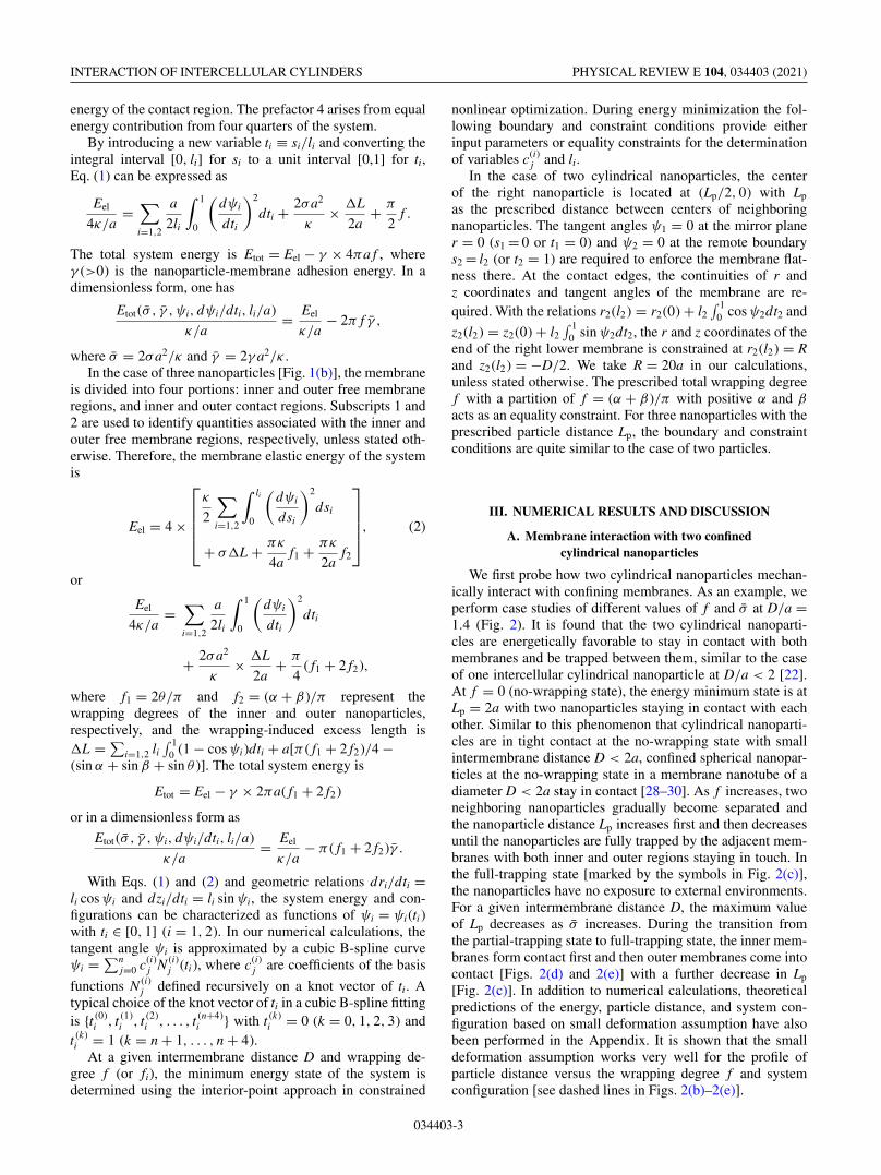
Based on the energy profile Etot ( f ) in Fig. 4, at equilibriumstate the relationship between f and γ̄ with given σ̄ and D/aand further the phase diagram of the nanoparticle-membraneinteraction for two confined cylindrical nanoparticles withrespect to γ̄ and σ̄ at given D/a can be obtained and areselectively shown in Fig. 5. It is found that the minimumadhesion energy γ̄min for partial trapping of two confinedcylindrical nanoparticles is almost equal to 1, similar to theresult for trapping of a single nanoparticle between adjacentmembranes [22]. At γ̄ < γ̄min, f = 0 and two particles stay incontact. As γ̄ exceeds γ̄min, finite f is reached and increaseswith increasing γ̄ until the full-trapping state marked by thesymbol is achieved [Fig. 5(a)]. At a given γ̄ (> γ̄min), thewrapping degree f increases as σ̄ or D decreases. Summa-rizing the relationship between f and γ̄ , one can determinephase boundaries between the no- and partial-wrapping statesas well as partial- and full-trapping states [Fig. 5(b)]. At agiven D/a, γ̄ required for full trapping is almost linearly
034403-4
INTERACTION OF INTERCELLULAR CYLINDERS PHYSICAL REVIEW E 104, 034403 (2021)
FIG. 3. Effects of the intermembrane distance D on the evolutionof the elastic energy Eel ( f ) and nanoparticle distance Lp( f ) in thecase of two confined nanoparticles at σ̄ = 4. The particle-membraneinteraction states of symbols are the full-trapping states with theouter membranes just in touch.
proportional to σ̄ . The dash-dotted lines in the partial-trappingregime denote the boundaries between the configurationswith and without contact between inner membrane parts[Fig. 5(b)].
The interaction phase diagram, except the boundariesmarking inner membrane touching, in Fig. 5(b) for two cylin-drical nanoparticles confined between membranes is almostthe same quantitatively as that for a single cylindrical nanopar-ticle confined between membranes (Fig. 6 in Ref. [22]). As theinteraction phase diagram is a reflection of the energy profile,the above observation suggests that the presence of a secondnanoparticle does not save the system elastic energy per par-ticle and there is no visible cooperative trapping in the senseof system energy evolution. These two nanoparticles simplyundergo individual trapping but in a synchronized manner ofsystem deformation. This conclusion is also confirmed by ob-serving that the elastic energy profiles Eel( f ) per nanoparticlesis almost the same as that for the case of a single cylindricalnanoparticle confined between membranes [22].
A related phenomenon is the wrapping of multiple con-fined spherical nanoparticles by soft membrane nanotubes
FIG. 4. Total energy change Etot of three nanoparticle-membraneinteraction states as a function of the wrapping degree f for differentvalues of γ̄ at σ̄ = 4 and D/a = 1.4. Solid symbols and the dottedred curve linking them correspond to the global minima at dif-ferent γ̄ .
[28–30]. In the case of two identical spherical nanoparticles ofradius a in a tense lipid membrane nanotube of finite length,the particle distance Lp increases from 2a to a maximumvalue and then decreases as the adhesion energy increasesor equivalently the wrapping degree increases [28], similar tothe trends observed in Figs. 2(c) and 3(b). In another case ofpacking multiple nanoparticles in a membrane nanotube withan infinite periodic pattern, it is revealed that there are twofundamental modes of interaction between these nanoparticlesand the soft nanotube, the cooperative wrapping and individ-ual wrapping [30]. At relatively small membrane tension, thecooperative wrapping is preferred [29,30]. At relatively largemembrane tension, confined spherical nanoparticles at smalland large wrapping degrees are cooperatively wrapped by themembrane nanotube, and particles are wrapped individuallyat intermediate wrapping degree [30].
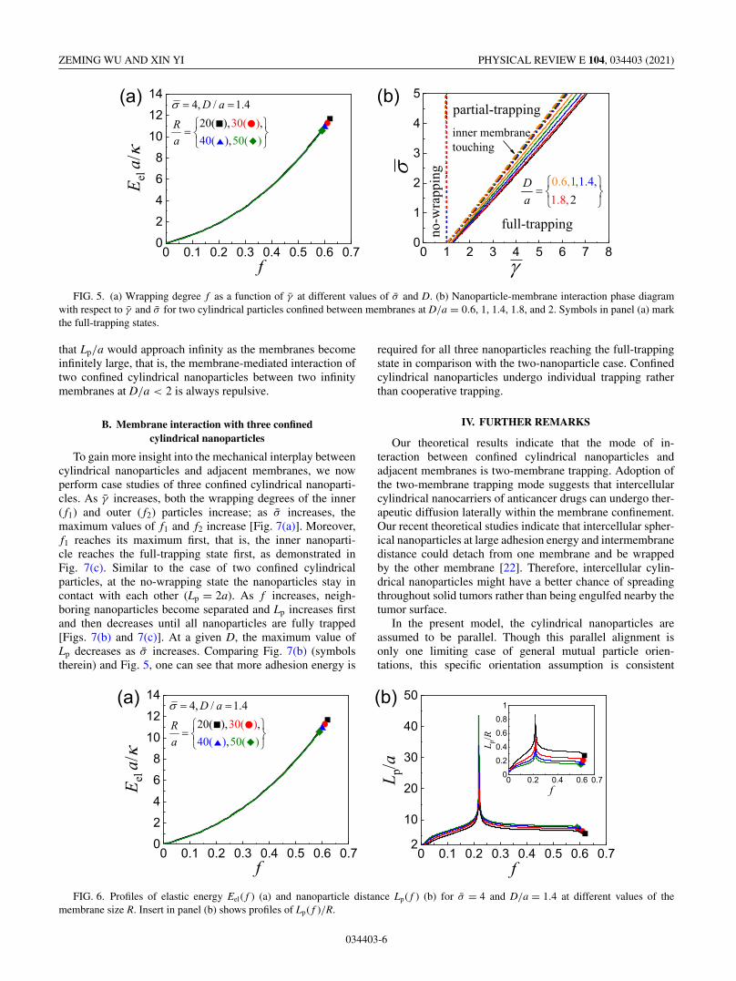
In the preceding calculations, R = 20a is taken and itis shown that there is a finite maximum value of Lp/a asf varies. Recalling that the membrane-mediated interactionof two cylindrical nanoparticles of circular [8–11] or ellip-tical [12] cross-sections attaching on the same side of aninfinitely large membrane is always repulsive, one mightwonder whether two cylindrical nanoparticles confined be-tween adjacent membranes would always repel each other atan infinity R/a. To understand the effects of the membranesize on the nanoparticle-membrane interaction and indirectparticle-particle interaction, profiles of elastic energy Eel( f )and nanoparticle distance Lp( f ) are investigated at differentvalues of R/a (Fig. 6). As a larger membrane leads to easierand larger deformation, the upper and lower membranes oflarger size come into contact at smaller f in the full-trappingstate [Fig. 6(a)]. In comparison with Eel( f ) in Fig. 6(a)showing insensitivity to R/a, the membrane size has strongereffect on Lp( f ). As R/a increases, the distance between thenanoparticles at equilibrium increases. One could anticipate
034403-5
ZEMING WU AND XIN YI PHYSICAL REVIEW E 104, 034403 (2021)
FIG. 5. (a) Wrapping degree f as a function of γ̄ at different values of σ̄ and D. (b) Nanoparticle-membrane interaction phase diagramwith respect to γ̄ and σ̄ for two cylindrical particles confined between membranes at D/a = 0.6, 1, 1.4, 1.8, and 2. Symbols in panel (a) markthe full-trapping states.
that Lp/a would approach infinity as the membranes becomeinfinitely large, that is, the membrane-mediated interaction oftwo confined cylindrical nanoparticles between two infinitymembranes at D/a < 2 is always repulsive.
B. Membrane interaction with three confinedcylindrical nanoparticles
To gain more insight into the mechanical interplay betweencylindrical nanoparticles and adjacent membranes, we nowperform case studies of three confined cylindrical nanoparti-cles. As γ̄ increases, both the wrapping degrees of the inner( f1) and outer ( f2) particles increase; as σ̄ increases, themaximum values of f1 and f2 increase [Fig. 7(a)]. Moreover,f1 reaches its maximum first, that is, the inner nanoparti-cle reaches the full-trapping state first, as demonstrated inFig. 7(c). Similar to the case of two confined cylindricalparticles, at the no-wrapping state the nanoparticles stay incontact with each other (Lp = 2a). As f increases, neigh-boring nanoparticles become separated and Lp increases firstand then decreases until all nanoparticles are fully trapped[Figs. 7(b) and 7(c)]. At a given D, the maximum value ofLp decreases as σ̄ increases. Comparing Fig. 7(b) (symbolstherein) and Fig. 5, one can see that more adhesion energy is
required for all three nanoparticles reaching the full-trappingstate in comparison with the two-nanoparticle case. Confinedcylindrical nanoparticles undergo individual trapping ratherthan cooperative trapping.
IV. FURTHER REMARKS
Our theoretical results indicate that the mode of in-teraction between confined cylindrical nanoparticles andadjacent membranes is two-membrane trapping. Adoption ofthe two-membrane trapping mode suggests that intercellularcylindrical nanocarriers of anticancer drugs can undergo ther-apeutic diffusion laterally within the membrane confinement.Our recent theoretical studies indicate that intercellular spher-ical nanoparticles at large adhesion energy and intermembranedistance could detach from one membrane and be wrappedby the other membrane [22]. Therefore, intercellular cylin-drical nanoparticles might have a better chance of spreadingthroughout solid tumors rather than being engulfed nearby thetumor surface.
In the present model, the cylindrical nanoparticles areassumed to be parallel. Though this parallel alignment isonly one limiting case of general mutual particle orien-tations, this specific orientation assumption is consistent
FIG. 6. Profiles of elastic energy Eel ( f ) (a) and nanoparticle distance Lp( f ) (b) for σ̄ = 4 and D/a = 1.4 at different values of themembrane size R. Insert in panel (b) shows profiles of Lp( f )/R.
034403-6
INTERACTION OF INTERCELLULAR CYLINDERS PHYSICAL REVIEW E 104, 034403 (2021)
FIG. 7. Membrane interaction with three confined rigid cylindrical nanoparticles at R/a = 20. The wrapping degrees for inner ( f1) andouter ( f2) nanoparticles (a) and nanoparticle distance Lp (b) as functions of adhesion energy γ̄ at different values of σ̄ and D/a. (c) Selectedsystem configurations for σ̄ = 1 and D/a = 1.4 at different values of γ̄ . Configurations in panel (c) from top to bottom correspond to the stateof no wrapping with Lp = 2a, partial-trapping state with maximum Lp, partial-trapping state with inner membranes in touch, and full-trappingwith both inner and outer membranes in touch, respectively. Symbols in panels (a) and (b) refer to the full-trapping states with the outermembranes just in touch.
with a recent theoretical study of membrane-mediated in-clusion interaction which shows that two strongly elongatedanisotropic membrane inclusions prefer a parallel mu-tual orientation via free-energy analysis [17]. A thoroughand firm study on the veiled orientational distribution ofconfined nanoparticles with minimal assumptions is chal-lenging and further detailed investigations are being calledfor.
The particle interaction mediated by the deformed mem-branes depends on the membrane size and wrapping degree.For infinitely large adjacent membranes at D/a < 2, theconfined nanoparticles always repel each other. A simi-lar repulsive membrane-mediated interaction between twocylindrical particles attaching on the same side of a singlemembrane holds [8–12]. For adjacent membrane patches offinite size, the distance between confined nanoparticles in-creases and then decreases as the wrapping degree increases,that is, there exists a maximum particle distance for finite-size membranes. The finite value of the particle distance at
the full-trapping state implies that the intercellular cylindricalnanoparticles could be distributed throughout the tumor.
A circumstance related to cylindrical nanoparticle interac-tion is the membrane-mediated interaction between sphericalnanoparticles either attaching on the membrane [7,8] or con-fined between adjacent membranes [21]. For the former case,theoretical studies and molecular dynamics simulations indi-cate that the interaction between two spherical nanoparticlesadhering to the same membrane side can be attractive or repul-sive depending on the extent of membrane deformation whichis regulated by the membrane bending and stretching as wellas nanoparticle binding strength [7,8]. For the case of multi-ple intercellular spherical nanoparticles, molecular dynamicssimulations demonstrate particle aggregation and so far nodevoted theoretical studies have been performed. Whether theinteraction is always attractive for confined spherical particlesis not answered, let alone confined ellipsoidal or irregu-lar nanoparticles. A minimal system for the confinement ofnanoparticles between lipid membranes is the particle-loaded
034403-7
ZEMING WU AND XIN YI PHYSICAL REVIEW E 104, 034403 (2021)
multilayered vesicles or capsosomes with liposomal subcom-partments. Understanding the interaction between membranesand confined nanoparticles is fundamentally beneficial to thedevelopment of therapeutic drug carriers.
V. CONCLUSIONS
Adopting the Canham-Helfrich membrane theory, a the-oretical analysis has been performed on the membrane-mediated mechanical interaction of parallel cylindricalnanoparticles confined between two adjacent lipid mem-branes. The cylindrical nanoparticles are trapped therein andhave contact with both membranes. The system energy atequilibrium and corresponding trapping configurations aredetermined using the interior-point method for constrainedoptimization. As the wrapping degree increases, neighbor-ing cylindrical nanoparticles gradually become separated andthe nanoparticle distance increases first and then decreasesuntil the nanoparticles are fully trapped by adjacent mem-brane patches of finite size. Comparing the energy profilesper nanoparticle in the cases of single and multiple confinedcylindrical nanoparticles, it is found that there is no visiblecooperative trapping for multiple cylindrical nanoparticles,different from the possible cooperative wrapping of sphericalnanoparticles in a membrane nanotube. The nanoparticle-membrane interaction phase diagram describing transitionbetween states of no wrapping, partial trapping, and full trap-ping is determined at different intermembrane distances. Itis shown that the normalized adhesion energy required forfull trapping is almost linearly proportional to the normalizedmembrane tension. Moreover, analytical predictions on thesystem energy and configurations based on the force balanceof the membranes at small deformation are obtained and agreewell with numerical solutions. Our results provide mechanis-tic insight into the mechanical behaviors of multiple cylindri-cal nanoparticles in cell junctions or gaps, and may serve asdesign guidelines for rational drug delivery in tumor tissues.
ACKNOWLEDGMENTS
This work was supported by the National Natural ScienceFoundation of China under Grants No. 12022207 and No.11872005. Computation resources supported by the High-performance Computing Platform at Peking University areacknowledged.
APPENDIX: ANALYTICAL SOLUTIONS AT SMALLMEMBRANE DEFORMATION
In this Appendix, we analytically investigate the particle-membrane interaction based on force balance of the deformedmembranes. Taking the lower membrane as example, the sub-scripts 1 and 2 are ignored unless they are necessary.
At zero spontaneous curvature, the local membrane forceper unit length can be given as [10,31]
�s = σ − κ
2c2 = σ − κ
2
(dψ
ds
)2
,
�n = −κdc
ds= −κ
d2ψ
ds2, (A1)
where c = dψ/ds is the curvature of the deformed membrane,�s denotes the in-plane membrane force along the arclength,and �n is the out-of-plane shear force along with the normaldirection.
Equations of the force balance along the vertical andhorizontal directions read �s sin ψ + �n cos ψ = Fz and�s cos ψ − �n sin ψ = Fr , respectively, with Fz and Fr ascomponents of force on the particle along the z axis and raxis by the deformed membrane of concern. With Eqs. (A1)one has
Fz(s)
κ= σ
κsin ψ − 1
2
(dψ
ds
)2
sin ψ − d2ψ
ds2cos ψ, (A2)
Fr (s)
κ= σ
κcos ψ − 1
2
(dψ
ds
)2
cos ψ + d2ψ
ds2sin ψ. (A3)
Since the outer free membrane cannot freely adjust its config-uration vertically with its fixed remote end, one has Fz �= 0 forthe outer free membrane. In contrast, for the inner membrane,one has Fz = 0 before the upper and lower inner membranesform contact, and the corresponding membrane configurationis exactly depicted by [10]
ψ1(s1) = −π − 2am
[s1
√Fr
κm− K (m), m
], (A4)
where m = 2Fr/(Fr + σ ), am(s, m) is the Jacobi amplitudewith parameter m, and K (m) is the complete elliptic integralof the first kind.
At small membrane deformation, one has
sin ψ ≈ dz
dr, cos ψ ≈ 1,
dψ
ds≈ d2z
dr2,
d2ψ
ds2≈ d3z
dr3. (A5)
With Eq. (A5), Eqs. (A2) and (A3) become
d3z
dr3− σ
κ
dz
dr+ Fz
κ= 0, (A6)
d3z
dr3
dz
dr− 1
2
(d2z
dr2
)2
+ σ − Fr
κ= 0. (A7)
The general solution of Eq. (A6) is
dz
dr= B1er
√σ/κ + B2e−r
√σ/κ+Fz
σ,
where B1, B2, and Fz could be determined from the followingboundary conditions.
For the outer free membrane, dz2/dr2 = tan β at the con-tact edge s2 = 0 (r2(0) = Lp/2 + a sin β ), and dz2/dr2 = 0and
∫ Rr2(0) (dz/dr)dr = D/2 − a(1 − cos β ) at the remote end
Here H is introduced as the height of the outer free membranepart.
The shape of the outer free membrane is then determinedas
z2 = z2(0) + Fz
σ[r2 − r2(0)] + B1
ω[eωr2 − eωr2(0)]
+ B2
ω[e−ωr2(0) − e−ωr2 ], (A8)
where z2(0) = −a cos β is the z coordinate of the outer mem-brane at the contact edge.
For the inner free membrane of Fz = 0, from Eq. (A6) wehave a general solution as
dz1
dr1= C1eωr1 + C2e−ωr1 . (A9)
From boundary conditions dz1/dr1 = − tan α at r1 = r1(s1 =l1) = Lp/2 − a sin α and dz1/dr1 = 0 at r1 = r1(0) = 0, C1,and C2 in Eq. (A9) are determined as
C1 = −C2 = − 12 tan α csch[ωr1(l1)],
and the inner membrane shape is
z1 = z1(0) + tan α
ω[1 − cosh(ωr1)] csch[ωr1(l1)], (A10)
with z1(0) = ω−1 tan α tanh[ωr1(l1)/2] − a cos α as the z co-ordinate of the inner membrane at s1 = 0.
Substituting Eqs. (A8) and (A10) into Eq. (A7), the balanceof horizontal forces Fr at r1 = 0 and r2 = R requires(
d2z1
dr21
)2∣∣∣∣∣r1=0
=(
d2z2
dr22
)2∣∣∣∣∣r2=R
. (A11)
At given particle distance Lp and wrapping degree f =(α + β )/π , contact angles α and β can be determined fromEq. (A11) numerically.
The particle distance Lp at a given f is determined byminimizing the total elastic energy Eel. The elastic energy ofthe membrane parts in contact regions is
Ec = 4 ×[πκ
2af + σa(π f − sin α − sin β )
].
For the right lower outer free membrane, the bending energyis
E (2)bend = κ
2
∫ l2
0
(dψ2
ds2
)2
ds2 ≈ κ
2
∫ R
r2(0)
(d2z2
dr22
)2
dr2,
and the tension energy is
E (2)ten = σ
∫ l2
0(1 − cos ψ2)ds2 ≈ σ
2
∫ R
r2(0)
(dz2
dr2
)2
dr2,
where cos ψ ≈ 1 − (dz/dr)2/2 is used.Then we have
E (2)bend = κω(A1 + A2)
4, E (2)
ten = A3 + 2σ 2(A1 − A2)
8ωσ,
where
A1 = [e−2ωr2(0) − e−2ωR](B2
1e2I1 + B22
), A2 = 4B1B2I2,
A3 = − 4F 2z I2 + 8σFz[e
−ωr2(0) − e−ωR](B2 + B1eI1 ),
I1 = ω[r2(0) + R], I2 = ω[r2(0) − R].
The energy of the outer membrane is Eouter = 4 × (E (2)bend +
E (2)ten ).
Similarly, the bending and tension energy of the right lowerinner free membrane are
E (1)bend = κ
2
∫ r1(l1 )
0
(d2z1
dr21
)2
dr1
and
E (1)ten = σ
2
∫ r1(l1 )
0
(dz1
dr1
)2
dr1,
respectively. Then we have
E (1)bend = κω tan2 α csch2[ωr1(l1)]
8
{2ωr1(l1)+ sinh[2ωr1(l1)]
},
E (1)ten = σ tan2 α
4ω{coth[ωr1(l1)] − ωr1(l1) csch2[ωr1(l1)]},
and the elastic energy of the inner membrane is Einner = 4 ×(E (1)
bend + E (1)ten ).
The total elastic energy at given Lp and f is
Eel(Lp, f ) = Ec + Eouter + Einner,
and the system at given f in equilibrium and correspondingLp are then determined by locating
Eel( f ) = min{Eel(Lp, f )} with Lp ∈ [2a, 2(R − a)]. (A12)
Comparison between theoretical results and numericalsolutions and in Figs. 2(b)–2(e) shows that the small deforma-tion assumption works very well for the profile of Lp- f andsystem configuration.
[1] I. Canton and G. Battaglia, Endocytosis at the nanoscale,Chem. Soc. Rev. 41, 2718 (2012).
[2] Y. Hui, X. Yi, F. Hou, D. Wibowo, F. Zhang, D. Zhao,H. Gao, and C.-X. Zhao, Role of nanoparticle mechanical
properties in cancer drug delivery, ACS Nano 13, 7410(2019).
[3] A. E. Nel, L. Mädler, D. Velegol, T. Xia, E. M. V. Hoek, P.Somasundaran, F. Klaessig, V. Castranova, and M. Thompson,
ZEMING WU AND XIN YI PHYSICAL REVIEW E 104, 034403 (2021)
Understanding biophysicochemical interactions at the nano–biointerface, Nat. Mater. 8, 543 (2009).
[4] S. Zhang, H. Gao, and G. Bao, Physical principles of nanopar-ticle cellular endocytosis, ACS Nano 9, 8655 (2015).
[5] X. Yi and H. Gao, Phase diagrams and morphological evolu-tion in wrapping of rod-shaped elastic nanoparticles by cellmembrane: A two-dimensional study, Phys. Rev. E 89, 062712(2014).
[6] C. Huang, P. J. Butler, S. Tong, H. S. Muddana, G. Bao, and S.Zhang, Substrate stiffness regulates cellular uptake of nanopar-ticles, Nano Lett. 13, 1611 (2013).
[7] B. J. Reynwar and M. Deserno, Membrane-mediated interac-tions between circular particles in the strongly curved regime,Soft Matter 7, 8567 (2011).
[8] Z. Yan, Z. Wu, S. Li, X. Zhang, X. Yi, and T. Yue, Curvature-mediated cooperative wrapping of multiple nanoparticles atthe same and opposite membrane sides, Nanoscale 11, 19751(2019).
[9] T. R. Weikl, Indirect interactions of membrane-adsorbed cylin-ders, Eur. Phys. J. E 12, 265 (2003).
[10] M. M. Müller, M. Deserno, and J. Guven, Balancing torques inmembrane-mediated interactions: Exact results and numericalillustrations, Phys. Rev. E 76, 011921 (2007).
[11] S. Mkrtchyan, C. Ing, and J. Z. Y. Chen, Adhesion of cylindricalcolloids to the surface of a membrane, Phys. Rev. E 81, 011904(2010).
[12] H. Tang, H. Ye, H. Zhang, and Y. Zheng, Wrapping of nanopar-ticles by the cell membrane: The role of interactions betweenthe nanoparticles, Soft Matter 11, 8674 (2015).
[13] T. Weikl, M. Kozlov, and W. Helfrich, Interaction of conicalmembrane inclusions: Effect of lateral tension, Phys. Rev. E 57,6988 (1998).
[14] J.-B. Fournier, Microscopic membrane elasticity and interac-tions among membrane inclusions: Interplay between the shape,dilation, tilt and tilt-difference modes, Eur. Phys. J. B 11, 261(1999).
[15] P. G. Dommersnes and J.-B. Fournier, N-body study ofanisotropic membrane inclusions: Membrane-mediated interac-tions and ordered aggregation, Eur. Phys. J. B 12, 9 (1999).
[16] V. I. Marchenko and C. Misbah, Elastic interaction of pointdefects on biological membranes, Eur. Phys. J. E 8, 477(2002).
[17] Y. Schweitzer and M. M. Kozlov, Membrane-mediatedinteraction between strongly anisotropic protein scaffolds,PLoS Comput. Biol. 11, e1004054 (2015).
[18] H. Cabral et al., Accumulation of sub-100 nm polymericmicelles in poorly permeable tumours depends on size,Nat. Nanotechnol. 6, 815 (2011).
[19] M. M. Falk, C. L. Bell, R. M. Kells Andrews, and S. A. Murray,Molecular mechanisms regulating formation, trafficking andprocessing of annular gap junctions, BMC Cell Biol. 17, S22(2016).
[20] J. Iwasa and W. Marshall, Karp’s Cell and Molecular Biology,9th ed. (Wiley, Hoboken, NJ, 2020).
[21] T. Yue, H. Zhou, H. Sun, S. Li, X. Zhang, D. Cao, X. Yi, and B.Yan, Why are nanoparticles trapped at cell junctions when thecell density is high? Nanoscale 11, 6602 (2019).
[22] Z. Wu and X. Yi, Mechanics of cell interaction withintercellular nanoparticles: Shape-dependent competition be-tween two-membrane trapping and single-membrane wrapping,Extreme Mech. Lett. 46, 101296 (2021).
[23] P. B. Canham, The minimum energy of bending as a possibleexplanation of the biconcave shape of the human red blood cell,J. Theor. Biol. 26, 61 (1970).
[24] W. Helfrich, Elastic properties of lipid bilayers: Theory andpossible experiments, Z. Naturforsch. C 28, 693 (1973).
[25] M. Deserno, Fluid lipid membranes: From differential geometryto curvature stresses, Chem. Phys. Lipids 185, 11 (2015).
[26] C. E. Morris and U. Homann, Cell surface area regulation andmembrane tension, J. Membr. Biol. 179, 79 (2001).
[27] J. Dai, M. P. Sheetz, X. Wan, and C. E. Morris, Membrane ten-sion in swelling and shrinking molluscan neurons, J. Neurosci.18, 6681 (1998).
[28] J. Z. Y. Chen, Y. Liu, and H.-J. Liang, Structure of a TubularMembrane Confining Spherical Particles, Phys. Rev. Lett. 102,168103 (2009).
[29] M. Raatz, R. Lipowsky, and T. R. Weikl, Cooperative wrappingof nanoparticles by membrane tubes, Soft Matter 10, 3570(2014).
[30] Z. Wu and X. Yi, Structures and mechanical behaviors of softnanotubes confining adhesive single or multiple elastic nanopar-ticles, J. Mech. Phys. Solids 137, 103867 (2020).
[31] J.-B. Fournier, On the stress and torque tensors in fluid mem-branes, Soft Matter 3, 883 (2007).