Just Accepted by Nanotoxicology
Physicochemical characterization and toxicological evaluation of plant based anionic polymers and their nanoparticulated system for ocular delivery
Deepa Pathak, prashant kumar, Gowthamarajan Kuppusamy, Ankur Gupta, Bhagyashree Kamble, Ashish Wadhwani
doi:10.3109/17435390.2013.834996
Abstract
The water soluble fractions of mucilages and gum from the seeds of fenugreek, isphagula and mango bark exudate were isolated, purified and characterized using X-ray diffraction spectrometry (XRD), fourier transform infrared spectroscopy (FT-IR), maldi/GC-MS, elemental analysis, 1D (1H and 13C) and 2D (HMQC, COSY) nuclear magnetic resonance spectroscopy (NMR). The fenugreek mucilage was identified to be a galactomannan chain consisting of 4 units of galactose attached to the backbone of 6 mannose units in 1:1.5 ratio. The isphagula mucilage was identified to be an arabinoxylan polysaccharide chain consisting of 4 units of arabinofuranose attached to the backbone of 9 xylopyrannose units in 1:3 ratio. The mango gum showed the presence of amylose, α-arabinofuranosyl and β-galactopyranosyl respectively.The characterterized mucilages and gum were individually formulated into nanoparticulate system using their complementarily charged polymer chitosan. The particles were observed to be spherical in shape in the range of 61.5-90 nm having zetapotential between 31-34 mV and PDI of 0.097-0.241. The prepared nanoparticles were observed to be non-irritant and non-toxic in vitro and in vivo upto 2000µg/ml .Therefore, these mucilages and gum can be the alternatives of anionic polymers for the ocular drug delivery system.
Informa UK, Ltd. This provisional PDF corresponds to the article as it appeared upon acceptance. Fully formatted PDF and full text (HTML) versions will be made available soon.
DISCLAIMER: The ideas and opinions expressed in the journal’s Just Accepted articles do not necessarily reflect those of Informa Healthcare (the Publisher), the Editors or the journal. The Publisher does not assume any responsibility for any injury and/or damage to persons or property arising from or related to any use of the material contained in these articles. The reader is advised to check the appropriate medical literature and the product information currently provided by the manufacturer of each drug to be administered to verify the dosages, the method and duration of administration, and contraindications. It is the responsibility of the treating physician or other health care professional, relying on his or her independent experience and knowledge of the patient, to determine drug dosages and the best treatment for the patient. Just Accepted articles have undergone full scientific review but none of the additional editorial preparation, such as copyediting, typesetting, and proofreading, as have articles published in the traditional manner. There may, therefore, be errors in Just Accepted articles that will be corrected in the final print and final online version of the article. Any use of the Just Accepted articles is subject to the express understanding that the papers have not yet gone through the full quality control process prior to publication.
Nan
otox
icol
ogy
Dow
nloa
ded
from
info
rmah
ealth
care
.com
by
Uni
vers
ity o
f C
alga
ry o
n 08
/29/
13Fo
r pe
rson
al u
se o
nly.
2
Title Page:
Physicochemical characterization and toxicological evaluation of plant based anionic polymers and
their nanoparticulated system for ocular delivery
Deepa Pathak1, Prashant Kumar1, Gowthamarajan Kuppusamy 1* Ankur Gupta2, Bhagyashree Kamble3,
Ashish Wadhwani4,
1. Department of Pharmaceutics, J.S.S. College of Pharmacy, Udhagamandalam, T.N.-643 001 (Off
campus J.S.S. University, Mysore), India.
2. Department of Pharmaceutical Chemistry, J.S.S. College of Pharmacy, Udhagamandalam, T.N.-643 001
(Off campus J.S.S. University, Mysore), India.
3. Department of Pharmacognosy, J.S.S. College of Pharmacy, Udhagamandalam, T.N.-643 001 (Off
campus J.S.S. University, Mysore), India.
4. Department of Pharmaceutical Biotechnology, J.S.S. College of Pharmacy, Udhagamandalam, T.N.-643
001 (Off campus J.S.S. University, Mysore), India.
* Author for Correspondence
Gowthamarajan Kuppusamy
Prof. & Head
Department of Pharmaceutics
J.S.S. College of Pharmacy
Udhagamandalam, T.N.-643 001
(J.S.S. University, Mysore), India.
Phone number: +91-9443089812
Email address: [email protected], [email protected]
Abstract
The water soluble fractions of mucilages and gum from the seeds of fenugreek, isphagula and mango bark
exudate were isolated, purified and characterized using X-ray diffraction spectrometry (XRD), fourier
transform infrared spectroscopy (FT-IR), maldi/GC-MS, elemental analysis, 1D (1H and 13C) and 2D
Nan
otox
icol
ogy
Dow
nloa
ded
from
info
rmah
ealth
care
.com
by
Uni
vers
ity o
f C
alga
ry o
n 08
/29/
13Fo
r pe
rson
al u
se o
nly.
3
(HMQC, COSY) nuclear magnetic resonance spectroscopy (NMR). The fenugreek mucilage was
identified to be a galactomannan chain consisting of 4 units of galactose attached to the backbone of 6
mannose units in 1:1.5 ratio. The isphagula mucilage was identified to be an arabinoxylan polysaccharide
chain consisting of 4 units of arabinofuranose attached to the backbone of 9 xylopyrannose units in 1:3
ratio. The mango gum showed the presence of amylose, α-arabinofuranosyl and β-galactopyranosyl
respectively.The characterterized mucilages and gum were individually formulated into nanoparticulate
system using their complementarily charged polymer chitosan. The particles were observed to be spherical
in shape in the range of 61.5-90 nm having zetapotential between 31-34 mV and PDI of 0.097-0.241. The
prepared nanoparticles were observed to be non-irritant and non-toxic in vitro and in vivo upto 2000µg/ml
.Therefore, these mucilages and gum can be the alternatives of anionic polymers for the ocular drug
delivery system.
Keywords: Anionic polymers isolation, Charcterization, Nanoparticle formulation, Ocular toxicity studies.
1. Introduction
Eyes are sensitive, delicate and vital organs of the human body. The most common diseases
associated with the eyes are conjunctivitis, cataract, glaucoma, diabetic retinopathy and age-related
macular degeneration. Various formulations such as eye drops, occuserts, intravitreal injection, ointment,
aqueous gel and routes either systemic or local route have been applied for the treatment of these
complications. Systemic administration requires higher dosage and frequent administration that results in
severe adverse effects. Local injections, particularly intravitreal and subconjunctival injections are
alternate strategies to achieve therapeutic concentration in the vitreo-retinal disorders (Pathak et al., 2012).
Nan
otox
icol
ogy
Dow
nloa
ded
from
info
rmah
ealth
care
.com
by
Uni
vers
ity o
f C
alga
ry o
n 08
/29/
13Fo
r pe
rson
al u
se o
nly.
4
However, to maintain the effective concentration repeated injections are required, which causes clinical
complications or patient discomfort (Kimura and Ogura, 2001).
The application of noninvasive polymeric drug delivery systems such as micro and nanoparticles,
microspheres, liposomes, hydrogels and ocular implants have permitted a targeted delivery of drugs to
specific sites in controlled manner. Apart from various advantageous effects of polymeric systems, these
systematic approach as may lead to high cost (Prow, 2010) and ocular toxicity due to size (Koster et al.,
1996, Sohaebuddin et al., 2010), charge (Hillegass et al., 2010) and metallic nature (Park, 2007, Theodore
et al., 2005).
Further, the polymers of natural and synthetic origin are widely employed for the preparation of
polymeric systems. The synthetic polymers are frequently used for its ability and usefulness in controlled
delivery of drugs and biological products. The disadvantages of synthetic polymers are their cost,
availability and sometimes toxic effect. Bejjani et al., 2005 and Tamboli et al., 2011 have reported non-
cytoxicity of poly (lactic) acid nanoparticles (PLA) for gene delivery in human and bovine retinal pigment
epithelial cells up to 4 mg/mL concentration but beyond this concentration they are toxic for eyes. Some
drugs show incompatibilities with different range of excipients. Marini et al., 2003 has reported atenolol-
PVP, atenolol-mg-stearate showed drug-excipient incompatibilities due to reaction between aldehydic
sugars and primary/secondary amines, leading to the formation of Schiff bases.
Polysaccharides of natural origin have recently been used as polymers for the preparation of
nanoparticles to overcome the problems associated with metallic and synthetic polymer based
nanoparticles. The natural polymers such as chitosan, sodium alginate, bovine serum albumin and gelatin
are widely employed in preparation of nanoparticles for ocular delivery due to their biodegradability,
mucoadhesive property, therapeutic potential, stability in the biological fluids and during storage.
(Motwani et al., 2008). However, with the mucoadhesive and biodegradability properties, these polymers
sometimes not suitable for ocular delivery due to adverse reaction in the eye for example albumin
microspheres (Hui and Robinson, 1985). The other example is of gelatin based aqueous delivery system
Nan
otox
icol
ogy
Dow
nloa
ded
from
info
rmah
ealth
care
.com
by
Uni
vers
ity o
f C
alga
ry o
n 08
/29/
13Fo
r pe
rson
al u
se o
nly.
5
which acts as a thermoreversible hydrogel with low mechanical strength which breaks at 30 °C and results
in drug loss. Glutaraldehyde is used as a crosslinking agent to provide stability toward thermal
degradation (Natu et al 2007) but it causes eye irritation. To avoid the use of toxic chemicals and high
cost polymers plant based polymers which are abundantly available in nature may be evaluated for their
composition, structure and toxicity.
Amelia M. Avachat et al., 2010 described various plant based mucilages and gums like fenugreek
mucilage, isphagula mucilage and mango gum etc. for their mucoadhesive and binding property for oral
drug delivery. The isolation and characterization of mucilages and gum from these sources are available
as mixture of water insoluble and soluble fraction (Nitalikar et al., 2003, Murthy et al., 2010, Singh et al.,
2010). Gowthamarajan et al., 2012 recently reported the use of natural gums in the preparation of
mucoadhesive tablet. Mehra et al., 2010, reported the use of tamarind gum based gel for ocular drug
delivery system.
To the best of our knowledge no reports are available on water soluble polymeric fractions of
mucilages and gum and their nanoformulation for ocular drug delivery. There are various advantages of
using water soluble factions for prepation of nanoparticles as, it excludes the use of organic solvents
(Chellat et al., 2000), it produces smaller particle size less than 350 nm which is required to cross blood
retinal barrier (Gaudana et al., 2010) and reach to posterior region of the eye and it reduces the production
cost.
Therefore, the current study has been carried out with the focus on isolation, purification,
characterization and toxicity evaluation of individual water soluble fractions from fenugreek, isphagula
seeds and mango bark exudates and their nanoformulation with chitosan for safe, feasible and non
invasive ocular drug delivery system.
2. Materials and methods
2.1. Materials
Nan
otox
icol
ogy
Dow
nloa
ded
from
info
rmah
ealth
care
.com
by
Uni
vers
ity o
f C
alga
ry o
n 08
/29/
13Fo
r pe
rson
al u
se o
nly.
6
The polymer chitosan (CS) (M.W:100,000-300,000 daltons, Deacetylation>80%) was procured
from Sigma-Aldrich (St. Louis, MO, USA). Fenugreek and Isphagula seeds were purchased from
Udhagamandalam local market (India). The mango bark exudate was purchased from Hyderabad local
market. The herbal samples were authenticated by Department of Pharmacognosy, J.S.S. College of
Pharmacy, Udhagamandalam, T.N.
2.2. Methods
2.2.1. Isolation, purification and preliminary tests of water soluble fractions
2.2.2.1. Extraction and purification of mucilage from fenugreek and isphagula seed
The seeds were collected and washed with water to remove the dirt and debris. The seeds (250 g)
were soaked in double distilled water (500 ml) overnight and then heated at 50 °C for 2h. The solution was
filtered through muslin cloth and to the filtrate equi-volume ratio of 90% v/v alcohol was added. The
obtained precipitate was filtered and dried in a hot air oven at 45ºC to obtain ≈150 g of powder. The
obtained powder was re-dissolved in 100 ml of water, filtered and centrifuged for 10 min at 3000 rpm. The
supernatant clear solution was collected, evaporated and dried. This process of purification was repeated
thrice. The purified solid mass was dried under reduced pressure at 40 °C, grounded and passed through
sieve no. 80 and stored in an airtight container.
2.2.2.2. Extraction and purification of mango gum
Crude plant exudates were collected in season of March to June. Mango gum was obtained from
the incised trunk of Mangifera indica. The Mango gum resin (250 g) was extracted by distilled water 500
ml on a water bath maintaining at 50 °C for 45 min with intermittent stirring, extraneous materials were
removed by straining through a several folds of muslin cloth. The gum was then precipitated by using 90%
Nan
otox
icol
ogy
Dow
nloa
ded
from
info
rmah
ealth
care
.com
by
Uni
vers
ity o
f C
alga
ry o
n 08
/29/
13Fo
r pe
rson
al u
se o
nly.
7
v/v alcohol followed by centrifugation at 3000 rpm. The extracted gum was filtered and dried in oven at
45 °C. The obtained powder was re-dissolved in 100 ml of water, filtered and centrifuged for 10 min at
3000 rpm ((Remi instruments ltd., Mumbai, India)). The supernatant clear solution was collected,
evaporated and dried. This process of purification was repeated thrice. The purified solid mass was dried
under reduced pressure at 40 °C, grounded and passed through sieve no. 80 and stored in an airtight glass
container.
2.2.2.3. Biochemical tests of isolated mucilages and gum
The purified samples of mucilages and gum were subjected to various biochemical tests for
carbohydrates, proteins, tannins, alkaloids, saponins, phenol and flavonoids as per official methods (Treas
& Evans, 1996).
2.2.2. Physico-chemical and structural characterization of mucilages and gum
2.2.2.1. X-Ray diffraction analysis (XRD)
In X-ray studies, an automatic X-ray diffractometer (Bruker, AXS/8, Berlin, Germany) equipped
with a PW R30 X-ray generator was used. The dry sample powder was pressed into pellets and X-ray
diffraction spectra were recorded using nickel-filtered Cu kα1 radiation having a wavelength of 1.5106 Å,
operating at 35 kW and 20 mA. X-ray diffractograms were obtained at a scanning rate of 1 degree (2θ) per
minute.
2.2.2.2. Elemental analysis
Elemental compositions of C, H and N were analyzed by using an elemental analyzer (Perkin
Elmer, 2400 Series, CHNSO Analyser, U.K). Accurately weighed 0.5 mg sample was heated to 1150 ºC
seperately and the corresponding element was determined by using a thermal conductivity detector.
2.2.2.3. Atomic absorbtion spectroscpic (AAS) analysis
The AAS instrumental parameters ((Shimadzu AA-6300, Serial No-A305245) for the estimation of
major, trace and heavy metals were set which include the lamp current, wavelength, slit width, lamp mode,
fuel flow rate, support gas flow rate, flame type and burner height were optimized. Deionized water was
Nan
otox
icol
ogy
Dow
nloa
ded
from
info
rmah
ealth
care
.com
by
Uni
vers
ity o
f C
alga
ry o
n 08
/29/
13Fo
r pe
rson
al u
se o
nly.
8
used for all the dilutions. All the plastic materials and glass wares were cleaned by soaking in dilute nitric
acid solution for 24 h and rinsed with distilled water followed by deionized water prior to use. The
calibration curves for the analyte ions were drawn after setting various parameters. All measurements were
performed in triplicate (n=3) and the standard deviation (SD) was recorded.
2.2.2.4. Fourier Transform Infrared spectroscopy (FT-IR)
The FT-IR spectrum (Shimadzu FTIR 84000 S, Japan) of samples were recorded on FT-IR
spectrophotometer. The dry sample (10 mg) was ground into fine powder using mortar and pestel then
pressed using potassium bromide (KBr 100 mg) disc technique under a hydraulic press at 10,000 psi. Each
KBr disc was scanned at 4 mm/s a resolution of 2 cm over a wave number region of 4000 – 400 cm-1.
2.2.2.5. Mass Spectroscopic analysis (MS)
The average molecular weight of sample was determined by matrix assisted laser
desorption/ionization-time of flight (Bruker Daltonik, Bremen, Germany, Reflex II MALDI-TOF
instrument) analyzer in the negative ionization mode. For the ionization, a nitrogen laser (337 nm, 3 ns
pulse width, 3 Hz) was used. Sample (5 mg) was dissolved in D2O solvent. 0.5 mL of the sample was
applied to the target followed by the addition of 1 mL of matrix solution (2,5-dihydroxybenzoic acid), and
dried under a gentle stream of air. All spectra were measured in the reflector mode using external
calibration.
2.2.2.6. 1D and 2D NMR spectroscopy
NMR spectra of 1H and 13C of sample were recorded in NMR spectrometer (Bruker, Bremen,
Germany). 100 mg of sample was dissolved in D2O and chemical shifts were reported in ppm relative to an
internal standard TMS (tetramethylsilane). NMR spectrum was obtained at a base frequency of 400MHz,
with 16 transitions and delay time 1.5 s using D2O as solvent. The chemical shifts were expressed in δ
(ppm) relative to the resonance of TMS.
Nan
otox
icol
ogy
Dow
nloa
ded
from
info
rmah
ealth
care
.com
by
Uni
vers
ity o
f C
alga
ry o
n 08
/29/
13Fo
r pe
rson
al u
se o
nly.
9
2D NMR spectrum was applied using double-quantum filtered correlated spectroscopy (DQF
COSY), hetero nuclear single-quantum coherence (HMQC) with a NMR spectrometer (Bruker, Bremen,
Germany).
2.2.2.7. Acid-base hydrolysis and FTIR, GC/MS analysis of mucilages and gum
2.2.2.7.1. Acid-base hydrolysis
The pH of the sample solution (300 mg/ml) was adjusted to 2.0 by adding 0.5 M sulfuric acid. The
solution was heated to 100 ºC for 24 h. After cooling, the pH of solution was adjusted to 10 by the addition
of 0.5 M NaOH and heated to 100 ºC for 24 h. After cooling the solution was neutralized with barium
carbonate and filtered. The solution was then dialyzed for 24 h against de-ionized water; freeze dried (De
Paula et al., 1998). The obtained hydrolysed dry samples were subjected for FTIR and Gas
chromatography-/mass spectrometry (GC-MS) studies.
2.2.2.7.2. FTIR and GC-MS analysis of hydrolysed fraction
The freezed dried hydrolysed fraction of mucilages and gum were subjected for FT-IR (method
described in section 2.2.2.4) and GC-MS analysis (Agilent Technologies, Palo Alto, USA). The GC-MS
was performed using HP-5 capillary column (0.25 mm x 30 m) linked to model TD800 Finningam ion trap
mass spectrometer (MS) operated at 70 eV. The columns were programmed from 50-220 ºC at 40 ºC min-1.
2.2.3. Pre-formulation studies of mucilages and gum
2.2.3.1. Determination of surface charge and pH
1% w/v solution of sample was prepared in distilled water and determined surface charge by
potentiometric analysis using potentiometer. The pH of same solutions (1% w/v) were determined by using
digital pH meter (pH meter pH/ /ion/MV meter, ion 510 bench pH, Eutech Instruments, Mumbai). All
measurements were performed in triplicate (n=3) and the standard deviation (SD) was recorded.
Nan
otox
icol
ogy
Dow
nloa
ded
from
info
rmah
ealth
care
.com
by
Uni
vers
ity o
f C
alga
ry o
n 08
/29/
13Fo
r pe
rson
al u
se o
nly.
10
2.2.3.2. In vitro cell viability assay
Cytotoxicity of the sample was determined by the MTT (3-(4,5-Dimethylthiazol-2-yl)-2,5-
diphenyltetrazolium bromide) assay using L929 mouse fibroblast cell line. 1x 10⁵cells/ml were plated in
each well of a 96 well microtitre plate in 100 µL of medium and incubated at 37 °C in CO2 incubator.
In the first six wells cells were left without any treatment as a positive control. Stock solutions of the
samples of different concentrations (0.05mg/mL, 0.5mg/mL, 1mg/mL, 2mg/mL and 5mg/mL) were made
in 1% DMSO and diluted with minimal essential medium (MEM) to a final concentration of 50, 500, 1000,
2000, 5000 µg/mL in the plate. Once the confluent monlolayer was ready, concentrations of the test
samples of 50 µL was added to each well in triplicate and incubated at 37 oC in a CO2 incubator for 72 h.
After incubation, 50 µL of MTT dye (2mg/mL) was added to each well and again incubated at 37 oC
for another 4 h. Formed formazan was washed and dissolved using 50 µL of isopropanol and optical
density was recorded with a microtitre plate reader at 550 nm (Scudiero et al., 1988). All the
measurements were performed in triplicate (n=3) and the standard deviation (SD) was recorded. Cell
viability was calculated by the formula:
………………………………………………………..Eq. 1
Where Isample is the absorbance of samples treated well and Icontrol is the absorbance of control wells
without samples.
2.2.3.3. Compatibility studies
The isolated mucilage-chitosan and isolated gum-chitosan (physical mixture 1:1 ratio) were
subjected for compatibility studies carried out by Differential scanning calorimetry (DSC, Water Q 200,
Bangalore) analysis to characterize the thermal behavior of the individual mucilages, polymer, gum and
their nanoparticles. Samples were crimped in standard aluminium pans and heated from 20 to 400 ºC at a
heating rate of 10 ºC/min under constant purging of dry nitrogen at 30mL/min. An empty pan, sealed in the
same way as the sample, was used as a reference.
Nan
otox
icol
ogy
Dow
nloa
ded
from
info
rmah
ealth
care
.com
by
Uni
vers
ity o
f C
alga
ry o
n 08
/29/
13Fo
r pe
rson
al u
se o
nly.
11
2.2.4. Formulation and Evaluation of nanoparticles
2.2.4.1. Preparation of polymeric nanoparticles
The purified mucilages and gum solution of 0.04% w/v was prepared by dissolving the individual
mucilages and gum in double distilled water. The pH of the solution was adjusted to 5.5 using 1 N
hydrochloric acid. The chitosan of 0.02% w/v was dissolved in a solution of 0.1% v/v acetic acid, volume
adjusted using distilled water and pH modified to 5.5 using 0.1 M NaOH. The solutions were filtered under
vacuum and were further taken for the preparation of nanoparticulate suspension by a modified
coacervation method at 3000 rpm for 2h using Remi stirrer (Douglas and Tabrizian, 2005). The formed
suspension was freeze dried using 0.5% w/v mannitol as lyoprotectant and stored in desiccator under
vacuum.
2.2.4.2. Characterization of nanoparticles
2.2.4.2.1. Transmission electron microscopic (TEM)
TEM (TEM, Philips CM-10, USA) was used to study the shape of nanoparticles. Samples of the
nanoparticles suspension (5-10 µl) were dropped onto formvar-coated copper grids. After complete drying,
the samples were stained using 2% w/v phosphotungstic acid. Digital Micrograph and soft imaging viewer
software were used to perform the image capture.
2.2.4.2.2. Particle size and zeta potential
Nanoparticles size distribution and zeta potential were determined using zetasizer photon
correlation spectroscopy (Malvern, Model No. 3000 HF, Malvern (U.K.). The size distribution analysis
was performed at a scattering angle of 90° and at a temperature of 25 °C using samples appropriately
diluted with filtered water (0.2 µm filter). Zeta potential was measured using a disposable zeta cuvette. For
Nan
otox
icol
ogy
Dow
nloa
ded
from
info
rmah
ealth
care
.com
by
Uni
vers
ity o
f C
alga
ry o
n 08
/29/
13Fo
r pe
rson
al u
se o
nly.
12
each sample, the mean diameter/zeta potential ± standard deviation of six determinations was calculated
applying multimodal analysis.
2.2.4.2.3. Sterility testing
The sterility testing was performed according to Indian Pharmacopoeia (IP), 2007 method on four
different media namely, fluid thioglycolate media (FTM), nutrient broth medium (NB), soyabean casein
digests medium (SCDM) and sorbitol-dextrose broth (SDB) to investigate the pesence or absence of
aerobic bacteria, anaerobic bacteria and fungi. FTM media was incubated at 37 ± 0.5 °C under aerobic
condition, NB at 30 ± 2.5 °C under anaerobic condition in a bacteriological incubator while SCDM and
SDB were incubated at 25 ± 0.5 °C under aerobic condition in a fungal incubator for 14 days. The
experiment was performed in triplicate.
2.2.4.2.4. In-vitro ocular irritation studies of nanoparticle (HET-CAM Test)
White Leghorn chicken eggs were obtained from the Poultry Research Centre, Kerala. White
Leghorn chicken eggs were selected for the study because they have no hereditary defects and yields
reproducible results. Eggs were incubated in the incubator for 9-10 days at 37 ºC. On day 9, eggs were
tested with candle light to ensure that all were viable. On day 10, the air cell was marked with a pencil and
removed the shell by tapered forceps. The membrane was carefully moistened with 0.9% NaCl solution
and the membrane was carefully removed without injuring any underlying blood vessels. 0.1 N NaOH and
sterile distilled water were used as positive and negative control/solvent control. The chorioallantoic
membrane (CAM) of each egg was applied directly with 0.3 mL of the positive/negative control and
different concentration of pure gum/mucilage and nanoparticles (50 µg/mL -5000 µg/mL). Two eggs for
controls and three for test substance were used for each assay. The reactions of hemorrhage, coagulation
and lysis on the CAM were observed over a period of 6h. The time for each reaction was recorded in 5 min
and an irritation score (IS) was calculated according to the formula given below. Each test was done in
triplicate and the mean score of the three eggs was determined. At the end of each assay the embryos were
Nan
otox
icol
ogy
Dow
nloa
ded
from
info
rmah
ealth
care
.com
by
Uni
vers
ity o
f C
alga
ry o
n 08
/29/
13Fo
r pe
rson
al u
se o
nly.
13
killed quickly by placing the eggs into a freezer at -20 ºC (Kishore et al., 2009). All measurements were
performed in triplicate (n=3) and the standard deviation (SD) was recorded.
H= hemorrhage; L = vessel lysis; C = coagulation; Min= 5 min. After treating for 6 h the main reaction
(hemorrhage, lysis or coagulation) was scored as follows: 0=no reaction; 1=slight reaction; 2=moderate
reaction; 3=severe reaction.
2.2.4.2.5. Haemocompatibility studies
Blood samples of healthy human volunteers were obtained from blood bank of government
hospital, Ooty in evacuated siliconized glass tube containing sodium citrate. Red blood cells were
separated by centrifugation at 1500 rpm for 10 min and then washed 3 times with phosphate buffer saline
pH 7.4. Stock solution of erythrocytes in PBS was prepared such that the cell count was 1×10⁸ cells/ml.
Equal volumes of RBC suspension and nanoparticles dispersion were suspended in a microcentrifuge tube
such that the final concentrations of pure mucilages/gum and nanoparticle dispersion were 50 µg/mL -5000
µg/mL and incubated at 37 ºC for 1 h. 1% v/v Triton X and PBS were used as positive and negative
controls respectively. After 1 h the tubes were centrifuged at 1500 rpm for 10 min and the hemoglobin
released in the supernatant was detected by UV absorbance at 540 nm. All measurements were performed
in triplicate (n=3) and the standard deviation (SD) was recorded. The percent haemolysis was calculated by
(Gulatia et al., 2010)
Where AbSsample is the absorbance of supernatant of erythrocyte and nanoparticles suspension. Abs0% is the
absorbance of supernatant of erythrocyte and PBS suspension. Abs100% is the absorbance of supernatant of
erythrocyte and Triton X suspension.
2.2.4.2.6. In-vivo ocular irritation studies
Nan
otox
icol
ogy
Dow
nloa
ded
from
info
rmah
ealth
care
.com
by
Uni
vers
ity o
f C
alga
ry o
n 08
/29/
13Fo
r pe
rson
al u
se o
nly.
14
Experimental animals were obtained from in house animal facility of JSSCP, Udhagamandalam.
All procedures using animals were reviewed and approved by the Institutional Animal Ethics Committee
(Proposal No.-JSSCP/IAEC/PH.D/PH-CEUTICS/01/2012-13).
Animals: The female New Zealand white rabbits, in the range of 2–3 kg body weight were
nulliparous and non-pregnant. The acute ocular irritation potential of gum nanoparticles were determined
according to OECD 404 and 405 test guidelines. The experiment was divided into groups (Positive control,
pure mucilages and gum, nanoparticles in 50 µg/mL-2000 µg/mL) and 3 animals were included in each
group.
Experiment: Both eyes of the experimental animals selected for testing were examined with an
ophthalmoscope 24 h prior to starting experiment. None of the animal’s selected for the study showed any
ocular defects or pre-existing corneal injury. Different concentrations of pure gum/mucilage and
nanoparticle solution (50 µg/mL-2000 µg/mL) were applied into the conjunctival sac of left eye of each
animal after gently pulling the lower lid away from the eyeball. The lids were then gently held together for
about 1 s in order to prevent loss of the material. The right eye which remained untreated served as
negative control. The eyes of the test animals were washed with distilled water at 24 h following the
application of test materials to remove the presence of residual test substance if any. The conjunctiva, iris,
and cornea of both treated and control eyes were evaluated at the end of 24, 48, and 72 h checked for the
sign of redness, lacrimation and inflammation by Draize’s scoring approach (Vinardell et al., 1994).
3. Results
3.1. Characterization of isolated mucilages and gum
The mucilages of Trigonella foenum graecum and Plantago ovata were isolated from the seeds
using water as solvent and alcohol (90% v/v) as non-solvent. After precipitation the water soluble fraction
of mucilages were obtained and purified by using water. The yield of water soluble fraction was found to
be 44% w/w and 42.5% w/w respectively.
Nan
otox
icol
ogy
Dow
nloa
ded
from
info
rmah
ealth
care
.com
by
Uni
vers
ity o
f C
alga
ry o
n 08
/29/
13Fo
r pe
rson
al u
se o
nly.
15
The gum from Magnifera indica was purified from crude mango gum exudates using water as
solvent and alcohol (90% v/v) as non-solvent. After precipitation of gum the water soluble faction was
obtained and purified using water. The yield of water soluble fraction of gum was found to be 43.8% w/w.
Mucilages and gum showed pink colour upon treatment with ruthenium red and a gelatinous mass
was obtained by heating and cooling. Various biochemical tests were performed to confirm the presence of
carbohydrates by Molisch’s test, polysaccharide iodine test, test for enzyme, tannins, alkaloids, glycoside,
alkaloids, saponins, flavonoids and various functional group tests.
No characteristic peaks in the spectrum were observed in powder XRD analysis. The elemental (C,
H, N, O % w/w) composition of fenugreek mucilage (FM), isphagula mucilage (IM) and mango gums
(MG), were found to be (FM) 41.12, 6.32, 0.19, 51.46; (IM) 38.29, 5.43, 0.40, 55.88, and (MG) 38.46,
6.11, 0.15, 55.28. The experimental results of mineral profile indicated that the fenugreek mucilage and
mango gum have high content of sodium and potassium where as isphagula mucilage contains high
concentration of potassium. The mucilages and gum were found to be consisted of low concentration of
calcium and absence of lead, cadmium, copper, mercury, iron and arsenic.
The major functional groups present in the FT-IR spectrum of mucilages and gum (FM) 3429.83
cm-1, (IM) 3433.72 cm-1, (MG) 3427.96 cm-1 indicate the stretching vibrations of free –OH groups; (FM)
2924.52, (IM) 2923.80, (MG) 2925.52/cm, corresponding to C-H stretching band; (FM) 1633.57 cm-1,
(IM) 1633.34 cm-1, (MG) 1633.57 cm-1 represents the carboxylate groups and (FM) 1161.45 cm-1, (IM)
1124.55 cm-1, (MG) 1051 cm-1 suggested the presence of C-O-C and –OH in the pyran structure (Yang et
al., 2006).
The MS analysis of FM, in the negative ion mode, showed the molecular ion peak at m/z 1699.5
daltons (M-H+Na+K), base peak at 1541.821 daltons (M-2H-Mannose+ Na+K) and a daughter ion peak at
704.014 daltons (M-4 mannose-2 galactose) (Figure 1a). Results of both 1H and 13C NMR studies gave a
complete and reliable assignment of 1H and 13C signals. The chemical shift in the 1H/13C NMR spectrum of
the fenugreek gum shows δH/C values for galactose, H-1/C-1 at 4.94/100.1, H-2/C-2 at 3.82/69.89, H-3/C-3
Nan
otox
icol
ogy
Dow
nloa
ded
from
info
rmah
ealth
care
.com
by
Uni
vers
ity o
f C
alga
ry o
n 08
/29/
13Fo
r pe
rson
al u
se o
nly.
16
at 3.93/70.66, H-4/C-4 at 4.00/69.89, H-5/C-5 at 3.90/72.19 and H-6/C-6 at 3.755/62.64. The δ values for
mannosyl residue, H-1/C-1 at 4.80/102.5, H-2/C-2 at 4.3/71.57, H-3/C-3 at 3.794/73.28, H-4/C-4 at
3.874/78.45, H-5/C-5 at 3.748/75.96 and C-6 at 68.39 indicating its O-6 substitution (Figure 1b,1c). The
assignment of the 1H resonances of mannose and galactose as being either due to α or β was also
compared with available data (Ramesh et al., 2001 and Andrews et al., 1952). The FTIR spectrum of the
acid-base hydrolysed fenugreek mucilage showed a prominent peak at 1710.31 cm-1 indicating the opening
of pyran ring and the presence of ester and –CHO group. The GC-MS analysis of the hydrolysed fraction
showed four peaks at retention time (RT) 10.12min, 11.02min, 12.15min and 13.68min with molecular ion
peaks of m/z ratio, 350.75, 424.23, 951.26 and 930.08 respectively consisted of 1 to 5 units of open
galactomannan chain (Figure 1d).
The MS analysis of IM, in the negative ion mode, showed the molecular ion peak at m/z 1967.972
daltons (M-H+6K) (Figure 2a). Results of 1H, 13C NMR and HMQC (Figure 2b) studies gave a complete
and reliable assignment of 1H and 13C signals. The chemical shift in the 1H/13C NMR spectrum of the
isphagula gum shows δH/C values for xylosepyranose unit, H-1/C-1 at 4.6/107.6, H-2/C-2 at 3.4/73.2, H-
3/C-3 at 3.5/76.16, H-4/C-4 at 3.65/69.86, H-5/C-5 at 3.91/64.8 and H-6/C-6 at 3.27/64.8. The δ values for
arabinofuranosyl residue, H-1/C-1 at 4.5/97.8, H-2/C-2 at 3.65/72.1, H-3/C-3 at 3.76/83.2, H-4/C-4 at
4.13/68.86, H-5/C-5 at 3.79/67.06 and H-6/C-6 at 3.67/67.06. The assignment of the 1H resonances of
xylopyrannose and arabinofuranose as being either due to α or β was also compared with available data
(Fischer et. al., 2004). The FTIR spectrum of the acid-base hydrolysed isphagula gum showed a prominent
peak at 1713.02 cm-1 indicating the opening of pyran ring and the presence of ester and –CHO group. The
GC-MS analysis of the hydrolysed fraction showed two peaks at retention time (RT) 10.57min, 11.60 min
with molecular ion peaks of m/z ratio, 884.23 and 850.17 respectively consisted of 4 units of open
xylopyrannose and 2 units of open arabinofuranose chain (Figure 2c).
The MS analysis of MG, in the negative ion mode, showed the molecular ion peak at m/z 1927.6
daltons (M-H+Na+K) (Figure 3a). The evidence for amylose in mango gum was characteristic signals in
Nan
otox
icol
ogy
Dow
nloa
ded
from
info
rmah
ealth
care
.com
by
Uni
vers
ity o
f C
alga
ry o
n 08
/29/
13Fo
r pe
rson
al u
se o
nly.
17
its 13C NMR spectrum at 100.6 ppm (C-1), 71.3 ppm (C-2), 74.1 ppm (C-3), 79.2 ppm (C-4), 72.3 ppm (C-
5) and 61.6 ppm (C-6) (Figure 3c). Signals at δ109.7 ppm and 104.1 ppm corresponding to C-1 of α-
arabinofuranosyl (A-1) and β-galactopyranosyl (GA-1) respectively (Iagher et. al., 2002). The FTIR
spectrum of the acid-base hydrolysed mango gum showed a prominent peak at 1707.12 cm-1 indicating the
opening of pyran ring and the presence of ester and –CHO group. The GC-MS analysis of the hydrolysed
fraction showed two peaks at retention time (RT) 9.35min, 10.05min with molecular ion peaks of m/z
ratio, 940.47 and 932.45 respectively consisted of 5 units of open amylose chain (Figure 3d).
3.2. Preformulation evaluation
The preformulation studies were carried out for the characterized mucilages and gum (FM, IM and
MG). Potentiometric analysis showed that these mucilages and gum at 1% w/v solution carry negative
surface charge of potential -25 mV (pH 5.68), -22 mV (pH 6.37), and -32 mV (pH 6.48) respectively.
Preliminary toxicity profile of the characterized mucilages and gum was confirmed by the in vitro cell
viability studies on L929 mouse fibroblast cell culture using MTT assay.
DSC thermograms of mucilages and gum were showed their respective endothermic peaks
according to their melting points ranging from 95-100 °C, 75-80 °C and 90-95 °C respectively.
3.3. Characterization of nanoparticles
The prepared nanoparticles were evaluated for their particle size, zeta potential, polydispersity
index (PdI) and surface morphology. The size of the particles formed with chitosan-fenugreek mucilage
complex (FM+C) was 90.01 nm, chitosan-isphagula mucilage (IM+C) complex was 81.80 nm and
chitosan-mango gum complex (MG+C) was 61.50 nm with the zeta potential/PdI 31.4 mV/0.139, 34.7
mV/0.097 and 39.4 mV/0.241 respectively. The shape of prepared nanoparticles was shown in Figure 4.
The amorphous powders of the prepared nanoparticles were re-dispersed in water for injection (5000
µg/mL) and sterilized using membrane filtration (0.2 µm). The sterility testing of the different suspensions
was carried out as per Indian Pharmacopoeia 2007 and no growth of bacteria and fungi were found after 14
days under aerobic and anaerobic condition.
Nan
otox
icol
ogy
Dow
nloa
ded
from
info
rmah
ealth
care
.com
by
Uni
vers
ity o
f C
alga
ry o
n 08
/29/
13Fo
r pe
rson
al u
se o
nly.
18
3.4 In-vitro and in-vivo ocular irritation studies
The in-vitro ocular irritation studies were carried out using HET-CAM method and results were
shown in Figure 5.
The RBC haemolysis testing is widely used as an important in-vitro tool for determining the
haemocompatibility of the nanoparticles depicted in table 1.
The in-vivo occular irritation studies were carried out using rabbit eye for the concentration 50
µg/mL-2000 µg/mL (selected on the basis of in-vitro studies) and determined for upto 72 h. The ocular
safety observation in the rabbit eyes adopting Draize’s scoring approach. The ocular safety score for the
mucilages and gum were found to be 0.23-0.25 and their naoparticles in the range of 0.27-0.31 (Table 2).
4. Discussion The preliminary biochemical tests of mucilages and gum confirmed the presence of carbohydrates,
reducing sugars and positive test for mucilage. All the mucilages and gum was found to be absence of
proteins, flavonoids, tannins, alkaloids, glycoside, saponins and flavonoids. XRD analysis indicated that
the isolated mucilages and gum were completely amorphous in nature.
The elemental analysis of indicated the composition of C, H, N, O. The experimental results of
mineral profile indicated that the FM and MG have high content of sodium and potassium where as IM
contains high concentration of potassium. The mucilages and gum were found to be consisted of low
concentration of calcium and absence of lead, cadmium, copper, mercury, iron and arsenic.
The major functional groups present in the FT-IR spectrum of mucilages and gum are free –OH
group, C-H stretching band, carboxylate groups and suggested the presence of C-O-C and –OH in the
pyran structure.
The FM was identified to be a galactomannan chain consisting of 4 units of galactose attached to
the backbone of 6 mannose units in 1:1.5 ratio (Brummer et al., 2003). The structure of the obtained
fenugreek mucilage has been proposed from the combined interpretation of elemental (C,H,N,O) analysis,
mineral element composition analysis, FT-IR, MALDI, 1H and 13C NMR (Figure 1).
Nan
otox
icol
ogy
Dow
nloa
ded
from
info
rmah
ealth
care
.com
by
Uni
vers
ity o
f C
alga
ry o
n 08
/29/
13Fo
r pe
rson
al u
se o
nly.
19
The IM was identified to be an arabinoxylan polysaccharide chain consisting of 4 units of
arabinofuranose attached to the backbone of 9 xylopyrannose units in 1:3 ratio. The structure of the
obtained isphagula mucilage has been proposed from the combined interpretation of elemental (C,H,N,O)
analysis, mineral element composition analysis, FT-IR, MALDI, 1H, 13C NMR and HMQC (Figure 2).
MG had 11 units of open amylose chain. The structure of the obtained MG has been proposed from
the combined interpretation of elemental (C,H,N,O) analysis, mineral element composition analysis, FT-
IR, MALDI, 1H, 13C NMR and HMQC (Figure 3).
The mucilages and gum were found to be negative surface charge and non toxic up to 5000 µg/mL
solution and therefore taken ahead for further studies.
Chitosan being a cationic polymer has been used for the production of nanoparticulate system by
modified coacervation method with negatively charged mucilages and gum (Calvo et al., 1997). The
Chitosan-mucilages/gum polyionic complexes are formed through the ionic gelation via interactions
between the –NH2 groups of chitosan and –OH groups of mucilages and gum which in DSC thermogram
showed merged endothermic peaks with chitosan (83.5 °C) ranging between 80-100 °C.
The shape of prepared nanoparticles was observed to be spherical (Figure 4). The amorphous
powders of the prepared nanoparticles were re-dispersed in water for injection (5000 µg/mL) and sterilized
using membrane filtration (0.2 µm). The sterility testing of the different suspensions was carried out as per
Indian Pharmacopoeia 2007 and was found to be sterile.
HET-CAM analysis the pure mucilages/gum and prepared nanoformulations were found to be
non-irritant and safe on blood vessels up to 2000 µg/mL solution with the irritation score of zero (Figure
5). Above this concentration dilation of blood vessels takes place. The irritation score for positive and
negative control was found to be 16.85 and zero indicated severe irritant and non-irritant respectively.
The RBC haemolysis testing is widely used as an important in-vitro tool for determining the
haemocompatibility of the nanoparticles. This method has been widely encouraged by various regulatory
agencies including OECD guidelines for determining biocompatibility to chemicals. The values of RBC
Nan
otox
icol
ogy
Dow
nloa
ded
from
info
rmah
ealth
care
.com
by
Uni
vers
ity o
f C
alga
ry o
n 08
/29/
13Fo
r pe
rson
al u
se o
nly.
20
haemolysis testing are shown in table 1. In this study less than 5% haemolysis is considered as an
acceptable limit for biocompatibility. The result indicated that mucilages/gum and prepared nanoparticle
were haemocompatible with insignificant toxicity up to 2000 µg/mL and above this concentration the
percentage haemolysis was found to be similar with nagative control causing haemolysis of blood vessels.
The in-vivo occular irritation studies were carried out using rabbit eye for the concentration 50
µg/mL-2000 µg/mL (selected on the basis of in-vitro studies) and determined for upto 72 h. The ocular
safety observation in the rabbit eyes adopting Draize’s scoring approach. The ocular safety score for the
mucilages and gum were found to be 0.23-0.25 and their naoparticles in the range of 0.27-0.31 considered
as practically non-irritating to rabbit eyes. (Table 2). These safety score were very insignificant when
compared to the maximum score of 110 and no symptoms of ocular irritation such as redness, tearing,
inflammation or swelling. Furthermore fluoresceine staining did not indicate corneal or conjunctival
epithelial defects. Thus, the developed ocular delivery systems were apparently free from any ocular
irritation and can be safely administered to human beings.
Conclusion
The nanoparticulated system of the characterized anionic mucilages and gum were formulated
using chitosan as a cationic polymer and were found to be non-irritant and non-toxic up to 2000µg/mL.
Therefore, these mucilages and gum can be used as an alternative of other anionic polymers for novel
ocular drug delivery system.
Acknowledgement
We would like to thank Department of Science and Technology, New Delhi, India for providing
financial assistance for carrying out this project. We also thank J.S.S. College of Pharmacy,
Udhagamandalm, India for providing facilities to carry out this research.
Nan
otox
icol
ogy
Dow
nloa
ded
from
info
rmah
ealth
care
.com
by
Uni
vers
ity o
f C
alga
ry o
n 08
/29/
13Fo
r pe
rson
al u
se o
nly.
21
References
1. Amelia MA, Dash RR, Shrotriya SN. 2011. Recent Investigations of Plant Based Natural
Gums, Mucilages and Resins in Novel Drug Delivery Systems. Ind J Pharm Edu Res 45: 86-99.
2. Andrews P, Hough L, Jones JKN. Mannose-containing polysaccharides. Part 1. 1952. The
galactomannan of fenugreek seed (Trigonella foenum-graecum). J A Chem Soc 74: 2744–2750.
3. Bejjani RA, BenEzra D, Cohen H, Rieger J, Andrieu C, Jeanny JC, Gollomb G, Behar-Cohen
FF 2005. Nanoparticles for gene delivery to retinal pigment epithelial cells. Mol Vis 11:124–
132.
4. Brummer Y, Cuib W, Wang Q. 2003. Extraction, purification and physicochemical
characterization of fenugreek gum, Food Hydrocoll 17: 229–236.
5. Calvo P, Vila-Jato JL, Alonso MJ. 1997. Evaluation of cationic polymer-coated nanocapsules
as ocular drug carriers. Int J Pharm 53: 41–50.
Nan
otox
icol
ogy
Dow
nloa
ded
from
info
rmah
ealth
care
.com
by
Uni
vers
ity o
f C
alga
ry o
n 08
/29/
13Fo
r pe
rson
al u
se o
nly.
22
6. Chellat F, Tabrizian M, Dumitriu S, Chornet E, Magny P, Rivard CH, Yahia L. 2000. In vitro
and in vivo biocompatibility of chitosan-xanthan polyionic complex. J Biomed Mater Res 51:
107–116.
7. De Paula RCM, Heatley F, Budd PM 1998. Characterization of Anacardium occidentale
exudate polysaccharide. Polymer Int 45: 27–35.
8. Douglas KL, Tabrizian MJ. 2005. Effect of experimental parameters on the formation of
alginate–chitosan nanoparticles and evaluation of their potential application as DNA carrier. J
Biomater Sci Polymer Edn 16: 43–56.
9. Evans WC (1996). Trease and Evans Pharmacognosy. W.B Saunders Company Ltd, London.
10. Fischer MH, Yu N, Gray GR, Ralph J, Anderson L, Marlett JA. 2004. The gel forming
polysaccharide of psyllium husk (Plantago ovata Forsk). Carbohydrate Res 339: 2009–
2017.
11. Gaudana R, Ananthula HK, Parenky A, Mitra AK. 2010. Ocular Drug Delivery, AAPS 12: 348-
360.
12. Gowthamarajan K. 2012. Development of buccal tablets for curcumin using Anacardium
occidentale gum. Carbohyd polymer 88: 1177-1183.
13. Gulatia N, Rastogi R, Dinda AK, Saxena R, Koul V. 2010. Characterization and cell material
interactions of PEGylated PNIPAAM nanoparticles. Colloids and Surfaces B: Biointerfaces 79:
164–173.
14. Hillegass JM, Shukla A, Lathrop SA, MacPherson MB, Fukagawa NK, Mossman BT. 2010.
Assessing nanotoxicity in cells in vitro. Nanomed Nanobiotechnol 2: 219–231.
15. Hui, H.W. and J.R. Robinson, 1985. Ocular drug 32. Giunchedia, P., E. Gavinia, P. Chetonib,
M. Cossua, delivery of progesterone using bioadhesive polymer. M.G. Alvarezc and M.
Fabrizio Saettoneb, 2004. International J. Pharmaceutics, 26: 203-213.
Nan
otox
icol
ogy
Dow
nloa
ded
from
info
rmah
ealth
care
.com
by
Uni
vers
ity o
f C
alga
ry o
n 08
/29/
13Fo
r pe
rson
al u
se o
nly.
23
16. Iagher F, Reicher F, Ganter JLM.S. 2002. Structural and rheological properties of
polysaccharides from mango (Mangifera indica L.) pulp. Int J Biol Macromol 31: 9-17.
17. Indian Pharmacopoeia (2007). Government of India ministry of health and family welfare,
volume II, controller of publication, New Delhi: A 117.
18. Kimura H and Ogura Y. 2001. Biodegradable polymers for ocular drug delivery. Opthalmol
215: 143-155.
19. Kishore AS, Surekha P, Balakrishna Murthy P. 2009. Assessment of the dermal and ocular
irritation potential of multi-walled carbon nanotubes by using in vitro and in vivo methods.
Toxicol Letters 191: 268–274.
20. Koster VS, Kuks PFM, Lange R, Talsma H. 1996. Particle size in parenteral fat emulsions,
what are the true limitations?. Int J Pharm 134: 235–238.
21. Marini A, Berbenni V, Pegoretti M. 2003. Drug excipient compatibility studies by
physicochemical techniques: The case of Atenolol. J The Anal Calorimetry 73: 547-561.
22. Mehra GR, Mathur M, Saroot R, Gilhotra N, Mishra DN 2010. Enhancement of miotic
potential of pilocarpine by tamarind gum based in-situ gelling ocular dosage form. Acta
Pharma Sciencia 52: 145-154.
23. Motwani SK, Chopra S, Talegaonkar S, Kohli K, Ahmad FJ, Khar RK. 2008. Chitosan–sodium
alginate nanoparticles as submicroscopic reservoirs for ocular delivery: Formulation,
optimisation and in vitro characterisation. Europ J Pharma Biopharm 68: 513–525.
24. Murthy KVR, Nyamathulla S, Parveen TD, Subramanyam GSV, Ganpaty S. 2010. Preparation
and evaluation of twice daily matrix tablets of tramadol HCL using ispaghula husk powder. Int.
J. Pharm. Tech. 2: 969-976.
25. Natu MV, Sardinha JP, Correia IJ, Gil MH. 2007. Controlled release gelatin hydrogels and
lyophilisates with potential application as ocular inserts. Biomed Mater 2:241–249.
Nan
otox
icol
ogy
Dow
nloa
ded
from
info
rmah
ealth
care
.com
by
Uni
vers
ity o
f C
alga
ry o
n 08
/29/
13Fo
r pe
rson
al u
se o
nly.
24
26. Nitalikar MM, Patil RA, Dhole SD, Sakarkar DM. 2003. Evaluation of fenufreek husk as tablet
binder. Int J Pharm Res Dev 2: 21-23.
27. Park B. (2007). Current and Future Applications of Nanotechnology. In: Harrison RM, Hester
RE editors.Consequences for Human Health and the Environment. The Royal Society of
Chemistry.
28. Pathak D, Gupta A, Kamble B, Gowthamarajan K, Suresh B. 2012. Oral targeting of Protein
kinase C receptor: Promising route for diabetic retinopathy?. Curr Drug Del 9:405-413.
29. Prow TW. 2010. Toxicity of nanomaterials. WIREs Nanomed Nanobiotech 2: 317-333.
30. Ramesh HP, Yamaki K, Ono H, Tsushida T. 2001. Two-dimensional NMR spectroscopic
studies of fenugreek (Trigonella foenum-graecum L.) galactomannan without chemical
fragmentation. Carbohydrate Pol 45: 69-77.
31. Scudiero DA, Shoemaker RH, Paull KD, Monks A, Tierney S, Nofziger TH, Currens MJ,
Seniff D, Boyd MR 1988. Evaluation of a soluble tetrazolium/formazan assay for cell growth
and drug sensitivity in culture using human and other tumor cell lines. Cancer Res 48:4827–
4833.
32. Singh AK, Shingala VK, Selvam RP, Shivakumar T. 2010. Evaluation of mangifera indica gum
as tablet binder. Int J Pharm Tech Res 2:2098-2100.
33. Sohaebuddin SK, Thevenot PT, Baker D, Eaton JW, Tang L. 2010. Nanomaterial cytotoxicity
is composition, size and cell type dependent. Particle and Fibre Toxicol 7:22-29.
34. Tamboli V, Mishra GP, Mitra AK. 2011. Polymeric vectors for ocular gene delivery. Ther
Deliv 2: 523–536.
35. Theodore LRGK. 2005. Nanotechnology/Environmental Overview. Nanotech Env Imp Sol
2:1–60.
36. Vinardell M, Macian M. 1994. Comparative study of HET-CAM test and the Draize eye test for
assessment of irritancy potential. Tocicol In Vitro 4:698-701.
Nan
otox
icol
ogy
Dow
nloa
ded
from
info
rmah
ealth
care
.com
by
Uni
vers
ity o
f C
alga
ry o
n 08
/29/
13Fo
r pe
rson
al u
se o
nly.
25
37. Yang B, Wang J, Zhao M, Liu Y, Wang W, Jiang Y. 2006. Identification of polysaccharides
from pericarp tissues of litchi (Litchi chinensis Sonn.) fruit in relation to their antioxidant
activities. Carbohydrate Res 341: 634-638.
Figure 1. (a) MALDI, (b) 1H, (c) 13C NMR of fenugreek mucilage and (d) GC-MS of hydrolysed fenugreek mucilage
Nan
otox
icol
ogy
Dow
nloa
ded
from
info
rmah
ealth
care
.com
by
Uni
vers
ity o
f C
alga
ry o
n 08
/29/
13Fo
r pe
rson
al u
se o
nly.
26
Figure 2. (a) MALDI, (b) HMQC analysis of isphagula mucilage and (c) GC-MS of hydrolysed isphagula mucilage
Nan
otox
icol
ogy
Dow
nloa
ded
from
info
rmah
ealth
care
.com
by
Uni
vers
ity o
f C
alga
ry o
n 08
/29/
13Fo
r pe
rson
al u
se o
nly.
27
Figure 3. (a) MALDI, (b) 1H, (c) 13C NMR of mango gum and (d) GC-MS of hydrolysed mango gum
Figure 4. TEM morphology of prepared nanoparticles
Nan
otox
icol
ogy
Dow
nloa
ded
from
info
rmah
ealth
care
.com
by
Uni
vers
ity o
f C
alga
ry o
n 08
/29/
13Fo
r pe
rson
al u
se o
nly.
28
HET-CAM analysis of following mucilages, gum and their nanoformulations
Nan
otox
icol
ogy
Dow
nloa
ded
from
info
rmah
ealth
care
.com
by
Uni
vers
ity o
f C
alga
ry o
n 08
/29/
13Fo
r pe
rson
al u
se o
nly.
29
Table 1: Haemocompatibility analysis of mucilages, gum and their nanoformulations
Nan
otox
icol
ogy
Dow
nloa
ded
from
info
rmah
ealth
care
.com
by
Uni
vers
ity o
f C
alga
ry o
n 08
/29/
13Fo
r pe
rson
al u
se o
nly.
30
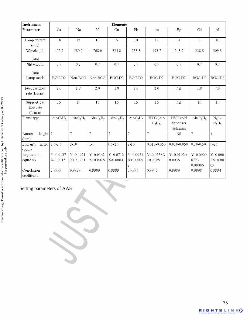
Ocular safety scores for mucilages, gum and their nanoparticles Reviewer 1: Comment 1: Introduction need to be broken in paragraphs. Clarification: Accepted and corrected. Comment 2: Specify the units of strength of alcohol used. Clarification: It was 90% v/v. Comment 3: Was the powder re-dissolved/re-suspended in water. Clarification: Obtained powder was re-dissolved. Comment 4: Mention the centrifuge used? Clarification: Remi centrifuge C-24 BL at 3000 rpm for 10 min. (Remi instruments ltd., Mumbai, India). Comment 5: Detail the setting parameters of atomic absorption spectroscopy. Clarification: The setting parameters of AAS are shown in table no 3. Table 3. Setting parameters of AAS
Elements Instrument Parameter Ca Na K Cu Pb As Hg Cd Al
Lamp current (mA)
10 12 10 6 10 12 4 8 10
Wavelength (nm)
422.7 589.0 768.0 324.8 283.3 193.7 243.7 228.8 309.3
Slit weidth (nm)
0.7 0.2 0.7 0.7 0.7 0.7 0.7 0.7 0.7
Lamp mode BGC-D2
Non-BCG
Non-BCG
BGC-D2 BGC-D2 BGC-D2 BGC-D2 BGC-D2 BGC-D2
Fuel gas flow rate (L/min)
2.0 1.8 2.0 1.8 2.0 2.0 Nil 1.8 7.0
Support gas flow rate (L/min)
15 15 15 15 15 15 Nil 15 15
Flame type Air-C2H2
Air-C2H2
Air-C2H2
Air-C2H2
Air-C2H2
HVG (Air-C2H2)
HVG cold Vaporizer technique
Air-C2H2
N2O-C2H2
Burner height (mm)
7 7 7 7 7 7 Nil 7 11
Linearity range (ppm)
0.5-2.5 2-10 1-5 0.5-2.5 2-10 0.010-0.050
0.010-0.050
0.10-0.50
5-25
Regression equation
Y=0.0137X-0.0015
Y=0.0921X+0.0261
Y=0.0142X+0.0028
Y=0.0712X-0.0064
Y=0.0021X+0.00092
Y=0.0238X+0.2108
Y=0.010X+0.0038
Y=0.000067X-0.00006
Y=0.0007X+0.0009
Correlation coefficient
0.9999 0.9989 0.9989 0.9999 0.9994 0.9945 0.9989 0.9998 0.9994
Comment 6: Correct HE-CAM test for HET-CAM test. Clarification: Accepted and corrected.
Nan
otox
icol
ogy
Dow
nloa
ded
from
info
rmah
ealth
care
.com
by
Uni
vers
ity o
f C
alga
ry o
n 08
/29/
13Fo
r pe
rson
al u
se o
nly.
31
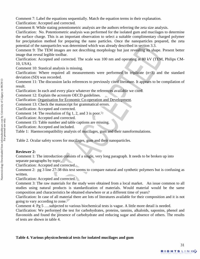
Comment 7: Label the equations sequentially. Match the equation terms in their explanation. Clarification: Accepted and corrected. Comment 8: While stating potentiometric analysis are the authors referring the zeta size analysis. Clarification: No. Potentiometric analysis was performed for the isolated gum and mucilages to determine the surface charge. This is an important observation to select a suitable complimentary charged polymer for precipitation method for preparing the nano particles. Once the nanoparticles prepared, the zeta potential of the nanoparticles was determined which was already described in section 3.3. Comment 9: The TEM images are not describing morphology but just revealing its shape. Present better image that reveal legible toolbar. Clarification: Accepted and corrected. The scale was 100 nm and operating at 80 kV (TEM, Philips CM-10, USA). Comment 10: Stastical analysis is missing. Clarification: Where required all measurements were performed in triplicate (n=3) and the standard deviation (SD) was recorded. Comment 11: The discussion lacks references to previously cited literature. It appears to be compilation of result. Clarification: In each and every place whatever the references available we cited. Comment 12: Explain the acronym OECD guidelines. Clarification: Organisation for Economic Co-operation and Development. Comment 13: Check the manuscript for grammatical errors. Clarification: Accepted and corrected. Comment 14: The resolution of fig 1, 2, and 3 is poor. Clarification: Accepted and corrected. Comment 15: Table number and table captions are missing. Clarification: Accepted and included. Table 1: Haemocompatibility analysis of mucilages, gum and their nanoformulations.
Table 2. Ocular safety scores for mucilages, gum and their nanoparticles.
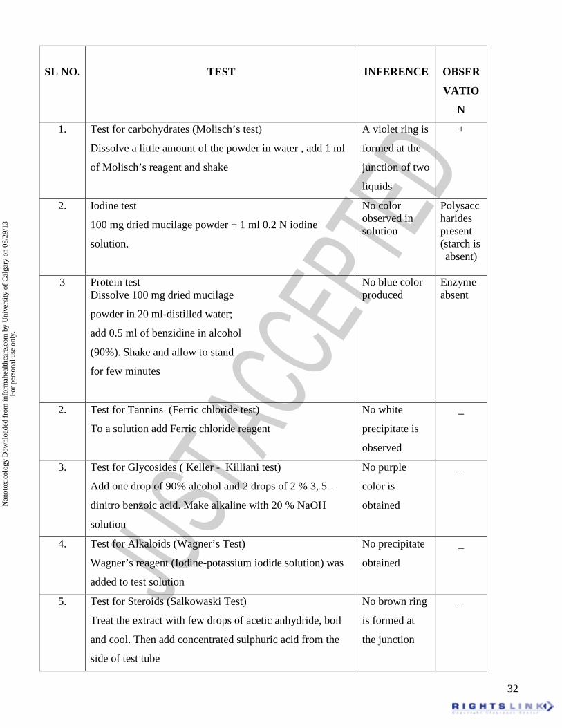
Reviewer 2: Comment 1: The introduction consists of a single, very long paragraph. It needs to be broken up into separate paragraphs by topic. Clarification: Accepted and corrected. Comment 2: pg 3 line 27-38 this text seems to compare natural and synthetic polymers but is confusing as written. Clarification: Accepted and corrected. Comment 3: The raw materials for the study were obtained from a local market. An issue common to all studies using natural products is standardization of materials. Would material would be the same composition and characteristics be obtained elsewhere or at a different time of years? Clarification: In case of all material there are lots of literatures available for their composition and it is not going to vary according to zone. Comment 4: Pg 5 ….subjected to various biochemical tests is vague. A little more detail is needed. Clarification: We performed the test for carbohydrates, proteins, tannins, alkaloids, saponins, phenol and flavonoids and found the presence of carbohydrate and reducing sugar and absence of others. The results of tests are shown in table 4. Table 4. Various physicochemical tests for isolated mucilages and gum
Nan
otox
icol
ogy
Dow
nloa
ded
from
info
rmah
ealth
care
.com
by
Uni
vers
ity o
f C
alga
ry o
n 08
/29/
13Fo
r pe
rson
al u
se o
nly.
32
SL NO.
TEST
INFERENCE
OBSER
VATIO
N
1. Test for carbohydrates (Molisch’s test)
Dissolve a little amount of the powder in water , add 1 ml
of Molisch’s reagent and shake
A violet ring is
formed at the
junction of two
liquids
+
2. Iodine test
100 mg dried mucilage powder + 1 ml 0.2 N iodine
solution.
No color observed in solution
Polysaccharides present (starch is absent)
3 Protein test Dissolve 100 mg dried mucilage
powder in 20 ml-distilled water;
add 0.5 ml of benzidine in alcohol
(90%). Shake and allow to stand
for few minutes
No blue color produced
Enzyme absent
2. Test for Tannins (Ferric chloride test)
To a solution add Ferric chloride reagent
No white
precipitate is
observed
_
3. Test for Glycosides ( Keller - Killiani test)
Add one drop of 90% alcohol and 2 drops of 2 % 3, 5 –
dinitro benzoic acid. Make alkaline with 20 % NaOH
solution
No purple
color is
obtained
_
4. Test for Alkaloids (Wagner’s Test)
Wagner’s reagent (Iodine-potassium iodide solution) was
added to test solution
No precipitate
obtained
_
5. Test for Steroids (Salkowaski Test)
Treat the extract with few drops of acetic anhydride, boil
and cool. Then add concentrated sulphuric acid from the
side of test tube
No brown ring
is formed at
the junction
_
Nan
otox
icol
ogy
Dow
nloa
ded
from
info
rmah
ealth
care
.com
by
Uni
vers
ity o
f C
alga
ry o
n 08
/29/
13Fo
r pe
rson
al u
se o
nly.
33
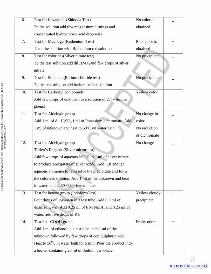
6. Test for flavanoids (Shinoda Test)
To the solution add few magnesium turnings and
concentrated hydrochloric acid drop wise
No color is
obtained
_
7. Test for Mucilage (Ruthenium Test)
Treat the solution with Ruthenium red solution
Pink color is
obtained
+
8. Test for chlorides(Silver nitrate test)
To the test solution add dil.HNO3 and few drops of silver
nitrate
No precipitate _
9. Test for Sulphate (Barium chloride test)
To the test solution add barium sulfate solution
No precipitate _
10. Test for Carbonyl compounds
Add few drops of unknown to a solution of 2,4 – dinitro
phenol
Yellow color +
11. Test for Aldehyde group
Add 1 ml of dil H2SO4 1 ml of Potassium dichromate. Add
1 ml of unknown and heat to 500C on water bath
No change in
color
No reduction
of dichromate
_
12. Test for Aldehyde group
Tollen’s Reagent (Silver mirror test)
Add few drops of aqueous NaOH to 1 ml of silver nitrate
to produce precipitate of silver oxide. Add just enough
aqueous ammonia to redissolve the precipitate and form
the colorless solution. Add 1 ml of the unknown and heat
in water bath at 500C for few minutes
No change _
13. Test for ketone group (IodoformTest)
Four drops of unknown in a test tube .Add 0.5 ml of
distilled water. Add 0.25 ml of 6 M NaOH and 0.25 ml of
water, add five drops of KI3
Yellow cloudy
precipitate
+
14.
Test for –COOH group
Add 1 ml of ethanol in a test tube; add 1 ml of the
unknown followed by few drops of con Sulphuric acid.
Heat to 500C in water bath for 5 min .Pour the product into
a beaker containing 20 ml of Sodium carbonate
Fruity odor +
Nan
otox
icol
ogy
Dow
nloa
ded
from
info
rmah
ealth
care
.com
by
Uni
vers
ity o
f C
alga
ry o
n 08
/29/
13Fo
r pe
rson
al u
se o
nly.
34
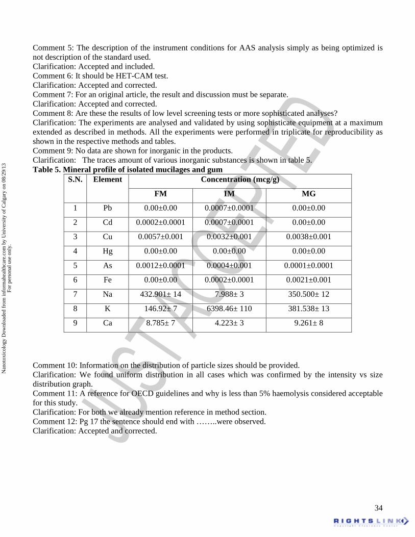
Comment 5: The description of the instrument conditions for AAS analysis simply as being optimized is not description of the standard used. Clarification: Accepted and included. Comment 6: It should be HET-CAM test. Clarification: Accepted and corrected. Comment 7: For an original article, the result and discussion must be separate. Clarification: Accepted and corrected. Comment 8: Are these the results of low level screening tests or more sophisticated analyses? Clarification: The experiments are analysed and validated by using sophisticate equipment at a maximum extended as described in methods. All the experiments were performed in triplicate for reproducibility as shown in the respective methods and tables. Comment 9: No data are shown for inorganic in the products. Clarification: The traces amount of various inorganic substances is shown in table 5. Table 5. Mineral profile of isolated mucilages and gum
Concentration (mcg/g) S.N. Element
FM IM MG
1 Pb 0.00±0.00 0.0007±0.0001 0.00±0.00
2 Cd 0.0002±0.0001 0.0007±0.0001 0.00±0.00
3 Cu 0.0057±0.001 0.0032±0.001 0.0038±0.001
4 Hg 0.00±0.00 0.00±0.00 0.00±0.00
5 As 0.0012±0.0001 0.0004±0.001 0.0001±0.0001
6 Fe 0.00±0.00 0.0002±0.0001 0.0021±0.001
7 Na 432.901± 14 7.988± 3 350.500± 12
8 K 146.92± 7 6398.46± 110 381.538± 13
9 Ca 8.785± 7 4.223± 3 9.261± 8
Comment 10: Information on the distribution of particle sizes should be provided. Clarification: We found uniform distribution in all cases which was confirmed by the intensity vs size distribution graph. Comment 11: A reference for OECD guidelines and why is less than 5% haemolysis considered acceptable for this study. Clarification: For both we already mention reference in method section. Comment 12: Pg 17 the sentence should end with ……..were observed. Clarification: Accepted and corrected.
Nan
otox
icol
ogy
Dow
nloa
ded
from
info
rmah
ealth
care
.com
by
Uni
vers
ity o
f C
alga
ry o
n 08
/29/
13Fo
r pe
rson
al u
se o
nly.
35
Setting parameters of AAS
Nan
otox
icol
ogy
Dow
nloa
ded
from
info
rmah
ealth
care
.com
by
Uni
vers
ity o
f C
alga
ry o
n 08
/29/
13Fo
r pe
rson
al u
se o
nly.
36
Nan
otox
icol
ogy
Dow
nloa
ded
from
info
rmah
ealth
care
.com
by
Uni
vers
ity o
f C
alga
ry o
n 08
/29/
13Fo
r pe
rson
al u
se o
nly.
37
Various physicochemical tests for isolated mucilages and gum
Mineral profile of isolated mucilages and gum Figure 1. (a) MALDI, (b) 1H, (c) 13C NMR of fenugreek mucilage and (d) GC-MS of hydrolysed fenugreek mucilage Figure 2. (a) MALDI, (b) HMQC analysis of isphagula mucilage and (c) GC-MS of hydrolysed isphagula mucilage Figure 3. (a) MALDI, (b) 1H, (c) 13C NMR of mango gum and (d) GC-MS of hydrolysed mango gum Figure 4. TEM morphology of prepared nanoparticles
Figure 5. HET-CAM analysis of following mucilages, gum and their nanoformulations
Table 1: Haemocompatibility analysis of mucilages, gum and their nanoformulations
Table 2. Ocular safety scores for mucilages, gum and their nanoparticles
Nan
otox
icol
ogy
Dow
nloa
ded
from
info
rmah
ealth
care
.com
by
Uni
vers
ity o
f C
alga
ry o
n 08
/29/
13Fo
r pe
rson
al u
se o
nly.