39
Physiology as the science. Bioelectrical phenomena in excitable tissues
| Date post: | 25-Dec-2015 |
| Category: |
Documents |
| Upload: | roxanne-richardson |
| View: | 233 times |
| Download: | 0 times |

Physiology as the science. Bioelectrical phenomena in
excitable tissues

Defining of “physiology” notion
• Physiology is the science about the regularities of organisms‘ vital activity in connection with the external environment.
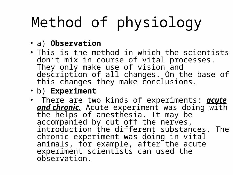
Method of physiology• a) Observation • This is the method in which the scientists don‘t mix
in course of vital processes. They only make use of vision and description of all changes. On the base of this changes they make conclusions.
• b) Experiment• There are two kinds of experiments: acute and
chronic. Acute experiment was doing with the helps of anesthesia. It may be accompanied by cut off the nerves, introduction the different substances. The chronic experiment was doing in vital animals, for example, after the acute experiment scientists can used the observation.

Method of physiology• c) Examination • This is the method of examine the patient with different
diseases, for example, with using the different apparatuses.
• d) Simulation• We can simulation different processes as a laboratory
simulation or realistic simulation, for example, apparatus of artificial kidney or apparatus of artificial circulation. It may be the simulation the different processes by means of computers.

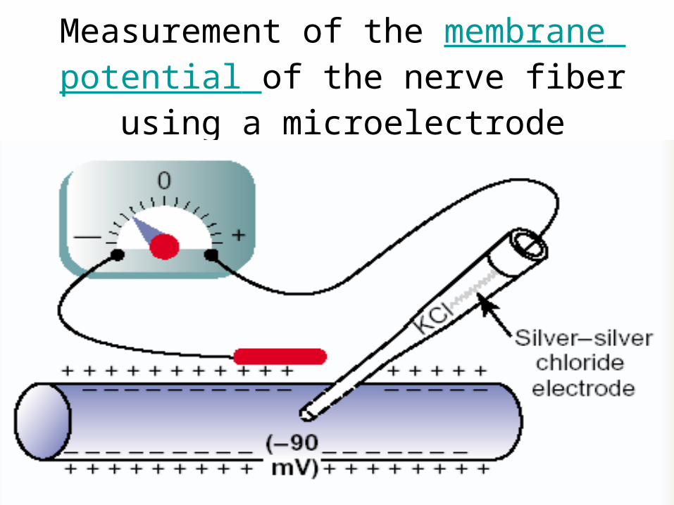
Measurement of the membrane potential of the nerve fiber using a microelectrode

Basic ConceptsForces that determine ionic movement
Electrostatic forcesOpposite charges attractIdentical charges repel
Concentration forcesDiffusion – movement of ions through
semipermeable membraneOsmosis – movement of water from region of
high concentration to low

Basic mechanisms of transport
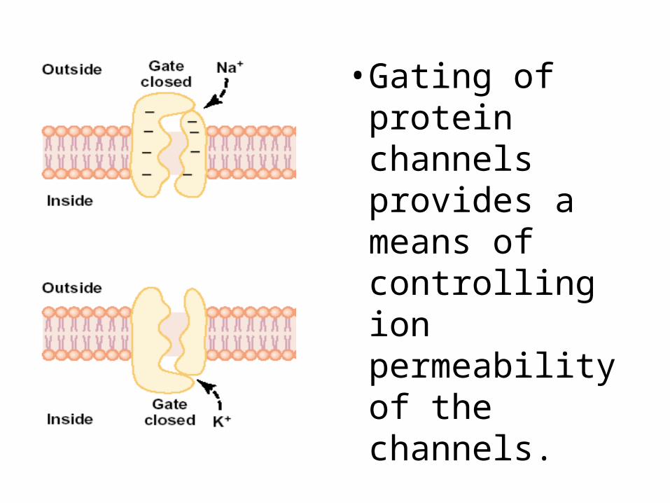
• Gating of protein channels provides a means of controlling ion permeability of the channels.
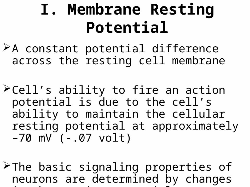
I. Membrane Resting Potential
A constant potential difference across the resting cell membrane
Cell’s ability to fire an action potential is due to the cell’s ability to maintain the cellular resting potential at approximately –70 mV (-.07 volt)
The basic signaling properties of neurons are determined by changes in the resting potential

Concept of Resting Potential (RP)
A potential difference across the cell membrane at the rest stage or when the cell is not stimulated.
Property: It is constant or stable It is negative inside relative to the outside Resting potentials are different in
different cells.

Resting Membrane Potential
Na+ and Cl- are more concentrated outside the cell
K+ and organic anions (organic acids and proteins) are more concentrated inside.
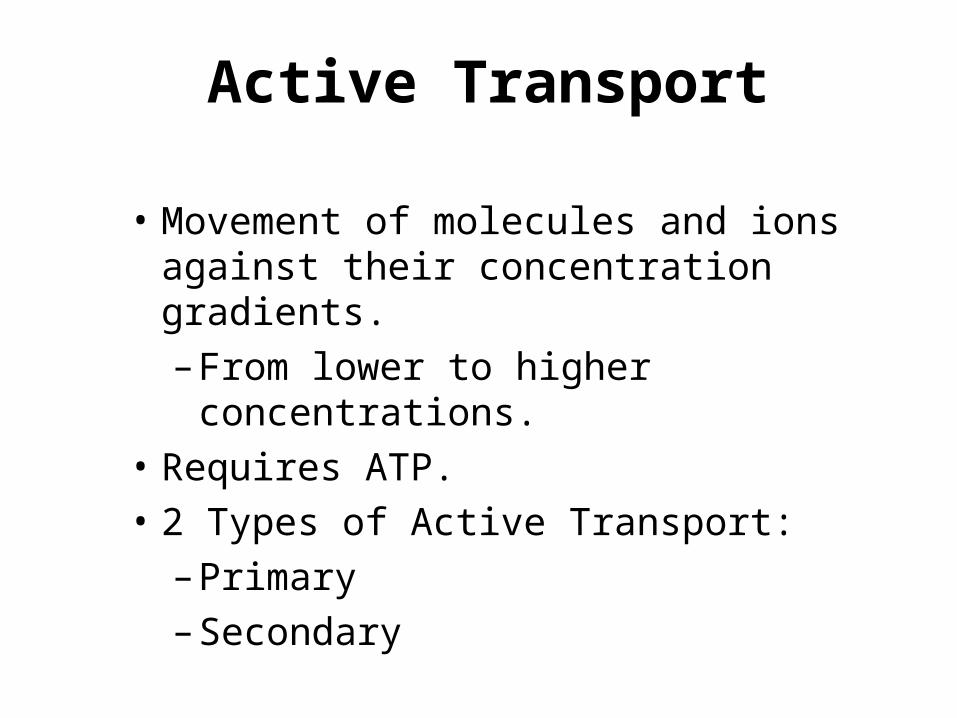
Active Transport
• Movement of molecules and ions against their concentration gradients.
– From lower to higher concentrations.
• Requires ATP.
• 2 Types of Active Transport:
– Primary
– Secondary
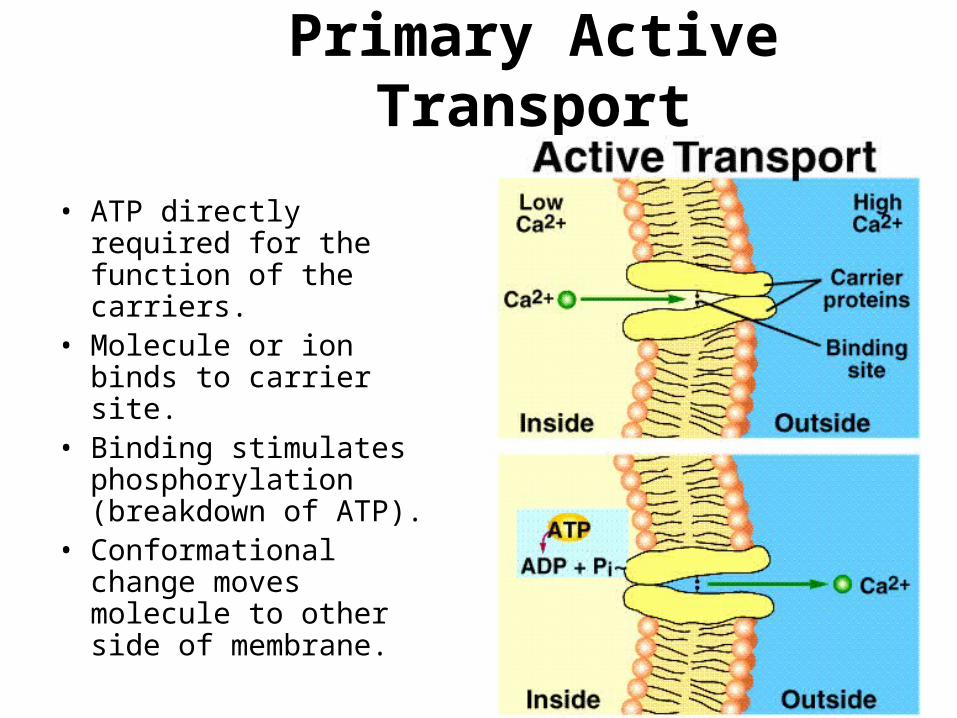
Primary Active Transport
• ATP directly required for the function of the carriers.
• Molecule or ion binds to carrier site.
• Binding stimulates phosphorylation (breakdown of ATP).
• Conformational change moves molecule to other side of membrane.

P
Extracellular fluid1. Three sodium ions (Na+) and adenosine triphosphate (ATP) bind to the carrier protein.
2. The ATP breaks down to adenosine diphosphate (ADP) and a phosphate (P) and releases energy.3. The carrier protein changes shape, and the Na+ are transported across the membrane.
4. The Na+ diffuse away from the carrier protein.
5. Two potassium ions (K+) bind to the carrier protein.
6. The phosphate is released.
7. The carrier protein changes shape, transporting K+ across the membrane, and the K+ diffuse away from the carrier protein. The carrier protein can again bind to Na+ and ATP.
Cytoplasm Na+
ATP
K+
Breakdown of ATP(releases energy)
Carrier protein changesshape (requires energy) ADP
1
3
2
Carrier protein resumesoriginal shape
Na+
Na+ K+
P
K+
45
6
7
ATP binding site
Carrier protein

Intracellular vs extracellular ion concentrations
Ion Intracellular Extracellular
Na+ 5-15 mM 145 mMK+ 140 mM 5 mMMg2+ 0.5 mM 1-2 mMCa2+ 10-7 mM 1-2 mMH+ 10-7.2 M (pH 7.2) 10-7.4 M (pH 7.4)
Cl- 5-15 mM 110 mM
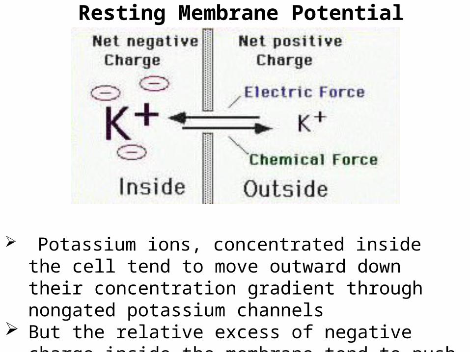
Resting Membrane Potential
Potassium ions, concentrated inside the cell tend to move outward down their concentration gradient through nongated potassium channels
But the relative excess of negative charge inside the membrane tend to push potassium ions out of the cell

Resting Membrane Potential
• But what about sodium?• Electrostatic and Chemical forces act together on
Na+ ions to drive them into the cell • The Na+ channel close during the resting state
Na+ is more concentrated outside than inside and therefore tends to flow into the cell down its concentration gradient
Na+ is driven into the cell by the electrical potential difference across the membrane.

Resting Potential

Postulated mechanism of the sodium-potassium pump
The formation of resting potential depends on:
Concentration difference of K+ across the membrane
Permeability of Na+ and K+ during the resting state
Na+-K+ pump
Factors that affect resting potential
Difference of K+ ion concentration across the membrane
Permeability of the membrane to Na+ and K+.
Action of Na+ pump
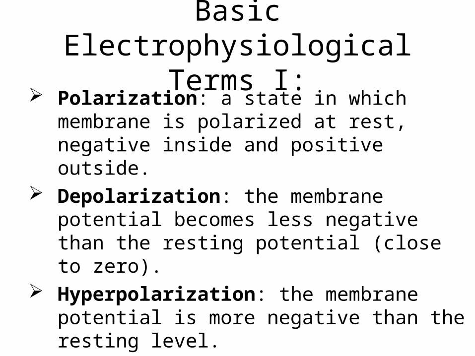
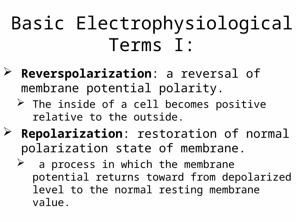
Basic Electrophysiological Terms I:
Polarization: a state in which membrane is polarized at rest, negative inside and positive outside.
Depolarization: the membrane potential becomes less negative than the resting potential (close to zero).
Hyperpolarization: the membrane potential is more negative than the resting level.
Basic Electrophysiological Terms I:
Reverspolarization: a reversal of membrane potential polarity.
The inside of a cell becomes positive relative to the outside.
Repolarization: restoration of normal polarization state of membrane.
a process in which the membrane potential returns toward from depolarized level to the normal resting membrane value.
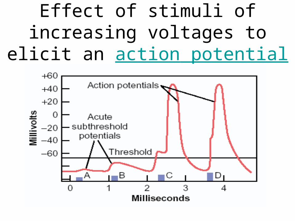
Effect of stimuli of increasing voltages to elicit an action potential
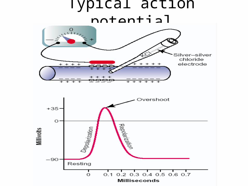
Typical action potential
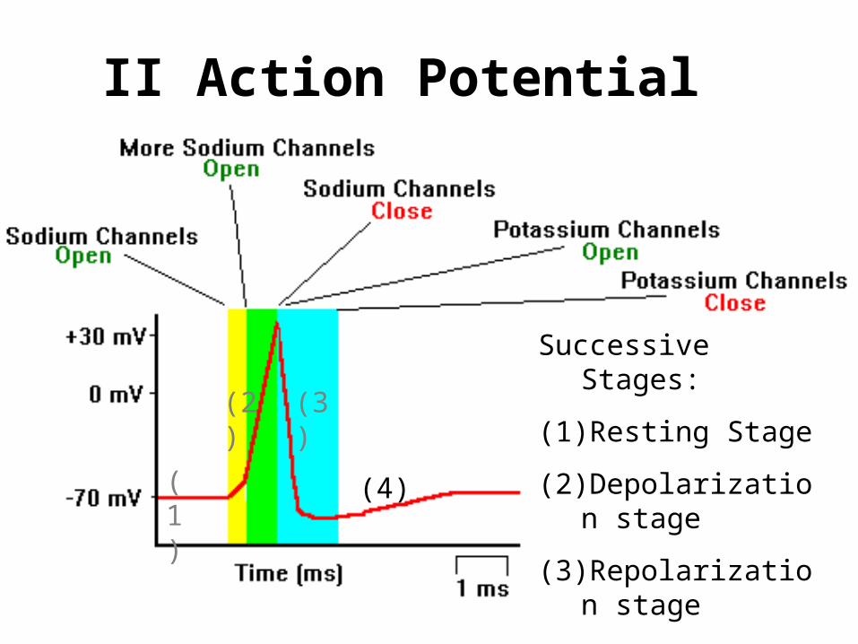
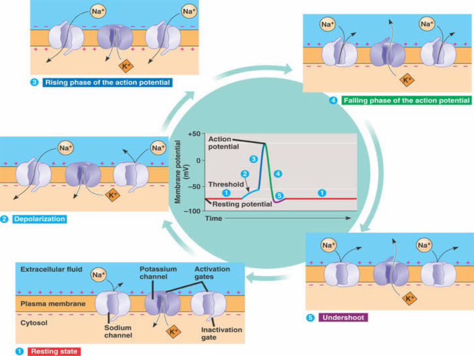
II Action Potential
Successive Stages:
(1) Resting Stage
(2) Depolarization stage
(3) Repolarization stage
(4) After-potential stage
(1)
(2) (3)
(4)


The Action Potential
• Components/characteristics
– RMP
– Depolarizing stimulus
– Threshold
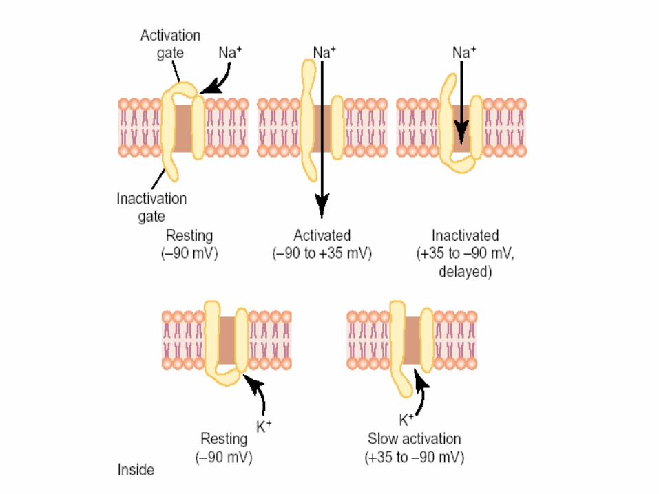
– Rapid Na+ entry (depolarization)
– Isopotential
– Overshoot
– Repolarization (K+ moves out)
– Undershoot (after-hyperpolarization)
– Absolute refractory period
– Relative refractory period
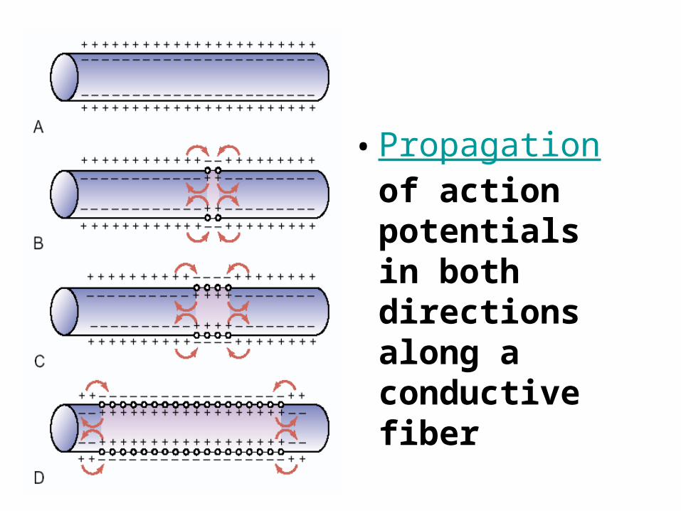
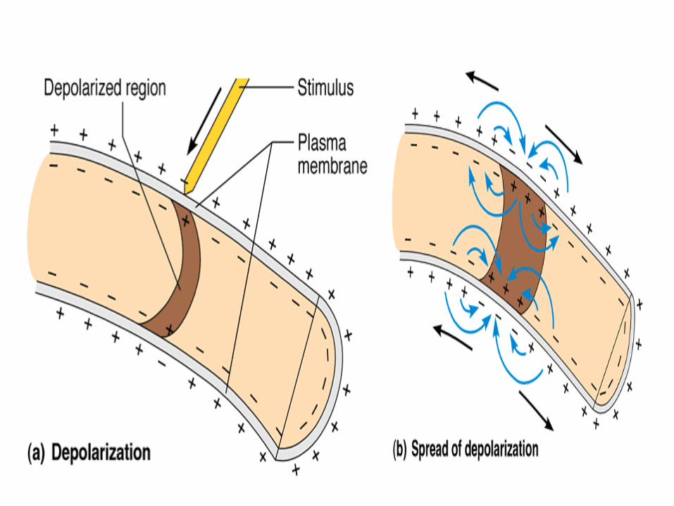
• Propagation of action potentials in both directions along a conductive fiber
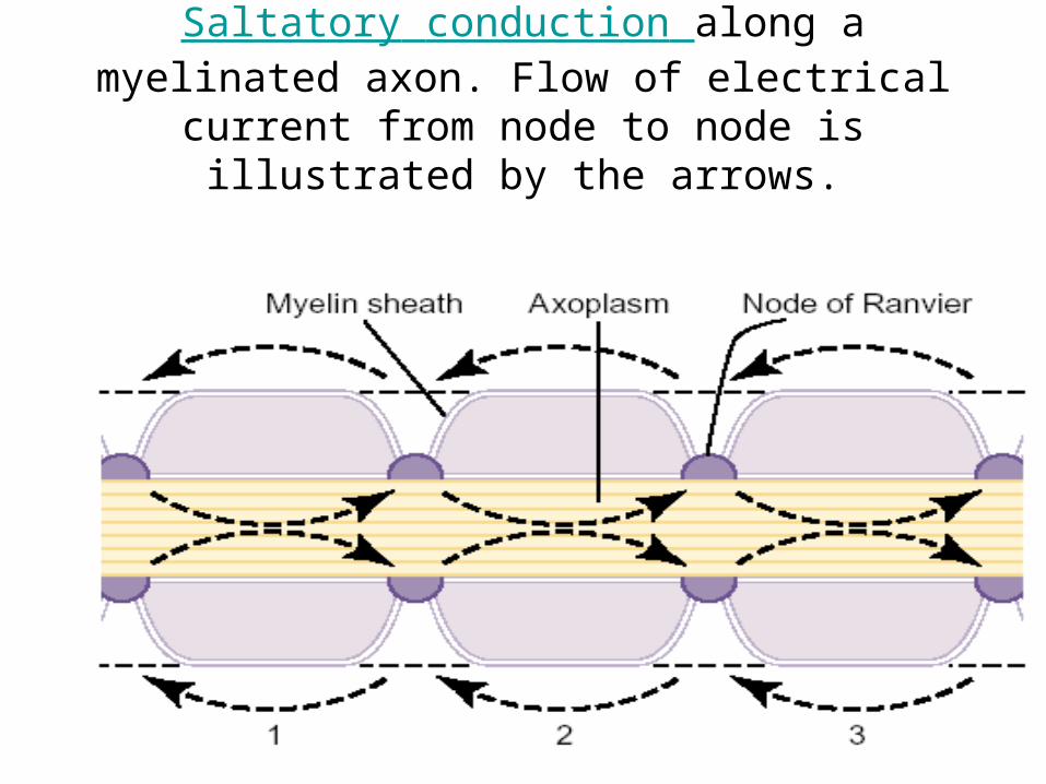
Saltatory conduction along a myelinated axon. Flow of electrical current from node to node is illustrated by the
arrows.


![Physiology of muscles and nerves - Al-Mustansiriya University10_09... · Describe rhythmicity of certain excitable tissues. ... have two essential properties: [1] ... Skeletal muscle](https://static.documents.pub/doc/80x56/5b2590fa7f8b9af7778b4871/physiology-of-muscles-and-nerves-al-mustansiriya-university-1009-describe.jpg)


