48
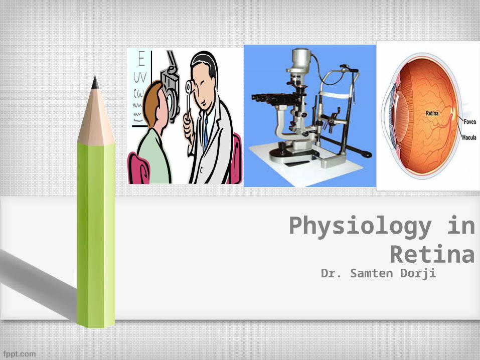
Physiology in Retina Dr. Samten Dorji
| Date post: | 16-Aug-2015 |
| Category: |
Health & Medicine |
| Upload: | samten-dorji |
| View: | 45 times |
| Download: | 4 times |
Physiology in RetinaDr. Samten Dorji
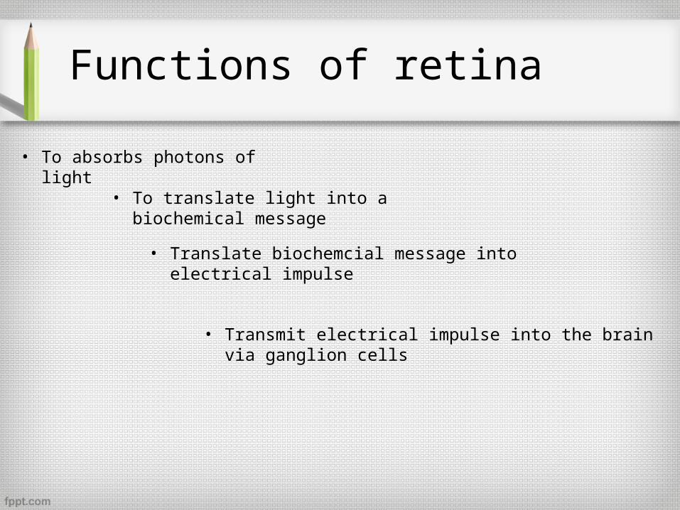
Functions of retina
• To absorbs photons of light
• To translate light into a biochemical message
• Translate biochemcial message into electrical impulse
• Transmit electrical impulse into the brain via ganglion cells
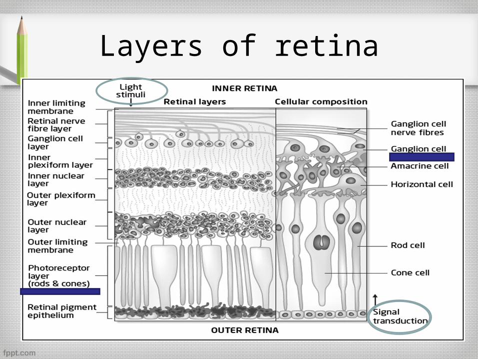
Layers of retina
Outline
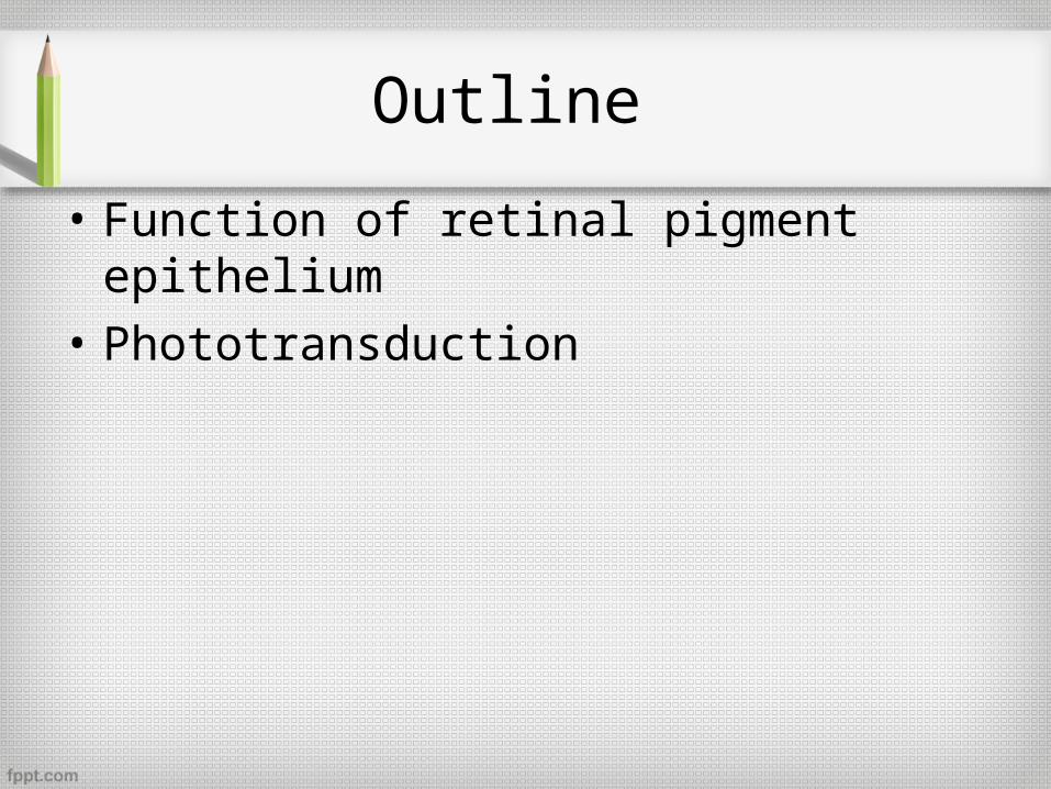
• Function of retinal pigment epithelium
• Phototransduction
Retinal pigment epithelium
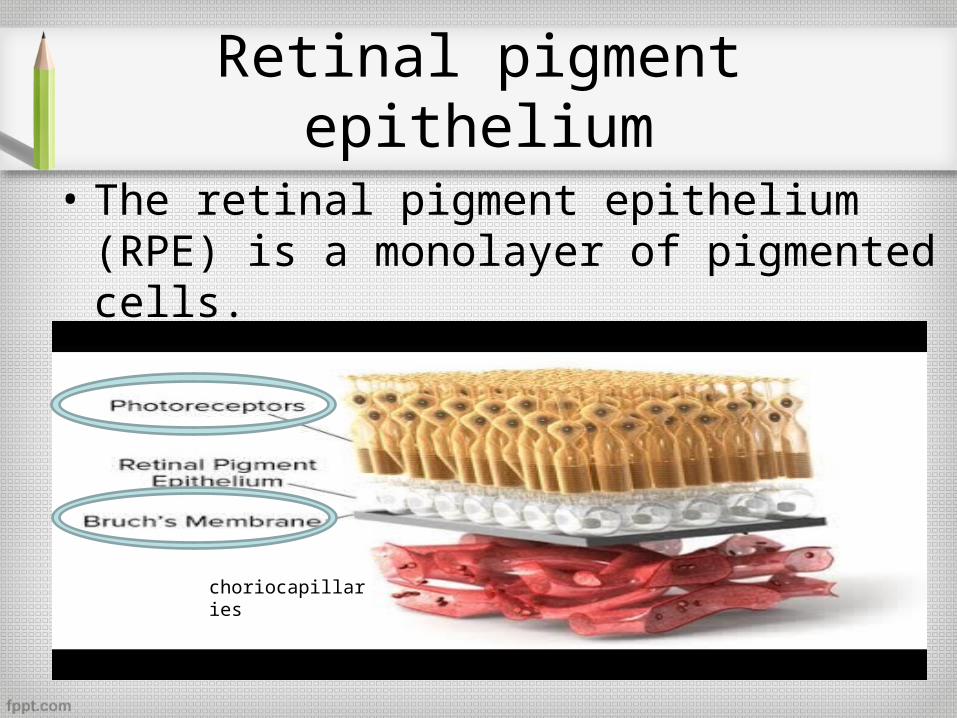
• The retinal pigment epithelium (RPE) is a monolayer of pigmented cells.
choriocapillaries
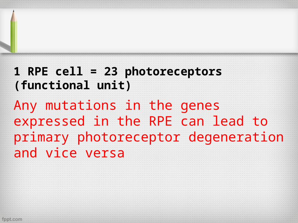
1 RPE cell = 23 photoreceptors (functional unit)
Any mutations in the genes expressed in the RPE can lead to primary photoreceptor degeneration and vice versa
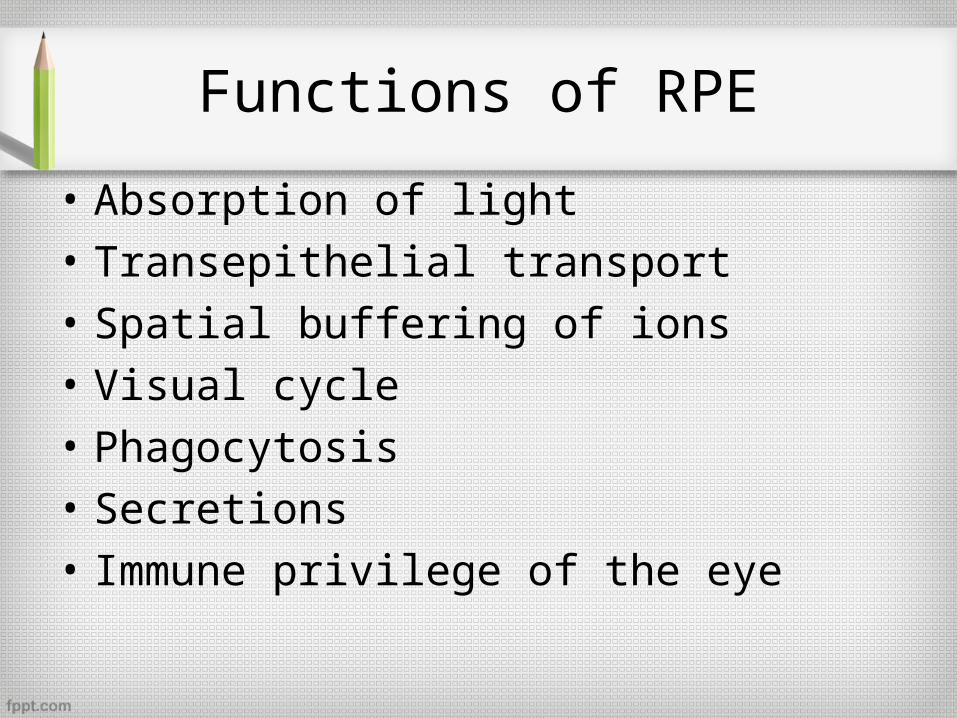
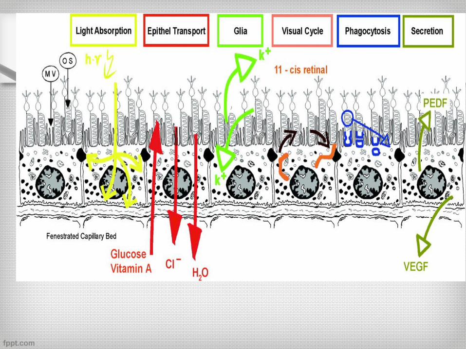
Functions of RPE
• Absorption of light
• Transepithelial transport
• Spatial buffering of ions
• Visual cycle
• Phagocytosis
• Secretions
• Immune privilege of the eye
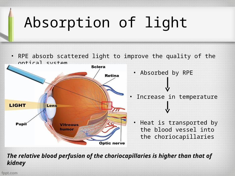
Absorption of light
• RPE absorb scattered light to improve the quality of the optical system.
• Absorbed by RPE
• Increase in temperature
• Heat is transported by the blood vessel into the choriocapillaries
The relative blood perfusion of the choriocapillaries is higher than that of kidney
Photo-oxidative damage
Photo-oxidative damage
Only small amounts of oxygen is used by the adjacent tissues which leads to increased oxygen saturation of the venous blood(>90%)
Large density of light energy+ overflow of oxygen Reactive oxygen species
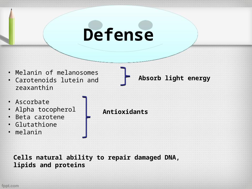
Defense Defense
• Melanin of melanosomes• Carotenoids lutein and zeaxanthin Absorb light energy
• Ascorbate• Alpha tocopherol• Beta carotene• Glutathione • melanin
Antioxidants
Cells natural ability to repair damaged DNA, lipids and proteins
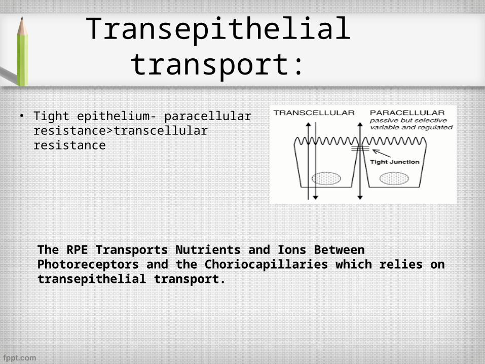
Transepithelial transport:
• Tight epithelium- paracellular resistance>transcellular resistance
The RPE Transports Nutrients and Ions Between Photoreceptors and the Choriocapillaries which relies on transepithelial transport.
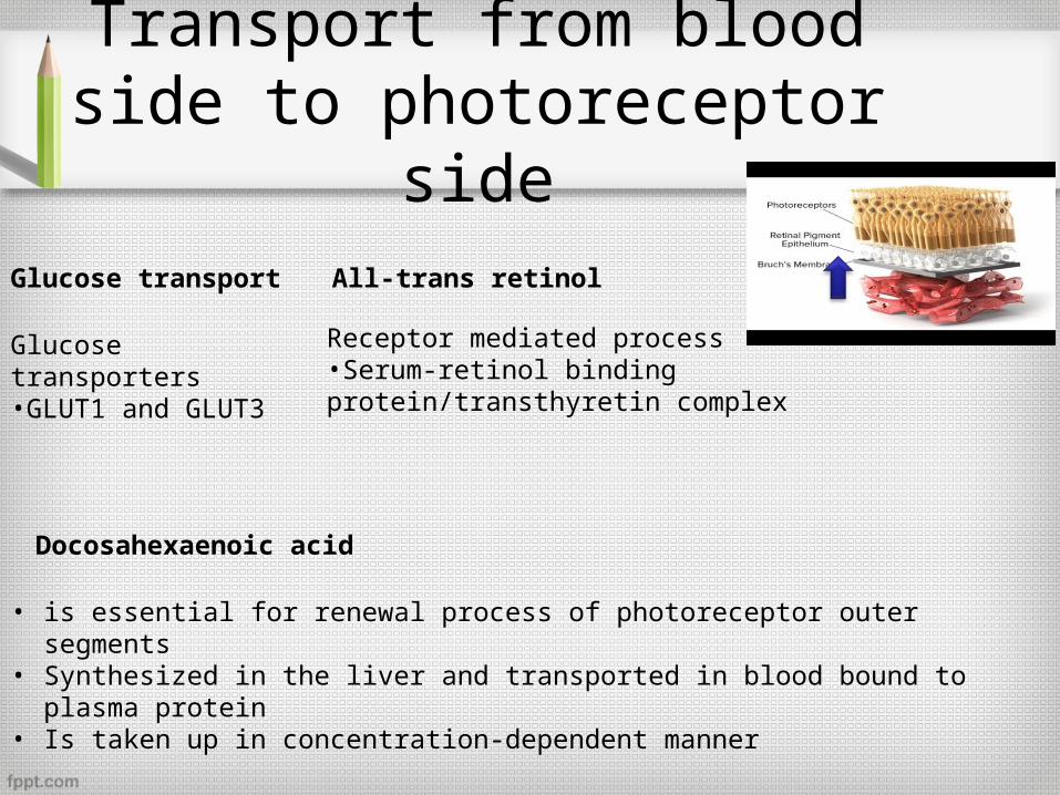
Transport from blood side to photoreceptor side
Glucose transport All-trans retinol
Docosahexaenoic acid
Glucose transporters•GLUT1 and GLUT3
Receptor mediated process•Serum-retinol binding protein/transthyretin complex
• is essential for renewal process of photoreceptor outer segments• Synthesized in the liver and transported in blood bound to plasma protein• Is taken up in concentration-dependent manner
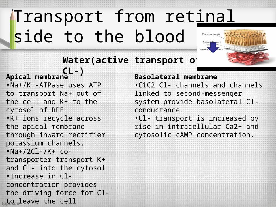
Transport from retinal side to the blood side
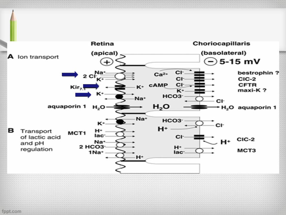
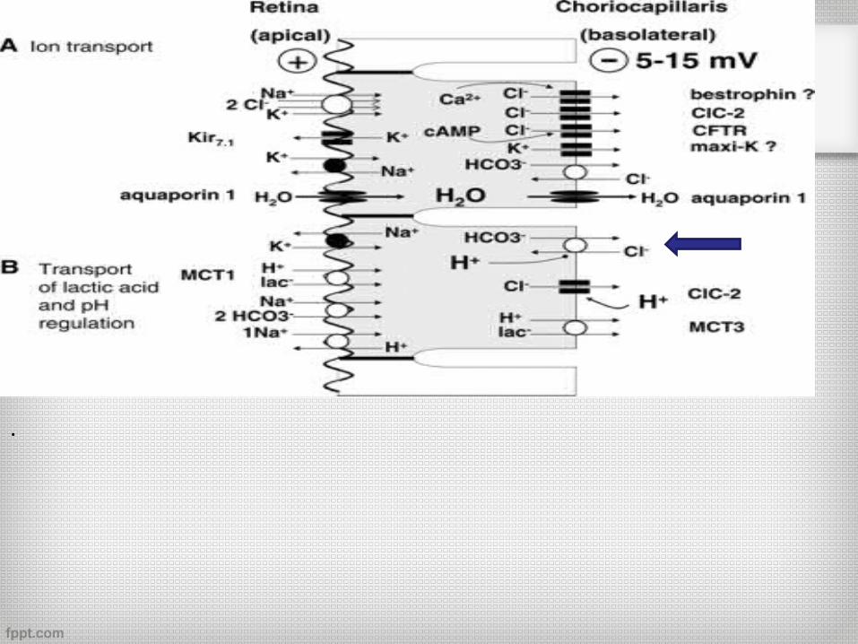
Water(active transport of CL-)
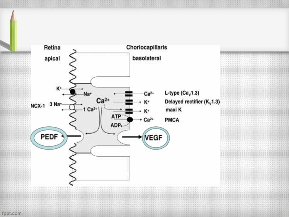
Apical membrane•Na+/K+-ATPase uses ATP to transport Na+ out of the cell and K+ to the cytosol of RPE•K+ ions recycle across the apical membrane through inward rectifier potassium channels.•Na+/2Cl-/K+ co-transporter transport K+ and Cl- into the cytosol•Increase in Cl- concentration provides the driving force for Cl- to leave the cell
Basolateral membrane•C1C2 Cl- channels and channels linked to second-messenger system provide basolateral Cl- conductance.•Cl- transport is increased by rise in intracellular Ca2+ and cytosolic cAMP concentration.
…….
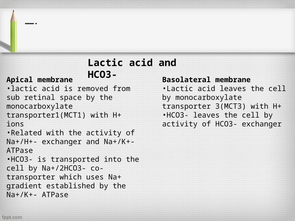
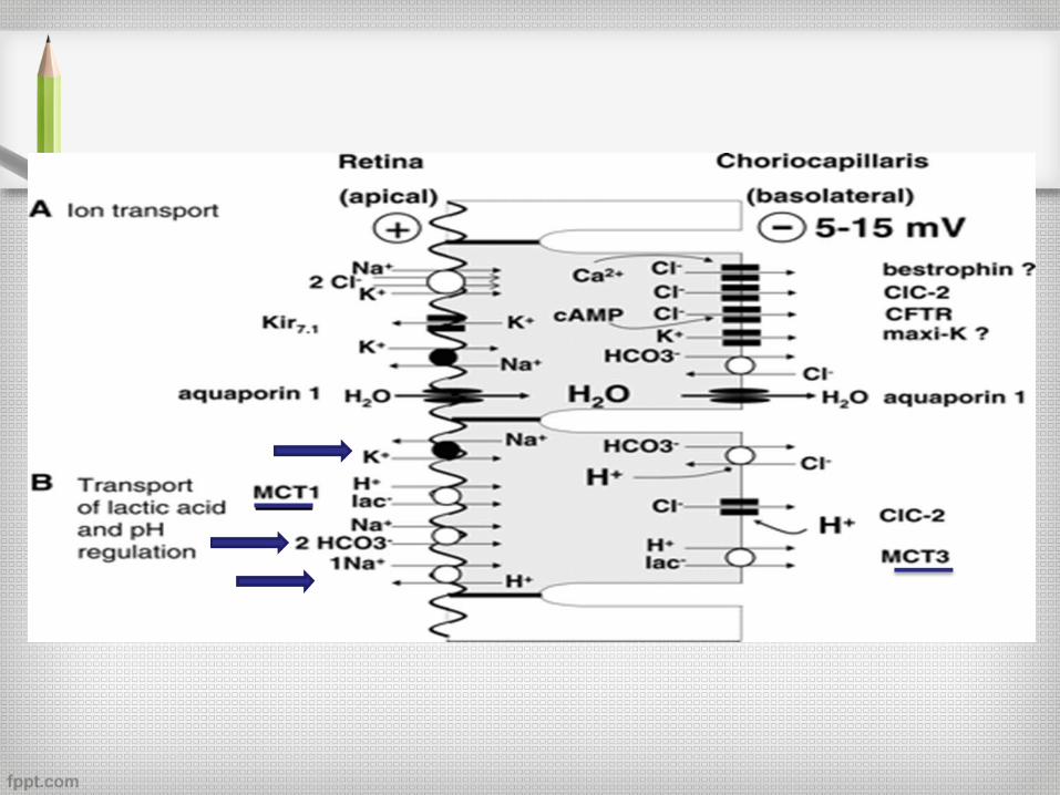
Lactic acid and HCO3-
Apical membrane•lactic acid is removed from sub retinal space by the monocarboxylate transporter1(MCT1) with H+ ions•Related with the activity of Na+/H+- exchanger and Na+/K+-ATPase•HCO3- is transported into the cell by Na+/2HCO3- co-transporter which uses Na+ gradient established by the Na+/K+- ATPase
Basolateral membrane•Lactic acid leaves the cell by monocarboxylate transporter 3(MCT3) with H+•HCO3- leaves the cell by activity of HCO3- exchanger
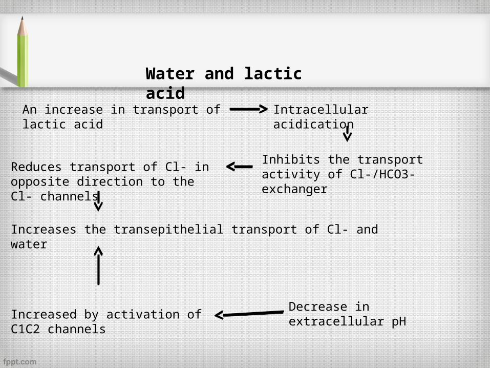
Water and lactic acid
An increase in transport of lactic acid Intracellular acidication
Inhibits the transport activity of Cl-/HCO3- exchanger
Reduces transport of Cl- in opposite direction to the Cl- channels
Increases the transepithelial transport of Cl- and water
Increased by activation of C1C2 channels
Decrease in extracellular pH
.
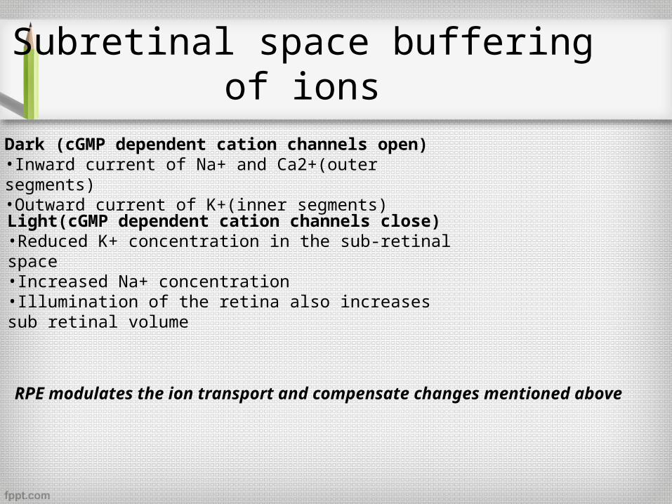
Subretinal space buffering of ions
Dark (cGMP dependent cation channels open)•Inward current of Na+ and Ca2+(outer segments)•Outward current of K+(inner segments)
Light(cGMP dependent cation channels close)•Reduced K+ concentration in the sub-retinal space•Increased Na+ concentration•Illumination of the retina also increases sub retinal volume
RPE modulates the ion transport and compensate changes mentioned above
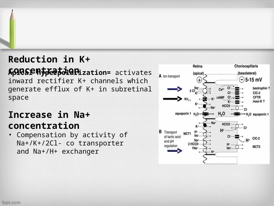
Reduction in K+ concentrationApical hyperpolarization= activates inward rectifier K+ channels which generate efflux of K+ in subretinal space
Increase in Na+ concentration
• Compensation by activity of Na+/K+/2Cl- co transporter and Na+/H+ exchanger
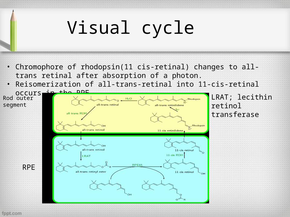
Visual cycle
• Chromophore of rhodopsin(11 cis-retinal) changes to all-trans retinal after absorption of a photon.
• Reisomerization of all-trans-retinal into 11-cis-retinal occurs in the RPE.
Rod outer segment
RPE
LRAT; lecithin retinol transferase
Phagocytosis
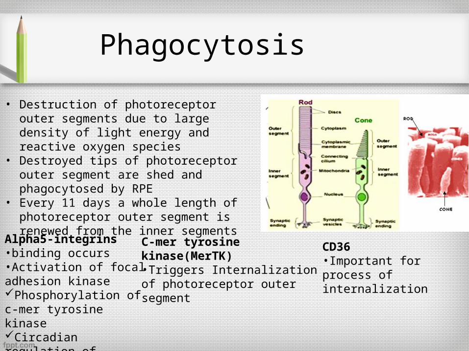
• Destruction of photoreceptor outer segments due to large density of light energy and reactive oxygen species
• Destroyed tips of photoreceptor outer segment are shed and phagocytosed by RPE
• Every 11 days a whole length of photoreceptor outer segment is renewed from the inner segments
Alpha5-integrins•binding occurs•Activation of focal adhesion kinasePhosphorylation of c-mer tyrosine kinaseCircadian regulation of phagocytosis
C-mer tyrosine kinase(MerTK)•Triggers Internalization of photoreceptor outer segment
CD36•Important for process of internalization
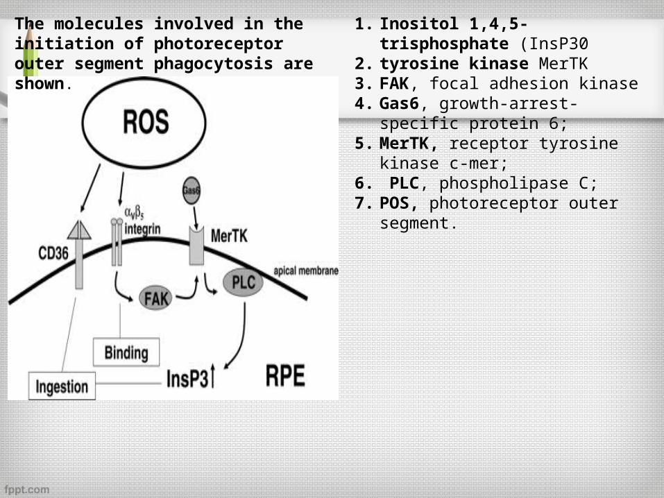
1. Inositol 1,4,5-trisphosphate (InsP302. tyrosine kinase MerTK 3. FAK, focal adhesion kinase 4. Gas6, growth-arrest-specific protein
6; 5. MerTK, receptor tyrosine kinase c-
mer;6. PLC, phospholipase C; 7. POS, photoreceptor outer segment.
The molecules involved in the initiation of photoreceptor outer segment phagocytosis are shown.
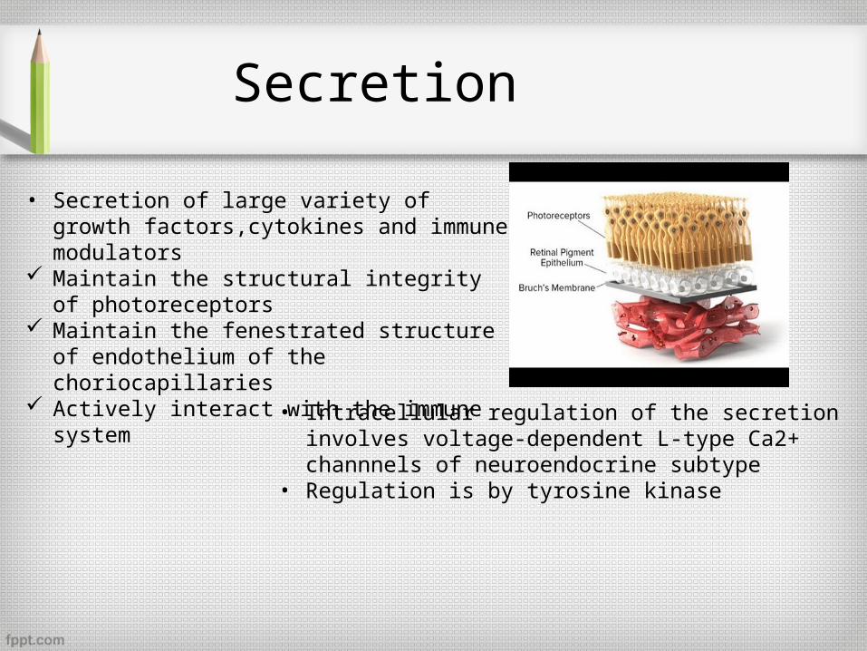
Secretion
• Secretion of large variety of growth factors,cytokines and immune modulators
Maintain the structural integrity of photoreceptors
Maintain the fenestrated structure of endothelium of the choriocapillaries
Actively interact with the immune system
• Intracellular regulation of the secretion involves voltage-dependent L-type Ca2+ channnels of neuroendocrine subtype
• Regulation is by tyrosine kinase
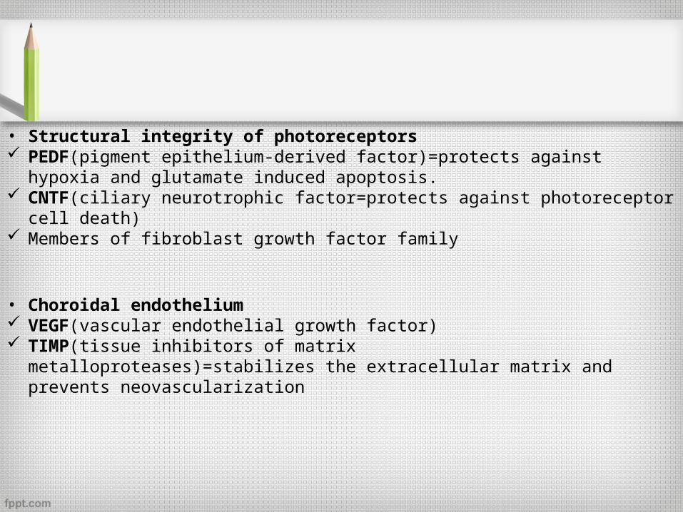
• Structural integrity of photoreceptors PEDF(pigment epithelium-derived factor)=protects against hypoxia and glutamate
induced apoptosis. CNTF(ciliary neurotrophic factor=protects against photoreceptor cell death) Members of fibroblast growth factor family
• Choroidal endothelium VEGF(vascular endothelial growth factor) TIMP(tissue inhibitors of matrix metalloproteases)=stabilizes the extracellular
matrix and prevents neovascularization
Immune Privilege of the Eye
• The RPE represents a mechanical and tight barrier which separates the inner space of the eye from the blood stream.
• It is able to communicate with the immune system in order to activate the immune system in the case of disease.

Phototransduction
• What is it? series of biochemical events • What does it do? a photon of light is changed
to an electrical signal • Where does it take place? in the photoreceptors.• Activation phase and
recovery phase
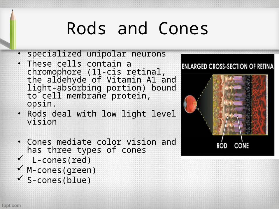
Rods and Cones• specialized unipolar neurons• These cells contain a chromophore (11-
cis retinal, the aldehyde of Vitamin A1 and light-absorbing portion) bound to cell membrane protein, opsin.
• Rods deal with low light level vision
• Cones mediate color vision and has three types of cones
L-cones(red) M-cones(green) S-cones(blue)
Rods and Cones
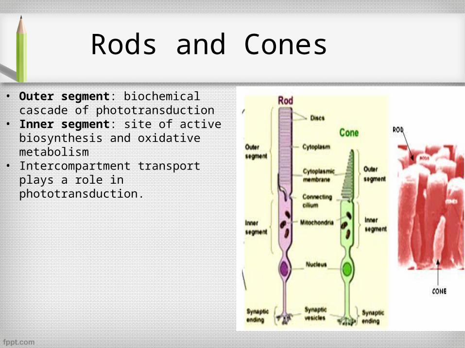
• Outer segment: biochemical cascade of phototransduction
• Inner segment: site of active biosynthesis and oxidative metabolism
• Intercompartment transport plays a role in phototransduction.
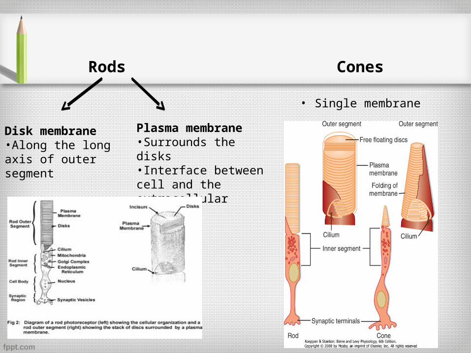
Rods Cones
Disk membrane•Along the long axis of outer segment
Plasma membrane•Surrounds the disks•Interface between cell and the extracellular space
• Single membrane
Dark- adapted rods
• Resting dark adapted state
• The activation phase of light response
• The recovery phase

Resting dark adapted state
• The membrane potential
• The dark current
• Ca2+ and the exchanger
• Control of cGMP
• Rhodopsin
• Lipid mileu
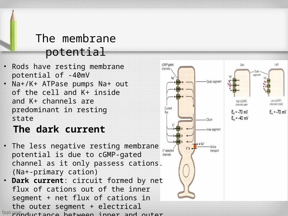
The membrane potential
• Rods have resting membrane potential of -40mV
• Na+/K+ ATPase pumps Na+ out of the cell and K+ inside and K+ channels are predominant in resting state
The dark current
• The less negative resting membrane potential is due to cGMP-gated channel as it only passess cations.(Na+-primary cation)
• Dark current: circuit formed by net flux of cations out of the inner segment + net flux of cations in the outer segment + electrical conductance between inner and outer segments
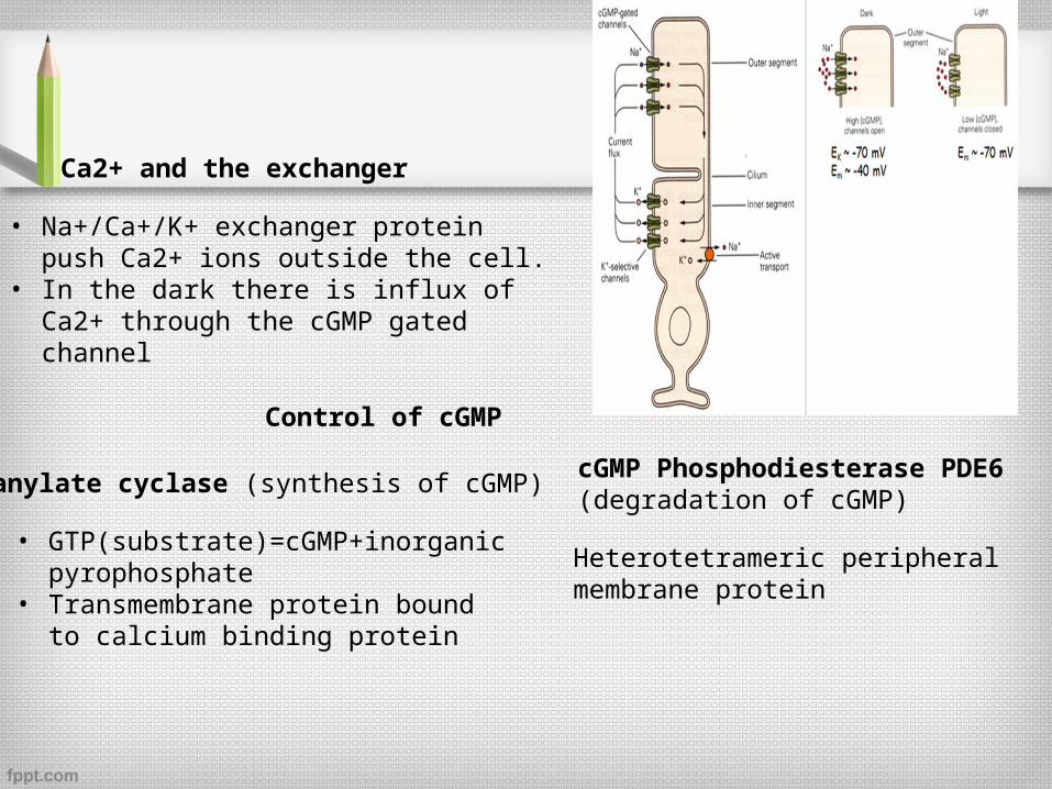
Ca2+ and the exchanger
• Na+/Ca+/K+ exchanger protein push Ca2+ ions outside the cell.
• In the dark there is influx of Ca2+ through the cGMP gated channel
Control of cGMP
Guanylate cyclase (synthesis of cGMP)
• GTP(substrate)=cGMP+inorganic pyrophosphate
• Transmembrane protein bound to calcium binding protein
cGMP Phosphodiesterase PDE6 (degradation of cGMP)
Heterotetrameric peripheral membrane protein
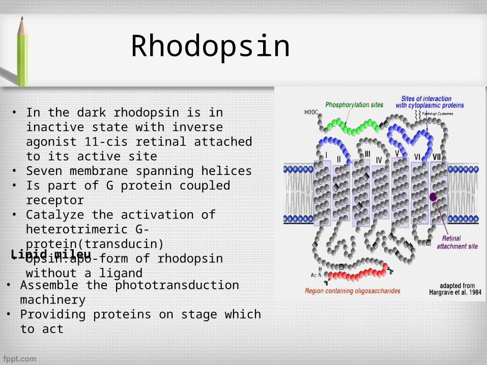
Rhodopsin
• In the dark rhodopsin is in inactive state with inverse agonist 11-cis retinal attached to its active site
• Seven membrane spanning helices• Is part of G protein coupled receptor• Catalyze the activation of heterotrimeric G-
protein(transducin)• Opsin:apo-form of rhodopsin without a ligand
Lipid mileu
• Assemble the phototransduction machinery• Providing proteins on stage which to act
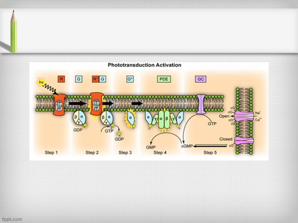
Activation phase of light response
• Photoisomerization of rhodopsin
• G- protein activation
• PDE6 activation
• Channel closing
• Slowing of neurotransmitter release
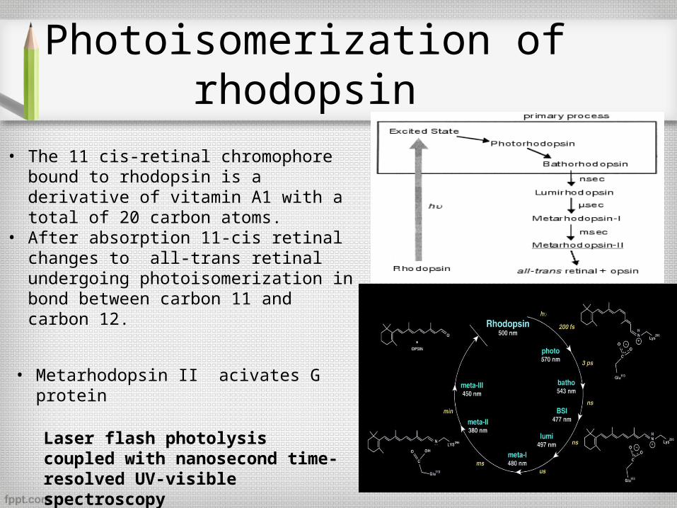
• The 11 cis-retinal chromophore bound to rhodopsin is a derivative of vitamin A1 with a total of 20 carbon atoms.
• After absorption 11-cis retinal changes to all-trans retinal undergoing photoisomerization in bond between carbon 11 and carbon 12.
Laser flash photolysis coupled with nanosecond time-resolved UV-visible spectroscopy
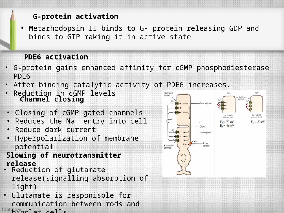
• Metarhodopsin II acivates G protein
Photoisomerization of rhodopsin
G-protein activation
• Metarhodopsin II binds to G- protein releasing GDP and binds to GTP making it in active state.
PDE6 activation
• G-protein gains enhanced affinity for cGMP phosphodiesterase PDE6• After binding catalytic activity of PDE6 increases.• Reduction in cGMP levels
Channel closing
• Closing of cGMP gated channels• Reduces the Na+ entry into cell• Reduce dark current• Hyperpolarization of membrane potential
Slowing of neurotransmitter release
• Reduction of glutamate release(signalling absorption of light)
• Glutamate is responisble for communication between rods and bipolar cells
Recovery phase
• Rhodopsin phosphorylation, retinoid cycling and regeneration
• Arrestin binding
• cGMP restoration by guanylate cyclase activation
• G-protein and PDE6 inactivation by RGS9
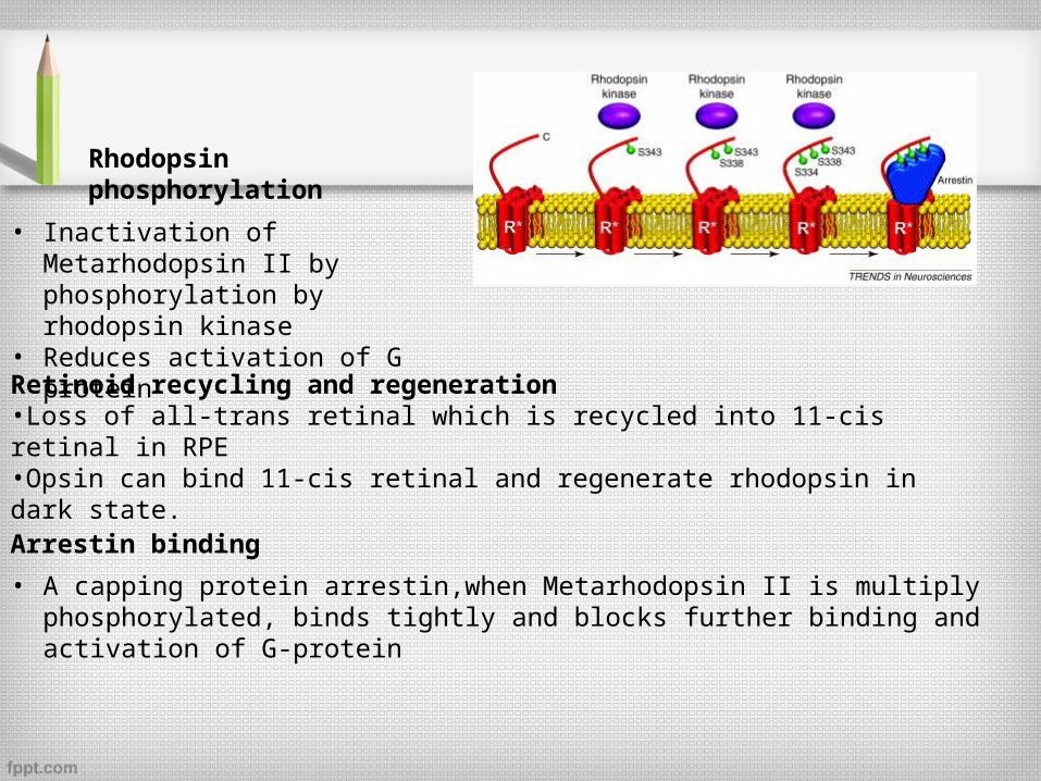
Rhodopsin phosphorylation
• Inactivation of Metarhodopsin II by phosphorylation by rhodopsin kinase
• Reduces activation of G protein
Retinoid recycling and regeneration•Loss of all-trans retinal which is recycled into 11-cis retinal in RPE•Opsin can bind 11-cis retinal and regenerate rhodopsin in dark state.
Arrestin binding
• A capping protein arrestin,when Metarhodopsin II is multiply phosphorylated, binds tightly and blocks further binding and activation of G-protein
cGMP restoration by guanylate cyclase activation
• Calcium feedback mechanism• Na+/Ca2+ exchanger pump Ca2+ out of cell and leakage of Ca2+ into the cell
through cGMP channel is blocked• Reduced calcium activates guanylate cyclase= balance the activation of PDE6
G-protein and PDE6 inactivation by RGS9-1
• RGS9-1: member of the regulator of G-protein signalling family of GTPase accelerating proteins
• Hydrolysis of bound GTP to G-protein to GDP= inactivated PDE6• Slowest step in recovery phase
Phototransduction and disease
• Retinal degeneration
• Night blindness
• Color blindness
• Achromatopsia
Summary
• Functions of retinal epithelium
• Visual phototransduction

Thank you
References
• Adlers physiology of the eye, seventh edition
• Adlers physiology of the eye, eleventh edition
• CLINICAL ANATOMY AND PHYSIOLOGY OF THE VISUAL SYSTEM, THIRD EDITION
• The Retinal Pigment Epithelium in Visual Function, Olaf Strauss
• Internet for images


