Page 1
Toxins 2012, 4, 1343-1366; doi:10.3390/toxins4111343
toxinsISSN 2072-6651
www.mdpi.com/journal/toxins
Review
PI3K/Akt/mTOR, a Pathway Less Recognized for Staphylococcal Superantigen-Induced Toxicity
Teresa Krakauer
Department of Immunology, Integrated Toxicology Division, United States Army Medical Research
Institute of Infectious Diseases, Fort Detrick, Frederick, MD 21702, USA;
E-Mail: [email protected] ; Tel.: +1-301-619-4733; Fax: +1-301-619-2348
Received: 27 September 2012; in revised form: 12 November 2012 / Accepted: 13 November 2012 /
Published: 15 November 2012
Abstract: Immunostimulating staphylococcal enterotoxin B (SEB) and related
superantigenic toxins cause diseases in humans and laboratory animals by activating cells
of the immune system. These toxins bind directly to the major histocompatibility complex
(MHC) class II molecules on antigen-presenting cells and specific Vβ regions of T-cell
receptors (TCR), resulting in hyperactivation of both T lymphocytes and
monocytes/macrophages. Activated host cells produce excessive amounts of
proinflammatory cytokines and chemokines, especially tumor necrosis factor α, interleukin
1 (IL-1), IL-2, interferon γ (IFNγ), and macrophage chemoattractant protein 1 causing
clinical symptoms of fever, hypotension, and shock. The well-explored signal transduction
pathways for SEB-induced toxicity downstream from TCR/MHC ligation and interaction
of cell surface co-stimulatory molecules include the mitogen-activated protein kinase
cascades and cytokine receptor signaling, culminating in NFκB activation. Independently,
IL-2, IFNγ, and chemokines from activated T cells signal via the phosphoinositide 3-kinase
(PI3K), the serine/threonine kinases, Akt and mammalian target of rapamycin (mTOR)
pathways. This article reviews the signaling molecules induced by superantigens in the
activation of PI3K/Akt/mTOR pathways leading to staphylococcal superantigen-induced
toxicity and updates potential therapeutics against superantigens.
Keywords: staphylococcal superantigens; inflammatory cytokines; signaling pathways;
PI3K; Akt; mTOR; therapeutics
OPEN ACCESS
Page 2
Toxins 2012, 4
1344
1. Introduction
Staphylococcus aureus is a ubiquitous gram-positive coccus that produces several exotoxins with
potent immunostimulating activities which contribute to its ability to cause disease in humans, most
notably food poisoning, toxic shock, and autoimmune diseases [1–7]. Staphylococcal enterotoxins A
through U (SEA-SEU) and toxic shock syndrome toxin 1 (TSST-1) were termed “superantigens” due
to their ability to polyclonally activate T cells at picomolar concentrations. Since then, many
structurally similar superantigens from Staphylococcus aureus and Streptococcus pyrogenes, as well as
those from other bacteria, virus, and fungal origins have been discovered [7]. Staphylococcal
superantigens induce a mitogenic response in T cells, stimulating a large proportion (5%–30%) of T
cells to proliferate compared to less than 0.01% of T-cell proliferation initiated by a conventional
antigen [8]. Superantigen binds outside the peptide-binding groove of the major histocompatibility
complex (MHC) class II and bypasses conventional antigen processing by antigen-presenting cells
(APC) [3,7,8]. By interacting with both MHC class II molecules on APC and specific elements within
the variable region of the Vβ chains of the T cell receptor (TCR), these microbial toxins perturb the
immune system and induce high levels of proinflammatory cytokines and chemokines [9–16]. Other
tissue damaging molecules such as matrix metalloproteinases (MMPs) and tissue factor are also
produced by superantigen-activated host cells, affecting both inflammatory and coagulation
pathways [17]. Activated neutrophils produce reactive oxygen species (ROS) which leads to increased
vascular permeability and lung injury [18]. Tumor necrosis factor α (TNFα) and interleukin 1 (IL-1)
are induced early after intoxication and are direct mediators of fever, hypotension, and shock [19–21].
In addition, IFNγ produced by activated T cells acts synergistically with TNFα and IL-1 to enhance
host defense and tissue injury by establishing an inflammatory environment for T cell activation and
differentiation. IL-2, another cytokine from superantigen-activated T cells is essential for T-cell
growth but excessive amounts cause vasodilation leading to vascular leak and edema [22].
SEB has historically been the most intensively studied superantigen and is listed as a category B
select agent by the Centers for Disease Control and Prevention (CDC), as it can be used as an
air-borne, food-borne, and water-borne toxin. Depending on the dose and route of exposure, SEB and
other SEs cause food poisoning, acute and fatal respiratory distress, autoimmune diseases, and toxic
shock [3,23–27]. Superantigens also enhance proinflammatory response and lethality by synergizing
with other bacterial products such as lipopolysaccharide (LPS), lipoproteins, and viruses [28–31].
Recent studies further indicate that superantigens upregulate toll-like receptor 2 (TLR2) and TLR4,
receptors for binding pathogen associated molecular patterns, further amplifying the immune response
to other microbial products [32,33]. Because it is common to encounter pathogens and their toxins
concomitantly in real life, superantigens can have profound toxic effects at extremely
low concentrations.
2. Staphylococcal Superantigen Structure and Binding
Staphylococcal enterotoxins (SEs) and TSST-1 are 22-kD to 30-kD single-chain proteins with
well-characterized secondary and tertiary structures [34]. Staphylococcal superantigens are grouped
based on their primary sequence homology with SEA, SED, and SEE as the first group sharing the
Page 3
Toxins 2012, 4
1345
highest sequence homology of 53% to 81% [5,7,35]. A second group consists of SEB, the SECs, and
SEG, which are 50% to 66% homologous. TSST-1 stands alone by itself in one group as it is distantly
related, with only 28% homology and has a distinct, shorter primary sequence of 194 amino acids with
no cysteines and a missing “disulfide loop” commonly found in SEs. A study with mutants of SEC2
indicated that the disulfide loop may be responsible for the emetic activity of SEs [36]. A newer
classification scheme of five bacterial superantigen groups including the streptococcal superantigens
was proposed based on their phylogenic relationships and similarities in modes of binding to MHC
class II molecules. Cross-reactivities of polyclonal and monoclonal antibodies to the SEs and TSST-1
indicate common epitopes exist among these toxins [37]. X-ray crystallography of SEA, SEB and
TSST-1 reveals similarities in the secondary-tertiary structure with two tightly packed domains
containing β-sheets and α-helices [34]. The relatively conserved TCR-binding site is located in the
shallow groove between these two domains [7,34,38,39].
There are two distinct sites on MHC class II molecules for superantigen binding; a common,
low-affinity binding site located on the α-chain of MHC class II and a high-affinity, zinc-dependent
binding site on the β-chain [7,40–43]. Superantigens in the SEA subfamily bind to both sites, whereas
SEB and TSST-1 bind only to the generic low-affinity site [41–45]. Individual toxin displays
preferential binding to distinct alleles of specific MHC isotypes accounting for differences in host
responses to SEs [45–48]. In general, HLA-DR binds SEs and TSST-1 better than HLA-DP or -DQ,
and murine IE molecules bind with higher affinity than IA [45,48].
The binding of superantigens to TCR Vβ is of low affinity (Kd = 10−4–10−6 M), similar to those with
conventional MHC/peptide/TCR [49,50]. However, each toxin binds to a distinct repertoire of TCR Vβ
chains revealing unique Vβ specificities of individual superantigen [7,51]. The binding contacts are
mostly between the side-chain atoms of the superantigen and the complementarity-determining regions
1 and 2 and the hypervariable region 4 within the Vβ chain. There are multiple modes of superantigen
binding to MHC and TCR. SEB and SEC crosslink MHC class II α chain and TCR Vβ whereas SEA
binds to both α and β chain of MHC class II to crosslink TCR Vβ [7]. The cooperative binding of
superantigen/MHC complex with TCR enables superantigen binding to TCR with a higher affinity
than with toxin alone [49]. A recent study suggests a third binding site for the co-stimulatory receptor
CD28 on T cells to SEB and peptides derived from the CD28 binding region protected mice from
SEB-induced lethality [52]. Receptor clustering and subsequent intracellular signaling in both T cells
and APC lead to excessive mediator release and specific pathways of cell activation [53,54].
Page 4
Toxins 2012, 4
1346
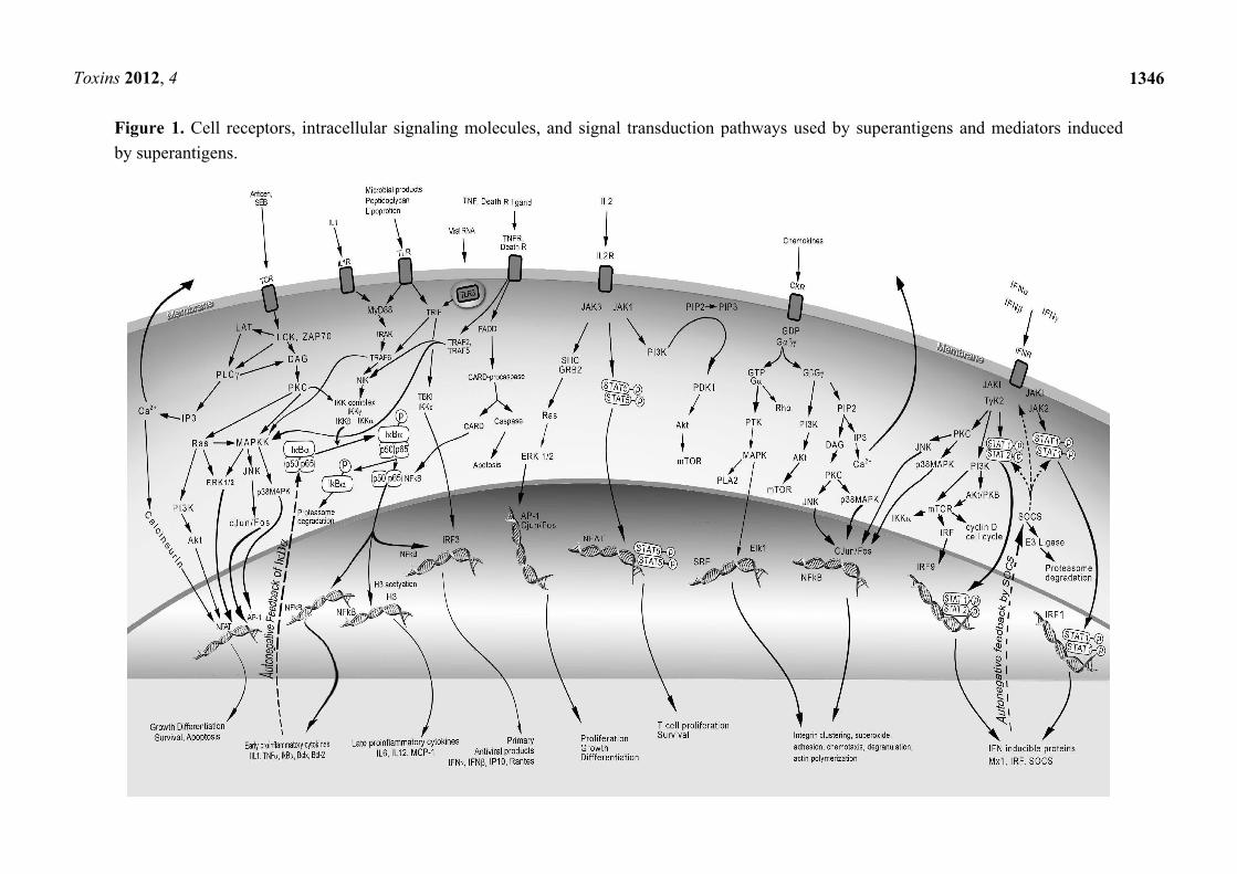
Figure 1. Cell receptors, intracellular signaling molecules, and signal transduction pathways used by superantigens and mediators induced
by superantigens.
Page 5
Toxins 2012, 4
1347
3. The Three Signals for T Cell Activation
Superantigens interact with both CD4+ and CD8+ T cells as well as mononuclear phagocytes
bearing MHC class II molecules [55–58]. Interaction of superantigen with TCR provides signal 1 for T
cell activation. As with conventional antigens, signal 2 comes from the engagement of co-stimulatory
molecules on APC and T cells upon superantigen binding and optimizes T cell activation [59,60].
Expression of intercellular adhesion molecule (ICAM) on APC promotes stable cell conjugate
formation and allows immunological synapse to occur. Initiation of TCR signaling by the formation of
supramolecular activation clusters comprising of TCR, co-stimulatory molecule CD28 and signaling
kinases is F-actin dependent [61]. The interactions of adhesion molecules and co-stimulatory
molecules, LFA-1/ICAM-1 and CD28/CD80, have both been implicated in SEA- and SEB-mediated
T-cell activation [60–62]. Activation of the CD28-regulated signal transduction pathway during SEA
stimulation of T cells enhanced IL-2 mRNA stability [62]. CD28 co-stimulation also increases T cell
survival by enhancing the expression of Bcl-xl [63]. Blocking CD28 with short synthetic peptides
corresponding to the binding region of CD28 inhibited TNFα, IFNγ, and IL-2 [52]. Other cell surface
molecules such as CD2, CD11a/ICAM-1, and ELAM facilitated optimal activation of endothelial cells
and T cells by SEB [60]. TCR and costimulatory receptors activate signaling kinases, protein kinase C
(PKC) and protein tyrosine kinases (PTKs) by the release of intracellular second messengers and
various intracellular adaptors [64–66]. PKC and PTK activation lead to other downstream signaling
pathways including mitogen–activated protein kinase (MAPK), extracellular signal regulated kinase
(ERK) and c-jun N-terminal kinase (JNK) pathways ultimately activating transcriptional factors NFκB,
NF-AT, and AP-1 [65–67]. Many proinflammatory cytokine genes contain NFκB binding sites in the
promotor/enhancer region and are induced by NFκB [68]. The cytokines IL-1, TNFα, IFNγ, IL-2, IL-6,
and chemokines, specifically MCP-1 are induced directly by superantigens, representing the third
signal for T cell activation. IL-1 and TNFα can also activate fibroblasts, epithelial, and endothelial
cells to produce other mediators providing inflammatory stimuli for activation of many different cell
types [21]. The mediators produced by superantigen-activated cells exert profound effects on the
immune and cardiovascular system, culminating in multi-organ dysfunction and lethal shock. PTKs
and T cell cytokines also activate the lipid kinase, phosphoinositide 3 kinase (PI3K) affecting many
intracellular processes including cell survival, growth, and migration [69]. PI3K consists of eight
isoforms, regulates many physiological and pathological processes, and plays a key role in cancer,
being constitutively active in malignancy and promotes growth factor independent growth in
tumor cells.
4. TCR and Costimulatory Receptors Activate the Phosphatidylinositol Pathway
T cell activation via the TCR-CD3 complex induces the activation of the Src family PTKs, LCK
and FYN, which in turn phosphorylate tyrosine-based motifs of the TCR intracellular components and
other cellular substrates [64–66]. LCK activates another PTK, ZAP-70, which then induces
tyrosine phosphorylation of the adaptors LAT (linker for activation of T cells) and SLP-76
(SH2-domain-containing leukocyte protein-76). These adaptors help to localize phospholipase C γ
(PLCγ) to the plasma membrane and activate PLCγ through phosphorylation by TCR-induced kinases,
Page 6
Toxins 2012, 4
1348
LCK and ZAP-70 (Figure 1) [64–66]. Phosphorylated and activated PLCγ cleaves phospholipid
phosphatidylinositol 4,5-bisphosphate, generating the second messengers diacylglycerol (DAG) and
inositol 1,4,5-trisphosphate (IP3). DAG activates protein kinase C θ (PKCθ) and indirectly the
protooncogene Ras whereas IP3 binds to its receptor on the surface of the endoplasmic reticulum and
induces an increase in intracellular calcium. PTKs also activate PI3K upon specific ligand binding to a
number of receptors besides the TCR, including CD28, IL-2 receptor (IL-2R), insulin receptor, growth
factor receptor, and G-protein-coupled receptor (GPCR). Activation of PI3K by PTK leads to the
generation of several inositol phospholipids including phosphatidylinositol 3,4-bisphosphate (PIP2)
and phosphatidylinositol 3,4,5-trisphosphate (PIP3) [64]. PIP3 recruits phosphoinositide-dependent
kinase 1 (PDK1) to the plasma membrane and activates it by phosphorylation. Activated PDK1
then phosphorylates Akt and PKCθ [70]. Although the activation of PKCθ isoform in
superantigen-activated cell has not been defined, PKCθ can be found at immunological synapse
formed after T cell activation by anti-CD3 and anti-CD28 [71]. Activation of PKCθ leads to the
phosphorylation of target genes, one of which is the activation of the inhibitor of κB (IκB) kinase
complex (IKK) [70]. IKK phosphorylation of IκB leads to its degradation, releasing NF-κB to be
translocated to the nucleus where it binds and activates many NFκB target genes. Another kinase
which is inducible by high cellular AMP/ATP ratio called AMP-activated protein kinase (AMPK) can
also phosphorylate PKCθ [72]. The multiple phosphorylation sites on PKCθ allow for its regulation by
at least three different kinases, LCK, PDK1 and AMPK, coordinating input from external stimuli.
The superantigen TSST-1 induces inositol phospholipid turnover, protein kinase C translocation,
and calcium mobilization in human T cells resembling responses from those of a mitogenic signal [73].
Various PTK inhibitors were used to study the PTK and PI3K pathways in mediating the effects of
superantigens. The production of IL-1 by TSST-1-stimulated human macrophage cell line was blocked
by three PTK inhibitors, genistein, tyrphostin, and herbimycin A [74]. However these inhibitors are not
very specific as genistein can also block the activity of PKA and PKC. The exact PTK or sites of
inhibition have not been identified with newer antibodies available for each specific PTK. Other PI3K
inhibitors, wortmannin and LY294004 have not been tested with superantigen-activated cells. In vivo
studies using these inhibitors on superantigen-induced shock models are lacking, perhaps due to
inherent toxicity, non-specificity, and the existence of different PI3K isoforms. Recently, the
superantigen SEE was shown to use an alternative LCK-independent pathway by activating PLCβ
signaling in T cells [75].
5. Regulation of Akt and Mammalian Target of Rapamycin (mTOR)
Downstream of PI3K is the serine/threonine kinase Akt which mediates many diverse biological
processes such as glucose transport, glycolysis, glycogen synthesis, cell proliferation, NFκB activation,
and inhibition of apoptosis [76,77] (Figure 2). Similar to PDK1, Akt can also be recruited to the
plasma membrane by the lipid messenger PIP3. The activation of Akt is controlled by two main
phosphorylation sites. Phosphorylation of the activation loop of Akt at Thr-308 by PDK1 is essential
for activation whereas phosphorylation of Ser-473 within the regulatory region further enhances its
activity. The role of Akt in SEB-mediated cellular effects has not been defined due to the lack of
specific inhibitors, but its activation downstream of PI3K indicates the importance of Akt upon the
Page 7
Toxins 2012, 4
1349
binding of several specific ligands as diverse as antigens/superantigens, IL-2, insulin, growth factor,
chemokines to their receptors TCR, IL-2R, insulin receptor, receptor tyrosine kinase (RTK), and
GPCR, respectively. Two potent cytokines from superantigen-stimulated T cells, IFNγ and IL-2 also
activate PI3K/Akt pathway via the transducer Janus kinase 1 (JAK1) after binding to the IFNγ and
IL-2 receptor, respectively [78,79].
Figure 2. The PI3K/Akt/mTOR pathway in superantigen activation.
One of the downstream targets of Akt in controlling cell proliferation and protein translation is
mTOR [80–82]. mTOR is a serine/threonine kinase that exists as two separate complexes, mTOR
complex1 (mTORC1) and mTORC2 and they do not interact directly. mTORC1 comprises of a kinase
component and two highly conserved proteins raptor and mLST8. A specific inhibitor, rapamycin,
binds to the immunophilin FK506-binding protein 12 (FKBP12) which then blocks mTORC1 activity
specifically [83]. Rapamycin has been used extensively to study the functions of mTORC1 and
mTORC2 in cell activation [83]. The action of rapamycin on mTORC2 is controversial, with earlier
reports of lack of inhibition to more recent studies indicating partial inhibition of mTORC2 with
prolonged treatment with rapamycin [84]. The most important function of mTORC2 lies upstream
since mTORC2 enhances Akt activity by phosphorylating Akt on Ser-473.
A critical protein complex in the regulation of Akt/mTOR is the TSC1/TSC2 (tuberous sclerosis
complex 1 and 2) which acts as a negative regulator of mTORC1 [80–86]. Phosphorylation of TSC2
by Akt results in the suppression of TSC1/TSC2 inactivation of the small GTPase, RHEB (Ras
homologue enriched in brain). Because RHEB is a potent activator of mTORC1, the effect of Akt on
TSC1/TSC2 is to promote mTORC1 activity. In contrast, TSC1/TSC2 associates with mTORC2 and
Page 8
Toxins 2012, 4
1350
promotes mTORC2 to phosphorylate and activate Akt. Cells deficient in TSC1/TSC2 complex are
defective in both mTORC2 and Akt activity. Activation of mTORC1 leads to phosphorylation and
activation of the ribosomal 40S protein p70S6 kinase (p70S6K) and the eukaryotic initiation factor
binding protein 1 (4EBP1) [80–83]. Phosphorylated p70S6K promotes mRNA translation and cell
growth by enhancing the biosynthesis machinery inside the cell. The phosphorylation of 4EBP1
prevents it from inhibiting the initiation factor EIF4E, thereby stimulating protein synthesis.
Rapamycin blocks mTORC1 and inhibits the translation of proteins that are essential to G1 to S phase
transition. mTORC2 can be stimulated by growth factors directly via PI3K promoting downstream
PKC activity through phosphorylation and activating Rho, culminating in actin reorganization.
mTORC1 and mTORC2 have distinct activities and act to coordinate signaling pathways from
mitogenic and superantigenic signals, growth factors and cytokines via PI3K/Akt. mTORC1 can also
be stimulated directly by nutrients such as amino acids and inactivated by oxidative stress or low
cellular energy levels via AMPK which activates TSC1/TSC2 by phosphorylation leading to mTORC1
inhibition [85,86]. Adding another level of regulation, Akt can inhibit AMPK through phosphorylation
and activates mTORC1. The cross-regulation of the components of the PI3K/Akt/mTOR pathway
allows for the tight control on energy levels, metabolism, proliferation, and growth.
Many excellent reviews have been written on the PI3K/Akt/mTOR pathway with original reference
citations to novel observations and the details of signaling molecules [80–86]. A critical role for
mTORC in SEB-induced signaling events is evident from the efficacy of the mTORC1-specific
inhibitor, rapamycin in rescuing mice from SEB-induced shock [87]. Rapamycin inhibited
SEB-induced T cell proliferation and was also a potent inhibitor of SEB-induced IL-2 and IFNγ
in vitro and in vivo. Furthermore, in comparison with dexamethasone treatment in the same mouse
model of SEB-mediated shock, early administration of dexamethasone post-SEB exposure as well as
longer duration of treatment was necessary to prevent lethality. The SEB-induced PI3K/Akt/mTORC
axis is found to be more effectively inhibited even when rapamycin was applied at a later time (24 h)
after SEB exposure with shorter duration of treatment sufficient to block SEB-induced shock [87].
6. Proinflammatory Mediators Signal via NFκB Activation
Excessive release of proinflammatory cytokines mediates the toxic effects of superantigens. The
proinflammatory cytokines IL-1 and TNFα can directly activate the transcriptional factor NFκB in
many cell types that include epithelial and endothelial cells thus perpetuating the inflammatory
response. The receptors, adaptors, and the signaling molecules used by IL-1, TNFα, IL-2, IL-6, and
IFNγ are vastly different and represent five different families of cytokine receptors.
IL-1 interacts with IL-1 receptor 1 (IL-1R1) requiring an additional receptor accessory protein for
subsequent activation of downstream signaling molecules, the adaptor myeloid differentiation factor
88 (MyD88), IL-1R-associated protein kinase 1 (IRAK1), and TNF receptor-associated factor 6
(TRAF-6) [88]. Another set of related receptors, the toll-like receptors (TLRs), signal with similar
intracellular adaptors and molecules as those used for IL-1R1 (Figure 1) but are not used for
superantigen signaling. However, SEB was reported to increase cellular expression of TLR2 and
TLR4 [32,33]. The TLRs are conserved type 1 transmembrane receptors used by pathogen associated
molecules to stimulate host innate immune responses and influence the adaptive immune
Page 9
Toxins 2012, 4
1351
response [89]. There is some specificity of individual TLR in recognition of specific molecular
structures of lipoproteins, peptidoglycan, dsRNA, LPS and viral RNA. Peptidoglycans from
gram-positive bacteria and LPS from gram-negative bacteria bind TLR2 and TLR4, respectively,
activate IκB kinases (IKK), and trigger NFκB activation through the MyD88-dependent pathway. The
phosphorylation of IκBα by IKK releases it from p50 and p65 of NFκB, allowing for nuclear
translocation of NFκB where it binds to promoter regions of many inflammatory genes [90].
Activation of NFκB leads to induction of proinflammatory genes as well as anti-apoptotic genes. An
auto-feedback loop exists to downregulate NFκB as IκBα is also induced by NFκB, thereby turning off
NFκB activation. TLR4 also signals through TRIF, the adaptor used by TLR3, to induce the expression
of IFN-mediated genes [89]. A recent report indicates that TLR2 signaling by cell wall
peptidoglycans of S. aureus downregulates T cell activation and likely reduces the risk of toxic
shock [91].
TNFα binds to TNF receptor 1 (TNFR1), TNFR2 and both receptors use intracellular TRAFs
different from those used by IL-1R1 or TLRs but ultimately activating NFκB, resulting in expression
of other cytokines, adhesion and co-stimulatory molecules [21,92]. The cytotoxic functions of TNFα
are mostly mediated by its binding to TNFR1. Cross-linking receptor chains and clustering upon
binding of TNFα results in recruitment of intracellular signaling molecules to the receptor. However,
the TNFR possesses death domains, commonly present in receptors of the TNFR superfamily, and
binding of TNFα to TNFR1 and TNFR2 also triggers cell death through caspase activation. In this
regard, there are common signaling molecules among the TNFR family which includes Fas
(CD95), the expression of which is induced by superantigens. Intracellular adaptors, TRADD
(TNFR-associated death domain), and FADD (Fas-associated death domain) are used by the TNFR
superfamily to activate the caspase 8 cascade, JNK, and NFκB, accounting for the diverse biological
effects of TNFα including apoptosis, cell activation, coagulation, inflammation, and host defense [92].
TNFα and IFNγ act synergistically on epithelial cells to increase ion transport, causing cell damage
and epithelial leakage [27]. The importance of TNFα in mediating the pathological effects in
SEB-induced lethality was recognized early on as anti-TNFα antibodies conferred protection from
SEB-induced shock in a D-galactoseamine sensitized mouse model [19].
7. T Cell Cytokines and Chemokines Activate the PI3K/Akt/mTOR Pathway
SEB binding induces TCR and co-stimulatory molecule CD28 activating PI3K/Akt/mTOR pathway
directly by membrane proximal components. In addition, the SEB-induced cytokines IFNγ, IL-2, and
chemokines binding to their respective receptors all activate PI3K activity. Diverse stimuli and
cytokines initiate the PI3K pathway with some common subsequent steps as well as multiple branch
points for regulation of Akt and mTOR, kinases downstream of PI3K.
IFNγ binds to IFNγR, which belongs to the family of interferon receptors, including the structurally
different receptors for type 1 interferons [93,94]. IFNRs use very different adaptors and signal
transducers from those for IL-1R, or TNFR, with signal transducer and activator of transcription 1
(STAT1) phosphorylation by JAK1 and JAK2 being critical for the IFNγR pathway to activate
antiviral responses and expression of other IFNγ-mediated genes. The binding of IFNγ to specific IFNγ
R triggers activation of receptor-associated PTK, JAK1 and JAK2. This leads to phosphorylation and
Page 10
Toxins 2012, 4
1352
activation of STAT1. Dimerization and translocation of STAT1 to the nucleus allows STAT1 to bind
and activate IFNγ-specific genes [95]. STAT1 activation is negatively regulated by suppressor of
cytokine signaling 1 (SOCS1) and SOCS3. The IFNγ-activated JAKs also activate PI3K in a STAT1
independent manner culminating in mTOR pathway activation, promoting protein translation [95].
IFNγ also activates PKC leading to MAPK pathway activation, which is commonly activated by IL-1,
TLR ligands, and TNFα through TRAFs. However, IFNγ induces apoptosis by the induction and
activation of death receptors such as Fas, activating FADD and caspase 8 signaling. The activation of
caspase 8 cascade results in cytochrome c release from mitochondria and DNA fragmentation. In vitro,
IFNγ induces MHC class II molecules, immunoproteasome components, and antigen-processing
protein transporters to enhance immune responses in host defense [95]. IFNγ dirupts epithelial barrier
function and ion transport in superantigen-activated cells and many of the interference of epithelial
barrier function in vitro can be duplicated with IFNγ with effects synergized by TNFα [96]. Anti-IFNγ
inhibited SEB-induced weight loss and hypoglycemia but had no effect on mortality in a
D-galactosamine-sensitized mouse model of SEB-mediated shock [97].
IL-2 binds to the IL-2R, which consists of three separate chains that heterodimerize and signal
through JAK1 and JAK3, activating PI3K and Ras [98]. The activation of the PI3K/Akt/mTOR axis
and Ras signaling controls proliferation, growth, and differentiation of many cell types. Ras activates
MAPK and ERK cascades leading to activation of AP-1, cJun/Fos and NFAT. IL-2 induces
vasodilation and increases microvascular permeability by suppressing endothelin-1, ultimately causing
perivascular edema seen in SEB-induced lung injury and shock models [99,100]. A recent study
demonstrates the prominent role of IL-2 as IL-2-deficient mice are resistant to SEB-induced toxic
shock [101].
IL-6, from both macrophages and activated T cells, has some overlapping activities with IL-1 and
TNFα, activates by binding to a different class of receptors belonging to the gp130 family [102].
Binding of IL-6 to its heterodimeric receptor activates JAK3 and Ras. Activated JAK3 phosphorylates
STAT3 which then dimerizes and translocates to the nucleus where it binds target genes essential for
gp130-mediated cell survival and G1 to S phase transition. The Ras-mediated pathway leads to MAPK
activation. In addition, IL-6R also signals through PI3K/Akt/mTOR to promote survival of cells.
Together and individually, IL-1, TNFα and IL-6 act on the liver to release acute phase proteins,
activate anti-apoptopic pathways, and decrease liver clearance function.
The chemokines, IL-8, MCP-1, MIP-1α, and MIP-1β, are induced directly by SEB or TSST-1 and
selectively act as chemoattractants and activate leukocytes and influence migration of neutrophils,
dendritic cells and leukocytes [13,21,103]. Chemokines bind to seven-transmembrane GPCR, induce
early Ca++ flux, activate PLC and signal via the PI3K pathway [21,103,104]. Cytokine- and
chemokine-activated neutrophils, recruited to sites of tissue injury and inflammation, produce ROS
and MMPs contributing to organ dysfunction. MMPs cause tissue degradation and change chemokine
interactions with the extracellular matrix creating a local gradient effect of chemokines [103].
Exudates from superantigen-injected air pouches were predominantly neutophils with some
macrophages [13]. Endothelial cells surrounding air pouches expressed ICAM-1, TNFα, MIP-2 (an
IL-8 related protein in mice), MIP-1α, and JE. Both systemic and intranasal administration of SEB
caused acute lung injury characterized by increased expression of adhesion molecules ICAM-1 and
Page 11
Toxins 2012, 4
1353
VCAM, increased neutrophils and mononuclear cells infiltrate, endothelial cell injury, and increased
vascular permeability [18,105].
The PI3K signaling pathway through Akt activation can directly and indirectly modulate mTOR
activation. Upstream positive regulators of mTORC1 include PI3K, PDK1, Akt, mTORC2, RHEB,
and nutrients leading to increase translation, cell proliferation, and survival. Negative regulators of
mTORC1 are AMPK, TSC1/TSC2, and AMP/ATP levels acting in concert to integrate signals
controlling cell metabolism, cell survival, and proliferation [80,81]. Since TCR, CD28, IL-2R, IFNγR
and chemokine receptors all signal through PI3K/Akt/mTOR, this pathway plays a dominant role in
superantigen-induced effects.
8. Therapeutic Antibodies against SEB
There is currently no available therapeutics for treatment of superantigen-induced shock except for
the use of intravenous human immunoglobulin [106]. Targeting superantigen directly by neutralization
of toxins is most suitable at the early stages of exposure before cell activation and release of
proinflammatory cytokines. Some of the neutralizing antibodies against one superantigen cross-react
and prevent the biological effects of a different superantigen [37]. Various monoclonal and
human-mouse chimeric antibodies against SEB have been used effectively to target SEB-induced T
cell activation [107–109]. A mixture of non-protective monoclonal antibodies was effective in rescuing
mice from SEB-mediated shock with one of the antibody inducing a structural change upon binding to
SEB which then allowed binding of a different antibody to neutralize SEB [109]. Recombinant
mutants of SEB with attenuated binding to MHC class II and devoid of superantigenicity were also
used successfully to vaccinate mice and monkeys against SEB-induced disease [110]. S. aureus
bacteremia triggers antibody response against superantigens and antibody titers increase during
infection thereby protecting the host [111]. Carriers previously exposed to S. aureus have high titers of
neutralizing antibodies specific for the superantigens expressed by their colonizing strain and are
protected against S. aureus septicemia [112].
9. Inhibitors of Cell Receptor-Toxin Interaction
Because the binding regions of SEB to MHC class II and TCR are known, small overlapping
peptides of SEB can also be used as antagonists to block the initial step of receptor-toxin interactions.
Conserved peptides corresponding to residues 150–161 of SEB blocked T cell activation and prevented
SEA-, SEB-, or TSST-1-induced lethal shock in mice [113]. This segment of SEB is not associated
with the classically defined MHC class II or TCR binding domains, but it may block co-stimulatory
signals necessary for T-cell activation. However other investigators found no inhibitory activities with
these peptides in vitro and in vivo [114,115]. Bi-specific chimeric inhibitors composed of the DRα1
domain of MHC class II and Vβ domain of the TCR connected by a flexible GSTAPPA)2 linker were
reported to bind SEB competitively and prevent its binding to MHC class II of APC and TCR on T
cells [116]. Both cell activation and IL-2 production was blocked by the use of these chimeras in
SEB-stimulated PBMC. A soluble TCR Vβ mutant with high affinity binding was engineered to
neutralize SEB and SPEA [117]. CTLA4-Ig, the synthetic ligand for CD28 inhibited TSST-1-induced
T cell proliferation in vitro and prevented lethal toxic shock in vivo [118]. The recent study of using
Page 12
Toxins 2012, 4
1354
novel peptides corresponding to the CD28 binding regions to block SEB-mediated effects underscores
the importance of co-stimulatory signals in T cell activation by superantigens [52]. Another approach
is the use of aptamers, basically peptides or single-stranded nucleic acid, obtained from recombinant
libraries, to bind SEB and block interaction with receptor [119].
10. Inhibitors of Signal Transduction
An important class of therapeutic compounds to be considered is inhibitors that can block signal
transduction pathways activated by superantigens, as these events are post-exposure and may be more
amenable to suppression and manipulation. The obvious advantage is that they are likely broad
spectrum, inhibiting many different superantigens or even pathogens that elicit similar host responses
or pathways. In vitro studies have shown that many of the genes including cell adhesion molecules,
cytokines, chemokines, acute phase proteins, and inducible nitric oxide synthase, implicated in
superantigen-induced lethal shock contain NFκB binding sites in the promotor/enhancer region [90].
The activation of NFκB, therefore, leads to the inducible expression of many of the mediators involved
in inflammation and tissue injury seen in SEB-induced lung injury and toxic shock models and
inhibiting NFκB may be beneficial in preventing superantigen-induced diseases. NFκB binding
activity is increased in patients with acute inflammation and sepsis, and can be correlated with clinical
severity and mortality [120]. A cell-permeable cyclic peptide targeting NFκB nuclear transport reduced
SEB-induced T cell responses and inflammatory cytokine production [121]. Decreased mortality rates
accompanied by an attenuation in liver apoptosis and hemorrhagic necrosis were seen in mice given
D-galactosamine plus SEB along with this NFκB inhibitor [99].
Another potent NFκB inhibitor is dexamethasone, a well-known FDA-approved
immunosuppressive corticosteriod used clinically to treat various inflammatory diseases [122].
Dexamethasone potently inhibited SEA-, and SEB-induced cytokine release, T-cell proliferation, and
cell activation marker expression in human PBMC [123]. Dexamethasone also significantly reduced
serum levels of TNFα, IFNγ, IL-1, IL-2, and IL-6 in the LPS-potentiated SEB model and the
un-potentiated SEB-only model of toxic shock [105,124]. Importantly, dexamethasone decreased
mortality in both of these mouse models was accompanied by attenuation of the hypothermic response
and weight loss induced by SEB. Another NFκB and proteosome inhibitor, bortezomib, attenuated
SEB-induced cytokine release but had no effect on SEB-induced lethality and liver necrosis [125].
Polyphenols such as epigallocatechin gallate (EGCG) from green tea and resveratrol (RES) from red
wine also reduced superantigen-induced T cell proliferation and cytokine release from human PBMC
by decreasing NFκB activity [126]. EGCG suppressed T cell activation, reducing IFNγ and TNFα from
SEB-stimulated human PBMC and murine lymph node cells and reduced IFNγ-induced epithelial
permeability increases [127]. RES blocked SEB-induced T cell activation, pulmonary permeability
increases, caspase 8-dependent apoptosis, and prevented SEB-induced lung injury in mice [128].
Recently, a synthetic mimetic (EM-163) to the BB-loop of MyD88 was found to inhibit TNFα, IFNγ,
IL-1, IL-2 and IL-6 in human PBMC activated by superantigens [129]. Furthermore, EM163 reduced
the level of cytokines in serum and protected mice from LPS plus SEB-induced shock [129,130].
Other signal transduction inhibitors include those directed against PKC and PTK. H7, a PKC
inhibitor and genistein, a tyrosine kinase inhibitor each blocked TNFα but not IL-1 production from
Page 13
Toxins 2012, 4
1355
TSST-1-stimulated PBMC [131]. D609, an inhibitor of PLC, which is activated upon superantigen
binding to TCR and CD28, blocked SEB-induced effects both in vitro and in vivo [132,133].
Curiously, the serum level of TNFα in mice treated with D609 and superantigen remained high despite
reduction in lethality [133]. Another natural feedback inhibitor of the various STATs used by IFNγ and
IL-2 signaling is SOCS3 which therefore controls the effects of these two cytokines [93]. In this
regard, a cell-penetrating form of SOCS3 protected animals from lethal effects of SEB and LPS by
reducing production of inflammatory cytokines and attenuating liver apoptosis and hemorrhagic
necrosis [134].
Two other potent immunosuppressant and calcineurin inhibitor used clinically for preventing
transplant rejection, cyclosporine A (CsA) and tacrolimus, did not protect superantigen-induced shock
in monkeys and human HLADR3-transgenic mice, respectively [135,136]. CsA inhibited SEB-induced
T cell proliferation in vitro and reduced serum IL-2, TNFα, and IFNγ, as well as attenuated pulmonary
inflammation which did not translated to a protective effect [135]. Tacrolimus suppressed
superantigen-induced T cell activation in vitro but did not confer protection from shock in vivo [136].
Recently, the mTORC1 specific inhibitor, rapamycin was shown to be efficacious even when given
24 h after SEB in a murine model of SEB-induced shock [87]. Rapamycin is a FDA-approved drug
currently used to prevent kidney graft rejection and is under clinically trials for cancer treatment.
Rapamycin works by suppressing mTOR activities resulting in inhibition of SEB-induced T cell
proliferation, reduced IL-2 and IFNγ. Another study indicates rapamycin was effective as an intranasal
drug, providing practical protection against SEB-induced shock even 17 h after toxin exposure [137].
Oxidative stress is another hallmark of SEB-intoxication as systemic administration of SEB causes
prolonged lung inflammation that is difficult to resolve [105]. Acute lung injury arises as SEB-,
cytokine- and chemokine-activated neutrophils infiltrate into lung areas, produce high levels of ROS
which in turn cause increase in vascular permeability and apoptosis [18]. One strategy is the use of
anti-oxidants such as N-acetyl cysteine (NAC) and pyrrolidine dithiocarbamate (PDTC) to mitigate
oxidative stress. Both NAC and PDTC are FDA-approved drugs for other indications and prevented
NF-κB signaling in superantigen-activated human PBMC [138].
Dexamethasone, rapamycin, cyclosporine A, tacrolimus, bortezomib, NAC, PDTC are
FDA-approved drugs currently used for other indications. The testing of FDA-approved drugs for
preventing superantigen-induced shock should speed up the approval process for biodefense use in
case of exposure. However, as seen from the various examples above, even knowing the mechanism of
action of these drugs is no guarantee for success as in vivo dosages, dosing routes and schedules as
well as animal models all affect the outcome. Rapamycin, by decreasing the levels and effects of IL-2
and IFNγ through mTOR inhibition, is proven to be effective to counter the toxic effects of SEB.
11. Inhibitors of Cytokines
Due to the pathophysiological complexities of toxic shock resulting from excessive
proinflammatory cytokine release from host cells responding to superantigens, therapeutics aimed at
inhibiting the release of these mediators overlap with inhibitors of signal transduction pathways used
by these cytokines. Most therapeutic testing in animal models of SEB-induced shock have targeted
proinflammatory cytokines, as there is a strong correlation between toxicity and increased serum levels
Page 14
Toxins 2012, 4
1356
of these inflammatory mediators [12–16]. Neutralizing antibodies against TNFα prevented
SEB-induced lethality in D-galactoseamine sensitized mouse model establishing the critical role of
TNFα in lethal shock [19]. The anti-inflammatory cytokine IL-10 also reduced lethality to
superantigen-induced toxic shock by reducing the production of IL-1, TNFα and IFNγ [139,140].
Niacinamide, a nitric oxide inhibitor, mitigated the effects of SEB by inhibiting the production of IL-2
and IFNγ, and improved survival of mice given LPS plus SEB [141]. Other drugs tested to block
cytokine release from superantigen-activated cells include doxycycline, an antibiotic, and
pentoxyfylline, a methylxanthine derivative. Doxycycline blocked SEB-induced proinflammatory
cytokines and chemokines and T-cell proliferation in human PBMC [142]. Pentoxyfylline, a
phophodiesterase inhibitor, is used clinically to treat peripheral vascular disease as it disrupts
intracellular regulatory pathways that affect leukocyte adhesion and cytokine production.
Pentoxyfylline reduced cytokines and T cell proliferation in SEB- or TSST-1-stimulated cells. It
prevented lethal shock accompanied by reduction in serum cytokines in the LPS plus SEB mouse
model [143].
Another strategy to attenuate IL-1 release from superantigen-activated cells is to target caspase 1, a
proteolytic enzyme that cleaves pro-IL-1 into active IL-1 [21]. The caspase 1 specific inhibitor,
Ac-YVAD-cmk, attenuated both IL-1 and MCP production in SEB- and TSST-1 stimulated PBMC
cultures but had no effect on other cytokines or T-cell proliferation [144]. Caspase 3 and caspase 8
inhibitors were also ineffective in down-regulating superantigen-activated cells or T cell proliferation.
In contrast, a pan-caspase inhibitor, Z-D-CH2-DCB, reduced the production of IL-1β, TNFα, IL-6,
IFNγ, MCP, MIP-1α, MIP-1β, and inhibited T-cell proliferation in SEB- and TSST-1-stimulated
PBMC [144].
Other compounds tested against superantigen-induced effects include herbal medicinal compounds,
tryptanthrin and baicalin. Tryptanthrin, derived from an Asian medicinal plant, Isatis tinctoria, reduced
IFNγ production by SEB-stimulated lymphocytes from Peyer’s patches [145]. Baicalin, a flavone
isolated from the Chinese medicinal herb Scutellaria baicalensis, attenuated IL-1, TNF, IL-6, IFNγ,
MCP-1, MIP-1α, MIP-1β mRNA and protein expression in SEB- and TSST-1-stimulated human
PBMC and blocked T cell proliferation [146]. Herbal compounds are usually less-well characterized
and are often used in combination with other medicinal herbs to be effective.
12. Summary
By binding to both MHC class II and TCR, superantigens stimulate T-cell proliferation and
excessive release of multiple inflammatory cytokines and chemokines. Similar to other shock
syndromes, extensive tissue inflammation and injury is the result of superantigen-induced
proinflammatory mediators via NFκB activation. The three signals for T cell activation after
superantigen binding all activate the PI3K/Akt/mTOR pathway by sequential phosphorylation steps
regulating proliferation, growth and survival. T cell cytokines, IFNγ and IL-2 and chemokines signal
via the PI3K/Akt/mTOR pathway, making this pathway even more important as a target for
intervention. The ability to stop the inflammatory and proliferation/survival signals initiated by
superantigens appears to be critical in preventing superantigen-mediated shock.
Page 15
Toxins 2012, 4
1357
Acknowledgments
I thank the Defense Threat Reduction Agency for generous support.
Conflict of Interest
The author declares no conflict of interest.
Disclaimer
The views expressed in this publication are those of the author and do not reflect the official policy
or position of the Department of the Army, the Department of Defense, or the U.S. Government.
References
1. Chesney, P.J.; Davis, P.J.; Purdy, W.K.; Wand, P.J.; Chesney, R.W. Clinical manifestations of
toxic shock syndrome. JAMA 1981, 246, 741–748.
2. Marrack, P.; Kappler, J. The staphylococcal enterotoxins and their relatives. Science 1990, 248,
705–709.
3. Kotzin, B.L.; Leung, D.Y.M.; Kappler, J.; Marrack, P. Superantigens and their potential role in
human disease. Adv. Immunol. 1993, 54, 99–166.
4. Kotb, M. Bacterial pyrogenic exotoxins as superantigens. Clin. Microbiol. Rev. 1995, 8,
411–426.
5. McCormick, J.K.; Yarwood, J.M.; Schlievert, P.M. Toxic shock syndrome and bacterial
superantigens: An update. Ann. Rev. Microbiol. 2001, 55, 77–104.
6. Proft, T.; Fraser, J.D. Bacterial superantigens. Clin. Exp. Immunol. 2003, 133, 299–306.
7. Fraser, J.D.; Proft, T. The bacterial superantigen and superantigen-like proteins. Immunol. Rev.
2008, 225, 226–243.
8. Choi, Y.; Kotzin, B.; Hernon, L.; Callahan, J.; Marrack, P.; Kappler, J. Interaction of
Staphylococcus aureus toxin “superantigens” with human T cells. Proc. Natl. Acad. Sci. USA
1989, 86, 8941–8945.
9. Jupin, C.; Anderson, S.; Damais, C.; Alouf, J.E.; Parant, M. Toxic shock syndrome toxin 1 as an
inducer of human tumor necrosis factors and gamma interferon. J. Exp. Med. 1988, 167,
752–761.
10. Trede, N.S.; Geha, R.S.; Chatila, T. Transcriptional activation of IL-1 beta and tumor necrosis
factor-alpha genes by MHC class II ligands. J. Immunol. 1991, 146, 2310–2315.
11. See, R.H.; Kum, W.W.; Chang, A.H.; Goh, S.H.; Chow, A.W. Induction of tumor necrosis factor
and interleukin-1 by purified staphylococcal toxic shock syndrome toxin 1 requires the presence
of both monocytes and T lymphocytes. Infect. Immun. 1992, 60, 2612–2618.
12. Miethke, T.; Wahl, C.; Heeg, K.; Echtenacher, B.; Krammer, P.H.; Wagner, H. Superantigen
mediated shock: A cytokine release syndrome. Immunobiology 1993, 189, 270–284.
13. Tessier, P.A.; Naccache, P.H.; Diener, K.R.; Gladue, R.P.; Neotem, K.S.; Clark-Lewis, I.;
McColl, S.R. Induction of acute inflammation in vivo by staphylococcal superantigens. II.
Critical role for chemokines, ICAM-1, and TNF-alpha. J. Immunol. 1998, 161, 1204–1211.
Page 16
Toxins 2012, 4
1358
14. Krakauer, T. The induction of CC chemokines in human peripheral blood mononuclear cells by
staphylococcal exotoxins and its prevention by pentoxifylline. J. Leukco. Biol. 1999, 66,
158–164.
15. Faulkner, L.; Cooper, A.; Fantino, C.; Altmann, D.M.; Sriskandan, S. The mechanism of
superantigen-mediated toxic shock: Not a simple Th1 cytokine storm. J. Immunol. 2005, 175,
6870–6877.
16. Krakauer, T.; Buckley, M.; Fisher, D. Proinflammatory mediators of toxic shock and their
correlation to lethality. Mediators Inflamm. 2010, doi:10.1155/2010/517594.
17. Mattsson, E.; Herwald, H.; Egsten, A. Superantigen from Staphylococcus aureus induce
procoagulant activity and monocyte tissue factor expression in whole blood and mononuclear
cells via IL-1β. J. Thromb. Haemost. 2003, 1, 2569–2575.
18. Neumann, B.; Engelhardt, B.; Wagner, H.; Holzmann, B. Induction of acute inflammatory lung
injury by staphylococcal enterotoxin B. J. Immunol. 1997, 158, 1862–1871.
19. Miethke, T.; Wahl, C.; Heeg, K.; Echtenacher, B.; Krammer, P.H.; Wagner, H. T cell-mediated
lethal shock triggered in mice by the superantigen staphylococcal enterotoxin B: Critical role of
tumor necrosis factor. J. Exp. Med. 1992, 175, 91–98.
20. Stiles, B.G.; Bavari, S.; Krakauer, T.; Ulrich, R.G. Toxicity of staphylococcal enterotoxins
potentiated by lipopolysaccharide: Major histocompatibility complex class II molecule
dependency and cytokine release. Infect. Immun. 1993, 61, 5333–5338.
21. Krakauer, T.; Vilcek, J.; Oppenheim, J.J. Proinflammatory Cytokines: TNF and IL-1 Families,
Chemokines, TGFβ and Others. In Fundamental Immunology, 4th ed.; Paul, W., Ed.;
Lippincott-Raven: Philadelphia, PA, USA, 1998; pp. 775–811.
22. Vial, T.; Descotes, J. Immune-mediated side-effects of cytokines in human. Toxicology 1995,
105, 31–57.
23. Holmberg, S.D.; Blake, P.A. Staphylococcal food poisoning in the United States. New facts and
old misconceptions. JAMA 1984, 251, 487–489.
24. Schwab, J.H.; Brown, R.R.; Anderle, S.K.; Schlievert, P.M. Superantigen can reactivate bacterial
cell wall-induced arthritis. J. Immunol. 1993, 150, 4151–4159.
25. Brocke, S.; Hausmann, S.; Steinmam, L.; Wucherpfennig, K.W. Microbial peptides and
superantigens in the pathogenesis of autoimmune diseases of the central nervous system.
Semin. Immunol. 1998, 10, 57–67.
26. Yarwood, J.M.; Leung, D.Y.; Schlievert, P.M. Evidence for the involvement of bacterial
superantigens in psoriasis, atopic dermatitis, and Kawasaki syndrome. FEMS Microbiol. Lett.
2000, 192, 1–7.
27. McKay, D.M. Bacterial superantigens: Provocateurs of gut dysfunction and inflammation?
Trends Immunol. 2001, 22, 497–501.
28. Sugiyama, H.; McKissic, E.M.; Bergdoll, M.S.; Heller, B. Enhancement of bacterial endotoxin
lethality by staphylococcal enterotoxin. J. Infect. Dis. 1964, 4, 111–118.
29. Sarawar, S.R.; Blackman, B.A.; Doherty, P.C. Superantigen shock in mice with an inapparent
viral infection. J. Infect. Dis. 1994, 170, 1189–1194.
Page 17
Toxins 2012, 4
1359
30. Zhang, W.J.; Sarawar, S.; Nguyen, P.; Daly, K.; Rehig, J.E.; Doherty, P.C.; Woodland, D.L.;
Blackman, M.A. Lethal synergism between influenza infection and staphylococcal enterotoxin B
in mice. J. Immunol. 1996, 157, 5049–5060.
31. Blank, C.; Luz, A.; Bendigs, S.; Erdmann, A.; Wagner, H.; Heeg, K. Superantigen and endotoxin
synergize in the induction of lethal shock. Eur. J. Immunol. 1997, 27, 825–833.
32. Hopkins, P.A.; Fraser, J.D.; Pridmore, A.C.; Russell, H.H.; Read, R.C.; Sriskandan, S.
Superantigen recognition by HLA class II on monocytes up-regulates toll-like receptor 4 and
enhances proinflammatory responses to endotoxin. Blood 2005, 105, 3655–3662.
33. Hopkins, P.A.; Pridmore, A.C.; Ellmerich, S.; Fraser, J.D.; Russell, H.H.; Read, R.C.;
Sriskandan, S. Increased surface toll-like receptor 2 expression in superantigen shock.
Crit. Care Med. 2008, 36, 1267–1276.
34. Papageorgiou, A.C.; Acharya, K.R. Microbial superantigens: From structure to function.
Trends Microbiol. 2000, 8, 369–375.
35. Monday, S.R.; Bohach, G.A. Properties of Staphylococcus aureus Enterotoxins and Toxic Shock
Syndrome Toxin-1. In The Comprehensive Sourcebook of Bacterial Protein Toxins; Alouf, J.E.,
Freer, J.H., Eds.; Academic: London, UK, 1999; pp. 589–610.
36. Wang, X.; Xu, M.; Cai, Y.; Yang, H.; Zhang, H.; Zhang, C. Functional analysis of the disulphide
loop mutant of staphylococcal enterotoxin C2. Appl. Microbiol. Biotechnol. 2009, 82, 861–871.
37. Bavari, S.; Ulrich, R.G.; LeClaire, R.D. Cross-reactive antibodies prevent the lethal effects of
Staphylococcus aureus superantigens. J. Infect. Dis. 1999, 180, 1365–1369.
38. Kappler, J.W.; Herman, A.; Clements, J.; Marrack, P. Mutations defining functional regions of
the superantigen staphylococcal enterotoxin B. J. Exp. Med. 1992, 175, 387–396.
39. Li, H.; Llera, A.; Tsuchiya, D.; Leder, L.; Ysern, X.; Schlievert, P.M.; Karjalainen, K.;
Mariuzza, R.A. Three-dimensional structure of the complex between a T cell receptor beta chain
and the superantigen staphylococcal enterotoxin B. Immunity 1998, 9, 807–816.
40. Mollick, J.A.; Chintagumpala, M.; Cook, R.G.; Rich, R.R. Staphylococcal exotoxin activation of
T cells. Role of exotoxin-MHC class II binding affinity and class II isotype. J. Immunol. 1991,
146, 463–468.
41. Chintagumpala, M.M.; Mollick, J.A.; Rich, R.R. Staphylococcal toxins bind to different sites on
HLA-DR. J. Immunol. 1991, 147, 3876–3882.
42. Ulrich, R.G.; Bavari, B.; Olson, M.A. Staphylococcal enterotoxins A and B share a common
structural motif for binding class II major histocompatibility complex molecules.
Nat. Struct. Biol.1995, 2, 554–560.
43. Hudson, K.R.; Tiedemann, R.E.; Urban, R.G.; Lowe, S.C.; Strominger, J.L.; Fraser, J.D.
Staphylococcal enterotoxin A has two cooperative binding sites on major histocompatibility
complex class II. J. Exp. Med. 1995, 182, 711–720.
44. Tiedemann, R.E.; Urban, R.J.; Strominger, J.L.; Fraser, J.D. Isolation of HLA-DR1.
(staphylococcal enterotoxins A) 2 trimers in solution. Proc. Natl. Acad. Sci. USA 1995, 92,
12156–12159.
45. Thibodeau, J.; Cloutier, I.; Lavoie, P.M.; Labrecque, N.; Mourad, W.; Jardetzky, T.; Sekaly, R.P.
Subsets of HLA-DR1 molecules defined by SEB and TSST-1 binding. Science 1994, 266,
1874–1878.
Page 18
Toxins 2012, 4
1360
46. Herrmann, T.; Acolla, R.S.; MacDonald, H.R. Different staphylococcal enterotoxins bind
preferentially to distinct MHC class II isotypes. Eur. J. Immunol. 1989, 19, 2171–2174.
47. Herman, A.; Croteau, G.; Sekaly, R.P.; Kappler, J.; Marrack, P. HLA-DR alleles differ in their
ability to present staphylococcal enterotoxins to T cells. J. Exp. Med. 1990, 172, 709–712.
48. Scholl, P.; Sekaly, R.; Diez, A.; Glimcher, L.; Geha, R. Binding of toxic shock syndrome toxin-1
to murine major histocompatibility complex class II molecules. Eur. J. Immunol. 1990, 20,
1911–1916.
49. Seth, A.; Stern, L.J.; Ottenhoff, T.H.; Engel, I.; Owen, M.J.; Lamb, J.R.; Klausner, R.D.;
Wiley, D.C. Binary and ternary complexes between T-cell receptor, class II MHC and
superantigen in vitro. Nature 1994, 369, 324–27.
50. Moza, B.; Varma, A.K.; Buonpane, R.A.; Zhu, P.; Herfst, C.A.; Nicholson, M.J.; Wilbuer, A.K.;
Seth, N.P.; Wucherpfennig, K.W.; McCormick, J.K.; Kranz, D.M.; Sundberg, E.J. Structural
basis of T-cell specificity and activation by the bacterial superantigen TSST-1.
EMBO J. 2007, 26, 1187–1197.
51. Ferry, T.; Thomas, D.; Perpoint, T.; Lina, G.; Monneret, G.; Mohammedi, I.; Chidiac, C.;
Peyramond, D.; Vandenesch, F.; Etienne, J. Analysis of superantigenic toxin Vbeta T-cell
signatures produced during cases of staphylococcal toxic shock syndrome and septic shock.
Clin. Microbiol. Infect. 2008, 14, 546–554.
52. Arad, G.; Levy, R.; Nasie, I.; Hillman, D.; Rotfogel, Z.; Barash, U.; Supper, E.; Shpilka, T.;
Minis, A.; Kaempfer, R. Binding of superantigen toxins into CD28 homodimer interface is
essential for induction of cytokine genes that mediate lethal shock. Plos Biol. 2012, 9, e1001149.
53. Germain, R.N. T-cell signaling: The importance of receptor clustering. Curr. Biol. 1997, 7,
R640–R644.
54. Chatila, T.; Geha, R.S. Signal transduction by microbial superantigens via MHC class II
molecules. Immunol. Rev. 1993, 131, 43–59.
55. Tiedemann, R.E.; Fraser, J.D. Cross-linking of MHC class II molecules by staphylococcal
enterotoxin A is essential for antigen-presenting cell and T cell activation. J. Immunol. 1996,
157, 3958–3966.
56. Carlsson, R.; Fischer, H.; Sjogren, H.O. Binding of staphylococcal enterotoxin A to accessory
cells is a requirement for its ability to activate human T cells. J. Immunol. 1998, 140, 2484–2488.
57. Anderson, M.R.; Tary-Lehmann, M. Staphylococcal enterotoxin-B-induced lethal shock in mice
is T-cell-dependent, but disease susceptibility is defined by the non-T-cell compartment.
Clin. Immunol. 2001, 98, 85–94.
58. Fleischer, B.; Schrezenmeier, H. T cell stimulation by staphylococcal enterotoxins. Clonally
variable response and requirement for major histocompatibility complex class II molecules on
accessory or target cells. J. Exp. Med. 1988, 167, 1697–1707.
59. Linsley, P.S.; Ledbetter, J.A. The role of the CD28 receptor during T cell responses to antigen.
Ann. Rev. Immunol. 1993, 11, 191–212.
60. Krakauer, T. Co-stimulatory receptors for the superantigen staphyloccoccal enterotoxin B on
human vascular endothelial cells and T cells. J. Leukco. Biol. 1994, 56, 458–463.
61. Cemerski, S.; Shaw, A. Immune synapses in T-cell activation. Curr. Opin. Immunol. 2006, 18,
298–304.
Page 19
Toxins 2012, 4
1361
62. Fraser, J.; Newton, M.; Weiss, A. CD28 and T-cell antigen receptor signal transduction
coordinately regulates interleukin 2 gene expression in response to superantigen stimulation.
J. Exp. Med. 1992, 175, 1131–1134.
63. Boise, L.H.; Minn, A.J.; Noel, P.J.; June, C.H.; Accavitti, M.A.; Lindsten, T.; Thompson, C.B.
CD28 costimulation can promote T cell survival by enhancing the expression of Bcl-xl. Immunity
1995, 3, 87–98.
64. Weiss, A.T. Lymphocyte Activation. In Fundamental Immunology, 4th ed.; Paul, W., Ed.;
Lippincott-Raven: Philadelphia, PA, USA, 1998; pp. 411–447.
65. Van Leeuwen, J.E.; Samelson, L.E. T cell-antigen receptor signal transduction.
Curr. Opin. Immunol. 1999, 11, 242–248.
66. Smith-Garvin, J.E.; Koretzky, G.A.; Jordan, M.S. T cell activation. Annu. Rev. Immunol. 2009,
27, 591–619.
67. Isakov, N.; Altman, A. PKC-theta-mediated signal delivery from the TCR/CD28 surface
receptors. Front. Immunol. 2012, 3, 273–284.
68. DiDonato, J.A.; Mercurio, F.; Karin, M. NFκB and the link between inflammation and cancer.
Immunol. Rev. 2012, 246, 379–400.
69. Deane, J.A.; Fruman, D.A. Phosphoinositide 3-kinase: Diverse roles in immune cell activation.
Annu. Rev. Immunol. 2004, 22, 563–598.
70. Park, S.G.; Schulze-Luehrman, J.; Hayden, M.S.; Hashimoto, N.; Ogawa, W; Kasuga, M.;
Ghosh, S.P. Phosphoinositide-dependent kinase 1 integrates T cell receptor and CD28
co-receptor signaling to effect NFκB induction and T cell activation. Nat. Immunol. 2009, 10,
158–166.
71. Cartwright, N.G.; Kashyap, A.K.; Schaefer, B.C. An active kinase domain is required for
retention of PKCθ at the immunological synapse. Mol. Biol. Cell 2011, 22, 3491–3497.
72. Carling, D.; Thornton, C.; Woods, A.; Sanders, M.J. AMP-activated protein kinase: New
regulation, new roles? Biochem. J. 2012, 445, 11–27.
73. Chatila, T.; Wood, N.; Parsonnet, J.; Geha, R.S. Toxic shock syndrome toxin-1 induces inositol
phospholipid turnover, protein kinase C translocation, and calcium mobilization in human T
cells. J. Immunol. 1988, 140, 1250–1255.
74. Scholl, P.R.; Trede, N.; Chatila, T.A.; Geha, R.S. Role of protein tyrosine phosphorylation in
monokine induction by the staphylococcal superantigen toxic shock syndrome toxin-1.
J. Immunol. 1992, 148, 2237–2241.
75. Bueno, C.; Lemke, C.D.; Criado, G.; Baroja, M.L.; Ferguson, S.S.; Rahman, A.K.;
Tsoukas, C.D.; McCormick, J.K.; Madrenas, J. Bacterial superantigens bypass Lck-dependent T
cell receptor signaling by activating a Galpha11-dependent, PLC-beta-mediated pathway.
Immunity 2006, 25, 67–78.
76. Manning, B.D.; Cantley, L.C. AKT/PBK signaling: Navigating downstream. Cell 2007, 129,
1261–1274.
77. Memmott, R.M.; Dennis, P.A. Akt-dependent and independent mechanisms of mTOR regulation
in cancer. Cell. Signal. 2009, 21, 656–664.
78. Ghoreschi, K.; Laurence, A.; O’Shea, J.J. Janus kinases in immune cell signaling. Immunol. Rev.
2009, 228, 273–287.
Page 20
Toxins 2012, 4
1362
79. Hulme, M.A.; Wasserfall, C.H.; Atkinson, M.A.; Brusko, T.M. Centrol role for interleukin-2 in
type 1 diabetes. Diabetes 2012, 61, 14–22.
80. Thomson, A.W.; Turnquist, H.R.; Raimondi, G. Immunoregulatory functions of mTOR
inhibition. Nat. Rev. Immunol. 2009, 9, 324–337.
81. Huang, J.; Manning, B.D. A complex interplay between Akt, TSC2 and the two mTOR
complexes. Biochem. Soc. Trans. 2009, 37, 217–222.
82. Laplante, M.; Sabatini, D.M. mTOR signaling at a glance. J. cell. Sci. 2009, 122, 3389–3394.
83. Abraham, R.T.; Wiederrecht, O.J. Immunopharmacology of rapamycin. Ann. Rev. Immunol.
1996, 14, 483–510.
84. Sarbassov, D.D.; Ali, S.M.; Sengupta, S.; Sheen, J.H.; Hsu, P.P.; Bagley, A.F.; Markhard, A.L.;
Sabatini, D.M. Prolonged rapamycin treatment inhibits mTORC2 assembly and Akt/PKB.
Mol. Cell 2006, 22, 159–168.
85. Wullschleger, S.; Loewith, R.; Hall, M.N. TOR signaling in growth and metabolism. Cell 2006,
124, 471–484.
86. Yang, O.; Guan, K.L. Expanding mTOR signaling. Cell Res. 2007, 17, 666–681.
87. Krakauer, T.; Buckley, M.; Issaq, H.J.; Fox, S.D. Rapamycin protects mice from staphylococcal
enterotoxin B-induced toxic shock and blocks cytokine release in vitro and in vivo.
Antimicrob. Agents Chemother. 2010, 54, 1125–1131.
88. Sims, J.E.; Smith, D.E. The IL-1 family: Regulators of immunity. Nat. Rev. Immunol. 2010, 10,
89–102.
89. Takeuchi, O.; Akira, S. Pattern recognition receptors and inflammation. Cell 2010, 140,
805–820.
90. Vallabhapurapu, S.; Karin, M. Regulation and function of NFκB transcription factors in the
immune system. Annu. Rev. Immunol. 2009, 27, 693–733.
91. Mele, T.; Madrenas, J. TLR2 signalling: At the crossroads of commensalism, invasive infections
and toxic shock syndrome by Staphylococcus aureus. Int. J. Biochem. Cell. Biol. 2010, 42,
1066–1071.
92. Keystone, E.C.; Ware, C.F. Tumor necrosis factor and anti-tumor necrosis factor therapies.
J. Rheumatol. 2010, 85, 27–39.
93. Dunn, G.P.; Koebel, C.M.; Schreiber, R.D. Interferons, immunity and cancer immunoediting.
Nat. Rev. Immunol. 2006, 6, 836–848.
94. Murray, P.J. The JAK-STAT signaling pathway: Input and output integration. J. Immunol. 2007,
178, 2623.
95. Ramana, C.V.; Gil, M.P.; Schreiber, R.D.; Stark, G.R. Stat-1-dependent and -independent
pathways in IFN-dependent signaling. Trends Immunol. 2002, 23, 96–101.
96. Lu, J.; Philpott, D.J.; Saunders, P.R.; Perdue, M.H.; Yang, P.C.; McKay, D.M. Epithelial ion
transport and barrier abnormalities evoked by superantigen-activated immune cells are inhibited
by interleukin-10 but not interleukin-4. J. Pharmacol. Exp. Ther. 1998, 287, 128–136.
97. Matthys, P.; Mitera, T.; Heremans, H.; van Damme, J.; Billiau, A. Anti-gamma interferon and
anti-interleukin-6 antibodies affect staphylococcal enterotoxin B-induced weight loss,
hypoglycemia, and cytokine release in D-galactosamine-sensitized and unsensitized mice.
Infect. Immun. 1995, 63, 1158–1164.
Page 21
Toxins 2012, 4
1363
98. Malek, T.R.; Castro, I. Interleukin-2 receptor signaling: At the interface between tolerance and
immunity. Immunity 2010, 33, 153–165.
99. Liu, D.; Zienkiewicz, J.; DiGiandomenico, A.; Hawiger, J. Suppression of acute lung
inflammation by intracellular peptide delivery of a nuclear import inhibitor. Mol. Ther. 2009, 17,
796–802.
100. Huzella, L.M.; Buckley, M.J.; Alves, D.A.; Stiles, B.G.; Krakauer, T. Central roles for IL-2 and
MCP-1 following intranasal exposure to SEB: A new mouse model. Vet. Res. Sci. 2009, 86,
241–247.
101. Khan, A.A.; Priya, S.; Saha, B. IL-2 regulates SEB induced toxic shock syndrome in BALB/c
mice. PLoS One 2009, 4, e8473.
102. Wang, X.; Lupardus, P.; LaPorte, S.L.; Garcia, K.C. Structural biology of shared cytokine
receptors. Annu. Rev. Immunol. 2009, 27, 27–60.
103. Sadik, C.D.; Kim, N.D.; Luster, A.D. Neutrophils cascading their way to inflammation.
Trends Immunol. 2011, 32, 452–460.
104. Zlotnik, A.; Yoshie, D. The chemokine superfamily revisited. Immunity 2012, 36, 705–716.
105. Krakauer, T.; Buckley, M.; Huzella, L.M.; Alves, D. Critical timing, location and duration of
glucocorticoid administration rescues mice from superantigen-induced shock and attenuates lung
injury. Int. Immunopharmacol. 2009, 9, 1168–1174.
106. Darenberg, J.; Soderquist, B.; Normark, B.H.; Norrby-Teglund, A. Differences in potency of
intravenous polyspecific immunoglobulin G against streptococcal and staphylococcal
superantigens: Implications for therapy of toxic shock syndrome. Clin. Infect. Dis. 2004, 38,
836–842
107. Tilahun, M.E.; Rajagopalan, G.; Shah-Mahoney, N.; Lawlor, R.G.; Tilahun, A.Y.; Xie, C.;
Natarajan, K.; Margulies, D.H.; Ratner, D.I.; Osborne, B.A.; Goldsby, R.A. Potent neutralization
of staphylococcal enterotoxin B by synergistic action of chimeric antibodies. Infect. Immun.
2010, 78, 2801–2811.
108. Larkin, E.A.; Stiles, B.G.; Ulrich, R.G. Inhibition of toxic shock by human monoclonal
antibodies against staphylococcal enterotoxin B. PLoS One 2010, 5, e13253.
109. Varshney, A.K.; Wang, X.; Cook, E.; Dutta, K.; Scharff, M.D.; Goger, M.J.; Fries, B.C.;
Generation, characterization, and epitope mapping of neutralizing and protective monoclonal
antibodies against staphylococcal enterotoxin B-induced lethal shock. J. Biol. Chem. 2011, 286,
9737–9747.
110. Bavari, S.; Dyas, B.; Ulrich, R.G. Superantigen vaccines: A comparative study of genetically
attenuated receptor-binding mutants of staphylococcal enterotoxin A. J. Infect. Dis. 1996, 174,
338–345.
111. Grumann, D.; Ruotsalainen, E.; Kolata, J.; Kuusela, P.; Jarvinen, A.; Kontinen, V.P.;
Broker, B.M.; Holtfreter, S. Characterization of infecting strains and superantigen-neutralizing
antibodies in Staphylococcus aureus bacteremia. Clin. Vaccine Immunol. 2001, 18, 487–493.
112. Holtfreter, S.; Roschack, K.; Eichler, P.; Eske, K.; Holtfreter, B.; Kohler, C.; Engelmann, S.;
Hecker, M.; Greinacher, A.; Broker, B.M. Staphylococcus aureus carriers neutralize
superantigens by antibodies specific for their colonizing strain: A potential explanation for their
improved prognosis in severe sepsis. J. Infect. Dis. 2006, 193, 1275–1278.
Page 22
Toxins 2012, 4
1364
113. Arad, G.; Levy, R.; Hillman, D.; Kaempfer, R. Superantigen antagonist protects against lethal
shock and defines a new domain for T-cell activation. Nat. Med. 2000, 6, 414–421.
114. Visvanathan, K.; Charles, A.; Bannan, J.; Pugach, P.; Kashfi, K.; Zabriskie, J.B. Inhibition of
bacterial superantigens by peptides and antibodies. Infect. Immun. 2001, 69, 875–884.
115. Rajagopalan, G.; Sen, M.M.; David, C.S. In vitro and in vivo evaluation of staphylococcal
superantigen peptide antagonists. Infect. Immun. 2004, 72, 6733–6737.
116. Geller-Hong, E.; Möllhoff, M.; Shiflett, P.R.; Gupta, G. Design of chimeric receptor mimics with
different TcRVβ isoforms: Type-specific inhibition of superantigen pathogenesis. J. Biol. Chem.
2004, 279, 5676–5684. 117. Wang, N.; Mattis, D.M.; Sundberg, E.J.; Schlievert, P.M.; Kranz, D.M. A single, engineered
protein therapeutic agent neutralizes exotoxins from both Staphylococcus aureus and Streptococcus pyogenes. Clin. Vacc. Immunol. 2010, 17, 1781–1789.
118. Saha, B.; Jaklic, B.; Harlan, D.M.; Gray, G.S.; June, C.H.; Abe, R. Toxic shock syndrome
toxin-1 induced death is prevented by CTLA4Ig. J. Immunol. 1996, 157, 3869–3875.
119. DeGrasse, J.A. A single-stranded DNA apttamer that selectively binds to Staphhylococcus
aureeus enterotoxin B. PLos One 2012, 7, e33410.
120. Tak, P.P.; Firestein, G.S. NF-κb, a key role in inflammatory diseases. J. Clin. Invest. 2001, 107,
7–11.
121. Liu, D.; Liu, X.Y.; Robinson, D.; Burnett, C.; Jackson, C.; Seele, L.; Veach, R.A.; Downs, S.;
Collins, R.D.; Ballard, R.W.; Hawiger, J. Suppression of staphylococcal enterotoxin B-induced
toxicity by a nuclear import inhibitor. J. Biol. Chem. 2004, 279, 19239–19246.
122. Sprung, C.L.; Goodman, S.; Weiss, Y.G. Steriod therapy of septic shock. Crit. Care Clin. 2009,
25, 825–834.
123. Krakauer, T. Differential inhibitory effects of interleukin-10, interleukin-4, and dexamethasone
on staphylococcal enterotoxin-induced cytokine production and T cell activation. J. Leukoc. Biol.
1995, 57, 450–454.
124. Krakauer, T.; Buckley, M. Dexamethasone attenuates staphylococcal enterotoxin B-induced
hypothermic response and protects mice from superantigen-induced toxic shock.
Antimicrob. Agents Chemother. 2006, 50, 391–395.
125. Tilahun, A.Y.; Theuer, J.E.; Patel, R.; David, C.S.; Rajagopalan, G. Detrimental effect of the
proteasome inhibitor, bortezomib in bacterial superantigen- and lipopolysaccharide-induced
systemic inflammation. Mol. Ther. 2010, 18, 1143–1154.
126. Krakauer, T. Comparative potency of green tea and red wine polyphenols in attenuating
staphylococcal superantigen-induced immune responses. Am. J. Biomed. Sci. 2012,
doi:10,5099/aj120200157.
127. Watson, J.L.; Vicario, M.; Wang, A.; Moreto, M.; McKay, D.M. Immune cell activation and
subsequent epithelial dysfunction by staphylococcal enterotoxin B is attenuated by the green tea
polyphenol (−)− epigallocatechin gallate. Cell. Immunol. 2005, 237, 7–16.
128. Rieder, S.A.; Nagarkatti, P.; Nagarkatti, M. Identification of multiple anti-inflammatory
pathways triggered by resveratrol leading to amelioration of staphylococcal enterotoxin
B-induced lung inflammation. Brit. J. Pharmacol. 2012, 167, 1244–1258.
Page 23
Toxins 2012, 4
1365
129. Kissner, T.L.; Ruthel, G.; Alam, S.; Mann, E.; Ajami, D.; Rebek, M.; Larkin, E.; Fernandez, S.;
Ulrich, R.G.; Ping, S.; et al. Therapeutic inhibition of pro-inflammatory signaling and toxicity to
Staphylococcal enterotoxin B by a synthetic dimeric BB-loop mimetic of MyD88. PLos One
2012, 7, e40773.
130. Kissner, T.L.; Moisan, L.; Mann, E.; Alam, S.; Ruthel, G.; Ulrich, R.G.; Rebek, M.;
Rebek, J., Jr.; Saikh, K.U. A small molecule that mimics the BB-loop in the Toll interleukin-1
(IL-1) receptor domain of MyD88 attenuates staphylococcal enterotoxin B-induced
pro-inflammatory cytokine production and toxicity in mice. J. Biol. Chem. 2011, 286,
31385–31396.
131. See, R.H.; Chow, A.W. Staphylococcal toxic shock syndrome toxin 1-induced tumor necrosis
factor alpha and interleukin-1β secretion by human peripheral blood monocytes and T
lymphocytes is differentially suppressed by protein kinase inhibitors. Infect. Immun. 1992, 60,
3456–3459.
132. Krakauer, T. Suppression of endotoxin- and staphylococcal exotoxin-induced cytokines and
chemokines by a phospholipase C inhibitor in human peripheral blood mononuclear cells.
Clin. Diagn. Lab. Immunol. 2001, 8, 449–453.
133. Tschaikowsky, K.J.; Schmidt, J.; Meisner, M. Modulation of mouse endotoxin shock by
inhibition of phosphatidylcholine-specific phospholipase C. J. Pharmacol. Exp. Therap. 1999,
285, 800–804.
134. Jo, D.; Liu, D.; Yao, S.; Collins, R.D.; Hawiger, J. Intracellular protein therapy with SOCS3
inhibits inflammation and apoptosis. Nat. Med. 2005, 11, 892–898.
135. Komisar, J.L.; Weng, C.F.; Oyejide, A.; Hunt, R.E.; Briscoe, C.; Tseng, J. Cellular and cytokine
responses in the circulation and tissue reactionsin the lung of rhesus monkeys (Macaca mulatta)
pretreated with cyclosporine A and challenged with staphylococcal enterotoxin B.
Toxicol. Pathol. 2001, 29, 369.
136. Tilahun, A.Y.; Karau, M.J.; Clark, C.R.; Patel, R.; Rajagopalan, G. The impact of tacrolimus on
the immunopathogenesis of with staphylococcal enterotoxin-induced systemic inflammatory
response syndrome and pneumonia. Microbes Infect. 2012, 14, 528–536.
137. Krakauer, T.; Buckley, B. Intranasal rapamycin rescues mice from staphylococcal
enterotoxin-induced shock. Toxins 2012, 4, 718–728.
138. Krakauer, T.; Buckley, M. The potency of anti-oxidants in attenuating superantigen-induced
proinflammatory cytokines correlates with inactivation of NFκB. Immunopharmacol.
Immunotoxic. 2008, 30, 163–179.
139. Bean, A.G.; Freiberg, R.A.; Andrade, S.; Menon, S.; Zlotnik, A. Interleukin 10 protects mice
against staphylococcal enterotoxin B-induced lethal shock. Infect. Immun. 1993, 61, 4937–4939.
140. Florquin, S.; Amraoui, Z.; Abramowicz, D.; Goldman, M. Systemic release and protective role of
IL-10 in staphylococcal enterotoxin B-induced shock in mice. J. Immunol. 1994, 153,
2618–2623.
141. LeClaire, R.D.; Kell, W.; Bavari, S.; Smith, T.; Hunt, R.E. Protective effects of niacinamide in
staphylococcal enterotoxin B induced toxicity. Toxicology 1996, 107, 69–81.
Page 24
Toxins 2012, 4
1366
142. Krakauer, T.; Buckley, M. Doxycycline is anti-inflammatory and inhibits staphylococcal
exotoxin-induced cytokines and chemokines. Antimicrob. Agents Chemother. 2003, 47,
3630–3633.
143. Krakauer, T.; Stiles, B.G. Pentoxifylline inhibits staphylococcal superantigen induced toxic
shock and cytokine release. Clin. Diagn. Lab. Immunol. 1999, 6, 594–598.
144. Krakauer, T. Caspase inhibitors attenuate superantigen-induced inflammatory cytokines,
chemokines and T-cell proliferation. Clin. Diagn. Lab. Immunol. 2004, 11, 621–624.
145. Takei, Y.; Kunikata, T.; Aga, M.; Inoue, S.; Ushio, S.; Iwaki, K.; Ikeda, M.; Kurimoto, M.
Tryptanthrin inhibits interferon-γ production by Peyer’s patch lymphocytes derived from mice
that had been orally administered staphylococcal enterotoxin. Biol. Pharm. Bull. 2003, 26,
365–367.
146. Krakauer, T.; Li, B.; Young, H. The flavonoid baicalin inhibits staphylococcal
superantigen-induced inflammatory cytokine and chemokine production in human peripheral
blood mononuclear cells. FEBS Lett. 2001, 500, 50–55.
© 2012 by the authors; licensee MDPI, Basel, Switzerland. This article is an open access article
distributed under the terms and conditions of the Creative Commons Attribution license
(http://creativecommons.org/licenses/by/3.0/).