1/02/2019 1 Pietro Maggi MD, PhD Neuroimmunology Unit, CHUV Lausanne, Switzerland The central vein sign for the differential diagnosis of multiple sclerosis BNS Research prize 2018 Brussels, December 8, 2018 MS diagnostic criteria McDonald et al. Annals of Neurology 2001 Schumacher et al. Annals New York Academy of Sciences 1965 • “On neurologic examination there must be evidence of involvement of 2 or more separate parts of the CNS” • “The involvement of neuraxis must have occurred temporally…in two or more episodes of worsening ” Polman et al. Annals of Neurology 2011 Reich D.S. et al. NEJM 2018 Thompson et al. Lancet Neurology 2018 Magnetic Resonance Imaging can substitute for clinical findings in the determination of DIS and DIT in patient with a typical CIS. MRI red flags: features atypical for MS but instead suggestive of an alternative diagnosis Solomon A.J et al. Neurology 2016; 87:1393-1399
Transcript
1/02/2019
1
Pietro Maggi
MD, PhD
Neuroimmunology Unit, CHUV
Lausanne, Switzerland
The central vein sign for the differential
diagnosis of multiple sclerosisBNS Research prize 2018
Brussels, December 8, 2018
MS diagnostic criteria
McDonald et al. Annals of Neurology 2001
Schumacher et al. Annals New York Academy of Sciences 1965
• “On neurologic examination there must be evidence of
involvement of 2 or more separate parts of the CNS”
• “The involvement of neuraxis must have occurred
temporally…in two or more episodes of worsening ”
Polman et al. Annals of Neurology 2011
Reich D.S. et al. NEJM 2018Thompson et al. Lancet Neurology 2018
Magnetic Resonance Imaging can substitute for clinical findings in the
determination of DIS and DIT in patient with a typical CIS.
MRI red flags: features atypical for MS but
instead suggestive of an alternative diagnosis
Solomon A.J et al. Neurology 2016; 87:1393-1399
1/02/2019
2
FLAIR* @CHUV
FLAIR* @CHUV
FLAIR* @CHUV
Imaging parenchymal veins in MS
Image courtesy of Martina Absinta (NIH)
The presence of a central vein within the lesion is a pathological hallmark of MS
100um
LFB-PAS
1. Absinta M. et al. Nature Review Neurology
• The perivenular topography of MS lesions has been recently visualized in vivo
using susceptibility based MRI at high field strength1: Magnitude T2* and Phase
• T2* relaxation: combination of “true T2 relaxation” and relaxation caused by
magnetic field inhomogeneities2
• Due to the deoxyhemoglobin (paramagnetic molecule), central veins appear
prominent within MS lesions.
2. Cavhan GB et al. Radiographics, RSNA 2009
• The susceptibility
effect is more
important at
higher field
strength (3T & 7T)
Image courtesy of Dr Pascal Sati NINDS, NIH
Imaging parenchymal veins in MS• Recently, the combination of FLAIR and T2*, so called FLAIR* image, allows to
achieve an excellent lesion/WM contrast (FLAIR) and vein detection (T2*)
Sati et al. Radiology, 265 (2012) 926-932.
Images courtesy of Dr. Daniel Reich and Pascal Sati Translational Neuroradiology Unit, NIB, NINDS, NIH –Bethesda US
Imaging parenchymal veins in MS
1/02/2019
3
Published 11 Nov
2016
Consensus criteria of the NAIMS cooperative Consensus criteria of the NAIMS cooperative
• Individuals with cerebral small vessel disease, migraine, AQP4-IgG-positive
NMOSD, Susac syndrome have a significantly lower proportion of brain
lesions with a central vein compared to MS.
The central vein in other diseases:
• Other MRI mimics of MS, such as SAD, neurosarcoidosis and Sjögren
syndrome, should also be investigated.
Sati et al. Nature Review Neurology 2016
Tallantyre et al. Neurology 2011
Mistry et al. JAMA Neurology 2013
Cortese et al. Neurology 2018
Accepted for publication in the Annals of Neurology on January 8, 2018
Multiple Sclerosis: Multiple Sclerosis:
Background
Recurrent focal neurological symptoms
associated to focal CNS lesions
Sagittal
Coronal
Image courtesy of
Dr. Martina Absinta (NIH, 2016)
FLAIR* MRI
@3Tesla
CNS vasculitis: CNS vasculitis:
Neurological presentation variable & non-specific
Possible recurrent focal neurological symptoms
Possible association to focal CNS lesions
Neurological presentation variable & non-specific
Possible recurrent focal neurological symptoms
Possible association to focal CNS lesions
Giannini et Al. Acta Neuropathologica 2012
FLAIR* MRI
@3Tesla
1/02/2019
4
Background
The differential diagnosis between MS and CNS vasculitis can be difficult because:
Focal neurological symptom
can be the first clinical
manifestation in SADs Sometimes MS and a SADs
can coexist in the same pt.
Limited specificity of current
MRI diagnostic techniques
Published online on November 11 2016 – Nature Review Neurology
is there a place for the “central vein sign” ?
CNS vasculitis
context of SADs
MS “flare-like”
clinical course
Clinical course Radiological presentation
MRI abnormalities can be undistinguishable
from those observed in MS (DIS criteria)
PACNS
MS “flare-like”
clinical course
• PACNS: primary angiitis of the CNS
• SADs: systemic inflammatory or autoimmune disorder
83 patients were included in this study:
Patients and Methods
31 patients with systemic autoimmune disease
and clinical/MRI evidence of brain involvement
or with PACNS (“inflammatory vasculopathies”)
52 patients with RRMS
according to McDonald’s criteria
Patients where recruited from:
• the Careggi Hospital (Florence)
• the Erasme-Brugmann Hospitals (Brussels)
• the San Raffaele Hospital (Milan)
vasculitis patients:
• 9 patients with SLE
• 7 patients with APS
• 10 patients with Behçet disease
• 2 patients with Sjögren Disease
• 3 patients with PACNS
Post processing:T2*w EPI FLAIR
FLAIR*
MRI scanners:
• 3T Philips Intera MRI scanners (Brussels and Milan)
• 1.5T Philips Achieva MRI scanner in Florence.
MRI acquisition protocol :
• 3D T2*-w EPI images acquired during Gd injection
• and 3D T2-FLAIR images acquired after Gd injection
MRI acquisition and image post-processing:
Sati P et al. Nature reviews Neurology. 2016;12(12):714-22.
Sati P et al. Radiology. 2012;265(3):926-32.
“central vein” assessment on FLAIR* images: MS vs. SADs with CNS involvment
Multiple Sclerosis Sjögren disease
APS SLE
Axial
Sagittal
Axial
Sagittal
Axial
Coronal
Axial
Coronal
Coronal Coronal
Sagittal Sagittal
Results: “central vein” assessment
Central vein sign assessment
Vasculitis MS
% p
eri
venula
rle
sio
ns
0
20
40
60
80
100
% p
eri
venula
rle
sio
ns
APS
SLE
Sjögr
en
Behçet
PACN
S MS
0
20
40
60
80
100
• The percentage of perivenular lesions was significantly higher in MS vs. vasculitis (p<0.0001)
• Among vasculitides, Behçet disease showed the highest percentage of perivenular lesions
• The separation between the two groups was complete when “50% rule” is applied.
• The frequency of perivenular lesions did not differ significantly between 1.5T and 3T MRI
88%
14%
50%
1/02/2019
5
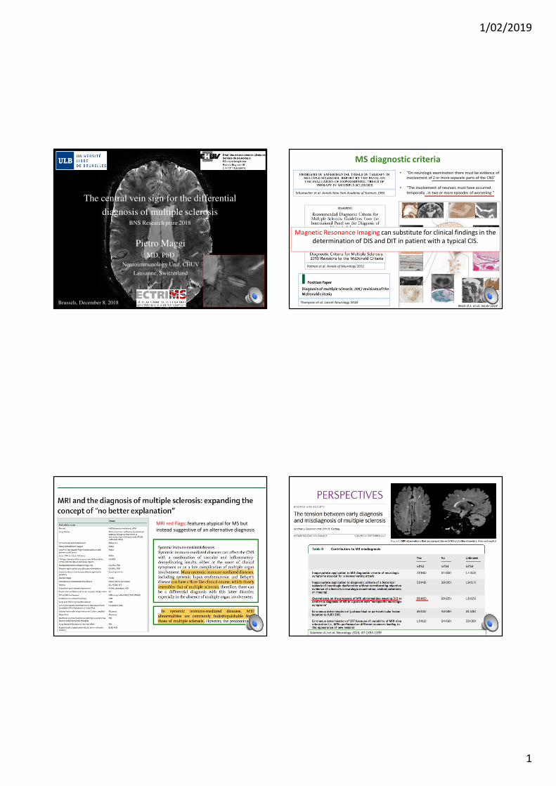
Results: Fulfillment of different MRI diagnostic criteria for MS Discussion
• When comparing MS and CNS vasculitis, the “central vein sign” alone or in combination
with the available MS diagnostic MRI criteria improves the diagnostic accuracy and
specificity without lowering the sensitivity of MS diagnosis.
• Why this is important ?
1. CNS vasculitis are difficult to diagnose accurately (biopsy and/or angiography)
• When dealing with chronic brain inflammatory conditions, the addition of the central
vein assessment to the existing clinical and radiological work up can reduce the risk of
misdiagnosis and aid therapeutic strategies.
High % of our vasculitis patients fulfilled the dissemination in space MRI criteria for MS
2. CNS vasculitis can have clinical and radiological presentations very similar to MS.
Acknowledgment
Vita-Salute San Raffaele UniversityNeuroimaging Research Unit
• Massimo Filippi
• Martina Absinta
Department of Neuroradiology
• Roberta Scotti
Department of Neurology
• Vittorio Martinelli
National Institutes of Health (NIH)Translational Neuroradiology Section, NINDS
• Martina Absinta
• Pascal Sati
• Daniel Reich
Université Libre de Bruxelles (ULB)Department of Neurology, CHU Brugmann
• Bernard Dachy
Department of Radiology, Hopital Erasme
• Niloufar Sadeghi
• Valentina Lolli
Department of Neurology, Hopital Erasme
• Gaetano Perrotta
• Massimo Pandolfo
Lausanne University Hospital (CHUV)Department Neurology CHUV
• Renaud Du Pasquier
• Marie Theaudin
• Team NIS
Department of Radiology
• Merixtell Bach Cuadra
• Hagmann Patric
Ecole Polytechnique de Lausanne (EPFL)Siemens Healthineers