NEOPLASTIC DISEASE Pituitary Null Cell Adenoma in a Domestic Llama (Lama glama) M. D. Chalkley * , M. Kiupel † and A. C. E. Draper * * Department of Veterinary Population Medicine, College of Veterinary Medicine, University of Minnesota, St Paul, MN and † Department of Pathobiology and Diagnostic Investigation/Diagnostic Center for Population and Animal Health, Michigan State University, East Lansing, MI, USA Summary Pituitary gland neoplasia has been reported rarely in camelids. A 12-year-old neutered male llama (Lama glama) presented with lethargy, inappetence and neurological signs. On physical examination, the llama was mentally dull and exhibited compulsive pacing and circling to the left. Complete blood count and serum biochemistry revealed haemoconcentration, mild hypophosphataemia, hyperglycaemia, hypercreatininaemia and hyperalbuminaemia. Humane destruction was elected due to rapid clinical deterioration and poor prog- nosis. Post-mortem examination revealed a pituitary macroadenoma and bilateral internal hydrocephalus. Microscopically, the pituitary tumour was composed of neoplastic chromophobic pituitary cells. Ultrastruc- tural studies revealed similar neoplastic cells to those previously described in human null cell adenomas. Im- munohistochemically, the neoplastic cells were strongly immunoreactive for neuroendocrine markers (synaptophysin and chromogranin A), but did not exhibit immunoreactivity for epithelial, mesenchymal, neuronal and all major pituitary hormone markers (adrenocorticotropic hormone, follicle stimulating hor- mone, growth hormone, luteinizing hormone, melanocyte-stimulating hormone, prolactin and thyroid stimu- lating hormone), consistent with the diagnosis of a pituitary null cell adenoma. This is the first report of pituitary neoplasia in a llama. Ó 2014 Elsevier Ltd. All rights reserved. Keywords: camelid; llama; null cell adenoma; pituitary gland Neoplastic diseases are relatively common in camelids (Valentine and Martin, 2007); however, pituitary neoplasia has been reported rarely (Gilsenan et al., 2012). In contrast, primary pituitary neoplasia is well documented in several other animal species, in particular, dogs, horses and cats (Capen, 2002), bud- gerigars (Langohr et al., 2012) and rats (Percy and Barthold, 2007). Adenomas of the adenohypophysis are the most prevalent pituitary tumours in animals. Adenohypophyseal pituitary adenomas are a hetero- geneous group of tumours for which subclassification is based on the hormone(s) produced and/or secreted by the constituent neoplastic cells (Kiupel et al., 2008). Null cell adenomas of the adenohypophysis are non-functional tumours composed of chromopho- bic cells that lack immunoreactivity for any of the ma- jor pituitary hormones (Kiupel et al., 2008). Because complete immunohistochemical and ultrastructural characterization of non-functional pituitary ade- nomas is lacking in many reports, the true prevalence of null cell adenomas in animals is difficult to deter- mine. Comparatively, human null cell pituitary ade- nomas are fairly well characterized and comprise a significant portion of pituitary macroadenomas (Lloyd et al., 2009). To our knowledge, the current case represents the first report of pituitary adenoma in a llama. Comprehensive clinical, morphological, ultrastructural and immunohistochemical character- ization of the tumour allowed subclassification as a null cell pituitary adenoma. A 12-year-old, 146 kg, neutered male llama (Lama glama) was presented for assessment of acute onset neurological signs. The llama was reportedly lethargic for 13 days prior to presentation. The Correspondence to: M. D. Chalkley (e-mail: [email protected]). 0021-9975/$ - see front matter Ó 2014 Elsevier Ltd. All rights reserved. http://dx.doi.org/10.1016/j.jcpa.2014.02.006 J. Comp. Path. 2014, Vol. -,1e6 Available online at www.sciencedirect.com ScienceDirect www.elsevier.com/locate/jcpa Please cite this article in press as: Chalkley MD, et al., Pituitary Null Cell Adenoma in a Domestic Llama (Lama glama), Journal of Comparative Pathology (2014), http://dx.doi.org/10.1016/j.jcpa.2014.02.006
Transcript
J. Comp. Path. 2014, Vol. -, 1e6 Available online at www.sciencedirect.com
ScienceDirect
www.elsevier.com/locate/jcpa
NEOPLASTIC DISEASE
Pituitary Null Cell Adenoma in a Domestic Llama(Lama glama)
Cor
002
http
Pl
Pa
M. D. Chalkley*, M. Kiupel† and A. C. E. Draper*
*Department of Veterinary Population Medicine, College of Veterinary Medicine, University of Minnesota, St Paul, MNand†Department of Pathobiology and Diagnostic Investigation/Diagnostic Center for Population and Animal Health, Michigan
State University, East Lansing, MI, USA
resp
1-99
://d
ease
tho
Summary
Pituitary gland neoplasia has been reported rarely in camelids. A 12-year-old neutered male llama (Lamaglama) presented with lethargy, inappetence and neurological signs. On physical examination, the llamawas mentally dull and exhibited compulsive pacing and circling to the left. Complete blood count and serumbiochemistry revealed haemoconcentration, mild hypophosphataemia, hyperglycaemia, hypercreatininaemiaand hyperalbuminaemia. Humane destruction was elected due to rapid clinical deterioration and poor prog-nosis. Post-mortem examination revealed a pituitary macroadenoma and bilateral internal hydrocephalus.Microscopically, the pituitary tumour was composed of neoplastic chromophobic pituitary cells. Ultrastruc-tural studies revealed similar neoplastic cells to those previously described in human null cell adenomas. Im-munohistochemically, the neoplastic cells were strongly immunoreactive for neuroendocrine markers(synaptophysin and chromogranin A), but did not exhibit immunoreactivity for epithelial, mesenchymal,neuronal and all major pituitary hormone markers (adrenocorticotropic hormone, follicle stimulating hor-mone, growth hormone, luteinizing hormone, melanocyte-stimulating hormone, prolactin and thyroid stimu-lating hormone), consistent with the diagnosis of a pituitary null cell adenoma. This is the first report ofpituitary neoplasia in a llama.
Neoplastic diseases are relatively common in camelids(Valentine and Martin, 2007); however, pituitaryneoplasia has been reported rarely (Gilsenan et al.,2012). In contrast, primary pituitary neoplasia iswell documented in several other animal species, inparticular, dogs, horses and cats (Capen, 2002), bud-gerigars (Langohr et al., 2012) and rats (Percy andBarthold, 2007). Adenomas of the adenohypophysisare the most prevalent pituitary tumours in animals.Adenohypophyseal pituitary adenomas are a hetero-geneous group of tumours for which subclassificationis based on the hormone(s) produced and/or secretedby the constituent neoplastic cells (Kiupel et al.,2008). Null cell adenomas of the adenohypophysisare non-functional tumours composed of chromopho-bic cells that lack immunoreactivity for any of the ma-
jor pituitary hormones (Kiupel et al., 2008). Becausecomplete immunohistochemical and ultrastructuralcharacterization of non-functional pituitary ade-nomas is lacking in many reports, the true prevalenceof null cell adenomas in animals is difficult to deter-mine. Comparatively, human null cell pituitary ade-nomas are fairly well characterized and comprise asignificant portion of pituitary macroadenomas(Lloyd et al., 2009). To our knowledge, the currentcase represents the first report of pituitary adenomain a llama. Comprehensive clinical, morphological,ultrastructural and immunohistochemical character-ization of the tumour allowed subclassification as anull cell pituitary adenoma.
A 12-year-old, 146 kg, neutered male llama (Lamaglama) was presented for assessment of acute onsetneurological signs. The llama was reportedlylethargic for 13 days prior to presentation. The
� 2014 Elsevier Ltd. All rights reserved.
Adenoma in a Domestic Llama (Lama glama), Journal of Comparative
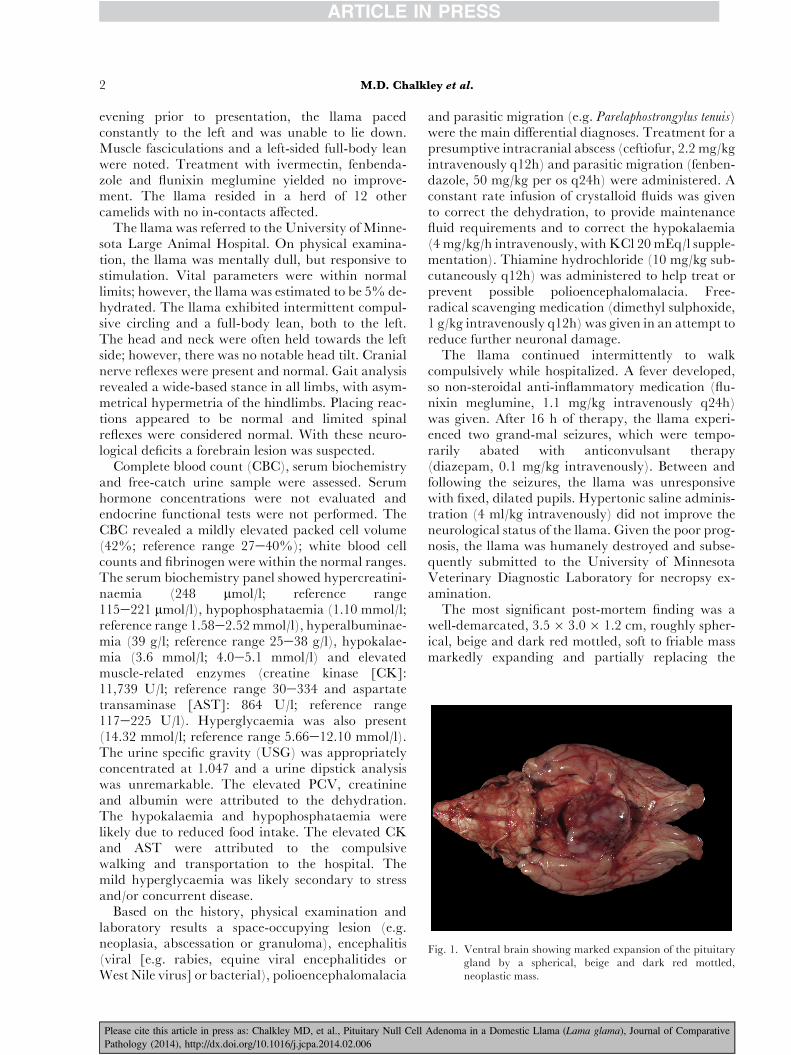
Fig. 1. Ventral brain showing marked expansion of the pituitarygland by a spherical, beige and dark red mottled,neoplastic mass.
2 M.D. Chalkley et al.
evening prior to presentation, the llama pacedconstantly to the left and was unable to lie down.Muscle fasciculations and a left-sided full-body leanwere noted. Treatment with ivermectin, fenbenda-zole and flunixin meglumine yielded no improve-ment. The llama resided in a herd of 12 othercamelids with no in-contacts affected.
The llama was referred to the University of Minne-sota Large Animal Hospital. On physical examina-tion, the llama was mentally dull, but responsive tostimulation. Vital parameters were within normallimits; however, the llama was estimated to be 5% de-hydrated. The llama exhibited intermittent compul-sive circling and a full-body lean, both to the left.The head and neck were often held towards the leftside; however, there was no notable head tilt. Cranialnerve reflexes were present and normal. Gait analysisrevealed a wide-based stance in all limbs, with asym-metrical hypermetria of the hindlimbs. Placing reac-tions appeared to be normal and limited spinalreflexes were considered normal. With these neuro-logical deficits a forebrain lesion was suspected.
Complete blood count (CBC), serum biochemistryand free-catch urine sample were assessed. Serumhormone concentrations were not evaluated andendocrine functional tests were not performed. TheCBC revealed a mildly elevated packed cell volume(42%; reference range 27e40%); white blood cellcounts and fibrinogen were within the normal ranges.The serum biochemistry panel showed hypercreatini-naemia (248 mmol/l; reference range115e221 mmol/l), hypophosphataemia (1.10 mmol/l;reference range 1.58e2.52mmol/l), hyperalbuminae-mia (39 g/l; reference range 25e38 g/l), hypokalae-mia (3.6 mmol/l; 4.0e5.1 mmol/l) and elevatedmuscle-related enzymes (creatine kinase [CK]:11,739 U/l; reference range 30e334 and aspartatetransaminase [AST]: 864 U/l; reference range117e225 U/l). Hyperglycaemia was also present(14.32 mmol/l; reference range 5.66e12.10 mmol/l).The urine specific gravity (USG) was appropriatelyconcentrated at 1.047 and a urine dipstick analysiswas unremarkable. The elevated PCV, creatinineand albumin were attributed to the dehydration.The hypokalaemia and hypophosphataemia werelikely due to reduced food intake. The elevated CKand AST were attributed to the compulsivewalking and transportation to the hospital. Themild hyperglycaemia was likely secondary to stressand/or concurrent disease.
Based on the history, physical examination andlaboratory results a space-occupying lesion (e.g.neoplasia, abscessation or granuloma), encephalitis(viral [e.g. rabies, equine viral encephalitides orWest Nile virus] or bacterial), polioencephalomalacia
Please cite this article in press as: Chalkley MD, et al., Pituitary Null Cell
and parasitic migration (e.g. Parelaphostrongylus tenuis)were the main differential diagnoses. Treatment for apresumptive intracranial abscess (ceftiofur, 2.2 mg/kgintravenously q12h) and parasitic migration (fenben-dazole, 50 mg/kg per os q24h) were administered. Aconstant rate infusion of crystalloid fluids was givento correct the dehydration, to provide maintenancefluid requirements and to correct the hypokalaemia(4 mg/kg/h intravenously, with KCl 20mEq/l supple-mentation). Thiamine hydrochloride (10 mg/kg sub-cutaneously q12h) was administered to help treat orprevent possible polioencephalomalacia. Free-radical scavenging medication (dimethyl sulphoxide,1 g/kg intravenously q12h) was given in an attempt toreduce further neuronal damage.
The llama continued intermittently to walkcompulsively while hospitalized. A fever developed,so non-steroidal anti-inflammatory medication (flu-nixin meglumine, 1.1 mg/kg intravenously q24h)was given. After 16 h of therapy, the llama experi-enced two grand-mal seizures, which were tempo-rarily abated with anticonvulsant therapy(diazepam, 0.1 mg/kg intravenously). Between andfollowing the seizures, the llama was unresponsivewith fixed, dilated pupils. Hypertonic saline adminis-tration (4 ml/kg intravenously) did not improve theneurological status of the llama. Given the poor prog-nosis, the llama was humanely destroyed and subse-quently submitted to the University of MinnesotaVeterinary Diagnostic Laboratory for necropsy ex-amination.
The most significant post-mortem finding was awell-demarcated, 3.5 � 3.0 � 1.2 cm, roughly spher-ical, beige and dark red mottled, soft to friable massmarkedly expanding and partially replacing the
Adenoma in a Domestic Llama (Lama glama), Journal of Comparative
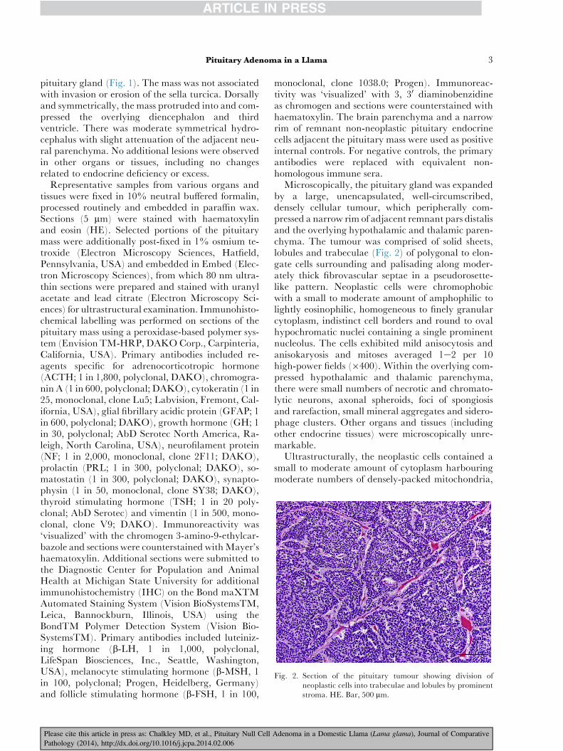
Fig. 2. Section of the pituitary tumour showing division ofneoplastic cells into trabeculae and lobules by prominentstroma. HE. Bar, 500 mm.
Pituitary Adenoma in a Llama 3
pituitary gland (Fig. 1). The mass was not associatedwith invasion or erosion of the sella turcica. Dorsallyand symmetrically, the mass protruded into and com-pressed the overlying diencephalon and thirdventricle. There was moderate symmetrical hydro-cephalus with slight attenuation of the adjacent neu-ral parenchyma. No additional lesions were observedin other organs or tissues, including no changesrelated to endocrine deficiency or excess.
Representative samples from various organs andtissues were fixed in 10% neutral buffered formalin,processed routinely and embedded in paraffin wax.Sections (5 mm) were stained with haematoxylinand eosin (HE). Selected portions of the pituitarymass were additionally post-fixed in 1% osmium te-troxide (Electron Microscopy Sciences, Hatfield,Pennsylvania, USA) and embedded in Embed (Elec-tron Microscopy Sciences), from which 80 nm ultra-thin sections were prepared and stained with uranylacetate and lead citrate (Electron Microscopy Sci-ences) for ultrastructural examination. Immunohisto-chemical labelling was performed on sections of thepituitary mass using a peroxidase-based polymer sys-tem (Envision TM-HRP, DAKOCorp., Carpinteria,California, USA). Primary antibodies included re-agents specific for adrenocorticotropic hormone(ACTH; 1 in 1,800, polyclonal, DAKO), chromogra-nin A (1 in 600, polyclonal; DAKO), cytokeratin (1 in25, monoclonal, clone Lu5; Labvision, Fremont, Cal-ifornia, USA), glial fibrillary acidic protein (GFAP; 1in 600, polyclonal; DAKO), growth hormone (GH; 1in 30, polyclonal; AbD Serotec North America, Ra-leigh, North Carolina, USA), neurofilament protein(NF; 1 in 2,000, monoclonal, clone 2F11; DAKO),prolactin (PRL; 1 in 300, polyclonal; DAKO), so-matostatin (1 in 300, polyclonal; DAKO), synapto-physin (1 in 50, monoclonal, clone SY38; DAKO),thyroid stimulating hormone (TSH; 1 in 20 poly-clonal; AbD Serotec) and vimentin (1 in 500, mono-clonal, clone V9; DAKO). Immunoreactivity was‘visualized’ with the chromogen 3-amino-9-ethylcar-bazole and sections were counterstained withMayer’shaematoxylin. Additional sections were submitted tothe Diagnostic Center for Population and AnimalHealth at Michigan State University for additionalimmunohistochemistry (IHC) on the Bond maXTMAutomated Staining System (Vision BioSystemsTM,Leica, Bannockburn, Illinois, USA) using theBondTM Polymer Detection System (Vision Bio-SystemsTM). Primary antibodies included luteiniz-ing hormone (b-LH, 1 in 1,000, polyclonal,LifeSpan Biosciences, Inc., Seattle, Washington,USA), melanocyte stimulating hormone (b-MSH, 1in 100, polyclonal; Progen, Heidelberg, Germany)and follicle stimulating hormone (b-FSH, 1 in 100,
Please cite this article in press as: Chalkley MD, et al., Pituitary Null Cell
monoclonal, clone 1038.0; Progen). Immunoreac-tivity was ‘visualized’ with 3, 30 diaminobenzidineas chromogen and sections were counterstained withhaematoxylin. The brain parenchyma and a narrowrim of remnant non-neoplastic pituitary endocrinecells adjacent the pituitary mass were used as positiveinternal controls. For negative controls, the primaryantibodies were replaced with equivalent non-homologous immune sera.
Microscopically, the pituitary gland was expandedby a large, unencapsulated, well-circumscribed,densely cellular tumour, which peripherally com-pressed a narrow rim of adjacent remnant pars distalisand the overlying hypothalamic and thalamic paren-chyma. The tumour was comprised of solid sheets,lobules and trabeculae (Fig. 2) of polygonal to elon-gate cells surrounding and palisading along moder-ately thick fibrovascular septae in a pseudorosette-like pattern. Neoplastic cells were chromophobicwith a small to moderate amount of amphophilic tolightly eosinophilic, homogeneous to finely granularcytoplasm, indistinct cell borders and round to ovalhypochromatic nuclei containing a single prominentnucleolus. The cells exhibited mild anisocytosis andanisokaryosis and mitoses averaged 1e2 per 10high-power fields (�400). Within the overlying com-pressed hypothalamic and thalamic parenchyma,there were small numbers of necrotic and chromato-lytic neurons, axonal spheroids, foci of spongiosisand rarefaction, small mineral aggregates and sidero-phage clusters. Other organs and tissues (includingother endocrine tissues) were microscopically unre-markable.
Ultrastructurally, the neoplastic cells contained asmall to moderate amount of cytoplasm harbouringmoderate numbers of densely-packed mitochondria,
Adenoma in a Domestic Llama (Lama glama), Journal of Comparative
4 M.D. Chalkley et al.
poorly developed rough endoplasmic reticulum andgolgi apparatus and scant, randomly dispersed cyto-plasmic secretory granules (Fig. 3). The majority ofsecretory granules were roughly spherical,100e200 mm in diameter, electron dense andmembranebound with a narrow submembranousspace. A smaller proportion of secretory granuleswere electron lucent and largely devoid of contents.
Immunohistochemically, the majority of neoplasticcells exhibited strong immunoreactivity for synapto-physin and weak to moderate immunoreactivity forchromogranin A. The neoplastic cells did not exhibitimmunoreactivity for ACTH, cytokeratin, FSH, GH,LH,MSH, NF, PRL, somatostatin, TSH or vimentin(Table 1). Cells of the remnant normal pituitarygland exhibited immunoreactivity for chromogranin,synaptophysin, ACTH, FSH, GH, LH, MSH, PRL,somatostatin and TSH (Table 1).
The combined clinical, morphological, ultrastruc-tural and immunohistochemical findings in this llamawere most consistent with a pituitary null cell ade-noma. Clinically, macroadenomas of the pituitarygland often present with signs consistent with prosen-cephalic dysfunction. Gait abnormalities can be pre-sent or absent (de Lahunta and Glass, 2009). Areport in an alpaca with a pituitary macroadenomadescribed similar signs to those reported here(Gilsenan et al., 2012). Hormonal dysfunction and hy-popituitarism were not apparent clinically in thisllama, although advanced testing was not performed.Central diabetes insipidus was excluded with aconcentrated USG, but has been reported in a sheepwith a pituitary macroadenoma (Zanolari et al.,
Fig. 3. Lowmagnification electron micrograph of a neoplastic cellwithin the pituitary mass containing moderate numbers ofmitochondria (arrows), poorly developed rough endo-plasmic reticulum (arrowheads) and scant, randomlydispersed, electron-dense and -lucent cytoplasmic secretorygranules. Transmission electron microscopy. Bar, 2 mm.
Please cite this article in press as: Chalkley MD, et al., Pituitary Null Cell
2004). Hypoglycaemia has been documented in agoat with pituitary macroadenomas (Reed andBauer, 2009); however, this was not a feature of thiscase or an alpaca with a similar tumour (Gilsenanet al., 2012). Anorexia was reported with this llamaand was the likely cause of the low phosphate in thiscase and in a similar report of a pituitary glandtumour in an alpaca (Gilsenan et al., 2012). However,persistent hypophosphataemia has been reported in ahuman being with a chromophobic pituitary tumourwhere no aetiology was postulated (de Oliveira,1953).
There are currently no reliable criteria for distin-guishing pituitary adenoma from carcinoma based onmorphological characteristics. Therefore, in bothman and animals, the diagnosis of pituitary adenomais based on the absence of cerebrospinal and systemicmetastases (Imboden et al., 2004; Kiupel et al., 2008).Although the spinal cord was not examined in thepresent case, the lack of local and distant metastasesin other organs/tissues was more compatible with thediagnosis of adenoma than carcinoma. Thesubclassification of this pituitary tumour as a null celladenoma was based on a combined morphological,ultrastructural and immunohistochemical diagnosticapproach. Consistent with the present case, null cellpituitary adenomas in both man and animals arecharacterized by histologically nondescriptchromophobic cells arranged variably inperisinusoidal sheets with or without pseudorosette-like structures (Saeger et al., 2007; Kiupel et al.,2008). Given the prominent pseudorosette-like patternin this case, a differential diagnosis of primitive neuro-ectodermal tumor (PNET) was entertained; however,other features consistent with PNET, such as truerosette formation, hyperchromatic ‘carrot-shaped’nuclei, neurosecretory-type cytoplasmic granules andimmunoreactivity for neurofilament protein (NF),were not present.
The ultrastructural appearance of pituitary ade-nomas has not been characterized in camelids, incontrast to man in which fairly reliable diagnosticcriteria have been developed (Kontogeorgos, 2005;Saeger et al., 2007; Lloyd et al., 2009). It is unknownat this time whether these criteria can beextrapolated to camelid pituitary adenomas. In thepresent case, the poorly developed endoplasmicreticulum and golgi apparatus, in conjunction withsparsely scattered cytoplasmic secretory granulesranging in size from 100 to 200 mm diameter, werecharacteristics comparable with human null celladenomas (Kontogeorgos, 2005; Saeger et al., 2007;Lloyd et al., 2009). The high density ofmitochondria in the neoplastic cells of the llama isan additional feature frequently observed in human
Adenoma in a Domestic Llama (Lama glama), Journal of Comparative
Table 1
Immunohistochemical labelling
Antigen Neoplastic cells Tumour stromal cells Normal pituitary
null cell adenomas, especially those exhibitingoncocytic transformation (Kontogeorgos, 2005;Saeger et al., 2007; Lloyd et al., 2009).Ultrastructurally, there is considerable overlap ofsome human gonadotroph, lactotroph and othersparsely granulated adenomas with null celladenomas, therefore, differentiation by IHC or in-situ hybridization is often necessary (Kontogeorgos,2005).
Compatible with the present case, null cell ade-nomas are devoid of immunoreactivity for the majoradenohypophyseal hormones, but are immunoreac-tive for synaptophysin and chromogranin A(Kontogeorgos, 2005; Saeger et al., 2007; Kiupelet al., 2008). Human null cell adenomas are oftenimmunoreactive for the free a-subunit ofglycoprotein pituitary hormones (a-subunit of FSH,LH and TSH). Additionally, gonadotropin subunit(a-subunit, b-FSH and b-LH) genes and thegonadotroph-associated transcription factor steroido-genic factor-1 (SF-1) have been demonstrated in thesetumours (Kumar et al., 1998). Although notconfirmed, this supports the hypothesis that null celladenomas are derived from gonadotrophs (Kumaret al., 1998). Alpha-subunit has been poorly charac-terized in animals and given the commercial unavail-ability of this immunohistochemical marker inanimals, a-subunit immunohistochemistry was notinvestigated in this llama.
In summary, pituitary tumours appear to be rare incamelids with only one case previously recorded in analpaca (Gilsenan et al., 2012). Despite this apparentrarity, the poor prognosis associated with any expan-sile intracranial neoplasm demands greater awareness
Please cite this article in press as: Chalkley MD, et al., Pituitary Null Cell
of this entity and such tumours should therefore beincluded in differential diagnostic lists for camelidswith neurological signs, particularly those presentingsimilarly to the llama in this case and the alpaca re-ported previously (Gilsenan et al., 2012).
Conflict of Interest Statement
The authors declared no potential conflicts of interestwith respect to the research, authorship and/or publi-cation of this article.
Acknowledgements
We thank R. Aho and J. Shivers, University of Min-nesota, for their histological and immunohistochem-ical assistance and D. Muldoon and D. Ariyakumerfor their electron microscopy assistance. The firstand last authors contributed equally to this manu-script.
References
Capen CC (2002) Tumors of the endocrine glands. In: Tu-
mors in Domestic Animals, 4th Edit., DJMeuten, Ed., IowaState Press, Ames, pp. 607e696.
de Lahunta A, Glass E (2009) Lower motor neuron: gen-eral somatic efferent, cranial nerve. In: Veterinary Neuro-
anatomy and Clinical Neurology, 3rd Edit., A de Lahunta,E Glass, Eds., Saunders, St Louis, p. 161.
de Oliveira HL (1953) Associated parathyroid and pitui-tary (chromophobe) adenomas: persisting hypophos-phatemia after the cure of the hyperparathyroidism.Journal of Clinical Endocrinology and Metabolism, 13,1129e1131.
Adenoma in a Domestic Llama (Lama glama), Journal of Comparative
Gilsenan WF, Habecker PL, Coyne TM, Johnson AL(2012) Neurologic disease attributed to a pituitary ade-noma in an alpaca. Journal of Veterinary Internal Medicine,26, 1073e1077.
Imboden PN, Borruat FX, De Tribolet N, Meagher-Villemure K, Pica A et al. (2004) Non-functioning pitu-itary carcinoma. Pituitary, 7, 149e156.
Kiupel M, Capen CC, Miller M, Smedley R (2008) Tu-mors of the hypophysis (pituitary gland) and sellar re-gion. In: Histological Classification of Tumors of the
Endocrine System of Domestic Animals, 2nd Edit.,FV Schulman, Ed., Armed Forces Institute of Pathol-ogy, Washington DC, pp. 17e25.
Kontogeorgos G (2005) Classification and pathology of pi-tuitary tumors. Endocrine, 28, 27e35.
Kumar TJ, Graham KE, Asa SL, Low ML (1998) Simianvirus 40 T antigen-induced gonadotroph adenomas: amodel of human null cell adenomas. Endocrinology, 139,3342e3351.
Langohr IM, Garner MM, Kiupel M (2012) Somatotrophpituitary tumors in budgerigars (Melopsittacus undulatus).Veterinary Pathology, 49, 503e507.
Lloyd RV, Scheithauer BW, Horvath E, Kovacs K (2009)Tumors of the pituitary gland. In: Surgical Pathology ofEndocrine and Neuroendocrine Tumors, A Khan, Ed., Hu-mana Press, Springer, New York, pp. 27e29.
Please cite this article in press as: Chalkley MD, et al., Pituitary Null Cell
Percy DH, Barthold SW (2007) Rat, pituitary gland tu-mors. In: Pathology of Laboratory Rodents and Rabbits, 3rdEdit., DH Percy, SW Barthold, Eds., Blackwell, Ames,pp. 173e174.
Reed SD, Bauer RW (2009) Pituitary acidophil macroade-noma in a pygmy goat (Capra hircus hircus). Journal of Vet-erinary Diagnostic Investigation, 21, 262e266.
Saeger W, L€udecke DK, Buchfelder M, Fahlbusch R,Quabbe HJ et al. (2007) Pathohistological classificationof pituitary tumors: 10 years of experience with theGerman Pituitary Tumor Registry. European Journal of
Endocrinology, 156, 203e216.Valentine BA, Martin JM (2007) Prevalence of neoplasia
in llamas and alpacas (Oregon State University,2001e2006). Journal of Veterinary Diagnostic Investigation,19, 202e204.
Zanolari P, Botteron C, Jaggy A, Meylan M (2004) Chro-mophobe adenocarcinoma of the pituitary gland in aram. Journal of Veterinary Internal Medicine, 18, 748e752.