ORIGINAL ARTICLE Placenta histopathology in SARS-CoV-2 infection: analysis of a consecutive series and comparison with control cohorts Luca Bertero 1 & Fulvio Borella 2 & Giovanni Botta 3 & Andrea Carosso 2 & Stefano Cosma 2 & Marialuisa Bovetti 2 & Marco Carosso 2 & Giancarlo Abbona 3 & Giammarco Collemi 1 & Mauro Papotti 4 & Paola Cassoni 1 & Chiara Benedetto 2 Received: 28 October 2020 /Revised: 14 March 2021 /Accepted: 30 March 2021 # The Author(s) 2021 Abstract Infection by SARS-CoV-2 has been shown to involve a wide range of organs and tissues, leading to a kaleidoscope of clinical conditions. Within this spectrum, an involvement of the fetal-maternal unit could be expected, but, so far, the histopathological evaluation of placentas delivered by women with SARS-CoV-2 infection did not show distinct hallmarks. A consecutive series of 11 placentas, delivered by 10 women with COVID-19 admitted to our Obstetrics and Gynecology clinic have been investigated and compared to a control cohort of 58 pre-COVID-19 placentas and 28 placentas delivered by women who had a previous cesarean section. Four out of eleven placentas showed changes consistent with chronic villitis/villitis of unknown etiology (VUE), while in one case, chronic histiocytic intervillositis was diagnosed. Thrombo-hemorrhagic alterations were observed in a subset of cases. Compared to the control cohort, chronic villitis/VUE (p < 0.001), chronic deciduitis (p = 0.023), microvas- cular thrombosis (p = 0.003), presence of infarction areas (p = 0.047) and of accelerated villous maturation (p = 0.005) showed higher frequencies in placentas delivered by women with COVID-19. Chronic villitis/VUE (p = 0.003) and accelerated villous maturation (p = 0.019) remained statistically significant by restricting the analysis to placentas delivered after a previous cesarean section. The observed differences in terms of pathological findings could be consistent with SARS-CoV-2 pathogenesis, but just a subset of alterations remained statistically significant after adjusting for a previous cesarean section. A careful consideration of potential confounders is warranted in future studies exploring the relationship between COVID-19 and pregnancy. Keywords COVID-19 . SARS-CoV-2 . Histopathology . Thrombosis . Inflammation . Obstetrics Introduction On March 11, 2020, due do the rapid escalation of the coro- navirus disease 2019 (COVID-19) outbreak, the World Health Organization (WHO) declared a pandemic [61]. COVID-19 is caused by the severe acute respiratory syndrome coronavirus 2 (SARS-CoV-2), a betacoronavirus similar to SARS-CoV and MERS-CoV, with multiple possible transmission routes and characterized by a high infectivity [59, 62]. In terms of maternal outcomes, data derived from initial cohorts suggested that COVID-19 clinical course in this group of patients is similar to non-pregnant women [11, 16, 17, 23, 28, 40], but not all data are reassuring. Seven maternal deaths were reported among nine pregnant women with severe COVID-19 [32], and an analysis of a Swedish cohort sug- gested an increased risk of being admitted to an intensive care unit [relative risk (RR): 5.4; 95% Confidence Interval (CI) 2.89–10.08] and of receiving invasive mechanical ventilation (RR: 4.0; 95%; CI 1.75–9.14) for pregnant women with Luca Bertero, Fulvio Borella, Paola Cassoni and Chiara Benedetto contributed equally to this work. * Paola Cassoni [email protected]1 Pathology Unit, Department of Medical Sciences, University of Turin and “Città della Salute e della Scienza di Torino” University Hospital, Turin, Italy 2 Obstetrics and Gynecology 1U, Department of Surgical Sciences, Sant’Anna Hospital, University of Turin and “Città della Salute e della Scienza di Torino” University Hospital, Turin, Italy 3 Pathology Unit, “Città della Salute e della Scienza di Torino” University Hospital, Turin, Italy 4 Pathology Unit, Department of Oncology, University of Turin and “Città della Salute e della Scienza di Torino” University Hospital, Turin, Italy https://doi.org/10.1007/s00428-021-03097-3 / Published online: 1 May 2021 Virchows Archiv (2021) 479:715–728
Transcript
ORIGINAL ARTICLE
Placenta histopathology in SARS-CoV-2 infection: analysisof a consecutive series and comparison with control cohorts
Luca Bertero1& Fulvio Borella2 & Giovanni Botta3 & Andrea Carosso2
Received: 28 October 2020 /Revised: 14 March 2021 /Accepted: 30 March 2021# The Author(s) 2021
AbstractInfection by SARS-CoV-2 has been shown to involve a wide range of organs and tissues, leading to a kaleidoscope of clinicalconditions. Within this spectrum, an involvement of the fetal-maternal unit could be expected, but, so far, the histopathologicalevaluation of placentas delivered bywomen with SARS-CoV-2 infection did not show distinct hallmarks. A consecutive series of11 placentas, delivered by 10 women with COVID-19 admitted to our Obstetrics and Gynecology clinic have been investigatedand compared to a control cohort of 58 pre-COVID-19 placentas and 28 placentas delivered by women who had a previouscesarean section. Four out of eleven placentas showed changes consistent with chronic villitis/villitis of unknown etiology(VUE), while in one case, chronic histiocytic intervillositis was diagnosed. Thrombo-hemorrhagic alterations were observedin a subset of cases. Compared to the control cohort, chronic villitis/VUE (p < 0.001), chronic deciduitis (p = 0.023), microvas-cular thrombosis (p = 0.003), presence of infarction areas (p = 0.047) and of accelerated villous maturation (p = 0.005) showedhigher frequencies in placentas delivered by women with COVID-19. Chronic villitis/VUE (p = 0.003) and accelerated villousmaturation (p = 0.019) remained statistically significant by restricting the analysis to placentas delivered after a previous cesareansection. The observed differences in terms of pathological findings could be consistent with SARS-CoV-2 pathogenesis, but justa subset of alterations remained statistically significant after adjusting for a previous cesarean section. A careful consideration ofpotential confounders is warranted in future studies exploring the relationship between COVID-19 and pregnancy.
On March 11, 2020, due do the rapid escalation of the coro-navirus disease 2019 (COVID-19) outbreak, theWorld HealthOrganization (WHO) declared a pandemic [61]. COVID-19 iscaused by the severe acute respiratory syndrome coronavirus2 (SARS-CoV-2), a betacoronavirus similar to SARS-CoVand MERS-CoV, with multiple possible transmission routesand characterized by a high infectivity [59, 62].
In terms of maternal outcomes, data derived from initialcohorts suggested that COVID-19 clinical course in this groupof patients is similar to non-pregnant women [11, 16, 17, 23,28, 40], but not all data are reassuring. Seven maternal deathswere reported among nine pregnant women with severeCOVID-19 [32], and an analysis of a Swedish cohort sug-gested an increased risk of being admitted to an intensive careunit [relative risk (RR): 5.4; 95% Confidence Interval (CI)2.89–10.08] and of receiving invasive mechanical ventilation(RR: 4.0; 95%; CI 1.75–9.14) for pregnant women with
Luca Bertero, Fulvio Borella, Paola Cassoni and Chiara Benedettocontributed equally to this work.
1 Pathology Unit, Department of Medical Sciences, University ofTurin and “Città della Salute e della Scienza di Torino” UniversityHospital, Turin, Italy
2 Obstetrics and Gynecology 1U, Department of Surgical Sciences,Sant’Anna Hospital, University of Turin and “Città della Salute edella Scienza di Torino” University Hospital, Turin, Italy
3 Pathology Unit, “Città della Salute e della Scienza di Torino”University Hospital, Turin, Italy
4 Pathology Unit, Department of Oncology, University of Turin and“Città della Salute e della Scienza di Torino” University Hospital,Turin, Italy
COVID-19 compared to non-pregnant women of similar age[19, 60].
The role of SARS-CoV-2 in the first trimester of pregnancyhas also been investigated and present data do not suggest anincreased spontaneous abortion risk [21], but the long-termconsequences are currently unknown and, in the endemicareas, tests for SARS-CoV-2 should be offered to all pregnantwomen to investigate this open question [20].
Concerning neonatal outcomes, fetal and respiratory dis-tress as well as thrombocytopenia accompanied by abnormalliver function and even death have been described [64]. Inaddition, evidence of a higher incidence of preterm birth hasbeen reported [25, 29, 63] as well as cases of SARS-CoV-2vertical transmission [5, 12–14, 26, 58], but this seems to bean uncommon occurrence [24, 25, 63].
High expression of proteins (ACE2/TMPRSS2) requiredfor SARS-CoV-2 cell entry has been observed in maternal-fetal interface tissues [42], and placental infection by SARS-CoV-2 has been demonstrated in a minority of cases [1, 2, 27,33, 55, 58].
To date, placental histopathology in COVID-19 has beeninvestigated [6, 7, 18, 22, 31, 33, 47, 48, 53, 54], but specificfeatures or hallmarks have not been identified. Nevertheless,the inflammatory activation and the increased thrombotic riskdescribed in patients with COVID-19 [35, 52, 57] make theplacenta a potential target of pathophysiological phenomenawhich could affect pregnancy outcomes.
The aim of this workwas to evaluate a consecutive series ofplacentas delivered at our institution from women withCOVID-19 infection and compare these data with a pre-COVID-19 control series.
Methods
Our study is based on the pathological analysis of 11 consec-utive placentas delivered from 10women affected byCOVID-19 and admitted to the Obstetrics and Gynecology clinic of S.Anna Hospital – “Città della Salute e delle Scienza di Torino”,University of Turin from March 22, 2020 to July 17, 2020.Sample collection was performed within the framework of the“SARS-CoV-2 infection during pregnancy and puerperium:an Italian Obstetric Surveillance System (ItOSS)” study bythe Italian National Institute of Health. All patients showedsymptomatic COVID-19 disease confirmed by a nasopharyn-geal swab according to World Health Organization (WHO)guidelines. SARS-CoV-2 RNA was detected using an auto-mated real-time RT-PCR assay [DiaSorin MolecularSimplexa™ COVID-19 Direct, target genes S and ORF1ab].All placentas and newborns were tested for SARS-CoV-2with the same assay while a rectal swab was only performedon the patient whose infant resulted positive to SARS-CoV-2to investigate the potential route of infection. Pathological
examination was performed according to routine proceduresand immunohistochemistry was set up on a BenchMarkULTRA platform (Ventana Medical Systems Inc., Tucson,AZ, USA).
Clinical records of our Pathology unit (“Città della Salute edella Scienza” University Hospital, Turin, Italy) weresearched to select a control cohort. Placentas submitted tohistopathological examination between March 2019 andJuly 2019 were screened and cases without significant mater-nal comorbidities (e.g., hypertension, diabetes mellitus, infec-tions during pregnancy) were collected (n = 60). Two caseswith a gestational age <30 weeks were excluded to match thegroups for this parameter. Since the rate of previous cesareansections was significantly higher in the COVID-19 group,control cases with this characteristic and without significantmaternal comorbidities or abnormal placental implantationwere selected to define a further control group. To reach a4:1 control to cases ratio for this group, the search was ex-panded to the same months of 2018.
The study was conducted in accordance with The Code ofEthics of the World Medical Association (Declaration ofHelsinki and following amendments) for studies involvinghumans and within the guidelines and regulations defined bythe Research Ethics Committee of the University of Turin.Patients signed a written informed consent. Data supportingstudy results are included within the manuscript.
Statistical analyses included the use of the t-test for un-paired variables and Fisher’s exact test for comparison of con-tinuous and categorical variables [Stata/MP 15.0 StatisticalSoftware (STATA, College Station, TX, USA)]. Statisticalsignificance was defined as a two-tailed p value of <0.05.
Results
Case series — general features
The clinical features of the 10 pregnant women affected byCOVID-19 and who delivered the 11 analyzed placentas arereported in Table 1. Placental weight percentiles were reportedusing the nomogram proposed by Almog et al. [3]. No patienthad a history of previous thrombosis, systemic autoimmunediseases, recurrent early miscarriages, fetal loss >10 week ofgestation, or other conditions suggestive of anti-phospholipidantibody syndrome [30]. All patients resulted negative for thefollowing pathogens: HIV, HBV, HCV, cytomegalovirus,Treponema pallidum and Toxoplasma gondii. Among thesix full-term and the four preterm patients (<37 weeks as de-fined by the WHO) [8] admitted to the hospital for delivery,one had preterm premature rupture of membranes (pPROM)and one a twin pregnancy with oligohydramnios. Four pa-tients complained of COVID-19 symptoms at the time of de-livery, one patient (Patient 1) became symptomatic one day
716 Virchows Arch (2021) 479:715–728
Table1
Clin
icalcharacteristicsof
thecase
series
CaseAge
(years)Previous
pregnanciesGestatio
nal
Age
(weeks)
Com
orbidities
COVID
-19
symptom
s/signs
Sym
ptom
s/signsonset
(days)*
NP
swab
date
(days)*
PROM
Delivery
mode
Indicatio
nforC-
section
Weight
(grams)
Neonatal
sex
APGAR
scores
at1’
ad5’
Placental
swab
Neonatal
nasopharyn-
gealsw
ab
128
137
None
Cough,fever
+1
+1
No
Vaginal
/3120
F9/9
Negative
Positive**
225
238
β-thalassem
iatrait
Fever
−30
No
C-section
Twoprevious
C-sectio
ns3010
F9/9
Negative
Negative
330
136
None
Cough
−8−8
No
Vaginal
/2390
M9/9
Negative
Negative
4***
351
30None
Fever,rhinorrhea
−11
0Yes
(27
wee-
ks)
C-section
Twin
pregnancy
(dichorionic/-
diam
niotic),one
previous
C-section
1090
and
950
FandM
Firsttwin:
5/9
Second
twin:
5/9
Negative
/Negative
Negative
/Negative
539
4****
32Chronichypertension,
severeobesity
(BMI:
50),gestational
diabetes,
polyhydram
nios
Rhinorrhea,coughand
feversince
aboutone
month.A
tadmittance:initial
respiratoryfatigue
with
interstitial
pneumonia
−39
−9No
C-section
Respiratory
distress,
twoprevious
C-sectio
ns
2100
F8/9
Negative
Negative
632
140
β-thalassem
iatrait
Asymptom
atic
Not ap
plicable
−6No
Vaginal
/3530
M9/9
Negative
Negative
737
237
None
Asymptom
atic
Not ap
plicable
−18
No
C-section
Previous
C-sectio
nandorthopedic
contraindication
(spondylolisthesis)
2469
F9/9
Negative
Negative
843
337
Intrahepaticcholestasis
ofpregnancy
Fever,cough
−85
−66
No
C-section
Three
previous
C-sectio
nsand
intrahepatic
cholestasisof
pregnancy
3130
M9/9
Negative
Negative
936
235
None
Asymptom
atic
Not ap
plicable
−95
No
C-section
Twin
pregnancy
(monochorionic/-
diaamniotic),
oligohydramnios
andprevious
C-sectio
n
2160
and
2060
FandF
Firsttwin:
6/6
Second
twin:
8/9
Negative/Ne-
gative
Negative
1032
237
None
Asymptom
atic
Not ap
plicable
−1No
C-section
Marginalp
lacenta
previa
2700
M9/9
Negative
Negative
*Day
0isconsidered
thedayof
thedeliv
ery
**In
thiscase
amaternalrectalswab
was
positiv
eforSA
RS-Cov-2
suggestin
gafecalcontaminationof
thenewborn
during
thevaginallabor
***S
ARS-CoV
-2infectionwas
foundat27
weeks
ofgestationalage
****Anewborn
died
onemonth
afterbirthbecauseof
causes
unrelatedto
pregnancy(infectio
usdisease)
NP,nasopharyngeal;BMI,BodyMassIndex;
C-sectio
n,cesarean
section;
IUGR,intrauterinegrow
threstriction;
PROM,prelaborruptureof
mem
branes;F
,fem
ale;M,m
ale
717Virchows Arch (2021) 479:715–728
after delivery, one patient (Patient 8) was no longer symptom-atic at the time of delivery (COVID-19 symptoms onset at day-95) and the four remaining patients were asymptomatic. Allplacental swabs were negative for SARS-Cov-2 and only oneneonatal nasopharyngeal swab was positive for the virus (asalready reported by our group) [12]. In this case, a maternalrectal swab was positive for SARS-Cov-2 suggesting the pos-sibility of a contamination of the newborn during the passagethrough the birth canal.
The overall relationship between COVID-19 onset and di-agnosis, hospital stay and time of delivery is reported on Fig.1, while a reference summary of pathological alterations ob-served in the COVID-19 series is presented in Table 2.
Case 1
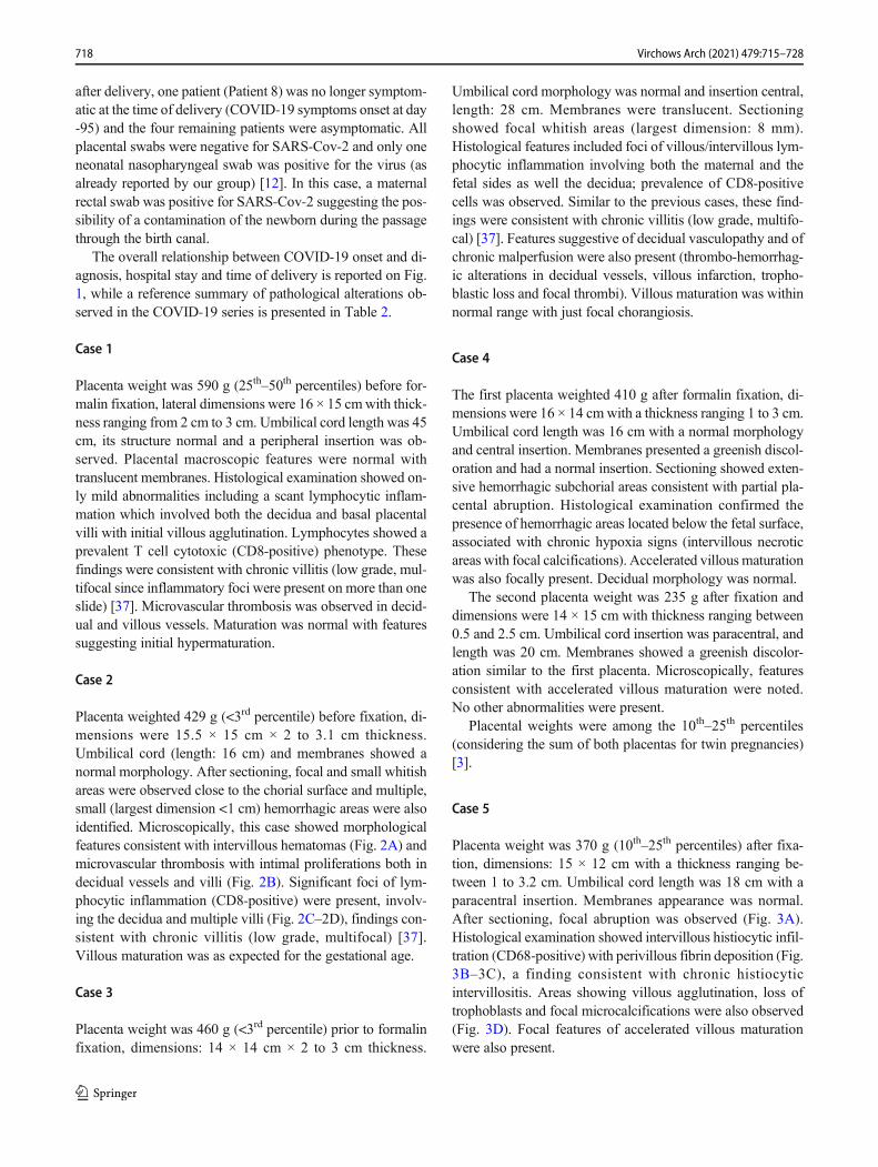
Placenta weight was 590 g (25th–50th percentiles) before for-malin fixation, lateral dimensions were 16 × 15 cmwith thick-ness ranging from 2 cm to 3 cm. Umbilical cord length was 45cm, its structure normal and a peripheral insertion was ob-served. Placental macroscopic features were normal withtranslucent membranes. Histological examination showed on-ly mild abnormalities including a scant lymphocytic inflam-mation which involved both the decidua and basal placentalvilli with initial villous agglutination. Lymphocytes showed aprevalent T cell cytotoxic (CD8-positive) phenotype. Thesefindings were consistent with chronic villitis (low grade, mul-tifocal since inflammatory foci were present on more than oneslide) [37]. Microvascular thrombosis was observed in decid-ual and villous vessels. Maturation was normal with featuressuggesting initial hypermaturation.
Case 2
Placenta weighted 429 g (<3rd percentile) before fixation, di-mensions were 15.5 × 15 cm × 2 to 3.1 cm thickness.Umbilical cord (length: 16 cm) and membranes showed anormal morphology. After sectioning, focal and small whitishareas were observed close to the chorial surface and multiple,small (largest dimension <1 cm) hemorrhagic areas were alsoidentified. Microscopically, this case showed morphologicalfeatures consistent with intervillous hematomas (Fig. 2A) andmicrovascular thrombosis with intimal proliferations both indecidual vessels and villi (Fig. 2B). Significant foci of lym-phocytic inflammation (CD8-positive) were present, involv-ing the decidua and multiple villi (Fig. 2C–2D), findings con-sistent with chronic villitis (low grade, multifocal) [37].Villous maturation was as expected for the gestational age.
Case 3
Placenta weight was 460 g (<3rd percentile) prior to formalinfixation, dimensions: 14 × 14 cm × 2 to 3 cm thickness.
Umbilical cord morphology was normal and insertion central,length: 28 cm. Membranes were translucent. Sectioningshowed focal whitish areas (largest dimension: 8 mm).Histological features included foci of villous/intervillous lym-phocytic inflammation involving both the maternal and thefetal sides as well the decidua; prevalence of CD8-positivecells was observed. Similar to the previous cases, these find-ings were consistent with chronic villitis (low grade, multifo-cal) [37]. Features suggestive of decidual vasculopathy and ofchronic malperfusion were also present (thrombo-hemorrhag-ic alterations in decidual vessels, villous infarction, tropho-blastic loss and focal thrombi). Villous maturation was withinnormal range with just focal chorangiosis.
Case 4
The first placenta weighted 410 g after formalin fixation, di-mensions were 16 × 14 cmwith a thickness ranging 1 to 3 cm.Umbilical cord length was 16 cm with a normal morphologyand central insertion. Membranes presented a greenish discol-oration and had a normal insertion. Sectioning showed exten-sive hemorrhagic subchorial areas consistent with partial pla-cental abruption. Histological examination confirmed thepresence of hemorrhagic areas located below the fetal surface,associated with chronic hypoxia signs (intervillous necroticareas with focal calcifications). Accelerated villous maturationwas also focally present. Decidual morphology was normal.
The second placenta weight was 235 g after fixation anddimensions were 14 × 15 cm with thickness ranging between0.5 and 2.5 cm. Umbilical cord insertion was paracentral, andlength was 20 cm. Membranes showed a greenish discolor-ation similar to the first placenta. Microscopically, featuresconsistent with accelerated villous maturation were noted.No other abnormalities were present.
Placental weights were among the 10th–25th percentiles(considering the sum of both placentas for twin pregnancies)[3].
Case 5
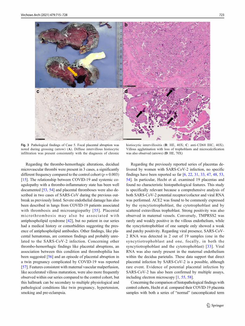
Placenta weight was 370 g (10th–25th percentiles) after fixa-tion, dimensions: 15 × 12 cm with a thickness ranging be-tween 1 to 3.2 cm. Umbilical cord length was 18 cm with aparacentral insertion. Membranes appearance was normal.After sectioning, focal abruption was observed (Fig. 3A).Histological examination showed intervillous histiocytic infil-tration (CD68-positive) with perivillous fibrin deposition (Fig.3B–3C), a finding consistent with chronic histiocyticintervillositis. Areas showing villous agglutination, loss oftrophoblasts and focal microcalcifications were also observed(Fig. 3D). Focal features of accelerated villous maturationwere also present.
718 Virchows Arch (2021) 479:715–728
Table 2 Pathological findings observed in the COVID-19 case series
Case Age Pathological findings
1 28 years • Weight: 590 g (before fixation), 25th–50th percentiles.• Dimensions: 16 cm × 15 cm × 2 cm to 3 cm thickness.• Translucent membranes.• Umbilical cord: 45 cm. Normal morphology and insertion.• Sectioning: no abnormalities.• Scant lymphocytic inflammation (CD8-positive), mainly located within the decidua and the basal half, consistent with
chronic villitis (low grade, multifocal).• Microvascular thrombosis in decidual vessels and villi.• Normal villous maturation with just focal signs of hypermaturation.
2 25 years • Weight: 429 g (before fixation), <3rd percentile.• Dimensions: 15.5 cm × 15 cm × 2 cm to 3.1 cm thickness.• Translucent membranes.• Umbilical cord: 16 cm. Normal morphology.• Sectioning: focal whitish areas close to the chorial surface and small (<1 cm) hemorrhagic foci.• Significant foci of lymphocytic inflammation (CD8-positive) involving the decidua andmultiple villi, consistent with chronic
villitis (low grade, multifocal).• Microvascular thrombosis in decidual vessels and villi.• Intervillous hematomas.• Normal villous maturation.
3 30 years • Weight: 370 g (before fixation), <3rd percentile.• Dimensions: 15 cm × 112 cm × 1 cm to 3 cm thickness.• Translucent membranes.• Umbilical cord: 28 cm. Normal morphology and insertion.• Sectioning: focal whitish areas (up to 8 mm).• Villous/intervillous lymphocytic (CD8-positive) inflammation involving the decidua and both the placental maternal and
fetal halves, consistent with chronic villitis (low grade, multifocal).• Thrombo-hemorrhagic alterations in decidual vessels, villous infarction, trophoblastic loss and focal thrombi, consistent with
decidual vasculopathy and chronic malperfusion.• Normal villous maturation with just focal chorangiosis.
4First placenta
35 years • Weight: 410 g (after fixation).• Dimensions: 16 cm × 14 cm × 1 cm to 3 cm thickness.• Greenish discoloration of membranes.• Umbilical cord: 16 cm. Normal morphology and insertion.• Sectioning: extensive subchorial hemorrhagic areas consistent with partial placental abruption.• Histological features consistent with partial placental abruption.• Hemorrhagic areas below the fetal surface, associated with chronic hypoxia signs (intervillous necrotic areas with focal
calcifications).• Focal features consistent with accelerated maturation.
4Second
placenta
• Weight: 235 g (after fixation).• Dimensions: 14 cm × 15 cm × 0.5 cm to 2.5 cm thickness.• Greenish discoloration of membranes.• Umbilical cord: 20 cm. Normal morphology and paracentral insertion.• Sectioning: no abnormalities.• Features consistent with accelerated maturation.
Placental weights were among the 10th–25th percentiles (considering the sum of both placentas for twin pregnancies).
5 39 years • Weight: 370 g (after fixation), 10th–25th percentiles.• Dimensions: 15 cm × 12 cm × 1 cm to 3.2 cm thickness.• Translucent membranes.• Umbilical cord: 18 cm. Normal morphology and paracentral insertion.• Sectioning: focal placental abruption.• Intervillous histiocytic infiltration (CD68-positive) with perivillous fibrin deposition.• Areas with villous conglutination, loss of trophoblasts and focal microcalcifications.• Focal accelerated villous maturation.
6 32 years • Weight: 490 grams (before fixation), 3rd–10th percentiles.• Dimensions: 17 cm × 17 cm × 1 cm to 3 cm thickness.• Translucent membranes.• Umbilical cord: 50 cm. Normal morphology and central insertion.• Sectioning: whitish areas (15 mm).• Irregular villous maturation with focal chorangiosis.
719Virchows Arch (2021) 479:715–728
Case 6
Placenta weighted 490 g (3rd–10th percentiles) before fixation,dimensions: 17 × 17 cm with a thickness ranging between 1 and3 cm. Placental macroscopic features were normal with translu-cent membranes. Umbilical cord length was 50 cm, with normalmorphology and central insertion. The histological examinationshowed irregular villous maturation with focal chorangiosis,intervillous infarction and villous calcifications.
Case 7
Placenta weight before fixation was 400 g (<3rd percen-tile), while its dimensions were 16 × 13 × 1 to 4 cmthickness. Membranes were translucent. The umbilicalcord exhibited a normal morphology and marginal
insertion with a total length of 18 cm. At sectioning,a focal congestion area was observed. Microscopically,features suggestive of umbilical blood flow restriction,like widespread chorangiosis, avascular villi with sclero-sis and vascular stenosis together with hypertrophy ofstem villi were noticed.
Case 8
Before fixation placenta weighted 520 g (10th–25th per-centiles), with total dimensions of 16 × 14 cm × 2 to3.5 cm thickness. Membranes were yellowish and um-bilical cord showed normal morphology and marginalinsertion, with a length of 37 cm. No abnormalitieswere microscopically observed, except for focal villoushypermaturation and focal villous agglutination.
7 37 years • Weight: 400 g (before fixation), <3rd percentile.• Dimensions: 16 cm × 13 cm × 1 cm to 4 cm thickness.• Translucent membranes.• Umbilical cord: 18 cm. Normal morphology and marginal insertion.• Sectioning: focal congestion area.• Widespread chorangiosis.• Avascular villi with sclerosis.• Vascular stenosis and hypertrophy of stem villi.• Findings consistent with umbilical blood flow restriction.
8 43 years • Weight: 520 g (before fixation), 10th–25th percentiles.• Dimensions: 16 cm × 14 cm × 2 cm to 3.5 cm thickness.• Yellowish membranes.• Umbilical cord: 37 cm. Normal morphology and marginal insertion.• Sectioning: no specific alterations.• Focal villous hypermaturation.• Focal villous agglutination.
9 36 years • Monochorionic diamniotic.• Weight: 665 g (before fixation), 3rd–10th percentiles for twin deliveries.• Dimensions: 19 cm × 16 cm × 3 cm to 4.5 cm thickness.• Translucent membranes.• First umbilical cord: 23 cm. Hypocoiled with focal edema and marginal insertion.• Second umbilical cord: 20 cm. Hypocoiled with marginal insertion.• Sectioning: no specific alterations.• Lymphocytic infiltration consistent with chronic villitis (low grade, multifocal).• Accelerated villous maturation associated with focal hypoplasia and increase of syncytial nodes.• Focal villous sclerosis consistent with fetal thrombotic vasculopathy.• Focal hemorrhagic area within basal decidua consistent with recent abruption.
10 32 years • Weight: 440 g (before fixation), 3rd–10th percentiles.• Dimensions: 16 cm × 15 cm × 1 cm to 3 cm thickness.• Translucent membranes.• Umbilical cord: 40 cm. Normal morphology and marginal insertion.• Sectioning: no specific alterations.• Villous hypoplasia.• Focal sclerosis consistent with previous placental infarction.
720 Virchows Arch (2021) 479:715–728
Case 9
This placenta was monochorionic diamniotic, with a totalweight of 665 g before fixation, (3rd–10th percentiles for twindeliveries) and dimensions of 19 × 16 cm with a thicknessranging between 3 and 4.5 cm. Membranes were translucent.The two umbilical cords were 23 and 20 cm in length, bothhypocoiled and with marginal insertion. The first was charac-terized by focal edema. Histological examination showed alymphocytic infiltration consistent with chronic villitis (lowgrade, multifocal). Accelerated villous maturation associatedwith focal hypoplasia and increase of syncytial nodes werealso detectable. Features suggestive of fetal thrombotic vascu-lopathy like focal villous sclerosis were observed. Focal hem-orrhagic area within basal decidua consistent with recentabruption was noted.
Case 10
Placenta weighted 440 g (3rd–10th percentiles) before fixation,with dimensions 16 × 15 cm × 1 to 3 cm thickness.Membranes were translucent. Umbilical cord, with normalmorphology and marginal insertion, was long 40 cm.Microscopically, this case showed villous hypoplasia and fo-cal sclerosis, a finding consistent with previous placentalinfarction.
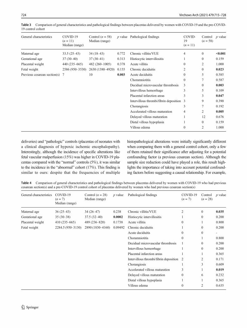
Comparison with pre-COVID-19 control cohorts
To ascertain the specificity of the observed alterations, a con-trol cohort of 58 pre-COVID-19 placentas was selectedamong placentas examined between March and July 2019,and this cohort was compared with COVID-19 placentas
(Table 3). General characteristics were similar between thetwo groups (p > 0.05) except for the rate of previous cesareansections which was higher in COVID-19 cases (p = 0.003).Regarding the observed pathological alterations, the incidenceof VUE (p < 0.001), chronic deciduitis (p = 0.023), microvas-cular thrombosis (p = 0.003), presence of infarction areas (p =0.047) and of accelerated villous maturation (p = 0.005)showed higher frequencies in placentas delivered by womenwith COVID-19.
Considering the significant difference in terms of previouscesarean sections, the COVID-19 series was compared with afurther pre-COVID-19 control series with this characteristic (n= 28). Characteristics of this cohort are reported in Table 4.Only differences in terms of presence of chronic villitis/VUE(p = 0.035) and of accelerated villous maturation (p = 0.019)retained their statistically significance.
Discussion
In this study we report the pathological findings observed ineleven consecutive placentas delivered by ten women affectedby COVID-19. Inflammatory and thrombo-hemorrhagic alter-ations were the most frequent and peculiar pathologicalchanges we observed in our series compared to control sam-ples, but only chronic villitis/VUE and accelerated villousmaturation retained statistical significance after restrictingthe analysis to placentas delivered after a previous cesareansection.
In 5 cases we found microscopic signs of inflammationinvolving the decidua and the chorial villi: four out of fivecases showed a CD8-positive T cell lymphocytic infiltrate,while in the remaining case a chronic histiocytic intervillositis
Fig. 1 Outline of COVID-19 clinical course in symptomatic patients andplacental pathological findings. Findings consistent with chronic villitiswere found in patients with shorter COVID-19 duration and mildersymptoms (blue), while chronic histiocytic intervillositis was diagnosedin Case 5 after long-standing and more severe symptoms (red). Patient 4
and patient 8 showed unspecific placental findings (yellow). Black arrow(Day 0): delivery time. Black rectangles: hospitalization. Colored boxes:days with COVID-19 symptoms. Striped boxes: time of nasopharyngealswab
721Virchows Arch (2021) 479:715–728
was identified. None of these five patients had comorbidities,other than SARS-CoV-2 infection, which could justify thepresence of placental inflammation. The presence of an in-flammatory lymphocytic infiltrate (cytotoxic CD8+ T celllymphocytes in particular, as observed in our series) and ofvillous damage is consistent with the diagnosis of chronicvillitis [10, 39]. Chronic villitis etiology can be related to anundergoing infection, although in most cases, it is not possibleto identify the specific etiopathogenetic agent: these cases aredefined as “villitis of unknown etiology” (VUE) and can befound in 2–33.8% of placentas [38]. Compared to our controlcohort, COVID-19 positive cases had a statistically significanthigher frequency of this condition (p < 0.001).
Viruses are the most frequent cause of chronic villitis sec-ondary to maternal infections: varicella zoster, herpes simplexvirus, cytomegalovirus, and influenza A/H1N1, have beenfound to be associated with this condition [4, 9, 45, 56].Little is known about the association between placental pa-thology and coronavirus infection. Seven placentas fromcoronavirus-infected women during the 2002–2004 SARSoutbreak have been previously described [49]: histologicalexamination found no abnormalities in 2/7, an increase insubchorionic and intervillous fibrin in 3/7, andmassive throm-botic vasculopathy associated with intra-uterine growth re-striction (IUGR) in the remaining 2/7 cases. No signs ofchronic villitis were detected in any case and the same is true
for MERS infections [36]. Moreover, considering experiencesderived from influenza A/H1N1 infections in pregnancy,characterized by the activation of cellular immune responsewith release of inflammatory cytokines that lead to indirectplacental damage [45], it stands to reason that SARS-CoV-2may play an equivalent role in these histological examina-tions, because of intrinsic active virus replication and/orthrough indirect activity of inflammatory cytokines [43].
In the remaining case with inflammatory abnormalities,features consistent with chronic histiocytic intervillositis wereidentified. This rare entity is usually associated with maternalimmunologic conditions like systemic lupus erythematosus,lupus anticoagulant and antiphospholipid syndrome [44, 46],while this patient’s significant comorbidities (chronic hyper-tension, severe obesity, and gestational diabetes) are not.Despite the different types of inflammatory infiltrate (histio-cytic versus lymphocytic) and of etiologies (chronic histiocyt-ic intervillositis is usually not related to viral infections); thispathological entity is frequently associated with chronicvillitis (30 to 50% of cases), supporting a potential overlapin terms of involved immunologic pathways [41, 50].Moreover, alveolar histiocytic infiltration is a frequentautoptic hallmark of SARS-CoV-2 infection [15]. Of note,despite its rarity, other cases of histiocytic intervillositis havebeen reported in placentas delivered by a SARS-CoV-2-positive women [34, 47].
Fig. 2 Pathological findings of Case 2. Placental intervillous hematomas(arrows) (A: HE, 20X) and microvascular thrombosis (arrows) (B: HE,100X) were observed. Significant foci of decidual and villous
inflammation were also present (arrows) (C: anti-CD8 IHC, 30X; D:anti-CD8 IHC, 80X), consistent with multifocal, low grade chronic villitis
722 Virchows Arch (2021) 479:715–728
Regarding the thrombo-hemorrhagic alterations, decidualmicrovascular thrombi were present in 3 cases, a significantlydifferent frequency compared to the control cohort (p = 0.003)[15]. The relationship between COVID-19 and systemic co-agulopathy with a thrombo-inflammatory state has been welldocumented [53, 54] and placental thromboses were also de-scribed in two cases of SARS-CoV during the previous out-break as previously listed. Severe endothelial damage has alsobeen described in lungs from COVID-19 patients associatedwith thrombosis and microangiopathy [55]. Placentalm ic ro th rombos i s may a l so be as soc i a t ed wi thantiphospholipid syndrome [42], but no patient in our serieshad a medical history or comorbidities suggesting the pres-ence of antiphospholipid antibodies. Other findings, like pla-cental hematomas, are common findings and probably unre-lated to the SARS-CoV-2 infection. Concerning otherthrombo-hemorrhagic findings like placental abruptions, anassociation between this condition and thrombophilia hasbeen suggested [56] and an episode of placental abruption ina twin pregnancy complicated by COVID-19 was reported[57]. Features consistent with maternal vascular malperfusion,like accelerated villous maturation, were also more frequentlyobserved within our series compared to the control cohort, butthis hallmark can be secondary to multiple physiological andpathological conditions like twin pregnancy, hypertension,smoking and pre-eclampsia.
Regarding the previously reported series of placentas de-livered by women with SARS-CoV-2 infection, no specificfindings have been reported so far [6, 22, 31, 33, 47, 48, 53,54]. In particular, Hecht et al. examined 19 placentas andfound no characteristic histopathological features. This studyis specifically relevant because a comprehensive analysis ofboth SARS-CoV-2 potential receptor/cofactor and viral RNAwas performed. ACE2 was found to be commonly expressedby the syncytiotrophoblast, the cytotrophoblast and byscattered extravillous trophoblast. Strong positivity was alsoobserved in maternal vessels. Conversely, TMPRSS2 wasrarely and weakly positive in the villous endothelium, whilethe syncytiotrophoblast of one sample only showed a weakand patchy positivity. Regarding viral presence, SARS-CoV-2 RNA was detected in 2 out of 19 samples (one in thesyncytiotrophoblast and one, focally, in both thesyncytiotrophoblast and the cytotrophoblast) [33]. ViralRNA was also rarely present in the maternal endotheliumwithin the decidua parietalis. These data support that directplacental infection by SARS-CoV-2 is a possible, althoughrare event. Evidence of potential placental infection bySARS-CoV-2 has also been confirmed by multiple assays,including electron microscopy [1, 55, 58].
Concerning the comparison of histopathological findingswithcontrol cohorts, Hecht et al. compared their COVID-19 placentasamples with both a series of “normal” (uncomplicated term
Fig. 3 Pathological findings of Case 5. Focal placental abruption wasnoted during grossing (arrow) (A). Diffuse intervillous histiocyticinfiltration was present consistently with the diagnosis of chronic
histiocytic intervillositis (B: HE, 40X; C: anti-CD68 IHC, 40X).Villous agglutination with loss of trophoblasts and microcalcificationwas also observed (arrows) (D: HE, 70X)
723Virchows Arch (2021) 479:715–728
deliveries) and “pathologic” controls (placentas of neonates witha clinical diagnosis of hypoxic ischemic encephalopathy).Interestingly, although the incidence of specific alterations likefetal vascular malperfusion (15%) was higher in COVID-19 pla-centas compared with the “normal” controls (5%), it was similarto the incidence in the “abnormal” cohort (17%). This finding issimilar to ours: despite that the frequencies of multiple
histopathological alterations were initially significantly differentwhen comparing them with a general control cohort, only a fewof them retained their significance after adjusting for a potentialconfounding factor (a previous cesarean section). Although thesample size reduction could have played a role, this result high-lights the importance of taking into account potential confound-ing factors before suggesting a causal relationship. For example,
Table 3 Comparison of general characteristics and pathological findings between placentas delivered by women with COVID-19 and the pre-COVID-19 control cohort
General characteristics COVID-19(n = 11)Median (range)
Table 4 Comparison of general characteristics and pathological findings between placentas delivered by women with COVID-19 who had previouscesarean section(s) and a pre-COVID-19 control cohort of placentas delivered by women who had previous cesarean section(s)
General characteristics COVID-19(n = 7)Median (range)
abnormal placental implantation is another potential confoundingvariable which should be considered in future studies. In ourcohort, only one case presented this finding (Case 10 –Marginal placenta previa).
Nevertheless, we detected significantly higher frequenciesof chronic villitis/VUE and accelerated villous maturationeven after accounting for a previous cesarean section.Recently, also Patberg et al. observed a higher incidence ofvillitis of unknown etiology (20.8 versus 7.1%) comparedwith a control group of term patients without COVID-19[51]. This result was also confirmed in a subgroup analysiscomparing asymptomatic COVID-19 patients with negativecontrols, but it did not reach statistical significance in a mul-tivariable comparison adjusting for multiple clinical character-istics. This finding further enhances the importance of analyz-ing larger series of cases, possibly through dedicated meta-analyses, and adjusting for the effect of potential confounders.Regarding vascular/thrombotic alterations, 2 out of the 3 de-cidual microvascular thromboses we observed amongCOVID-19 cases were present in placentas delivered bywom-en who had no previous cesarean section and thus they wereindependent of this potentially confounding variable.Therefore, the lack of significance observed by restrictingthe analysis to COVID-19 placentas delivered after a previouscesarean section could be at least partially due to sample sizereduction.
Concerning our knowledge about the clinical outcomes ofpregnancy in COVID-19 patients, Chen et al. [17] reported theoutcomes of a relatively large series of pregnant women af-fected by COVID-19 observing a high rate (14/68, 21%) ofpreterm delivery. A systematic review evaluating 41 pregnan-cies affected by COVID-19 confirmed this observation,reporting preterm birth (<37 weeks) in above 40% of preg-nancies [25]. Finally, even the largest systematic review avail-able on 441 pregnant patients with COVID-19 also found thatpreterm birth is the most common adverse neonatal outcome(reported in the 21% of women who delivered) [29].However, it should be noted that this higher rate of pretermdeliveries could be at least partially explained by a differentattitude during the early months of the pandemic favoringearly delivery by cesarean section because of fear of potentialCOVID-19 complications.
Finally, the limited sample size and the intrinsically unspe-cific etiology of most of the observed placental alterations arethe main limitations of our study as well of many of thosepublished so far. Larger, multi-institutional prospective stud-ies taking into account the whole set of variables related toSARS-CoV-2 infection (timing, severity, duration…), placen-tal histopathological findings, potential confounders andmaterno-fetal outcomes are warranted to provide a definitiveassessment of the relationship between COVID-19 andpregnancy.
Author contribution GB, PC and CB designed the research protocol; allauthors contributed to data collection and analysis; LB, FB, GB, AC andSC drafted the paper; all authors edited the paper and approved the finalversion.
Funding The present study was kindly supported by a grant from “Bancadel Piemonte” to PC dedicated to COVID-19 research and by the ItalianMinistry of University and Research/University of Turin (ex 60%) toLB. Open access funding provided by Università degli Studi di Torinowithin the CRUI-CARE Agreement.
Declarations
Conflict of interest The authors declare no competing interests.
Open Access This article is licensed under a Creative CommonsAttribution 4.0 International License, which permits use, sharing, adap-tation, distribution and reproduction in any medium or format, as long asyou give appropriate credit to the original author(s) and the source, pro-vide a link to the Creative Commons licence, and indicate if changes weremade. The images or other third party material in this article are includedin the article's Creative Commons licence, unless indicated otherwise in acredit line to the material. If material is not included in the article'sCreative Commons licence and your intended use is not permitted bystatutory regulation or exceeds the permitted use, you will need to obtainpermission directly from the copyright holder. To view a copy of thislicence, visit http://creativecommons.org/licenses/by/4.0/.
References
1. Algarroba GN, Hanna NN, Rekawek P, Vahanian SA, Khullar P,Palaia T, Peltier MR, Chavez MR, Vintzileos AM (2020)Confirmatory evidence of the visualization of severe acute respira-tory syndrome coronavirus 2 invading the human placenta usingelectron microscopy. Am J Obstet Gynecol 223:953–954. https://doi.org/10.1016/j.ajog.2020.08.106
2. Algarroba GN, Hanna NN, Rekawek P, Vahanian SA, Khullar P,Palaia T, Peltier MR, Chavez MR, Vintzileos AM (2020)Confirmatory evidence of visualization of SARS-CoV-2 virus in-vading the human placenta using electron microscopy. Am J ObstetGynecol 223:953–954. https://doi.org/10.1016/j.ajog.2020.08.106
3. Almog B, Shehata F, Aljabri S, Levin I, Shalom-Paz E, Shrim A(2011) Placenta weight percentile curves for singleton and twinsdeliveries. Placenta 32:58–62. https://doi.org/10.1016/j.placenta.2010.10.008
4. Altshuler G, Russell P (1975) The human placental villitides: areview of chronic intrauterine infection. Curr Top Pathol 60:64–112
5. Alzamora MC, Paredes T, Caceres D, Webb CM, Valdez LM, LaRosa M (2020) Severe COVID-19 during pregnancy and possiblevertical transmission. Am J Perinatol 37:861–865. https://doi.org/10.1055/s-0040-1710050
6. Baergen RN, Heller DS (2020) Placental pathology in Covid-19positive mothers: preliminary findings. Pediatr Dev Pathol 23:177–180. https://doi.org/10.1177/1093526620925569
7. BaudD, GreubG, Favre G, Gengler C, Jaton K,Dubruc E, Pomar L(2020) Second-trimester miscarriage in a pregnant woman withSARS-CoV-2 infection. JAMA. https://doi.org/10.1001/jama.2020.7233
8. Beck S, Wojdyla D, Say L, Betran AP, Merialdi M, Requejo JH,Rubens C, Menon R, Van Look PF (2010) The worldwide inci-dence of preterm birth: a systematic review of maternal mortalityand morbidity. Bull World Health Organ 88:31–38. https://doi.org/10.2471/BLT.08.062554
9. Becroft DM, Thompson JM, Mitchell EA (2005) Placental villitisof unknown origin: epidemiologic associations. Am J ObstetGynecol 192:264–271. https://doi.org/10.1016/j.ajog.2004.06.062
10. Boog G (2008) Chronic villitis of unknown etiology. Eur J ObstetGynecol Reprod Biol 136:9–15. https://doi.org/10.1016/j.ejogrb.2007.06.018
11. Breslin N, Baptiste C, Gyamfi-Bannerman C,Miller R,Martinez R,Bernstein K, Ring L, Landau R, Purisch S, Friedman AM, Fuchs K,Sutton D, AndrikopoulouM, Rupley D, Sheen JJ, Aubey J, Zork N,Moroz L, Mourad M, Wapner R, Simpson LL, D'Alton ME,Goffman D (2020) COVID-19 infection among asymptomaticand symptomatic pregnant women: two weeks of confirmed pre-sentations to an affiliated pair of New York City hospitals. Am JObstet Gynecol MFM:100118. https://doi.org/10.1016/j.ajogmf.2020.100118
12. Carosso A, Cosma S, Borella F, Marozio L, Coscia A, Ghisetti V,Di Perri G, Benedetto C (2020) Pre-labor anorectal swab for SARS-CoV-2 in COVID-19 patients: is it time to think about it? Eur JObstet Gynecol Reprod Biol 249:98–99. https://doi.org/10.1016/j.ejogrb.2020.04.023
13. Carosso A, Cosma S, Serafini P, Benedetto C, Mahmood T (2020)How to reduce the potential risk of vertical transmission of SARS-CoV-2 during vaginal delivery? Eur J Obstet Gynecol Reprod Biol250:246–249. https://doi.org/10.1016/j.ejogrb.2020.04.065
14. Carosso AR, Cosma S, Benedetto C (2020) Vaginal delivery inCOVID-19 pregnant women: anorectum as a potential alternativeroute of SARS-CoV-2 transmission. Am J Obstet Gynecol 223:612. https://doi.org/10.1016/j.ajog.2020.06.012
15. Carsana L, Sonzogni A, Nasr A, Rossi R, Pellegrinelli A, Zerbi P,Rech R, Colombo R, Antinori S, Corbellino M, Galli M, Catena E,Tosoni A, Gianatti A, Nebuloni M (2020) Pulmonary post-mortemfindings in a large series of COVID-19 cases from Northern ItalymedRxiv:2020.2004.2019.20054262. https://doi.org/10.1101/2020.04.19.20054262
16. Chen H, Guo J, Wang C, Luo F, Yu X, ZhangW, Li J, Zhao D, XuD, Gong Q, Liao J, Yang H, Hou W, Zhang Y (2020) Clinicalcharacteristics and intrauterine vertical transmission potential ofCOVID-19 infection in nine pregnant women: a retrospective re-view of medical records. Lancet 395:809–815. https://doi.org/10.1016/S0140-6736(20)30360-3
17. Chen L, Li Q, Zheng D, Jiang H, Wei Y, Zou L, Feng L, Xiong G,Sun G, Wang H, Zhao Y, Qiao J (2020) Clinical characteristics ofpregnant women with Covid-19 in Wuhan, China. N Engl J Med382:e100. https://doi.org/10.1056/NEJMc2009226
18. Chen S, Huang B, Luo DJ, Li X, Yang F, Zhao Y, Nie X, HuangBX (2020) Pregnant women with new coronavirus infection: a clin-ical characteristics and placental pathological analysis of threecases. Zhonghua Bing Li Xue Za Zhi 49:E005. https://doi.org/10.3760/cma.j.cn112151-20200225-00138
19. Collin J, Bystrom E, Carnahan A, Ahrne M (2020) Pregnant andpostpartumwomenwith SARS-CoV-2 infection in intensive care inSweden. Acta Obstet Gynecol Scand 99:819–822. https://doi.org/10.1111/aogs.13901
20. Cosma S, Borella F, Carosso A, Sciarrone A, Cusato J, Corcione S,Mengozzi G, Preti M, Katsaros D, Di Perri G, Benedetto C (2020)The "scar" of a pandemic: cumulative incidence of COVID-19 dur-ing the first trimester of pregnancy. J Med Virol 93:537–540.https://doi.org/10.1002/jmv.26267
21. Cosma S, Carosso AR, Cusato J, Borella F, Carosso M, Bovetti M,Filippini C, D'Avolio A, Ghisetti V, Dip G, Benedetto C (2020)COVID-19 and first trimester spontaneous abortion: a case-control
study of 225 pregnant patients. Am J Obstet Gynecol 224:391.e1–391.e7. https://doi.org/10.1016/j.ajog.2020.10.005
22. Cribiu FM, Croci GA, Del Gobbo A, Rizzuti T, Iurlaro E, TondoM, Viscardi A, Bosari S, Ferrero S (2020) Histological characteri-zation of placenta in COVID19 pregnant women. Eur J ObstetGynecol Reprod Biol 252:619–621. https://doi.org/10.1016/j.ejogrb.2020.06.041
23. Dashraath P, Wong JLJ, Lim MXK, Lim LM, Li S, Biswas A,Choolani M, Mattar C, Su LL (2020) Coronavirus disease 2019(COVID-19) pandemic and pregnancy. Am J Obstet Gynecol222:521–531. https://doi.org/10.1016/j.ajog.2020.03.021
24. Della Gatta AN, Rizzo R, Pilu G, Simonazzi G (2020) COVID19during pregnancy: a systematic review of reported cases. Am JObstet Gynecol 223:36–41. https://doi.org/10.1016/j.ajog.2020.04.013
25. Di Mascio D, Khalil A, Saccone G, Rizzo G, Buca D, Liberati M,Vecchiet J, Nappi L, Scambia G, Berghella V, D'Antonio F (2020)Outcome of coronavirus spectrum infections (SARS, MERS,COVID 1 -19) during pregnancy: a systematic review and meta-analysis Am J Obstet Gynecol MFM:100107. https://doi.org/10.1016/j.ajogmf.2020.100107
26. Dong L, Tian J, He S, Zhu C, Wang J, Liu C, Yang J (2020)Possible vertical transmission of SARS-CoV-2 from an infectedmother to her newborn. JAMA. https://doi.org/10.1001/jama.2020.4621
27. Facchetti F, Bugatti M, Drera E, Tripodo C, Sartori E, Cancila V,Papaccio M, Castellani R, Casola S, Boniotti MB, Cavadini P,Lavazza A (2020) SARS-CoV2 vertical transmission with adverseeffects on the newborn revealed through integrated immunohisto-chemical, electron microscopy and molecular analyses of Placenta.EBioMedicine 59:102951. https://doi.org/10.1016/j.ebiom.2020.102951
28. Ferrazzi EM, Frigerio L, Cetin I, Vergani P, Spinillo A, Prefumo F,Pellegrini E, Gargantini G (2020) COVID-19 Obstetrics TaskForce, Lombardy, Italy: executive management summary and shortreport of outcome. Int J Gynaecol Obstet 149:377–378. https://doi.org/10.1002/ijgo.13162
29. Gajbhiye R, Modi D, Mahale S (2020) Pregnancy outcomes, new-born complications and maternal-fetal transmission of SARS-CoV-2 in women with COVID-19: a systematic review of 441 casesmedRxiv:2020.2004.2011.20062356. https://doi.org/10.1101/2020.04.11.20062356
30. Garcia D, Erkan D (2018) Diagnosis and management of theantiphospholipid syndrome. N Engl J Med 378:2010–2021.https://doi.org/10.1056/NEJMra1705454
31. Gulersen M, Prasannan L, Tam HT, Metz CN, Rochelson B,Meirowitz N, Shan W, Edelman M, Millington KA (2020)Histopathological evaluation of placentas after diagnosis of mater-nal SARS-CoV-2 infection Am J Obstet Gynecol MFM:100211.https://doi.org/10.1016/j.ajogmf.2020.100211
32. Hantoushzadeh S, Shamshirsaz AA, Aleyasin A, Seferovic MD,Aski SK, Arian SE, Pooransari P, Ghotbizadeh F, Aalipour S,Soleimani Z, Naemi M, Molaei B, Ahangari R, Salehi M, OskoeiAD, Pirozan P, Darkhaneh RF, Laki MG, Farani AK, Atrak S, MiriMM, Kouchek M, Shojaei S, Hadavand F, Keikha F, Hosseini MS,Borna S, Ariana S, Shariat M, Fatemi A, Nouri B, NekooghadamSM, Aagaard K (2020) Maternal death due to COVID-19 disease.Am J Obstet Gynecol 223:109.e1–109.e16. https://doi.org/10.1016/j.ajog.2020.04.030
33. Hecht JL, Quade B, Deshpande V, Mino-Kenudson M, Ting DT,Desai N, Dygulska B, Heyman T, Salafia C, Shen D, Bates SV,Roberts DJ (2020) SARS-CoV-2 can infect the placenta and is notassociated with specific placental histopathology: a series of 19placentas from COVID-19-positive mothers. Mod Pathol 33:2092–2103. https://doi.org/10.1038/s41379-020-0639-4
34. Hosier H, Farhadian SF,Morotti RA, DeshmukhU, Lu-Culligan A,Campbell KH, Yasumoto Y, Vogels CB, Casanovas-Massana A,Vijayakumar P, Geng B, Odio CD, Fournier J, Brito AF, Fauver JR,Liu F, Alpert T, Tal R, Szigeti-Buck K, Perincheri S, Larsen C,Gariepy AM, Aguilar G, Fardelmann KL, Harigopal M, TaylorHS, Pettker CM, Wyllie AL, Cruz CD, Ring AM, Grubaugh ND,Ko AI, Horvath TL, Iwasaki A, Reddy UM, Lipkind HS (2020)SARS-CoV-2 infection of the placenta. J Clin Invest 130:4947–4953. https://doi.org/10.1172/JCI139569
35. Huang C,Wang Y, Li X, Ren L, Zhao J, HuY, Zhang L, Fan G, XuJ, Gu X, Cheng Z, Yu T, Xia J,Wei Y,WuW, Xie X, YinW, Li H,Liu M, Xiao Y, Gao H, Guo L, Xie J, Wang G, Jiang R, Gao Z, JinQ, Wang J, Cao B (2020) Clinical features of patients infected with2019 novel coronavirus in Wuhan, China. Lancet 395:497–506.https://doi.org/10.1016/S0140-6736(20)30183-5
36. Jeong SY, Sung SI, Sung JH, Ahn SY, Kang ES, Chang YS, ParkWS, Kim JH (2017) MERS-CoV infection in a pregnant woman inKorea. J Korean Med Sci 32:1717–1720. https://doi.org/10.3346/jkms.2017.32.10.1717
37. Khong TY, Mooney EE, Ariel I, Balmus NC, Boyd TK, BrundlerMA, Derricott H, Evans MJ, Faye-Petersen OM, Gillan JE, HeazellAE, Heller DS, Jacques SM, Keating S, Kelehan P, Maes A,McKay EM, Morgan TK, Nikkels PG, Parks WT, Redline RW,Scheimberg I, Schoots MH, Sebire NJ, Timmer A, Turowski G,van der Voorn JP, van Lijnschoten I, Gordijn SJ (2016) Samplingand definitions of placental lesions: Amsterdam PlacentalWorkshop Group Consensus Statement. Arch Pathol Lab Med140:698–713. https://doi.org/10.5858/arpa.2015-0225-CC
38. Khong TY, Mooney EE, Nikkels PGJ, Morgan TK, Gordijn SJ(2019) Pathology of the placenta: a practical guide. Springer,Cham, Switzerland
39. Kim CJ, Romero R, Chaemsaithong P, Kim JS (2015) Chronicinflammation of the placenta: definition, classification, pathogene-sis, and clinical significance. Am J Obstet Gynecol 213:S53–S69.https://doi.org/10.1016/j.ajog.2015.08.041
40. Knight M, Bunch K, Vousden N, Morris E, Simpson N, Gale C,O'Brien P, Quigley M, Brocklehurst P, Kurinczuk JJ (2020)Characteristics and outcomes of pregnant women hospitalised withconfirmed SARS-CoV-2 infection in the UK: a national cohortstudy using the UK Obstetric Surveillance System (UKOSS)medRxiv:2020.2005.2008.20089268. https://doi.org/10.1101/2020.05.08.20089268
42. Li M, Chen L, Zhang J, Xiong C, Li X (2020) The SARS-CoV-2receptor ACE2 expression of maternal-fetal interface and fetal or-gans by single-cell transcriptome study. PLoS One 15:e0230295.https://doi.org/10.1371/journal.pone.0230295
43. Liu H, Wang LL, Zhao SJ, Kwak-Kim J, Mor G, Liao AH (2020)Why are pregnant women susceptible to COVID-19? An immuno-logical viewpoint. J Reprod Immunol 139:103122. https://doi.org/10.1016/j.jri.2020.103122
44. Marchaudon V, Devisme L, Petit S, Ansart-Franquet H, Vaast P,Subtil D (2011) Chronic histiocytic intervillositis of unknown eti-ology: clinical features in a consecutive series of 69 cases. Placenta32:140–145. https://doi.org/10.1016/j.placenta.2010.11.021
45. Meijer WJ, Wensing AM, Bruinse HW, Nikkels PG (2014) Highrate of chronic villitis in placentas of pregnancies complicated byinfluenza A/H1N1 infection. Infect Dis Obstet Gynecol 2014:768380–768385. https://doi.org/10.1155/2014/768380
46. Mekinian A, Costedoat-Chalumeau N, Masseau A, Botta A,Chudzinski A, Theulin A, Emmanuelli V, Hachulla E, De CarolisS, Revaux A, Nicaise P, Cornelis F, Subtil D, Montestruc F,Bucourt M, Chollet-Martin S, Carbillon L, Fain O, Snfmi, the
European Forum of APS (2015) Chronic histiocytic intervillositis:outcome, associated diseases and treatment in a multicenter pro-spective study. Autoimmunity 48:40–45. https://doi.org/10.3109/08916934.2014.939267
47. Mongula JE, Frenken MWE, van Lijnschoten G, Arents NLA, deWit-Zuurendonk LD, Schimmel-de Kok APA, van RunnardHeimel PJ, Porath MM, Goossens S (2020) COVID-19 duringpregnancy: non-reassuring fetal heart rate, placental pathologyand coagulopathy Ultrasound. Obstet Gynecol 56:773–776.https://doi.org/10.1002/uog.22189
48. Mulvey JJ, Magro CM, Ma LX, Nuovo GJ, Baergen RN (2020)Analysis of complement deposition and viral RNA in placentas ofCOVID-19 patients. Ann Diagn Pathol 46:151530. https://doi.org/10.1016/j.anndiagpath.2020.151530
49. Ng WF, Wong SF, Lam A, Mak YF, Yao H, Lee KC, Chow KM,Yu WC, Ho LC (2006) The placentas of patients with severe acuterespiratory syndrome: a pathophysiological evaluation. Pathology38:210–218. https://doi.org/10.1080/00313020600696280
50. Parant O, Capdet J, Kessler S, Aziza J, Berrebi A (2009) Chronicintervillositis of unknown etiology (CIUE): relation between pla-cental lesions and perinatal outcome. Eur J Obstet Gynecol ReprodBiol 143:9–13. https://doi.org/10.1016/j.ejogrb.2008.06.012
51. Patberg ET, Adams T, Rekawek P, Vahanian SA, Akerman M,Hernandez A, Rapkiewicz AV, Ragolia L, Sicuranza G, ChavezMR, Vintzileos AM, Khullar P (2020) Coronavirus disease 2019infection and placental histopathology in women delivering at term.Am J Obstet Gynecol 224:382.e1–382.e18. https://doi.org/10.1016/j.ajog.2020.10.020
52. Schett G, Sticherling M, Neurath MF (2020) COVID-19: risk forcytokine targeting in chronic inflammatory diseases? Nat RevImmunol 20:271–272. https://doi.org/10.1038/s41577-020-0312-7
53. Shanes ED,Mithal LB, Otero S, Azad HA,Miller ES, Goldstein JA(2020) Placental pathology in COVID-19. Am J Clin Pathol 154:23–32. https://doi.org/10.1093/ajcp/aqaa089
54. Sharps MC, Hayes DJL, Lee S, Zou Z, Brady CA, Almoghrabi Y,Kerby A, Tamber KK, Jones CJ, Adams Waldorf KM, HeazellAEP (2020) A structured review of placental morphology and his-topathological lesions associated with SARS-CoV-2 infection.Placenta 101:13–29. https://doi.org/10.1016/j.placenta.2020.08.018
55. Taglauer E, Benarroch Y, Rop K, Barnett E, Sabharwal V,Yarrington C, Wachman EM (2020) Consistent localization ofSARS-CoV-2 spike glycoprotein and ACE2 over TMPRSS2 pre-dominance in placental villi of 15 COVID-19 positive maternal-fetal dyads. Placenta 100:69–74. https://doi.org/10.1016/j.placenta.2020.08.015
57. Tang N, Li D, Wang X, Sun Z (2020) Abnormal coagulation pa-rameters are associated with poor prognosis in patients with novelcoronavirus pneumonia. J Thromb Haemost 18:844–847. https://doi.org/10.1111/jth.14768
58. Vivanti AJ, Vauloup-Fellous C, Prevot S, Zupan V, Suffee C, DoCao J, Benachi A, De Luca D (2020) Transplacental transmission ofSARS-CoV-2 infection. Nat Commun 11:3572. https://doi.org/10.1038/s41467-020-17436-6
59. Wang W, Xu Y, Gao R, Lu R, Han K, Wu G, Tan W (2020)Detection of SARS-CoV-2 in different types of clinical specimens.JAMA. https://doi.org/10.1001/jama.2020.3786
60. Westgren M, Pettersson K, Hagberg H, Acharya G (2020) Severematernal morbidity and mortality associated with COVID-19: therisk should not be down-played. Acta Obstet Gynecol Scand 99:815–816. https://doi.org/10.1111/aogs.13900
61. World Health Organization (2020) WHO announces COVID-19outbreak a pandemic (http://www.euro.who.int/en/health-topics/health-emergencies/coronavirus-covid-19/news/news/2020/3/who-announces-covid-19-outbreak-a-pandemic). http://www.euro.who.int/en/health-topics/health-emergencies/coronavirus-covid-19/news/news/2020/3/who-announces-covid-19-outbreak-a-pandemic. Accessed 25/09/2020
62. Xiao F, Tang M, Zheng X, Liu Y, Li X, Shan H (2020) Evidencefor gastrointestinal infection of SARS-CoV-2. Gastroenterology.https://doi.org/10.1053/j.gastro.2020.02.055
63. Yan J, Guo J, Fan C, Juan J, Yu X, Li J, Feng L, Li C, Chen H,Qiao Y, Lei D, Wang C, Xiong G, Xiao F, He W, Pang Q, Hu X,
Wang S, Chen D, Zhang Y, Poon LC, Yang H (2020) Coronavirusdisease 2019 (COVID-19) in pregnant women: a report based on116 cases. Am J Obstet Gynecol 223:111.e1–111.e14. https://doi.org/10.1016/j.ajog.2020.04.014
64. Zhu H, Wang L, Fang C, Peng S, Zhang L, Chang G, Xia S, ZhouW (2020) Clinical analysis of 10 neonates born to mothers with2019-nCoV pneumonia. Transl Pediatr 9:51–60. https://doi.org/10.21037/tp.2020.02.06
Publisher’s note Springer Nature remains neutral with regard to jurisdic-tional claims in published maps and institutional affiliations.