Toxicology and Applied Pharmacology 276 (2014) 220–230
Contents lists available at ScienceDirect
Toxicology and Applied Pharmacology
j ourna l homepage: www.e lsev ie r .com/ locate /ytaap
Placental oxidative stress and decreased global DNA methylation arecorrected by copper in the Cohen diabetic rat
Zivanit Ergaz a,⁎, Claire Guillemin b, Meytal Neeman-azulay a, Liza Weinstein-Fudim a, Christopher J. Stodgell c,Richard K. Miller c, Moshe Szyf b, Asher Ornoy a
a Hebrew University Hadassah Medical School, Jerusalem, Israelb Department of Pharmacology and Therapeutics, McGill University, Montreal, Canadac Department of Obstetrics and Gynecology, University of Rochester, Rochester, USA
Abbreviations: CAT, Catalase like activity; CDr, Cohen ddiabetic sensitive rat; FGR, fetal growth restriction;HIF1a,HSD, high sucrose low copper diet; LUMA, luminomemalondialdehyde; RD, regular diet; SOD, super oxide dismlial growth factor.⁎ Corresponding author at: Department of Neonatolog
Fetal GrowthRestriction (FGR) is a leading cause for long termmorbidity. The Cohendiabetic sensitive rats (CDs),originating fromWistar, develop overt diabetes when fed high sucrose low copper diet (HSD) while the originaloutbred Sabra strain do not. HSD induced FGR and fetal oxidative stress, more prominent in the CDs, that was al-leviatedmore effectively by copper than by the anti-oxidant vitamins C and E.Our aimwas to evaluate the impactof copper or the anti-oxidant Tempol on placental size, protein content, oxidative stress, apoptosis and total DNAmethylation. Animalsweremated following onemonth of HSD or regular chowdiet and supplemented through-out pregnancy with either 0, 1 or 2 ppm of copper sulfate or Tempol in their drinking water. Placental weight onthe 21st day of pregnancy decreased in dams fed HSD and improved upon copper supplementation. Placental/fetal weight ratio increased among the CDs. Protein content decreased in Sabra but increased in CDs fed HSD.Oxidative stress biochemical markers improved upon copper supplementation; immunohistochemistry foroxidative stress markers was similar between strains and diets. Caspase 3 was positive in more placentae of damsfed HSD than those fed RD. Placental global DNA methylation was decreased only among the CDs dams fed HSD.We conclude that FGR in this model is associated with smaller placentae, reduced DNA placental methylation,and increased oxidative stress that normalized with copper supplementation. DNA hypomethylation makes ourmodel a unique method for investigating genes associated with growth, oxidative stress, hypoxia and copper.
Fetal growth restriction (FGR) is a leading cause for long term mor-bidity in adult life causing increased incidence of hypertension, obesity,non-insulin-dependent diabetes, cardiovascular disease and osteoporo-sis (Gluckman et al., 2008). Embryonic and fetal growth is dictated bythe interaction between genetic and environmental factors. The extentof fetal growth restriction (FGR) depends on a variety of causes includ-ing maternal factors such as inadequate nutrition, chronic maternaldiseases, multiple births, parental genetics, placental pathology, mainlyplacental vascular damage and fetal anomalies such as congenital infec-tions, genetic and chromosomal aberrations (Ergaz et al., 2005).
Pre-gestational and gestational diabetesmay cause an increased rateof perinatal morbidities, and lifelong effects including metabolicsyndrome (Taricco et al., 2009). In most animal studies the fetus isgrowth restricted (Ornoy and Zusman, 1991) as may happen inwomen suffering from pregestational diabetes complicated byvasculopathy and/or nephropathy (Haeri et al., 2008).
The Cohen diabetic rats, originally inbred from Wistar strain, haveone outbred strain, Sabra and two contrasting sub-strains: the CD sensi-tive rat (CDs) that, when fed a high sucrose low copper diet (HSD),develops overt diabetes mellitus and the CD resistant rat (CDr), whichremains healthy and unaffected even when exposed to the same HSDdiet. It is a non-obese experimental rodent model of type 2 diabetesand fetal growth restriction. Cohen's selection criteria were based onan oral glucose tolerance test with blood glucose levels at 2 h ofN180 mg/dl for Cohen diabetic-sensitive (CDs) and b140 mg/dl forCohen diabetic-resistant (CDr) rats. Offspring with abnormal glucosetolerance were brother–sister mated and developed hyperglycemia,glucosuria, and diabetic complications (Cohen, 1990). Selection by theglucose response was continued for bringing overall inbreeding to N50generations. This model is unique in that it allows for the study ofthe important interactions between the genetic background and
221Z. Ergaz et al. / Toxicology and Applied Pharmacology 276 (2014) 220–230
environmental-nutritional factors (Weksler-Zangen et al., 2001).Previously we demonstrated that all strains fed HSD diet suffered fromgrowth restriction but the highest growth reduction was in the CDsanimals and lowest in the Sabra. The FGR in CDs rats resembles FGRfound in the poorly controlled diabetic pregnancy often complicatedby maternal vasculopathy (Ornoy et al., 2009b).
We documented that diabetic embryopathy induced by hyperglyce-mia can be aggravated by fetal and yolk sac hypoxia, as noted in ratembryos (10.5 days) cultured in hyperglycemicmedium resulting in in-creased oxidative stress in accordance with elevated Hypoxia InducingFactor 1 alfa (HIF1a) (Ornoy et al., 2010). Further, previous in vivo stud-ies confirmed that when dams were fed HSD the CDs became hypergly-cemic and their fetuses and placentae when evaluated on day 21 ofpregnancy suffered from increased oxidative stress (Ornoy et al.,2009a). The super oxide dismutase (SOD) activity was significantlylower, and the Catalase like activity was significantly higher in theanimals fed HSD. The addition of vitamins C and E (antioxidants) tothe food reduced the signs of enhanced oxidative stress in all animalsbut only partially restored fetal growth and only in the diabetic CDsrats.Malondialdehyde (MDA) resulting from lipid peroxidationwas sig-nificantly increased only in the CDs dams fed HSD (Ornoy et al., 2009b).The repeated observations of only partial improvement of fetal growthby antioxidants despite oxidative stress improvement in the offspringimplies that the oxidative stress in the Cohen model is only one of thecomponents in the mechanisms leading to FGR.We, further, discoveredthat copper supplementation alleviated the diabetes in the CDs dams,prevented the growth restriction in the fetuses of the 3 strains andimproved oxidative stress parameters in the maternal liver in a dosedependent mode; however, the mechanism of action was not clear.We hypothesized that since copper deficiency by itself can be harmfulto the developing embryo and fetus and that it contributes to oxidativestress and/or hypoxia produced by diabetes (Ergaz et al., 2012), it mayalso have detrimental effect on the chorio placenta and extraembryonicmembranes.
The establishment of appropriate vascularization in the chorioplacenta provides the uterine vasculature that is required for metabolicand gas exchange to provide adequate nutrition for fetal growth. Thealterations in thematernal nutritional environment and nutrient supplymay lead tometabolic and genetic changes (imprinting/methylation) inorder to adapt to pregnancy perturbations.
Human placentae from pregnancies complicated by growth restric-tion had lesions of utero-placental insufficiency or chronic villitis(Salafia et al., 1995). Diabetes during human pregnancy was associatedwith higher placental weight and lower fetal/placental weights ratio,increased rate of fibrinoid necrosis, chorangiosis, nucleated red bloodcells and villous immaturity (Daskalakis et al., 2008; Ornoy et al.,1976). Placentae frommurine diabetic pregnancies have increased ath-erosclerosis, excessive syncytial knots and perivillous fibrin depositionthat may induce placental hypercapillarization (Eriksson et al., 1991).
The antioxidant Tempol (4-hydroxy-2,2,6,6-tetramethylpiperidine-N-oxyl) is a water soluble superoxide dismutase (SOD) mimetic(Wilcox and Pearlman, 2008). Previous studies established a beneficialeffect of Tempol in the diabetic environment in murine models. Wedemonstrated reductions in anomalies of rat embryos cultured inhyperglycemic culture medium supplemented with Tempol (Ryuet al., 2007) and Chess et al. demonstrated improved cardiac hypertro-phy and blunted lipid peroxidation among rats fed a high fructose diet(Chess et al., 2008).Weadded the Tempol on the eighth day of pregnan-cy since we wanted to evaluate the role of oxidative stress on postimplantation embryonic development and not on earlier phases. The7.5 day conceptus is at the egg cylindrical stage which is a very earlystage of organogenesis (Brown and Fabro, 1981).
DNAmethylation is an importantmechanism that regulates genomefunction by acting at specific sites in regulatory regions of genes tomodulate gene expression. However, one of the hallmarks of chronicdiseases such as cancer and autoimmune diseases is also global
hypomethylation (Szyf et al., 2004) (Sekigawa et al., 2002). Global hy-pomethylation is independent of the local changes in DNA methylationin regulatory regions of genes as promoter methylation accounts foronly a fraction of the global DNA methylation in the genome. Geneticand cellular data support the concept that global DNA methylation isinvolved in higher order organization of genomic function aswell as ge-nome integrity and that global hypomethylation could lead to chromo-somal aberrations (Goelz et al., 1985; Sekigawa et al., 2002). Previousstudies in rats (Ke et al., 2006; MacLennan et al., 2004; Park et al.,2008) and baboons (Unterberger et al., 2009) have shown that insuffi-cient nutrition during pregnancy results in global changes in the levelof DNA methylation. In addition, a high-fat diet during pregnancy hasmodified the expression of imprinted genes, as well as local and globalmethylation pattern in mice placenta (Gallou-Kabani et al., 2010).
The placenta is the primary means of communication and nutrientdelivery to the fetus and is, therefore, an appropriate organ for investi-gating how it is affected by differences in maternal food composition.We hypothesized that the improvement of maternal hyperglycemiaand fetal growth by copper supplementation will be followed byimprovement in placental oxidative stress, apoptosis and global DNAmethylation level, with different effects on the control and inbredstrains. We were also interested to see whether there is a similarity be-tween the placental biochemical and immunohistochemical changesand those already described by us in the fetal liver (Ergaz et al., 2012)and how much placental changes can reflect the changes in the fetus.
The placental DNA methylation level and antioxidant system werestudied in dams fed a regular diet (RD) andHSD andwhen fed HSD sup-plemented in the drinking water with copper. To further elucidate theimpact of oxidative stress on the placenta, dams fed either RD or HSDhad their diet supplemented with Tempol in the drinking water.
Methods
Experimental animals
One month old inbred CDs female rats, or outbred females of the“Sabra” Wistar derived rats, were kept on regular chow (RD, HarlanLaboratories Inc.) containing 7–8 ppm of copper or HSD. The HSD,prepared by our laboratory, contains 72% sucrose, less than 1 ppm cop-per, and fat, vitamins and minerals in concentrations similar to thatfound in the regular chow (Weksler-Zangen et al., 2003). Animals fedregular diet were maintained on tap water (pH-7) while animals fedHSD were maintained on double distilled water (pH-5.5). Animalswere kept on the diet for 1 month prior to mating and the presence orabsence of diabetes was verified by a 2 hour postprandial glucoseblood test (Ergaz et al., 2012). All CDs animals fed HSD were diabeticat the beginning of the mating trials, while the Sabra dams were not.Following the one-month specific diets, the animals of all groups weremated with male rats of the same strain fed RD. The animals weremated overnight with male rats of the same strain fed RD for 14–16 h,after which vaginal smear was evaluated by light microscopy forsperm. The day the sperm and documented and considered day zeroof pregnancy. On themorning of the 21st day of pregnancy, the animalswereweighed, euthanized via a high dose of chloral hydrate, andmater-nal blood was removed for further studies. The fetuses and placentaewere removed and weighed. Placentae were kept frozen at −70 °C forbiochemical analysis (Weksler-Zangen et al., 2003; Zangen et al.,2006), histology, iron staining, immunohistochemistry and DNA meth-ylation analysis. In a second set of experiments, the studieswere repeat-ed in similar groups of animals fed RD or HSD. From the beginning ofpregnancy until delivery, the animals on HSD received in their distilledwater either 1 or 2 ppmof copper sulfate (pH-5.5) (hydrate form CupricSulfate 5-Hydrate, J.T. Baker Chemicals Co. Phillipsburg; NJ). We knewfrom previous studies that normal copper concentrations in the HSDthat is 6 ppm prevent the development of diabetes in the CDs rats.Supplementation of copper sulfate in the drinking water was chosen
222 Z. Ergaz et al. / Toxicology and Applied Pharmacology 276 (2014) 220–230
due to its safety and lack of clinical signs of toxicity (Hebert et al., 1993).In a third set of experiments animals fed RD or HSD had their dietsupplemented from day 8 of pregnancy with the anti-oxidant Tempol(4-hydroxy-2,2,6,6-tetramethylpiperidine-N-oxyl) 175 mg/L in thedrinking water. Tempol was chosen due to its water solubility (Wilcoxand Pearlman, 2008) and our previous finding of its protection againstdiabetes-induced teratogenicity in rat embryos cultured in hyperglyce-mic medium (Ryu et al., 2007). Since Tempol in high doses may have apro-oxidant effect and reduce blood pressure among the dams (Wilcoxand Pearlman, 2008) we chose to use a lower dosage than most previ-ous studies (1 mmol/L) (Stanley et al., 2012). Additionally, a previoustrial to supplement our dams' diet with a double Tempol dosage result-ed in increased embryonic resorption. Previous studies documentedthat Tempol supplementation did not changewater intake during preg-nancy in mice (Stanley et al., 2012). Animals drank ad libitum; theamount of drinking water consumed by each group was not measured.
Eighty-one pregnant rats and 521 placentae and fetuses were evalu-ated; 1–14 fetuses were obtained from each dam, depending on thelitter size, as specified in Table 1. Fetuses that were exposed duringpregnancy to maternal RD, HSD and supplementation with copperwere evaluated for growth and oxidative stress in the liver in our previ-ous work (Ergaz et al., 2012). Fetuses supplemented with Tempol werenot included in our previous work.
Animals were handled according to the NIH specifications with theapproval of the committee for experimentation on animals of theHebrew University.
Protein content
Protein was measured in the crude homogenate of the placentaeaccording to Bradford using bovine serum albumin as a standard(Bradford, 1976).
Oxidative stress
Oxidative stress parameters: superoxide dismutase (SOD) activity,catalase like activity (CAT), andmalondialdehyde (MDA) were evaluatedin the placenta as stated below.
Superoxide dismutase (SOD) activity. SOD activity was evaluated via themethod described by McCord and Friedovich (McCord and Fridovich,1988) and us (Ryu et al., 2008). The superoxide radical produced xan-thine oxidase reduced the cytochrome C that wasmeasured by spectro-photometry at 550 nm. The addition of SOD reduced the amount ofsuperoxide radicals, thereby reducing the amount of reduced cyto-chrome C detected by the spectrophotometer. Total SOD activity (i.e.the activity ofMnSODand Cu/ZnSOD)was calculated from the slope ob-tained when OD was plotted versus time.
Table 1Maternal weight gain in pregnancy, litter size and resorption rate.
Catalase like activity. Catalase decomposes hydrogen peroxide to waterand oxygen. The method for measuring catalase activity was describedby Thurman et al. (Thurman et al., 1972) and our team (Ryu et al.,2008). In brief, it measures the red complex that is formed by hydrogenperoxide, ferrous ammonium sulfate and thiocyanate. The concentra-tion of the complex, which is directly related to the concentration of hy-drogen peroxide in the tested solution, is read by a spectrophotometerat 480 nm (PowerWave X340, BioTek Instruments, Inc.).
Lipid peroxidation assay (malondialdehyde (MDA)). The extent of lipidperoxidation was assayed by measuring thiobarbituric acid reactivesubstances (TBARS) (Mihara and Uchiyama, 1978). Sixty microlitersof TCA (60%) was added to 300 ml of fetal placentae homogenate in1.5-ml microtubes. Following vigorous mixing, the tubes were centri-fuged for 10min at 14,000 g. Supernatant samples (200ml)were placedinto 96-well plates, and 80 ml of thiobarbituric acid (1.3%, dissolvedin NaOH 0.3%) was added to the samples in the wells. The platewas wrapped with SaranTM (Ziploc, Indianapolis, IN), incubatedfor 20 min at 90 °C bath, and then cooled on ice. The samples werespectrally analyzed in the plate at 532 nm.
Preparation of microscopic sections for immunohistochemistry and irondeposition
Paraffin-embedded sections of the placentae and fetal liver werestained with hematoxylin and eosin for histological analysis and withPrussian blue for iron deposits. Paraffin embedded sections of theplacentae were stained with Prussian blue for iron deposits.
Immunohistochemistry. Paraffin-embedded sections of the placentaewere used for the study of the following: Hypoxia inducing factor1alpha (HIF1alpha), nitrotyrosine, nuclear factor kappa-light-chain-enhancer of activated B cells (NFkB), inhibitor of NFkB (IkB) and caspase3. The immunohistochemical studies were performed with animmunoperoxidase technique (Histomouse, max kit, Zymed Lab., Inc.,San Francisco, CA) using the following primary antibodies: polyclonalanti-rabbit NFkB-p65 (NeoMarkers, Fremont, CA), goat monoclonalanti-IkB (Santa Cruz Biotechnology, Santa Cruz, CA), monoclonal anti-mouse HIF-1alpha (NeoMarkers, Fremont, CA) mouse monoclonalanti-nitrotyrosine antibody (abcam, AB-ab78163, mouth monoclonal2A82 to nitrotyrosine). Anti-active caspase 3 antibody (abcamab2302) was performed with specific blocker (normal horse blockingserum) and secondary antibody (Impress, Vector). These techniqueshave been described in detail (HIF1alpha (Dai et al., 2004); NFkB andIkB (Zhang et al., 2000); nitrotyrosine (Jawerbaum et al., 2005). Heat in-duced antigen exposure by water bath of 95 °C for 5–7min (Yamashita,2007) was performed for caspase 3. All antibodies were used in a dilu-tion of 1/100, except caspase 3 with a dilution of 1/20 in CAS block
223Z. Ergaz et al. / Toxicology and Applied Pharmacology 276 (2014) 220–230
solution.We used peroxidase-conjugated secondary antibodies, and thecounter-stain was H&E. Tissues were cut to a depth of 4 μm/section. Allimmuno-stainswere performed on 1–2 slides, from the same block thathad at least 6 serial sections. Via this method, we could follow theimmuno-staining of practically the same sections. Negative control sec-tions were used in the absence of the primary antibodies. The immuno-staining for HIF1alpha, nitrotyrosine, NFkB, and IkB was evaluated byDAB staining. The intensity of the immunostaining for caspase 3 wasgiven a score from 0 to 2 according to the number of areas with apopto-sis seen by light microscopy (magnified 1:200): 0: only 1–2 areaspositive for caspase 3; 1: 3–5 areas, 2: more than 6 areas.
All biochemical analyses and immunohistochemical sections wereexamined without foreknowledge as to the experimental group of thefetuses.
DNA methylation
Extraction of genomic DNA. Homogenized placenta samples were incu-bated in DNA extraction buffer (2 mM EDTA; 10 mM Tris–HCl, pH 8.0;1% SDS; 400 mM NaCl) containing proteinase K (20 mg/ml; Roche,Basel, Switzerland) at 56 °C for 12 h. Sampleswere treatedwith RNAaseA (50 U/mg; 30 min; Roche) and phenol-chloroform (1:1). To precipi-tate DNA, ethanol [95% (vol/vol)] was added. The DNA pellet waswashed and redissolved in TE buffer [TrisHCL (10 mm) and EDTA(1 mm)]. DNA purity and concentration were assessed using spectro-photometric analysis. DNA integrity was verified using agarose gel [1%(wt/vol)].
LUMA assay. Luminometric methylation assay (LUMA) is based on rec-ognition and cleavage of 5′-CCGG-3′ sequences by themethylation sen-sitive restriction enzyme (HpaII) and its methylation insensitiveisoschizomer (MspI) in parallel reactions. Additionally, EcoRI is includedin all reactions to normalize the amount of DNA input (Karimi et al.,2006a) (Karimi et al., 2006b).We used themethod optimized by PilsnerJR and colleagues (Pilsner et al., 2010) with some slight variations. Foreach sample, genomic DNA (1 μg) was digested with HpaII + EcoRIand MspI + EcoRI in parallel reactions containing 2 μL of buffer (NEB),5 U of HpaII or MspI, 5 U of EcoRI and ddH2O to a final volume of20 μL. After the incubation, 15 μL of pyrosequencing annealing buffer(Qiagen) was added to 15 μL of the digested samples and themixed so-lution was transferred to 24 well pyrosequencing plates. The remainingdigested DNA was used to check the digestion by agarose gelelectrophoresis. The extent of restriction cleavage is measured bybioluminometic polymerase extension via pyrosequencing (Qiagen).The original LUMA assay has been modified by changing nucleotide-dispensing order with a series of dTTPs and dGTPs before measurementto eliminate any background caused by any non-digested 5' overhangsthat could occur during DNA purification (Bjornsson et al., 2008). Thefollowing nucleotide dispensation order was used: GTGTCACATGTGTG. Peak heights of nucleotide incorporation from the resultingprograms were used to calculate % genomic DNA methylation usingthe formula: 1-[(peak height HpaII/peak height EcoRI) / (peak height
MspI / peak height EcoRI)] × 100. All samples were run at least intriplicate with 4 to 5 different animals per group.
Statistical analysis
Mean, SD and range are presented where appropriate. Meancomparisons between the six diet groups of embryo total organs andplacental oxidative stress parameters were performed using one wayANOVA (Tables 2–3). When comparing two diets for each rat type,Tu–Kramer multiple comparison test was used.
When comparing between CDs and Sabra, we performed mixedlinear model. The model enabled us to control for the maternal randomeffect and to better characterize the association between rat type andembryo total organs and placental oxidative stress parameters. Datastorage and analysis were performed using SAS 9.1e package (SASInstitute Inc., Cary, North Carolina). All p values were 2-tailed andp b 0.05 was considered statistically significant.
Results
The HSD given to the pregnant rats at least a month before matingproduced diabetes with glucose levels over 200 mg% only in the CDsstrain. However, when we added 1 ppm of copper to the drinkingwater during pregnancy, the glucose blood levels were reduced buttheywere still above normal, whereaswhenwe added 2 ppmof copper,post-prandial blood glucose levels in the CDs rats returned to normalranging between 57 and 114mg%. The addition of Tempol did not affectmaternal glucose blood levels.
Maternal weight gain, litter size and resorption rate
Maternalweight gain, net weight gain (excluding fetal and placentalweights), and number of live and resorbed fetuses are presented inTable 1. Although of similar age, the initial weight of the Sabra rats(ranging 237–442,median 319 g)was higher than the CDs rats (ranging154–329, median 248 g), p b 0.0001. The weight gain and the netweight gain during pregnancy (when the weight of the fetuses andtheir placentae, but not of the amniotic fluid, were excluded) did notdiffer among the groups. In a few animals in most groups, includingthose fed RD, there was a negative net weight gain (Table 1); howevertherewere no signs of dehydration. As already observed in our previouswork, the mean litter size was lower among the CDs animals comparedto the Sabra strain, and in each group, the animals fed HSD diet resultedin a smaller litter size (Ergaz et al., 2012; Ornoy et al., 2009a). Amonganimals fed HSD, litter size increased with the copper supplementation.The CDs animals had some of the embryos resorbed, even when fed RDor supplemented with 1 or 2 ppm of copper or Tempol (Table 1).
Fetal growth
As previously published (Ergaz et al., 2012) the CDs dams fed HSDhad fetuses with decreased length and lower total and organ weight(brain, heart, liver) that improved after copper supplementation. In
224 Z. Ergaz et al. / Toxicology and Applied Pharmacology 276 (2014) 220–230
each strain, Tempol supplementation had no effect on fetal growthparameters in animals fed RD except for improved fetal length(p b .0001), and reduced liver weight (p = 0.018) among the Sabrastrain fed RD when supplemented with Tempol. Both strains hadimproved length (CDs p = 0.011, Sabra p = 0.016), but total andbrain weight improved only among the Sabra strain fed HSD andsupplemented with Tempol (total weight p b .0001, brain p b .0001).
Placental weight
Placental weight did not vary between the Sabra and CDs when fedRD, and decreased in dams fed HSD in both strains in a similar fashion(p = 0.839). Copper supplementation improved placental weightwith dose. Supplementation with 1 ppm copper improved placentalweight in the Sabra (p = 0.023 but not in the CDs (p = 0.438), andwith 2 ppm in both (Sabra, p b .0001, CDs p b .0001). Tempol supple-mentation did not affect placental weight of the dams fed RD (CDsp = 0.992, Sabra, p = 0.845), or fed HSD (p b 0.081) (Table 2).
Placental fetal weight ratio did not vary in each strain regardless ofthe diets but was significantly increased among the CDs compared tothe Sabra on all diets (Table 2).
Placental protein content per mg tissuePlacental protein content per mg tissue was increased among CDs
dams fed HSD compared to RD (p= b .0001) (Table 3). Copper supple-mentation reduced the protein content to levels that were not differentfrom the levels of CDs dams fed RD. Tempol supplementation did not af-fect placental protein content: CDs dams supplemented with Tempolhad higher placental protein content when fed HSD compared to RD(p b .0001); however, there was no difference in protein contentamong CDs dams fed RD or HSD with and without Tempol (RD, p =0.375; HSD, p = 0.199).
There was no difference in placental protein content between theSabra dams fed RD and HSD (p= 0.208) (Table 3). Copper supplemen-tation did not affect placental protein content. Tempol supplementationdid not affect placentae from Sabra dams fed RD (p = 0.998, butincreased protein content among placentae of dams fed HSD andsupplemented with Tempol (p = 0.015).
When comparing the CDs to the Sabra, CDs had lower protein con-tent compared to the Sabra when fed RD (p = 0.0003) that increasedto similar levels when fed HSD (p = 0.636). Copper supplementationdecreased the placental protein content with non-significant differenceamong the strains upon supplementation with 1 ppm (p = 0.143), tostatistically higher protein among the CDs compared to the Sabra with2 ppm (p= 0.001) (on both diets Tempol supplementationwas associ-ated with increased protein content among the Sabra compared to theCDs (RD p = 0.002, HSD p b .0001).
Oxidative stress parameters
We chose to present our results as activity in relation to mg protein.Similar results were found for mg placenta but we thought that
placental weight may be influenced by inflammatory process oredema so that it is more accurate to give the results in comparison tothe protein level which represents cellular activity.
SOD activity/mg proteinSOD activity/mg protein did not vary between CDs dams fed RD and
HSD (p = 0.297). Copper supplementation increased the SOD activitystatistically with 1 ppm (p = b .0001) and non-significantly with 2 ppm(p = 0.399). Tempol supplementation did not affect SOD among damsfed RD (p = 1.000), or HSD (p = 1.000) (Table 3).
SOD activity/mg protein was significantly lower among the Sabradams fed HSD compared to RD (p b .0001). Supplementation with 1ppm copper increased SOD activity/mg protein (p b .0001), thatdropped to levels lower than RD (p = 0.002) and similar to HSD upon2 ppm supplementation (p = 0.998). Tempol supplementation had noeffect on Sabra dams fed RD (p = 0.390) and significantly increasedthe SOD activity in dams fed HSD (p = 0.022).
SOD activity/protein was significantly higher in the Sabra comparedto the CDs when fed RD (p= 0.004), and non-significantly lower in theSabra compared to CDs in dams fed HSD (p = 0.087). There was nodifference between the strains after 1 ppm (p = 0.822) and 2 ppm(p = 0.175). Tempol supplementation did not affect dams fed RD(p = 0.941) and significantly increased SOD levels/mg protein amongthe Sabra fed HSD compared to the CDs (p = 0.048) (Table 3).
CAT activity/mg proteinCAT activity/mg protein was significantly increased among CDs
dams fed HSD compared to RD (p = 0.001) with no effect of coppersupplementation (1 ppm, p = 0.997, 2 ppm, p = 0.953). Tempolsupplementation increased activity in dams fed RD (p = 0.001) andhad no effect on those fed HSD (p = 0.097).
CAT activity/mg protein did not differ between Sabra dams fed RDand HSD (p= 0.224), and was not affected by copper supplementation(1 ppm p= 0.923, 2 ppm p= 0.457). Tempol supplementation had noeffect on dams fed RD (p= 0.390), but significantly decreased CAT/mgprotein among those fed HSD (p = 0.022).
The Sabra compared to the CDs had significantly higher CAT/protein(p b .0001) when fed RD, but not when fed HSD (p = 0.659). Coppersupplementation was associated with higher CAT activity in the Sabra(1 ppm p b .0001, 2 ppm p = 0.009) (Table 3).
MDAMDA/protein was significantly increased in the CDs fed HSD
compared to RD (p= 0.0008), was not changed upon supplementationwith 1 ppm copper (p= 0.923), but decreased significantlywith 2 ppm(p= 0.004). Tempol supplementation significantly increased the MDAin dams fed RD (p= 0.0006), and had no effect on dams fed HSD (p=0.388) (Table 3).
MDA/protein did not differ among the Sabra regardless of the strainand diet (p = 0.066).
MDA/mg protein was significantly higher among the CDs comparedto the Sabrawhen fedHSD (p= 0.039) but did not differ between those
225Z. Ergaz et al. / Toxicology and Applied Pharmacology 276 (2014) 220–230
fed RD (p = 0.2798) or supplemented with copper (1 ppm p = 0.083,2 ppm p = 0.257) or Tempol (RD p = 0.653, HSD p = 0.896).
Placental histology
Placental histology was appropriate for the gestational age (deRijk et al., 2002) and did not differ between dams regardless of thestrain and diet. There were no differences in the microscopic appear-ance of the placental villi, yolk sac epithelium, cytotrophoblastic cellsor giant cell. Nor was any difference in the appearance of the deciduanoted.
Immunohistochemistry
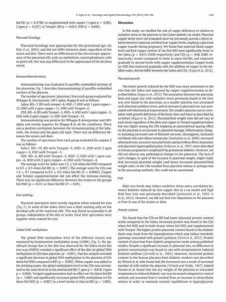
Immunostaining was evaluated in paraffin-embedded sections ofthe placentae. Fig. 1 describes Immunostaining of paraffin-embeddedsections of the placenta.
The number of specimens (placentae) fromeach group evaluated forNFkappa B, nitrotyrosine, HIF1 alpha, IKappa B and as follows:
Sabra: RD—7, RDwith tempol—8, HSD—7, HSDwith 1 ppm copper—12, HSD with 2 ppm copper—11, HSD with Tempol—2.
CDS: RD—6, RDwith Tempol—5, HSD—4, HSDwith 1 ppm copper—5,HSD with 2 ppm copper—5, HSD with Tempol—15.
Immunostaining was positive for NFkappa B nitrotyrosine and HIF1alpha and mostly negative for IKappa B in all the conditions. Therewas a positive correlation between the immunostaining of the laby-rinth, the stroma and the giant cell layer. There was no difference be-tween the strains and diets.
The number of specimens from each group evaluated for caspase 3was as follows:
Sabra: RD—10, RD with Tempol—5, HSD—2, HSD with 2 ppmcopper—2, HSD with Tempol—5.
CDS: RD—6, RD with Tempol—5, HSD—3, HSD with 1 ppm cop-per—6, HSD with 2 ppm copper—4, HSD with Tempol—4.
The average score for Sabra was 1.2 ± 0.6 when fed HSD comparedto 0.5 ± 0.5 when fed RD (p = 0.007). The average score for CDs was1.5 ± 0.7 compared to 0.5 ± 0.5 when fed RD (P = 0.0002). Copperand Tempol supplementation did not affect the immuno-staining.There was no significant difference between the strains in the groupsfed HSD (p = 0.33) or those fed RD (P = 0.95).
Iron staining
Placental specimens were mostly negative when stained for iron(Fig. 2). In some of the slides there was a faint staining only on thedecidual cells of the maternal side. This was found occasionally in allgroups, independent of the diet or strain. Fetal liver specimens werenegative when stained for iron.
Global DNA methylation
The global DNA methylation level of the different tissues wasmeasured by luminometric methylation assay (LUMA) (Fig. 3). No sig-nificant change due to the diet was observed for the Sabra strain butone-way ANOVA revealed a significant effect of the diet on global DNAmethylation in CDs damplacenta (p b 0.001). Post hoc analysis revealeda significant decrease in global DNA methylation in the placenta of CDsdams fed HSD compared to RD (p= 0.002).When copperwas added tothe drinkingwater, the globalmethylation level in the CDs was normal-ized to the same level as in the animals fed RD (1 ppmp= 0.018, 2 ppmp= 0.004). Tempol supplementation had no effect on CDs dams fed RD(p= 1.000) and significantly increased global DNA methylation in CDsdams fed HSD (p= 0.007) to a level similar to that on RD (p= 1.000).
Discussion
In this study, we clarified the role of copper deficiency in relation tooxidative stress in the placenta in the Cohen diabetic rat model. Placentalcopper levels were not evaluated since we previously proved a direct re-lation betweenmaternal and fetal liver copper levels, implying aplacentalcopper transfer during pregnancy. We found that maternal blood copperlevel and fetal copper content of rats fed HSD were significantly lower inthe Sabra (p = 0.013, 0.039 respectively) and CDs (p = 0.06, 0.001 re-spectively) strains compared to those in dams fed RD, and improvedgradually to normal levels with copper supplementation. Copper levelson HSD that improved gradually with the addition of copper to the dis-tilledwater, did not differ between the Sabra and CDs. (Ergaz et al., 2012).
Placental growth
The lower growth induced by the HSD was more prominent in theCDs than the Sabra and improved by copper supplementation as de-scribed before (Ergaz et al., 2012). The association between the placental,fetal and organ size with oxidative stress previously found in the liverwas now found in the placentae, as a smaller placenta was associatedwith placental oxidative stress, and an increase in placental sizewas asso-ciatedwith biochemical improvement. The smaller placentawas in corre-lationwith growth deficiency of the brain, liver and heart as described byus before (Ergaz et al., 2012). Placental/fetal weight ratio did not vary ineach strain regardless of the diets and copper or Tempol supplementationbut was higher among the CDs implying an asymmetrical sparing effecton the placenta or an increase in placental damage. Inflammatory chang-es including increased rate of fibrinoid necrosis, chorangiosis, nucleatedred blood cells and villous immaturity (Daskalakis et al., 2008), increasedatherosclerosis, excessive syncytial knots andperivillousfibrin depositionand placental hypercapillarization (Eriksson et al., 1991) were describedin human pregnancies complicated by gestational diabetes. However, wedid not observe any pathological changes in the placentae. The lack ofsuch changes, in spite of the increase in placental weight, might implythat increased placental weight (and hence increased placental/fetalweight ratio) in the CDs resulted from placental edema or perhaps dueto the processing methods; this could not be ascertained.
Iron
High iron levels may induce oxidative stress and a correlation be-tween diabetes induced by low copper diet in a rat model and highfetal liver iron was previously described (Jankowski et al., 1993; Liet al., 2012). However, we did not find iron depositions in the placentaor liver in any of the strains or diets.
Protein content
We found that the CDs on RD had lower placental protein contentlevels compared to the Sabra. Increased protein was found in the CDswhen fed HSD and in both strains when fed HSD and supplementedwith Tempol. The higher protein placental content found in the diabeticdams may result from the hyperglycemia which may induce metabolicpathways associated with protein synthesis (Clark et al., 2012). Proteincontent of placentae from diabetic pregnancies varies among publishedstudies. Despite a significant increase in placental size, no difference inprotein concentration was found in rats with streptozotocin-induceddiabetes mellitus (Gewolb et al., 1983). However, increased proteincontent in the human placenta from diabetic mothers was describedbyWinick et al. who found that the increment was a result of increasednumber of cells within the placenta (Winick and Noble, 1967). IndeedHusain et al. found that the dry weight of the placenta in untreatedstreptozotocin induced diabetic rats was increased compared to controlanimals and assumed that it resulted from an increase in intracellularsolutes in order to maintain osmotic equilibrium in hyperglycemic
0
0.2
0.4
0.6
0.8
1
1.2
1.4
1.6
1.8
2
RD
RD+Tem
pol
HSD
HSD+CU1
HSD+CU2
HSD+Tem
pol
Sabra Score CDs Scoreg
Fig. 1. Immunostaining of paraffin-embedded sections of the placenta. a. Phosphate buffer saline (PBS), negative control, Sabra high sucrose diet, labyrinth. b. Nitrotyrosine, Sabra, highsucrose diet, labyrinth. c. Nitrotyrosine CDs, high sucrose diet, labyrinth. d. NFkappa B, Sabra, high sucrose diet, labyrinth. e. NFkappa B, CDs, high sucrose diet, labyrinth. f. Caspase 3,CDs, high sucrose diet labyrinth. g. Average score, immunohistochemistry for Caspase 3. The intensity of the immunostaining for caspase 3 was given a score from 0 to 2 according tothe number of areas with apoptosis seen by light microscopy (magnified 1:200): 0: only 1–2 areas positive for caspase 3; 1: 3–5 areas, 2: more than 6 areas.(minimal number of samplesper group n = 3).
226 Z. Ergaz et al. / Toxicology and Applied Pharmacology 276 (2014) 220–230
milieu (Husain et al., 2001). Increased glycogen content (Shafrir andBarash, 1991) edema, cystic degeneration, fibrosis and ischemia werefound in the placentae of streptozotocin induced diabetic pregnant
rats who also had smaller fetal weights (Szalay and Gaal, 1975). The dif-ferences in protein content are probably the result of different animalmodels, diets and maternal blood glucose levels.
Fig. 2. Paraffin embedded section of theplacentae: Placental tissue negatively stainedwith Prussian blue for iron deposits, arrow: erythrocytes positively stainedwith Prussian blue for irondeposits, CDs regular diet.
227Z. Ergaz et al. / Toxicology and Applied Pharmacology 276 (2014) 220–230
Oxidative stress
Copper supplementation: A dose response improvement in growthwith copper supplementation was associated with an effect of over-correction in SOD and CAT activity with 1 ppm copper supplementationthat normalized with full recovery of placental and organ size with 2ppm copper supplementation. It is supported by the gradual decreasein levels of MDA in the CDs fed HSD when copper supplementationincreased from 1 ppm to 2 ppm. This supports our hypothesis of a roleof oxidative stress in the disturbed fetal growth in this model.
Tempol supplementation: To further elucidate the role of oxidativestress we used Tempol and found that it did not improve placentalweight and partially improved fetal length in both strains and weight
Fig. 3.Global DNA methylation of the placenta from CDs and Sabra dams fed regular diet, h(CU1), or 2 ppm (CU2) or Tempol (TPL). Box plots were used to show the global DNAmethy25th and 75th percentiles. Whiskers denote the 5th and 95th percentiles. Differences betwequality of variances and Bonferroni post hoc test (*p b 0.05).
in the Sabra fetuses. Thiswas in linewith our previous findings of failureto restore to normal the growth of the fetuses in this model by vitaminsC and E (Ornoy et al., 2009a). As mentioned above, we also previouslydemonstrated that Tempol reduced the degree of oxidative stress andanomalies in cultured rat embryos (Ryu et al., 2007). Stanley et al. re-ported improved fetal weight and crown–rump length associated withlower placental weight and reduced vascular density in the placentalbed after oral supplementation with Tempol in a murine model ofFGR. Contrary to our results, the placentae of the FGR mice hadincreased SOD production, but as observed in the CDs, Tempol supple-mentation had no effect on placental SOD activity or the immunostain-ing for nitrotyrosine (Stanley et al., 2012). Clark et al. described in amurine model for reduced placental vasculature that leads to pre-
igh sucrose diet (HSD) or HSD complemented in the drinking water by copper: 1 ppmlation level. The line within each box denotes themedian. Limits of the box denote theeen methylation level were statistically analyzed by one-way ANOVA after testing for
228 Z. Ergaz et al. / Toxicology and Applied Pharmacology 276 (2014) 220–230
eclampsia and FGR, that oral supplementation with Tempol improvedplacental spiral artery luminal area evaluated at day 12.5 of pregnancy.This was associated with improved fetal growth and reduction inplacental apoptosis (Clark et al., 2012). The lack of improvement in pla-centalweight in ourmodel ismost probably due to strain difference andthe combined effect of oxidative stress, hypoxia and copper deficiencywhich harmed the placenta so that the anti-oxidant treatment alonecould not fully correct the growth restriction.
Immunohistochemistry
We found nodifferences among strains and diets in immunostainingfor oxidative stress parameters despite the biochemical evidence foroxidative stress in the dams fed HSD especially the CDs. The markedstaining for NFkB from both strains regardless of the diet can resultfrom its high levels, so thatmore dilutions could emphasize a difference.Why NIT levels were increased regardless of the strain and diet is notcurrently understood.
HypoxiaThe role of hypoxia in the placenta was evaluated by immunohisto-
chemical staining for HIF 1 alpha. Compared to the intense staining forHIF 1 alpha found in the liver of the CDs strain fed HSD, positive stainingwas found in the placentae of all strains and diets. This may be due tonecrosis of giant trophoblast cells surrounding the embryo-fetal tissueand aggregation of inflammatory cells that are seldom found towardsthe end of rat pregnancy (de Rijk et al., 2002). Increased oxidative stressand hypoxia in accordance with inflammatory changes were noted in18.5 day placentae of mice fed high fat diet during pregnancy thatdeveloped insulin resistance (Li et al., 2013).
ApoptosisWe found specimens positive for caspase 3 among all groups,
however the number of positive cells was higher among fetuses ofdams fed HSD compared to those fed RD. The higher rate of specimenspositive for caspase 3 among both strains fed HSD is in correlationwith the growth restriction and increase in oxidative stress markersamong the Sabra fed HSD. Caspases are cysteine proteases involved inthe initiator and effector phases of apoptosis. Caspase-3 represents acommon final pathway of the execution of apoptosis in highly divergentsystems, and its activity is used to assess the level of apoptotic activity(Stennicke and Salvesen, 1998). Previous studies found a correlationbetween diabetic pregnancy and an increase in markers for apoptosis.An increase in caspase 3 was found in embryos of streptozotocininjected diabetic rats at days 9 and 11 of pregnancy (El-Bassiouniet al., 2005) and in malformed embryos of streptozotocin injected dia-betic mice on day 11 (Yang et al., 2008) of gestation. Immunostainingof human placentae for caspase 3 was higher than in controls amongwomen suffering from preeclampsia and/or fetal growth restriction(Cali et al., 2013) (Barrio et al., 2009).
DNA methylation
The placenta has an inherently lower level ofmethylation than othertissueswhichmay render this organ highly sensitive to the effects of theenvironmental factors affecting the DNA methylation pattern (Razinet al., 1984).We found a reduction of global DNAmethylation of the pla-centa in CDs dams fed HSD. We measured changes in the global DNAmethylation level of placenta by LUMA. The LUMA technique is basedon the HpaII/MspI recognition sequence, 5′-CCGG-3′, which is fairlywell interspersed through the genome. Approximately 50% of theCCGG sites are located in repetitive DNA sequences and 50% in uniquesequences (Fazzari and Greally, 2004), and even though they areaccumulated at CpG islands, CpG islands only comprise about 1% ofthe genome (Bjornsson et al., 2008). Therefore, methylation effectsmeasured by LUMA are dominated by methylation outside of CpG
islands, a fact that is also supported by its linear correlation with LINE-1 analysis (Romermann et al., 2008). The level of global DNA methyla-tionmeasured here does not imply that other forms of DNAmethylationalterations such as region-specific or site-specific DNA methylationdifferences are not present. Global demethylation is characteristic of se-rious diseases such as cancer (Feinberg, 1988; Feinberg and Vogelstein,1983) and autoimmune diseases. Moreover, global demethylation isbelieved to affect long-range genome regulation and genome stability(Espada and Esteller, 2007) that could have an important effect onplacental function during gestation.
Hypomethylation of vascular endothelial growth factor (VEGF)promoter and consequent upregulation of VEGF mRNA levels werefound in human placentae of women with preeclampsia. The morespecific evaluation demonstrated incremental increase, no change, orreduction in methylation in accordance with gestational age implyingthe dynamic process in which the methylation is influenced by theintrauterine surroundings (Sundrani et al., 2013).
We observed a reduction of global DNAmethylation of the placentaonly in CDs dams fed HSD. The supplementation of copper in the drink-ing water corrected the diabetes phenotype and most of the problemsinduced by HSD and the global methylation level as well. The additionof Tempol, was not sufficient to correct the diabetic phenotype in CDsHSD dams but improved fetal weight and length and was able to bringthe level of global DNA methylation back to normal. This suggests thatthe global methylation level is not affected merely by the diabetes butis probably also sensitive to the resulting oxidative stress. Decreasedmethylation in genes associated with adult morbid obesity (MESTgene, glucocorticoid receptor NR3C1 gene, interspersed ALU repeats)was found in human placentae from women suffering from gestationaldiabetes treated through diet and insulin dependent compared tocontrols (El Hajj et al., 2013).
Resemblance to liver
The resemblance in the oxidative stress parameters between theplacenta and livermay allow a simpleway to evaluate the fetus. Liver bi-opsymay give a better representation of the hypoxic process in the fetusbut is hard to achieve and is associated with complications. Evaluationof the placenta in this model may substitute for liver analysis. Thismay allow us in an experimental animal model to evaluate the impactof oxidative stress during pregnancy and further evaluate its impactlater in life without either euthanizing some of the fetuses in eachgroup after delivery or endangering their lives through biopsies.
Conclusion
Fetal growth restriction in the CohenDiabetic ratmodel is associatedwith smaller placental size but with relative weight sparing comparedto the fetus. Fetal growth and oxidative stress improved with Tempolbut were corrected only with copper supplementation. This partialrecovery despite the correction in DNA methylation implies thatoxidative stress is a component in a more complicated biologicalprocess that is copper dependent. The correlation between theoxidativestress parameters in the placenta with our previous findings in the livermay facilitate the documentation of oxidative stress in the fetus moreeasily by assessing the placenta in future studies of the offspring. The as-sociation between hypomethylation of genes and adultmorbidity foundalso in human gestational diabetes makes our model a unique methodfor investigating genes associated with growth, oxidative stress, hypox-ia and copper.
Conflict of interest
The authors declare that there are no conflicts of interest.
229Z. Ergaz et al. / Toxicology and Applied Pharmacology 276 (2014) 220–230
Acknowledgments
Supported by grant number 0374352 from the American IsraelBinational Science Foundation and by a grant from the Ministry ofEconomic Development, Innovation and Export Trade of Quebec(grant PSR-SIIRI-181).
References
Barrio, E., Calvo, M.T., Miramar, M.D., Lorente, F., Rodriguez, A., Labarta, J.I., Mayayo, E.,Longas, A.F., 2009. Study of apoptosis and related proteins, CRH and hpGH inplacentas of newborns small for gestational age (SGA). Pediatr. Endocrinol. Rev. 6(Suppl. 3), 337–342.
Bradford, M.M., 1976. A rapid and sensitive method for the quantitation of microgramquantities of protein utilizing the principle of protein-dye binding. Anal. Biochem.72, 248–254.
Brown, N.A., Fabro, S., 1981. Quantitation of rat embryonic development in vitro: a mor-phological scoring system. Teratology 24, 65–78.
Cali, U., Cavkaytar, S., Sirvan, L., Danisman, N., 2013. Placental apoptosis in preeclampsia,intrauterine growth retardation, and HELLP syndrome: an immunohistochemicalstudy with caspase-3 and bcl-2. Clin. Exp. Obstet. Gynecol. 40, 45–48.
Chess, D.J., Xu, W., Khairallah, R., O'Shea, K.M., Kop, W.J., Azimzadeh, A.M., Stanley, W.C.,2008. The antioxidant tempol attenuates pressure overload-induced cardiac hyper-trophy and contractile dysfunction in mice fed a high-fructose diet. Am. J. Physiol.Heart Circ. Physiol. 295, H2223–H2230.
Clark, P.A., Brown, J.L., Li, S., Woods, A.K., Han, L., Sones, J.L., Preston, R.L., Southard, T.L.,Davisson, R.L., Roberson, M.S., 2012. Distal-less 3 haploinsufficiency results in elevat-ed placental oxidative stress and altered fetal growth kinetics in the mouse. Placenta33, 830–838.
Cohen, A., 1990. Development of the model. In: AM, C., Rosenmann, E. (Eds.), The CohenDiabetic Rat. Karger, Basel, pp. 1–9.
Dai, S.Y., Kanenishi, K., Ueno, M., Sakamoto, H., Hata, T., 2004. Hypoxia-inducible factor-2alpha is involved in enhanced apoptosis in the placenta from pregnancies withfetal growth restriction. Pathol. Int. 54, 843–849.
Daskalakis, G., Marinopoulos, S., Krielesi, V., Papapanagiotou, A., Papantoniou, N.,Mesogitis, S., Antsaklis, A., 2008. Placental pathology in women with gestationaldiabetes. Acta Obstet. Gynecol. Scand. 87, 403–407.
de Rijk, E.P., van Esch, E., Flik, G., 2002. Pregnancy dating in the rat: placental morphologyand maternal blood parameters. Toxicol. Pathol. 30, 271–282.
El Hajj, N., Pliushch, G., Schneider, E., Dittrich, M., Muller, T., Korenkov, M., Aretz, M., Zechner,U., Lehnen, H., Haaf, T., 2013. Metabolic programming of MEST DNA methylation by in-trauterine exposure to gestational diabetes mellitus. Diabetes 62, 1320–1328.
El-Bassiouni, E.A., Helmy, M.H., Abou Rawash, N., El-Zoghby, S.M., Kamel, M.A., Abou Raya,A.N., 2005. Embryopathy in experimental diabetic gestation: assessment of PGE2level, gene expression of cyclooxygenases and apoptosis. Br. J. Biomed. Sci. 62,161–165.
Ergaz, Z., Avgil, M., Ornoy, A., 2005. Intrauterine growth restriction-etiology and conse-quences: what do we know about the human situation and experimental animalmodels? Reprod. Toxicol. 20, 301–322.
Ergaz, Z., Shoshani-Dror, D., Guillemin, C., Neeman-Azulay, M., Fudim, L., Weksler-Zangen,S., Stodgell, C.J., Miller, R.K., Ornoy, A., 2012. The effect of copper deficiency on fetalgrowth and liver anti-oxidant capacity in the Cohen diabetic rat model. Toxicol.Appl. Pharmacol. 265, 209–220.
Feinberg, A.P., 1988. Alterations in DNAmethylation in colorectal polyps and cancer. Prog.Clin. Biol. Res. 279, 309–317.
Feinberg, A.P., Vogelstein, B., 1983. Hypomethylation distinguishes genes of some humancancers from their normal counterparts. Nature 301, 89–92.
Gallou-Kabani, C., Gabory, A., Tost, J., Karimi, M., Mayeur, S., Lesage, J., Boudadi, E., Gross,M.S., Taurelle, J., Vige, A., Breton, C., Reusens, B., Remacle, C., Vieau, D., Ekstrom, T.J.,Jais, J.P., Junien, C., 2010. Sex- and diet-specific changes of imprinted gene expressionand DNA methylation in mouse placenta under a high-fat diet. PLoS One 5, e14398.
Gluckman, P.D., Hanson, M.A., Cooper, C., Thornburg, K.L., 2008. Effect of in utero andearly-life conditions on adult health and disease. N. Engl. J. Med. 359, 61–73.
Goelz, S.E., Vogelstein, B., Hamilton, S.R., Feinberg, A.P., 1985. Hypomethylation of DNAfrom benign and malignant human colon neoplasms. Science 228, 187–190.
Haeri, S., Khoury, J., Kovilam, O., Miodovnik, M., 2008. The association of intrauterinegrowth abnormalities in women with type 1 diabetes mellitus complicated by vascu-lopathy. Am. J. Obstet. Gynecol. 199 (278), e271–e275.
Hebert, C.D., Elwell, M.R., Travlos, G.S., Fitz, C.J., Bucher, J.R., 1993. Subchronic toxicity ofcupric sulfate administered in drinking water and feed to rats and mice. Fundam.Appl. Toxicol. 21, 461–475.
Husain, S.M., Frost, R., Mughal, Z.M., 2001. Effect of diabetes mellitus on rat placentacellularity. Early Hum. Dev. 60, 207–214.
Jankowski, M.A., Uriu-Hare, J.Y., Rucker, R.B., Keen, C.L., 1993. Effect of maternal diabetesand dietary copper on fetal development in rats. Reprod. Toxicol. 7, 589–598.
Jawerbaum, A., Higa, R., White, V., Capobianco, E., Pustovrh, C., Sinner, D., Martinez, N.,Gonzalez, E., 2005. Peroxynitrites and impaired modulation of nitric oxide concentra-tions in embryos from diabetic rats during early organogenesis. Reproduction 130,695–703.
Karimi, M., Johansson, S., Ekstrom, T.J., 2006a. Using LUMA: a luminometric-based assayfor global DNA-methylation. Epigenetics 1, 45–48.
Karimi, M., Johansson, S., Stach, D., Corcoran, M., Grander, D., Schalling, M., Bakalkin, G.,Lyko, F., Larsson, C., Ekstrom, T.J., 2006b. LUMA (LUminometric Methylation Assay)—a high throughput method to the analysis of genomic DNA methylation. Exp. CellRes. 312, 1989–1995.
Li, X., Li, H., Lu, N., Feng, Y., Huang, Y., Gao, Z., 2012. Iron increases liver injury throughoxidative/nitrative stress in diabetic rats: involvement of nitrotyrosination of glucoki-nase. Biochimie 94, 2620–2627.
Li, H.P., Chen, X., Li, M.Q., 2013. Gestational diabetes induces chronic hypoxia stress and ex-cessive inflammatory response in murine placenta. Int. J. Clin. Exp. Pathol. 6, 650–659.
MacLennan, N.K., James, S.J., Melnyk, S., Piroozi, A., Jernigan, S., Hsu, J.L., Janke, S.M., Pham,T.D., Lane, R.H., 2004. Uteroplacental insufficiency alters DNA methylation, one-carbonmetabolism, and histone acetylation in IUGR rats. Physiol. Genomics 18, 43–50.
McCord, J.M., Fridovich, I., 1988. Superoxide dismutase: the first twenty years (1968–1988). Free Radic. Biol. Med. 5, 363–369.
Mihara, M., Uchiyama, M., 1978. Determination of malonaldehyde precursor in tissues bythiobarbituric acid test. Anal. Biochem. 86, 271–278.
Ornoy, A., Zusman, I., 1991. Embryotoxic effects of diabetes on pre-implantation embryos.Isr. J. Med. Sci. 27, 487–492.
Ornoy, A., Crone, K., Altshuler, G., 1976. Pathological features of the placenta in fetal death.Arch. Pathol. Lab. Med. 100, 367–371.
Ornoy, A., Tsadok, M.A., Yaffe, P., Zangen, S.W., 2009a. The Cohen diabetic rat as a modelfor fetal growth restriction: vitamins C and E reduce fetal oxidative stress but do notrestore normal growth. Reprod. Toxicol. 28, 521–529.
Ornoy, A., Tsadok, M.A., Yaffe, P., Zangen, S.W., 2009b. The Cohen diabetic rat as a modelfor fetal growth restriction: vitamins C and E reduce fetal oxidative stress but do notrestore normal growth. Reprod. Toxicol. 28, 521–529.
Ornoy, A., Rand, S.B., Bischitz, N., 2010. Hyperglycemia and hypoxia are interrelated intheir teratogenic mechanism: studies on cultured rat embryos. Birth Defects Res. BDev. Reprod. Toxicol. 89, 106–115.
Park, J.H., Stoffers, D.A., Nicholls, R.D., Simmons, R.A., 2008. Development of type 2 diabe-tes following intrauterine growth retardation in rats is associated with progressiveepigenetic silencing of Pdx1. J. Clin. Invest. 118, 2316–2324.
Pilsner, J.R., Lazarus, A.L., Nam, D.H., Letcher, R.J., Sonne, C., Dietz, R., Basu, N., 2010.Mercury-associated DNA hypomethylation in polar bear brains via the LUminometricMethylation Assay: a sensitive method to study epigenetics in wildlife. Mol. Ecol. 19,307–314.
Razin, A., Webb, C., Szyf, M., Yisraeli, J., Rosenthal, A., Naveh-Many, T., Sciaky-Gallili, N.,Cedar, H., 1984. Variations in DNA methylation during mouse cell differentiationin vivo and in vitro. Proc. Natl. Acad. Sci. U. S. A. 81, 2275–2279.
Romermann, D., Hasemeier, B., Metzig, K., Gohring, G., Schlegelberger, B., Langer, F.,Kreipe, H., Lehmann, U., 2008. Global increase in DNA methylation in patients withmyelodysplastic syndrome. Leukemia 22, 1954–1956.
Ryu, S., Kohen, R., Samuni, A., Ornoy, A., 2007. Nitroxide radicals protect cultured ratembryos and yolk sacs from diabetic-induced damage. Birth Defects Res. A Clin.Mol. Teratol. 79, 604–611.
Ryu, S., Ornoy, A., Samuni, A., Zangen, S., Kohen, R., 2008. Oxidative stress in Cohendiabetic rat model by high-sucrose, low-copper diet: inducing pancreatic damageand diabetes. Metabolism 57, 1253–1261.
Salafia, C.M., Minior, V.K., Pezzullo, J.C., Popek, E.J., Rosenkrantz, T.S., Vintzileos, A.M.,1995. Intrauterine growth restriction in infants of less than thirty-two weeks'gestation: associated placental pathologic features. Am. J. Obstet. Gynecol. 173,1049–1057.
Sekigawa, I., Yoshiike, T., Iida, N., Hashimoto, H., Ogawa, H., 2002. Allergic disorders insystemic lupus erythematosus: prevalence and family history. Lupus 11, 426–429.
Shafrir, E., Barash, V., 1991. Placental glycogen metabolism in diabetic pregnancy. Isr.J. Med. Sci. 27, 449–461.
Stanley, J.L., Andersson, I.J., Hirt, C.J., Moore, L., Dilworth, M.R., Chade, A.R., Sibley, C.P.,Davidge, S.T., Baker, P.N., 2012. Effect of the anti-oxidant tempol on fetal growth ina mouse model of fetal growth restriction. Biol. Reprod. 87 (25), 21–28.
Stennicke, H.R., Salvesen, G.S., 1998. Properties of the caspases. Biochim. Biophys. Acta1387, 17–31.
Sundrani, D.P., Reddy, U.S., Joshi, A.A., Mehendale, S.S., Chavan-Gautam, P.M., Hardikar,A.A., Chandak, G.R., Joshi, S.R., 2013. Differential placentalmethylation and expressionof VEGF, FLT-1 and KDR genes in human term and preterm preeclampsia. Clin.Epigenetics 5, 6.
Szalay, J., Gaal, M., 1975. Clinical and morphological studies in streptozotocin diabeticpregnant rats. Acta Med. Acad. Sci. Hung. 32, 35–41.
Szyf, M., Pakneshan, P., Rabbani, S.A., 2004. DNA demethylation and cancer: therapeuticimplications. Cancer Lett. 211, 133–143.
Taricco, E., Radaelli, T., Rossi, G., Nobile de Santis, M.S., Bulfamante, G.P., Avagliano, L.,Cetin, I., 2009. Effects of gestational diabetes on fetal oxygen and glucose levelsin vivo. BJOG 116, 1729–1735.
230 Z. Ergaz et al. / Toxicology and Applied Pharmacology 276 (2014) 220–230
Thurman, R.G., Ley, H.G., Scholz, R., 1972. Hepatic microsomal ethanol oxidation. Hydro-gen peroxide formation and the role of catalase. Eur. J. Biochem. 25, 420–430.
Unterberger, A., Szyf, M., Nathanielsz, P.W., Cox, L.A., 2009. Organ and gestational ageeffects of maternal nutrient restriction on global methylation in fetal baboons.J. Med. Primatol. 38, 219–227.
Weksler-Zangen, S., Yagil, C., Zangen, D.H., Ornoy, A., Jacob, H.J., Yagil, Y., 2001. The newlyinbred Cohen diabetic rat: a nonobese normolipidemic genetic model of diet-inducedtype 2 diabetes expressing sex differences. Diabetes 50, 2521–2529.
Weksler-Zangen, S., Yaffe, P., Ornoy, A., 2003. Reduced SOD activity and increased neuraltube defects in embryos of the sensitive but not of the resistant Cohen diabetic rats cul-tured under diabetic conditions. Birth Defects Res. A Clin. Mol. Teratol. 67, 429–437.
Wilcox, C.S., Pearlman, A., 2008. Chemistry and antihypertensive effects of tempol andother nitroxides. Pharmacol. Rev. 60, 418–469.
Winick, M., Noble, A., 1967. Cellular growth in human placenta. II. Diabetes mellitus.J. Pediatr. 71, 216–219.
Yang, P., Zhao, Z., Reece, E.A., 2008. Activation of oxidative stress signaling that is implicat-ed in apoptosis with a mouse model of diabetic embryopathy. Am. J. Obstet. Gynecol.198 (130), e131–e137.
Zangen, S.W., Ryu, S., Ornoy, A., 2006. Alterations in the expression of antioxidant genesand the levels of transcription factor NF-kappa B in relation to diabetic embryopathyin the Cohen diabetic rat model. Birth Defects Res. A Clin. Mol. Teratol. 76, 107–114.