Engelsdorf et al., Sci. Signal. 11, eaao3070 (2018) 26 June 2018
S C I E N C E S I G N A L I N G | R E S E A R C H A R T I C L E
1 of 14
P L A N T B I O L O G Y
The plant cell wall integrity maintenance and immune signaling systems cooperate to control stress responses in Arabidopsis thalianaTimo Engelsdorf1*, Nora Gigli-Bisceglia1, Manikandan Veerabagu1†, Joseph F. McKenna2‡, Lauri Vaahtera1, Frauke Augstein1§, Dieuwertje Van der Does3‖, Cyril Zipfel3, Thorsten Hamann1¶
Cell walls surround all plant cells, and their composition and structure are modified in a tightly controlled, adaptive manner to meet sometimes opposing functional requirements during growth and development. The plant cell wall integrity (CWI) maintenance mechanism controls these functional modifications, as well as responses to cell wall damage (CWD). We investigated how the CWI system mediates responses to CWD in Arabidopsis thaliana. CWD induced by cell wall–degrading enzymes or an inhibitor of cellulose biosynthesis elicited similar, turgor-sensitive stress responses. Phenotypic clustering with 27 genotypes identified a core group of receptor-like kinases (RLKs) and ion channels required for the activation of CWD responses. A genetic analysis showed that the RLK FEI2 and the plasma membrane–localized mechanosensitive Ca2+ channel MCA1 functioned downstream of the RLK THE1 in CWD perception. In contrast, pattern-triggered immunity (PTI) signaling components, including the receptors for plant elicitor peptides (AtPeps) PEPR1 and PEPR2, repressed responses to CWD. CWD induced the expression of PROPEP1 and PROPEP3, which encode the precursors of AtPep1 and AtPep3, and the release of PROPEP3 into the growth medium. Application of AtPep1 and AtPep3 repressed CWD-induced phytohormone accumulation in a concentration-dependent manner. These results suggest that AtPep-mediated signaling suppresses CWD-induced defense responses controlled by the CWI mechanism. This suppression was alleviated when PTI signaling down-stream of PEPR1 and PEPR2 was impaired. Defense responses controlled by the CWI maintenance mechanism might thus compensate to some extent for the loss of PTI signaling elements.
INTRODUCTIONPlants adapt to diverse environments by modifying their architecture. The cell walls surrounding all plant cells are key elements enabling this adaptability and consist of different components including pro-teins and polysaccharides such as cellulose, hemicelluloses, pectins, and lignin (1). These components are synthesized in different sub-cellular compartments and have specialized functions. Cellulose is synthesized by plasma membrane–localized cellulose synthase com-plexes and released into the adjacent extracellular space as strands that form microfibrils before being incorporated into the wall where they function as the main load-bearing element. The walls are also essential elements underlying growth, development, and resistance to biotic and abiotic stresses, all of which influence crop yield (2, 3). This is illustrated by mutations that improve yields of staple crops like maize and rice by affecting cell wall biosynthesis, homeostasis, poly-saccharide modifications, and signaling components (4, 5).
Cell wall plasticity describes the ability of plant cell walls to adapt to dynamic and challenging growth conditions. Plasticity and the re-sulting recalcitrance in cell wall biochemistry and structure against targeted manipulation also represent a major challenge to produc-ing energy from lignocellulosic biomass (6). The available evidence suggests that the plant cell wall integrity (CWI) maintenance system forms an integral element of cell wall plasticity (7–9). This mecha-nism seems to involve receptor-like kinases (RLKs) and ion chan-nels that constantly monitor the state of the cell wall and initiate adaptive changes in both cellular and cell wall metabolism in response to cell wall damage (CWD) (10–12). Here, we refer to any changes to cell wall structure or composition that impair CWI as CWD. Because CWD may be induced by various means, both ligand-mediated mecha-nisms and mechanoperception could be involved in CWD detection. For example, pathogen-derived enzymes break down cell walls, which releases cell wall–derived fragments. This could lead to cell wall weak-ening, deformation, and displacement of the cell wall relative to the plasma membrane and can eventually result in cell bursting due to the high turgor pressure of the cell (13, 14). The cell wall fragments, such as cellobiose or oligogalacturonides (OGs; fragments of pectic polysaccharides), can activate plant immune responses (15, 16). Al-though OGs are detected through wall-associated receptor kinases (WAKs), the receptors for cellobiose have not been identified (7). Mechanosensitive systems may also be activated by CWD that com-promises the structural integrity of the cell wall (17). In addition to the enzymatic actions of pathogens and mechanical damage caused by breakage or grazing, defects in cell wall biosynthetic processes can also cause CWD by preventing the production of load-bearing structural elements (18).
Although several candidate genes have been implicated in CWD perception, experimental evidence confirming their involvement is
1Department of Biology, Høgskoleringen 5, Realfagbygget, Norwegian University of Science and Technology, 7491 Trondheim, Norway. 2Department of Biology, Imperial College London, South Kensington Campus, SW7 2AZ London, UK. 3The Sainsbury Laboratory, Norwich Research Park, Norwich NR4 7UH, UK.*Present address: Division of Plant Physiology, Department of Biology, Philipps University of Marburg, 35043 Marburg, Germany.†Present address: Department of Plant Sciences, Norwegian University of Life Sci-ences, N-1432 Ås, Norway.‡Present address: Plant Cell Biology, Department of Biological and Medical Sciences, Oxford Brookes University, Oxford OX3 0BP, UK.§Present address: Department of Organismal Biology, Physiological Botany, Evolu-tionary Biology Centre and Linnean Centre for Plant Biology, Uppsala University, Ullsv. 24E, SE-75651 Uppsala, Sweden.‖Present address: 2Blades Foundation/BecA-ILRI Hub, P.O. Box 30709, Nairobi 00100, Kenya.¶Corresponding author. Email: [email protected]
Engelsdorf et al., Sci. Signal. 11, eaao3070 (2018) 26 June 2018
S C I E N C E S I G N A L I N G | R E S E A R C H A R T I C L E
2 of 14
scarce (7, 10, 19). Among the candidates identified in Arabidopsis thaliana are two homologs of plasma membrane–localized RLK1-like proteins originally in Catharanthus roseus (CrRLK1Ls): THESEUS1 (THE1) and FERONIA (FER) (20, 21). Additionally, the leucine-rich repeat (LRR) RLK MALE DISCOVERER 1 (MDIS1)–INTERACTING RECEPTOR-LIKE KINASE 2 (MIK2), WALL-ASSOCIATED KINASE 1 (WAK1) and WAK2, as well as the putatively stretch-activated, mecha-nosensitive Ca2+ channel MATING PHEROMONE INDUCED DEATH 1 (MID1)–COMPLEMENTING ACTIVITY 1 (MCA1) have also been implicated in CWI maintenance in Arabidopsis (22–26). MCA1 was originally identified through its ability to partially com-plement a Saccharomyces cerevisiae strain deficient for MID1-CCH1 (calcium channel homolog 1), which is required for CWI maintenance in this yeast (22, 27). Homologs of MCA1 and THE1 have been identi-fied in Oryza sativa (OsMCA1), Zea mays (NOD), and Marchantia polymorpha (MpTHE), suggesting that these proteins may participate in cell wall maintenance across the plant kingdom (28–30). A charac-teristic feature of THE1, FER, WAK1, and WAK2 is the presence of domains that may bind cell wall–derived epitopes or ligands (8, 10, 31). However, binding to cell wall components has been confirmed only for WAK1, WAK2, and FER (8, 21, 32). THE1, FER, and MIK2 are required for resistance to the fungal pathogen Fusarium oxysporum f. sp. conglutinans, implicating CWI signaling also in biotic stress responses (25, 33).
CWD induced by the inhibition of cellulose production stimulates the compensatory production of the cell wall components callose and lignin; accumulation of the hormones jasmonic acid (JA), salicylic acid (SA), and ethylene; generation of reactive oxygen species (ROS); and activation of Ca2+-based signaling, implicating all of these pro-cesses in CWI maintenance (11, 23, 34–37). CWD in A. thaliana pro-motes the generation of ROS in the apoplast—the space adjacent to the plasma membrane that contains the cell wall—by RESPIRATORY BURST OXIDASE HOMOLOG D (RBOHD) (23). The activity of RBOHD is regulated by both Ca2+-dependent and Ca2+-independent mechanisms, with the latter requiring BOTRYTIS-INDUCED KINASE 1 (BIK1), which is a substrate of BRASSINOSTEROID INSENSITIVE 1 (BRI1)–ASSOCIATED KINASE 1 (BAK1) and other RLKs during pattern-triggered immunity (PTI) (38, 39). BAK1 acts as a co-receptor for LRR-RLKs and, as such, plays an important role in responses to pathogen-associated molecular patterns (PAMPs) such as flagellin, the flagellin-derived epitope flg22, elongation factor–thermo unstable (EF-Tu), and the EF-Tu–derived epitope elf18, as well as damage- associated molecular patterns (DAMPs) such as the AtPep1 and AtPep3 peptides (40–42). AtPep1 and AtPep3 precursor peptides are encoded by the PROPEP1 and PROPEP3 genes, which are induced by pathogen infection and wounding (43). The precursor peptides are reportedly released into the apoplastic space where they are likely processed to give rise to the active form (43). Application of AtPep peptides en-hances both expression of their own PROPEP genes, creating a posi-tive feedback loop, and PTI-controlled defense responses. PTI and CWI maintenance may complement each other during plant defense, but such regulatory interactions between CWI and PTI signaling have not been characterized (44–46).
Here, we investigated the responses to different types of CWD to understand the cellular events underlying CWD perception. We an-alyzed CWD responses in 27 A. thaliana genotypes to establish the functions of candidate genes in CWI maintenance and performed genetic analyses to assess whether key CWI signaling elements belong to one or more signaling cascades. We found that CWD induced the
expression of AtPROPEP1 and AtPROPEP3 as well as the release of a PROPEP3 fusion protein. In contrast, application of AtPep1 and AtPep3 repressed CWD-induced phytohormone production, thus identifying a mechanism through which PTI signaling and the CWI maintenance mechanism cooperate to regulate defense responses.
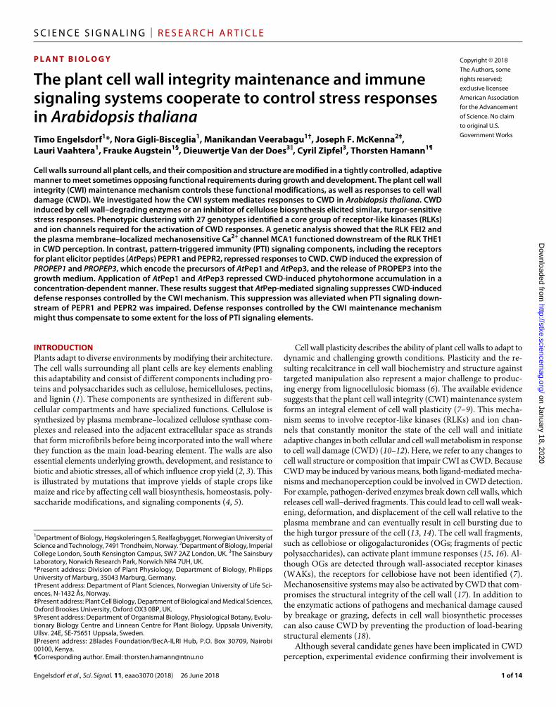
RESULTSCWD responses induced by different stimuli are osmosensitiveWe used an Arabidopsis seedling–based model system to investigate how plants respond to different types of CWD and elucidate further the role of turgor pressure in CWD perception (11). CWD was induced using either Driselase, a mix of several cell wall–degrading enzymes from Basidiomycetes sp., or isoxaben (ISX), a herbicide that blocks cellulose biosynthesis (13, 18). We chose Driselase because this enzyme mix is similar to the enzyme cocktail released by fungal pathogens during infection (47–49). Furthermore, the enzymes lead to cell wall fragmentation, thus directly causing CWD regardless of cell type, dif-ferentiation stage, or turgor pressure. We chose ISX because it inhibits cellulose production only in actively elongating cells (for example, in the root elongation zone). It causes CWD in conjunction with the naturally high turgor pressure of plant cells because it reduces the number of load-bearing cellulose microfibrills in the walls, thus making the cell wall susceptible to failure. This is illustrated by the suppression of ISX-induced lignin, callose, JA, and SA accumulation; tissue lesion formation; and redistribution of carbohydrates by addition of osmotica like sorbitol or mannitol (11, 50). Similar effects have also been reported in yeast cells exposed to CWD, indicating the importance of turgor pressure in CWI maintenance (51).
First, we investigated the effect of ISX or Driselase treatment on the morphology of Wave 131Y seedlings, which ubiquitously express membrane-localized yellow fluorescent protein (YFP), in the presence or absence of an osmoticum (sorbitol) in time course experiments (52). Whole seedlings were grown submerged in liquid culture (11), and the medium was exchanged for fresh medium containing the ISX solvent dimethyl sulfoxide (DMSO), sorbitol, Driselase, ISX, Driselase with sorbitol, or ISX with sorbitol at the 0-hour time point. At 7 hours, epidermal cells in the root elongation zone of ISX-treated seedlings exhibited a swollen phenotype, which was reduced by cotreatment with sorbitol (fig. S1A). Driselase treatment resulted in degradation of the root tip (including the elongation zone) after 7 hours, leaving behind only larger, already fully elongated cells for visualization (fig. S1B). This degradation was possibly enhanced by the addition of osmoticum. Because the effects on roots were so pronounced after 7 hours of treat-ment, we did not investigate the phenotypic effects in roots at later time points. In cotyledons of ISX-treated seedlings, the plasma mem-brane marker signal was similar to that in cotyledons from DMSO controls after 7 hours but was lost in patches after 24 hours (fig. S2, A and B). Sorbitol treatment alone had no effect on membrane marker intensity but, when coadministered with ISX, restored the marker signal at the 24-hour time point. Driselase treatment resulted in the formation of patches lacking the membrane marker after 7 hours (fig. S2C). These patches seemed to be more pronounced after 24 hours and were not affected by the addition of sorbitol (fig. S2, C and D).
After establishing the dynamics of CWD responses, we used the same experimental setup to investigate cell death and the deposition of lignin and callose in cotyledons and the accumulation of JA and SA in whole seedlings (11). We also included isoxaben resistant1-1
Engelsdorf et al., Sci. Signal. 11, eaao3070 (2018) 26 June 2018
S C I E N C E S I G N A L I N G | R E S E A R C H A R T I C L E
3 of 14
(ixr1-1) and bak1-5 mutant seedlings in the studies. The ixr1-1 mu-tation causes an amino acid substitution in CELLULOSE SYNTHASE A3 (CESA3) that renders the protein resistant to inhibition by ISX (53), thus providing a control for ISX specificity. Plants carrying the bak1-5 allele are only impaired in immune responses triggered by LRR-RLKs but not in brassinosteroid-dependent signaling, making the plants a suitable control for detecting the involvement of either DAMPs (for example, AtPep1) generated by CWD or PAMPs that are possibly present as contaminants in Driselase (54) and perceived by LRR-RLKs. Col-0 (wild type) and bak1-5 seedlings that were mock (DMSO)–treated or treated with boiled (inactivated) Driselase ex-hibited no cell death in cotyledons (fig. S3, A and B). ixr1-1 seed-lings treated in the same manner exhibited a slight increase in cell death compared to Col-0. Treatment with ISX induced cell death in Col-0 and bak1-5, but not in ixr1-1, cotyledons compared to mock- treated controls. Driselase treatment induced cell death in all geno-types examined. Sorbitol addition suppressed ISX-induced cell death but had no effect on Driselase-induced cell death.
We also analyzed compensatory lignin deposition in the cotyledons of seedlings that were treated with DMSO, ISX, boiled Driselase, or Driselase, with or without sorbitol, for 24 hours (fig. S3C). Lignin deposition was detectable after ISX treatment in vascular tissue areas in Col-0 and bak1-5, but not in ixr1-1, seedlings. Driselase-treated Col-0, bak1-5, and ixr1-1 seedlings exhibited more ubiquitous lignin deposition. The addition of sorbitol reduced lignin deposition in all cases examined. bak1-5 and ixr1-1 cotyledons seemed more sensitive to Driselase treatment than Col-0 cotyledons based on cell death and lignin deposition phenotypes (fig. S3, A and C).
Next, we investigated compensatory callose deposition in the coty-ledons of seedlings treated in the same manner. Whereas there was no detectable callose deposition in mock- or boiled Driselase–treated Col-0 and bak1-5 cotyledons, callose was deposited in ixr1-1 cotyle-dons subjected to these same control treatments (fig. S3D). Sorbitol alone had no effect on callose deposition in Col-0 and bak1-5 coty-ledons but reduced callose deposition in both ixr1-1 treatment groups. ISX treatment induced callose deposition strongly in Col-0 seedlings, moderately in bak1-5 seedlings, but not in ixr1-1 seedlings. Sorbitol cotreatment with ISX reduced callose deposition in Col-0 and bak1-5. Driselase treatment induced callose deposition in Col-0, but not in bak1-5, cotyledons and did not induce more callose deposition in ixr1-1 seedlings than did boiled Driselase treatment (fig. S3D). bak1-5 plants also exhibit reduced flg22-induced callose deposition, suggesting that the lack of induction observed here is part of a more general defect (55). The lack of significant increase in callose deposition in ixr1-1 was possibly caused by the combination of the substantial amount of basal callose deposition in mock conditions and limited callose in-duction by Driselase.
We next quantified phytohormones in Col-0, bak1-5, and ixr1-1 seedlings treated in the same manner as before. JA and SA abundances were low in mock-treated Col-0 and bak1-5 seedlings and slightly increased in ixr1-1 seedlings (Fig. 1, A and B). ISX treatment induced JA accumulation in bak1-5 seedlings more than in Col-0 seedlings, but no induction was observed in ixr1-1 seedlings (Fig. 1A). ISX in-duced SA accumulation in both Col-0 and bak1-5 seedlings to a simi-lar degree, but not in ixr1-1 seedlings (Fig. 1B). Cotreatment with sorbitol repressed ISX-induced JA and SA accumulation in Col-0 and bak1-5. Phytohormone amounts were lower in ixr1-1 seedlings treated with sorbitol or a combination of ISX and sorbitol than in mock- treated ixr1-1 seedlings, suggesting that sorbitol reduced stress in these
plants (Fig. 1, A and B). Driselase treatments induced JA and SA ac-cumulation in Col-0, bak1-5, and ixr1-1 seedlings to different degrees compared to treatment with boiled Driselase (Fig. 1, C and D). In contrast to ISX treatments, Driselase treatment reduced SA accumu-lation in bak1-5 compared to Col-0 (Fig. 1D). Although induction of both JA and SA by Driselase was less pronounced than induction by ISX in Col-0 and bak1-5 (Fig. 1, A to D), sorbitol cotreatments nevertheless reduced or prevented accumulation of JA and SA in all genotypes examined. The results of these experiments suggest that responses to CWD are not restricted to a particular cell type (exem-plified by lignin deposition in both vascular and epidermal tissues). Despite apparent differences in damage caused by ISX and Driselase, Col-0 seedlings exhibited similar osmosensitive responses to both types of CWD with respect to callose, lignin deposition, as well as JA and SA accumulation. These observations suggest that both mechanoper-ception and osmoperception may be required for induction of CWD responses. bak1-5 seedlings, which have defects in PTI, exhibited dis-tinct differences in their responses to the two CWD-inducing stimuli, suggesting some cross-regulation between PTI and CWI signaling.
Osmosensitivity distinguishes CWI signaling from DAMP- and PAMP-dependent responsesTo investigate the regulatory processes responsible for the observed CWD-induced phenotypes, we first performed expression analysis of the defense marker PLANT DEFENSIN1.2 (PDF1.2) because it encodes a defense peptide that is involved in both CWD- and PTI- mediated processes (37, 56). In both Col-0 and bak1-5 seedlings, treatment with boiled or active Driselase induced PDF1.2 expression; however, PDF1.2 was less highly expressed in boiled Driselase–treated bak1-5 seedlings compared to boiled Driselase–treated Col-0, and it was more highly induced by Driselase in bak1-5 than in Col-0 seedlings (Fig. 1E). This supports the hypothesis that BAK1 might contribute to the PTI-mediated recognition of factors in the enzyme preparation, which are not removed by boiling, but represses the response to CWD elicited by the active enzymes. To investigate the role of mechanoper-ception in the response to CWD, we analyzed the expression of a marker for mechanical stimulation [TOUCH4 (TCH4)] in ISX- and Driselase-treated Col-0 seedlings (57). ISX and active Driselase in-duced TCH4 expression, whereas sorbitol cotreatments reduced it, providing support for an involvement of mechanoperception in the detection of CWD (Fig. 1F).
The results from the phytohormone measurements in bak1-5 seed-lings treated with ISX and Driselase with or without sorbitol, in con-junction with the results from the PDF1.2 expression analysis, suggested that DAMP or PAMP signaling, or both, might also be sensitive to turgor changes. Therefore, we investigated a possible involvement of OG-induced signaling in turgor-sensitive CWI maintenance using gene expression analysis and phytohormone measurements (58). We examined the expression of RETICULINE OXIDASE (RET-OX) and CYTOCHROME P450, FAMILY 81, SUBFAMILY F, POLYPEPTIDE 2 (CYP81F2), both of which are induced by OG and the flagellin de-rivative flg22 (58), in Col-0 seedlings that had been treated with two different concentrations of OGs in the absence or presence of the os-moticum sorbitol. Expression of both genes was induced by OG treat-ments but was not sensitive to sorbitol cotreatment (fig. S4, A and B). We also quantified JA and SA in seedlings treated in the same manner. Both OG and sorbitol treatments resulted in only minor changes in phy-tohormone amounts (fig. S4, C and D). These results showed that OGs can be perceived by the seedlings in our assay and that OG-induced
Engelsdorf et al., Sci. Signal. 11, eaao3070 (2018) 26 June 2018
S C I E N C E S I G N A L I N G | R E S E A R C H A R T I C L E
4 of 14
responses, unlike ISX- and Driselase-induced responses, are not os-mosensitive, suggesting that CWD responses analyzed here do not involve OG-dependent signaling.
Next, we quantified RET-OX and CYP81F2 expression and the amounts of JA and SA in seedlings treated with flg22, sorbitol, or both. RET-OX and CYP81F2 expression was not significantly increased by sorbitol treatment alone, increased moderately by flg22 treatment, and increased greatly in seedlings treated with flg22 plus sorbitol (fig. S4, E and F). Treatments with sorbitol and flg22 resulted in changes in JA amounts that were at the lower limit of detection, although both sorbitol alone and flg22 plus sorbitol promoted JA accumulation (fig. S4G). Flg22 induced SA accumulation after 3 and 7 hours of treatment, and cotreatment with sorbitol enhanced SA accumulation after 7 hours (fig. S4H). These results showed that flg22-induced gene expression and JA and SA accumulation are turgor-sensitive. However, sorbitol treatments seem to enhance the flg22-induced responses, contrary to what we observed with the responses to Driselase and ISX. These results suggest that turgor pressure is relevant for flg22-induced re-sponses but that the underlying regulatory process is distinct from CWI maintenance signaling.
ISX and cell wall–degrading enzymes induce similar osmosensitive responsesDriselase is a complex mix of enzymes that degrade several different cell wall polymers (59). To investigate whether the effects observed in Driselase-treated seedlings can be assigned to particular enzymatic activities, we obtained homogeneous preparations of the individual
enzymes (xylanase, cellulase, and pectinase) that, according to the manufacturer, account for most of the enzymatic activities in com-mercially prepared Driselase. Initially, we treated seedlings with in-creasing concentrations of the individual enzymes and measured phytohormone accumulation to establish optimal experimental con-ditions (fig. S5, A and B). Xylanase did not induce phytohormone production, whereas cellulase induced only SA accumulation. Pectin-ase treatment increased the abundance of both SA and JA in seed-lings in a concentration-dependent manner. On the basis of these tests, we focused primarily on pectinase and cellulase. JA or SA did not accumulate in seedlings treated with boiled enzymes or sorbitol (Fig. 1, G to J). Pectinase treatment induced accumulation of both JA and SA in an osmosensitive manner (Fig. 1, G and H). Because cellulase treatment alone did not induce JA accumulation, we quan-tified hormone accumulation after combining cellulase with either xylanase or pectinase. Seedlings treated with cellulase plus xylanase exhibited no increase in JA and a moderate increase in SA accumu-lation, similar to cellulase alone (Fig. 1, I and J, and fig. S5B). Cellulase plus pectinase elicited JA accumulation, which was higher than in the seedlings treated with the individual enzymes (Fig. 1, G and I, and fig. S5A). SA amounts were lower than in pectinase-treated seed-lings but higher than in those treated with cellulase alone (Fig. 1, H and J, and fig. S5B). Sorbitol addition reduced phytohormone accu-mulation in all enzyme treatments examined. These results suggested that the combination of cellulase and pectinase is mainly responsible for the observed JA and SA accumulation in Driselase-treated seed-lings. The differences in the effects of individual enzymes suggest
0
0.5
1
1.5
2
2.5
3
0
0.5
1
1.5
2
2.5
3
3.5
0 2 4 6 8
10 12 14 16 18 A
B
0
0.5
1
1.5
2
2.5
3
3.5
4
0
10
20
30
40
50
60
70
80
µg J
A /g
DW
µg
SA
/gD
W
Col-0 bak1-5 ixr1-1
moc
k m
ock
+ S
ISX
IS
X +
S
moc
k m
ock
+ S
ISX
IS
X +
S
moc
k m
ock
+ S
ISX
IS
X +
S
a a a
b
a a a
b
b b a a
a a a
b
a a a
b
b b a a
** ***
ns **
***
bDri
+ S
bDri
+ S
bDri
+ S
bDri
Dri
Dri
+ S
bDri
Dri
Dri
+ S
bDri
Dri
Dri
+ S
Col-0 bak1-5 ixr1-1
C
D
µg J
A /g
DW
µg
SA
/gD
W
a a b
c
a a
b
c
a a a
b
a a
b
c
a b
d
c b a
c
d
**
** ns
0
0.5
1
1.5
2
2.5
0
1
2
3
4
5
6
7
µg J
A /g
DW
µg
SA
/gD
W
G
H
0
2
4
6
8
10
12
0
0.5
1
1.5
2
2.5
3
µg J
A /g
DW
zaa
µg
SA
/gD
W
I
J
a a a
b
a a b
c
a a a a a b
c
d
a ab
ab ab ab b
c
d
bP
bP +
S P
P +
S
bC +
bP
bC +
bP
+ S
C +
P
C +
P +
S
bC +
bX
bC +
bX
+ S
C +
X
C +
X +
S
Rel
ativ
e ex
pres
sion
F
ab
d
a a
bc
c
d
c
TCH4
NT
bDri
Dri
NT
bDri
Dri
Rel
ativ
e ex
pres
sion
0.02 E
Col-0 bak1-5
0
0.01
PDF1.2
a
b b
b
a
c
moc
k m
ock
+ S
ISX
IS
X +
S
bDri
+ S
bDri
Dri
Dri
+ S
Fig. 1. Different types of CWD induce similar osmosensitive responses. Quantification of (A) JA and (B) SA, expressed as microgram per gram dry weight (gDW), in Col-0, bak1-5, and ixr1-1 seedlings that had been treated with DMSO (mock), DMSO and sorbitol (S), ISX, or ISX and sorbitol (ISX + S). (C) JA and (D) SA quantification in Col-0, bak1-5, and ixr1-1 seedlings treated with boiled (inactive) Driselase (bDri), bDri and sorbitol (bDri + S), Driselase (Dri), or Driselase and sorbitol (Dri + S). (E) Relative expression of PDF1.2 as determined by quantitative reverse transcription polymerase chain reaction (qRT-PCR) in Col-0 and bak1-5 seedlings treated with bDri or Dri compared to untreated (NT) seedlings. (F) Relative expression of TCH4 in Col-0 seedlings treated as indicated. (G) JA and (H) SA quantification in Col-0 seedlings treated with boiled pectinase (bP), boiled pectinase and sorbitol (bP + S), pectinase (P), or pectinase and sorbitol (P + S). (I) JA and (J) SA quantification in Col-0 seedlings treated with the indicated combinations of boiled (b) or active preparations of cellulase (C), pectinase (P), xylanase (X), and sorbitol (S). All values represent means with error bars indicating SD. n = 4 (A to D) and n = 3 (E to J). Letters a to d (A to J) indicate statistically significant differences according to one-way analysis of variance (ANOVA) and Tukey’s HSD (honestly significantly different) test ( = 0.05) between treatments for each genotype. Asterisks (A to D) indicate statistically significant differences to the wild type (Student’s t test, **P < 0.01; ***P < 0.001; ns, not significant).
Engelsdorf et al., Sci. Signal. 11, eaao3070 (2018) 26 June 2018
S C I E N C E S I G N A L I N G | R E S E A R C H A R T I C L E
5 of 14
that particular types of CWD may induce distinct phytohormone responses.
It is conceivable that a factor that is released or secreted from cells upon ISX or Driselase treatment is responsible for activation of the CWD responses observed. To test this hypothesis, we measured JA and SA accumulation in (i) ixr1-1 seedlings that had been incu-bated with supernatants from Col-0 seedlings pretreated with ISX for 12 or 24 hours and (ii) Col-0 seedlings incubated with boiled supernatants from Col-0 seedlings pretreated with Driselase for 12 or 24 hours (fig. S6, A and B). JA accumulation was barely above the detection limit in the seedlings treated with the different supernatants (fig. S6C). With respect to SA accumulation, only minor changes were detected compared to mock-treated samples (fig. S6D). These results suggested that ISX and Driselase treatments do not cause the release of a factor into the medium that is capable of inducing JA and SA accumulation. Together, the osmosensitivity and similarities in seedling responses to different types of CWD (enzymatic versus ISX) suggest that different causes of CWD may stimulate cells simi-larly (or even in the same way), which, in turn, activates the same cellular responses.
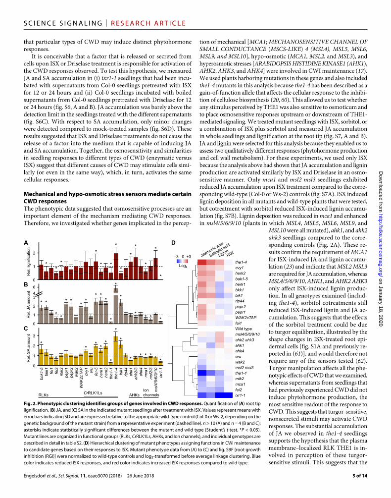
Mechanical and hypo-osmotic stress sensors mediate certain CWD responsesThe phenotypic data suggested that osmosensitive processes are an important element of the mechanism mediating CWD responses. Therefore, we investigated whether genes implicated in the percep-
tion of mechanical [MCA1; MECHANOSENSITIVE CHANNEL OF SMALL CONDUCTANCE (MSCS-LIKE) 4 (MSL4), MSL5, MSL6, MSL9, and MSL10], hypo-osmotic (MCA1, MSL2, and MSL3), and hyperosmotic stresses [ARABIDOPSIS HISTIDINE KINASE1 (AHK1), AHK2, AHK3, and AHK4] were involved in CWI maintenance (17). We used plants harboring mutations in these genes and also included the1-4 mutants in this analysis because the1-4 has been described as a gain-of-function allele that affects the cellular response to the inhibi-tion of cellulose biosynthesis (20, 60). This allowed us to test whether any stimulus perceived by THE1 was also sensitive to osmoticum and to place osmosensitive responses upstream or downstream of THE1- mediated signaling. We treated mutant seedlings with ISX, sorbitol, or a combination of ISX plus sorbitol and measured JA accumulation in whole seedlings and lignification at the root tip (fig. S7, A and B). JA and lignin were selected for this analysis because they enabled us to assess two qualitatively different responses (phytohormone production and cell wall metabolism). For these experiments, we used only ISX because the analysis above had shown that JA accumulation and lignin production are activated similarly by ISX and Driselase in an osmo-sensitive manner. Only mca1 and msl2 msl3 seedlings exhibited reduced JA accumulation upon ISX treatment compared to the corre-sponding wild-type (Col-0 or Ws-2) controls (fig. S7A). ISX induced lignin deposition in all mutants and wild-type plants that were tested, but cotreatment with sorbitol reduced ISX-induced lignin accumu-lation (fig. S7B). Lignin deposition was reduced in mca1 and enhanced in msl4/5/6/9/10 (plants in which MSL4, MSL5, MSL6, MSL9, and
MSL10 were all mutated), ahk1, and ahk2 ahk3 seedlings compared to the corre-sponding controls (Fig. 2A). These re-sults confirm the requirement of MCA1 for ISX-induced JA and lignin accumu-lation (23) and indicate that MSL2 MSL3 are required for JA accumulation, whereas MSL4/5/6/9/10, AHK1, and AHK2 AHK3 only affect ISX-induced lignin produc-tion. In all genotypes examined (includ-ing the1-4), sorbitol cotreatments still reduced ISX-induced lignin and JA ac-cumulation. This suggests that the effects of the sorbitol treatment could be due to turgor equilibration, illustrated by the shape changes in ISX-treated root epi-dermal cells [fig. S1A and previously re-ported in (61)], and would therefore not require any of the sensors tested (62). Turgor manipulation affects all the phe-notypic effects of CWD that we examined, whereas supernatants from seedlings that had previously experienced CWD did not induce phytohormone production, the most sensitive readout of the response to CWD. This suggests that turgor-sensitive, nonsecreted stimuli may activate CWD responses. The substantial accumulation of JA we observed in the1-4 seedlings supports the hypothesis that the plasma membrane–localized RLK THE1 is in-volved in perception of these turgor- sensitive stimuli. This suggests that the
0
1
2
3
4 6
0
1
2
3
A
C
B
Rel
. JA
amou
nt
Rel
. SA
amou
nt
*
* *
* * *
*
* * *
*
* * * * *
*
*
*
*
* *
* * *
*
* * * * *
*
the1-4
cvy1
herk2
bak1-5
herk1
bkk1
bik1
rlp44
pepr2
pepr1
WAK2cTAP
fei1
Wild type msl4/5/6/9/10
ahk2 ahk3
ahk1
ahk4
eru
wak2
msl2 msl3
the1-1
mik2
mca1
fei2
ixr1-1
D
–3 0 +3
Log2
RLKs CrRLK1Ls AHKs Ion channels
th
e1
-4
cvy1
he
rk2
ba
k1
-5
he
rk1
bkk1
bik
1
rlp
44
pe
pr2
pe
pr1
WA
K2
cTA
P
fe
i1
fe
r-5
msl4
/5
/6
/9
/1
0
ah
k2
/3
ah
k1
ah
k4
eru
wa
k2
msl2
/3
th
e1
-1
mik
2
mca
1
fe
i2
ixr1
-1
0
1
2
*
* *
* *
*
* *
* *
*
*
*
Rel
. lig
nific
atio
n
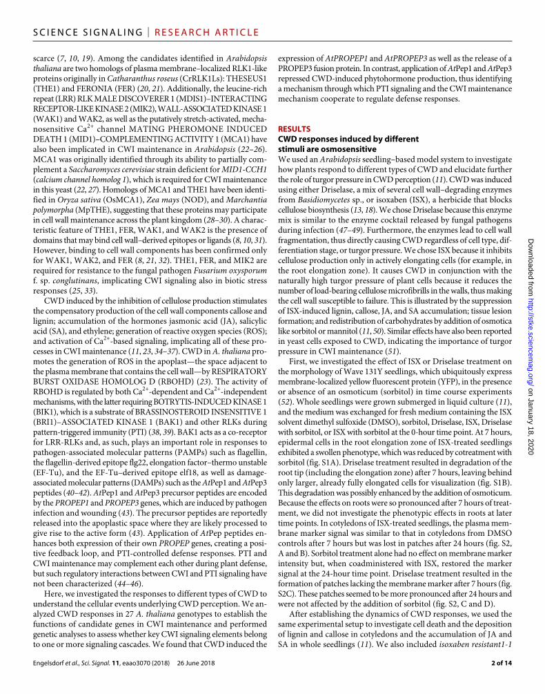
Fig. 2. Phenotypic clustering identifies groups of genes involved in CWD responses. Quantification of (A) root tip lignification, (B) JA, and (C) SA in the indicated mutant seedlings after treatment with ISX. Values represent means with error bars indicating SD and are expressed relative to the appropriate wild-type control (Col-0 or Ws-2, depending on the genetic background of the mutant strain) from a representative experiment (dashed line). n ≥ 10 (A) and n = 4 (B and C); asterisks indicate statistically significant differences between the mutant and wild type (Student’s t test, *P < 0.05). Mutant lines are organized in functional groups (RLKs, CrRLK1Ls, AHKs, and Ion channels), and individual genotypes are described in detail in table S2. (D) Hierarchical clustering of mutant phenotypes assigning functions in CWI maintenance to candidate genes based on their responses to ISX. Mutant phenotype data from (A) to (C) and fig. S9F [root growth inhibition (RGI)] were normalized to wild-type controls and log2-transformed before average linkage clustering. Blue color indicates reduced ISX responses, and red color indicates increased ISX responses compared to wild type.
Engelsdorf et al., Sci. Signal. 11, eaao3070 (2018) 26 June 2018
S C I E N C E S I G N A L I N G | R E S E A R C H A R T I C L E
6 of 14
stimuli indicating compromised CWI may consist of cell wall–bound epitopes that change conformation. Alternatively, mechanical dis-tortion or displacement of the plasma membrane against the cell wall upon CWD, similar to the processes activating the CWI maintenance mechanism in S. cerevisiae, is conceivable as stimuli (51).
Phenotypic clustering identifies a core group of RLKs and ion channels mediating CWI maintenanceGenes required for cell elongation, fertilization, and immunity have been implicated in CWI maintenance (3, 10, 19, 63). To gain further insight into the molecular mode of action of CWI maintenance and to establish which of the candidate genes are required and assess their relative importance in the process, we investigated knockout or gain-of-function alleles for 15 RLKs and 1 RLP [THE1, CURVY 1 (CVY1), FER, HERCULES RECEPTOR KINASE 1 (HERK1), HERK2, ERULUS (ERU), WAK2, FEI1, FEI2, MIK2, BAK1, BAK1-LIKE 1 (BKK1), PEPR1, PEPR2, BIK1, and RECEPTOR-LIKE PROTEIN 44 (RLP44)]. The specific alleles of each gene, including a T-DNA in-sertion allele of WAK2 that we designated as WAK2-12 (fig. S8, A to C), are noted in the figures and summarized in table S2. We mea-sured JA and SA accumulation in mock- and ISX-treated seedlings of these genotypes, as well as in the osmosensing and mechanosens-ing ahk1, ahk2 and ahk3 (ahk2/3), mca1, msl2 and msl3 (msl2/3), and msl4/5/6/9/10 mutants and in the ISX-resistant ixr1 mutant (Fig. 2, B and C, and fig. S9, A to D). JA and SA accumulation was similar to the corresponding wild-type controls in all mock-treated genotypes with the exception of fer-5 seedlings, which already exhibited increased JA and SA accumulation in the mock-treated samples, in line with the multifunctional nature of FER (fig. S9, A to D) (9, 64). Moreover, ISX-induced JA and SA accumulation was strongly increased in fer-5 compared to wild type, suggesting that FER is not essential for per-ception of ISX- induced CWD (fig. S9, B and D). We also investigated root growth and ISX resistance, which could potentially distort the analyses performed here, in each genotype and found no substantial deviations from wild type, with the exception of bak1-5 seedlings showing somewhat shorter roots than wild-type seedlings and irx1-1 seedlings being resistant to ISX, as expected. (fig. S9, E and F). We quantified lignin deposition in the root tip area using an image analysis– based approach to generate quantitative data that could be normalized and used for subsequent hierarchical clustering (Fig. 2A). The quantitative data for JA, SA, and lignin accumulation were inte-grated through hierarchical clustering to generate a global, standardized overview, allowing assessment of both relative importance and func-tions of individual candidates in CWI maintenance (Fig. 2D). Data for fer-5 were not included in the hierarchical clustering to avoid dis-tortion during data integration due to the increased amounts of phyto-hormones in mock- treated seedlings (fig. S9, A and C). The results showed that knockouts in five PTI signaling elements (BAK1, BKK1, BIK1, PEPR1, and PEPR2) enhanced JA and SA accumulation in re-sponse to ISX treatment. Whereas the WAK2cTAP dominant-active allele exhibited increased JA accumulation, wak2 seedlings showed only a slight and statistically insignificant reduction in JA accumula-tion, which might be caused by redundancy within this gene family (8). In parallel, fei2 and mik2 seedlings exhibited significant reductions in the CWD responses examined, implicating (in the case of FEI2) or con-firming [in the case of MIK2; (25)] their involvement in CWI mainte-nance. Seedlings in which the CrRLK1L family members CVY1, HERK1, and HERK2 had been knocked out exhibited enhanced hormone re-sponses, whereas eru seedlings were not strongly affected, and the1-1
seedlings exhibited reduced responses, implying functional divergence within the CrRLK1L family. Loss of RLP44, which is involved in cell wall– mediated activation of brassinosteroid signaling (63), did not affect the responses analyzed, suggesting that RLP44 is not required for responses to ISX-induced CWD. In summary, the hierarchical clustering showed that among the genes we tested, MIK2, MCA1, MSL2/3, FEI2, and THE1 are the most important ones for activation of ISX-induced CWD responses. Several of these proteins have been implicated in turgor perception and mechanoperception and are lo-cated in the plasma membrane or plastid envelope, both of which are subcellular compartments that are particularly sensitive to changes in turgor and mechanical stimuli (19, 65).
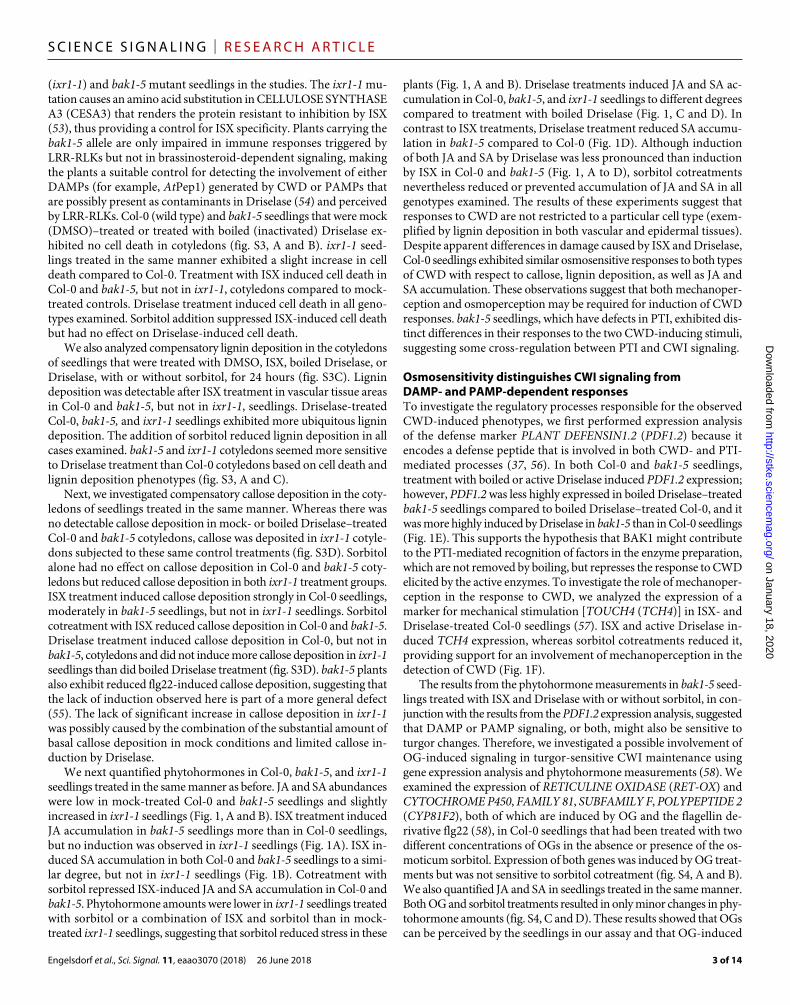
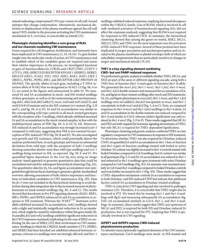
THE1 is a key signaling element mediating CWD- but not PAMP-induced responsesWe performed a genetic analysis to establish whether THE1, MCA1, and FEI2 are part of the same or different signaling cascade, using both a THE1 loss-of-function (the1-1) and a gain-of-function (the1-4) allele. We generated the mca1 fei2, the1-1 mca1, the1-1 fei2, the1-4 mca1, and the1-4 fei2 double mutants and measured the accumulation of JA, SA, and lignin in these mutant seedlings after mock and ISX treatments. JA, SA, and lignin phenotypes in mca1 fei2, the1-1 mca1, and the1-1 fei2 seedlings were not additive, but fei2 was epistatic to mca1, and the1-1 was epistatic to both mca1 and fei2 (Fig. 3, A to C). Next, we compared responses in the1-4 mca1 and the1-4 fei2 seedlings to the1-4 alone. JA and SA accumulation in the double mutants was reduced compared to the1-4 and similar to Col-0, whereas relative lignification was only re-duced in the1-4 mca1 (Fig. 3, D to F). These results suggested that MCA1 and FEI2 are both required for hormone signaling downstream of THE1, but only MCA1 is required for THE1-dependent lignification.
Phenotypic clustering and genetic analyses confirmed THE1 as a key regulatory component in CWI maintenance in response to ISX treatment. To determine whether THE1 was also required for Driselase- induced CWD, we quantified JA and SA accumulation in the1-1 loss- of-function and the1-4 gain-of-function seedlings treated with boiled or active Driselase. SA content was slightly increased in the1-4 controls treated with boiled Driselase compared to Col-0 seedlings, but JA content was similar in all genotypes (Fig. 3, G and H). JA accumulation was reduced in the1-1 and enhanced in the1-4 seedlings upon treatment with active Driselase compared to Col-0 seedlings (Fig. 3G). SA amounts were increased simi-larly in Col-0 and the1-4 seedlings compared to boiled Driselase controls and even further increased in the1-1 (Fig. 3H). These results suggest that a THE1-dependent mechanism controls JA accumulation in response to both Driselase- and ISX-induced CWD but indicate that additional factors control SA accumulation in Driselase- treated seedlings.
THE1 is critical for CWI signaling and also involved in pathogen resistance (25). Therefore, it is conceivable that THE1 might also be involved in PTI. We tested this by treating the1-1 and the1-4 seed-lings with flg22 and measuring subsequently SA accumulation (fig. S10). SA accumulated similarly in Col-0, the1-1, and the1-4 seed-lings. In summary, these results suggest that THE1 acts upstream of MCA1 and FEI2, is required for both ISX and Driselase-induced JA production, but is not required for PTI, implying that THE1 is spe-cifically involved in CWI signaling.
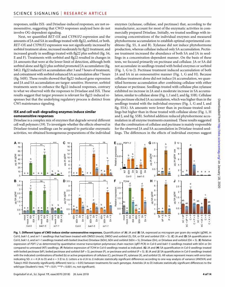
AtPEP1 and AtPEP3 repress CWD-induced phytohormone productionTo identify transcriptionally regulated elements of the CWI mainte-nance mechanism, Col-0 seedlings were mock- or ISX-treated and
Engelsdorf et al., Sci. Signal. 11, eaao3070 (2018) 26 June 2018
S C I E N C E S I G N A L I N G | R E S E A R C H A R T I C L E
7 of 14
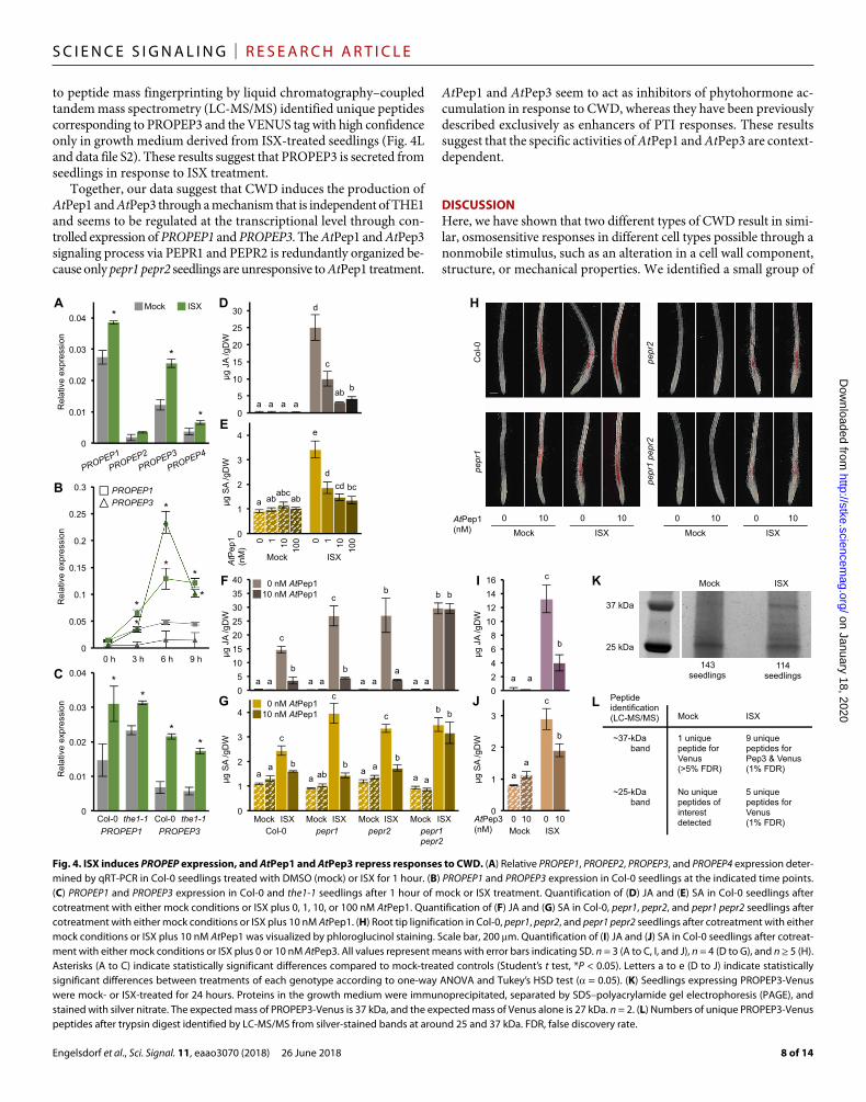
analyzed by RNA sequencing (RNA-seq) using mock- or ISX-treated ixr1-1 seedlings as controls. In Col-0 seedlings treated with ISX for 1 hour, 109 transcripts exhibited statistically significant differences from mock-treated controls (data file S1). Although mock-treated ixr1-1 seedlings exhibited differences compared to mock-treated Col-0 at the transcriptome level, none of the ISX-regulated transcripts in Col-0 were differentially expressed in ISX-treated ixr1-1 seedlings (data file S1). Gene Ontology (GO) enrichment analysis detected an overrepresentation of genes implicated in phytohormone-dependent stress responses in ISX-treated Col-0 seedlings (table S1). Among the differentially expressed transcripts were PROPEP1, PROPEP2, PROPEP3, and PROPEP4, which encode the precursors of the signaling pep-tides AtPep1 to AtPep4 (fig. S11A) (43). PEPR1 has been shown to bind AtPep1 to AtPep4, whereas PEPR2 binds only AtPep1 and AtPep2 (43). This observation was intriguing given that pepr1 and pepr2 seed-lings exhibit enhanced JA accumulation upon ISX treatment (Fig. 2B). Gene expression analysis by qRT-PCR showed that PROPEP1 and PROPEP3 expression were particularly strongly induced by ISX (Fig. 4A). Time course expression analysis of PROPEP1 and PROPEP3 detected increases in expression over time in ISX-treated seedlings, suggesting that AtPep1 and AtPep3 might accumulate in response to ISX treatment (Fig. 4B). Expression of PROPEP1 and PROPEP3 was still increased in ISX-treated the1-1 seedlings, indicating that their induction was inde-
pendent of THE1-mediated processes (Fig. 4C). AtPep1 enhances JA, SA, and ethylene accumulation in response to wounding (66). To in-vestigate whether AtPep1 also enhanced CWD responses, Col-0 seed-lings were treated with different concentrations of AtPep1 alone or in combination with ISX before JA, SA, and lignin accumulation were measured. AtPep1 treatments alone did not induce JA and SA accu-mulation (Fig. 4, D and E). Seedlings cotreated with ISX and AtPep1 exhibited reductions in ISX-induced JA and SA accumulation in a manner that depended on the concentration of AtPep1 (Fig. 4, D and E). AtPep1 induced lignin deposition in Col-0 seedling roots in a distinctly different pattern than ISX treatment did, whereas lignin deposition seemed to be additive in cotreated root tips compared to roots treated with either AtPep1 or ISX alone (fig. S11B).
To exclude indirect effects and determine whether the observed effects on AtPep1 were mediated by the established AtPep1 receptors, the experiments were repeated with Col-0, pepr1, pepr2, and pepr1 pepr2 seedlings. ISX-induced JA and SA accumulation was reduced in pepr1 and pepr2 seedlings upon cotreatment with ISX and AtPep1, similarly to the Col-0 seedlings (Fig. 4, F and G). However, this was not the case in pepr1 pepr2 seedlings, in which cotreatment with AtPep1 did not counteract ISX-induced accumulation of JA and SA, suggest-ing that AtPep1 can inhibit ISX-induced phytohormone production through either PEPR1 or PEPR2. Analysis of lignin deposition in seed-
lings treated with AtPep1, ISX, or both showed that PEPR2 is essential for AtPep1- induced lignin deposition, but PEPR1 is not (Fig. 4H). This provided further sup-port for differences between PEPR1 and PEPR2 with respect to signaling activities. We also investigated AtPep3, which binds to PEPR1 but not to PEPR2, and found that AtPep3 cotreatment with ISX had similar effects as AtPep1 cotreatment on JA and SA accumulation in Col-0 seed-lings (Fig. 4, I and J). However, AtPep3 did not induce lignin production as did AtPep1 (fig. S11C), which can be explained by the inability of AtPep3 to bind to PEPR2 (42). Because the fusion protein PROPEP3- Venus is secreted upon treatment with AtPep2 (67), we tested whether ISX treatment also induced PROPEP3-Venus secretion. Seedlings stably expressing pPROPEP3:: PROPEP3-Venus were mock- or ISX- treated, and we immunoprecipitated the fusion protein from the growth medium (68). On a silver-stained polyacrylamide gel containing the protein preparations from the growth medium, bands of around 25 kDa were visible in preparations from both mock- and ISX-treated samples, con-sistent with the fusion protein being pro-cessed after secretion into the apoplast to release Venus alone (27 kDa) (Fig. 4K). A band corresponding to the size of the full-length fusion protein (PROPEP3- Venus, 37 kDa) was only detected in the preparation from ISX-treated seedlings. Subjecting the proteins in these bands
0
20
40
60
0
2
4
6
Col
-0 0
1
2
3
4
5
0
10
20
30
40 B
µg J
A /g
DW
µg
SA
/gD
W
A
E
µg J
A /g
DW
µg
SA
/gD
W
D
d
e
a
c bc ab ab
c
d
a
b b
a a
0 0.2 0.4 0.6 0.8
1 1.2 1.4
F C
0
2
4
6
8
10
12
0
0.5
1
1.5
2
2.5
3
3.5
4
Co
l-0
th
e1
-1
th
e1
-4
Co
l-0
th
e1
-1
th
e1
-4
H
µg J
A /g
DW
µg
SA
/gD
W
G bDri
Dri
Col
-0
Col
-0
*
**
***
b
a a a
c
b a ab
0
1
2
3
Rel
. lig
nific
atio
n
b
a
b
a
**
mca
1
fe
i2
mca
1 fe
i2
th
e1
-1
th
e1
-1
fe
i2
th
e1
-1
m
ca
1
Rel
. lig
nific
atio
n
a a
a a a a
b
th
e1
-4
th
e1
-4
m
ca
1
th
e1
-4
fe
i2
Col
-0
Fig. 3. THE1 functions upstream of MCA1 and FEI2 and promotes responses to different types of CWD. Quanti-fication of (A) root tip lignification, (B) JA, and (C) SA after ISX treatment in wild-type (Col-0) and mutant seedlings carrying the indicated loss-of-function mutations. Quantification of (D) root tip lignification, (E) JA, and (F) SA after ISX treatment in Col-0 and mutant seedlings carrying the gain-of-function allele the1-4 or the1-4 in combination with the loss-of-function allele mca1 or fei2. Quantification of (G) JA and (H) SA in Col-0, the1-1 (loss-of-function), and the1-4 (gain-of-function) seedlings treated with boiled Driselase (bDri) or Driselase (Dri). n ≥ 17 (lignin) and n = 4 (JA and SA). Letters a to e (A to F) indicate statistically significant differences between genotypes according to one-way ANOVA and Tukey’s HSD test ( = 0.05). Asterisks (G and H) indicate statistically significant differences compared to the Col-0 control (Student’s t test, *P < 0.05; **P < 0.01; ***P < 0.001).
Engelsdorf et al., Sci. Signal. 11, eaao3070 (2018) 26 June 2018
S C I E N C E S I G N A L I N G | R E S E A R C H A R T I C L E
8 of 14
to peptide mass fingerprinting by liquid chromatography–coupled tandem mass spectrometry (LC-MS/MS) identified unique peptides corresponding to PROPEP3 and the VENUS tag with high confidence only in growth medium derived from ISX-treated seedlings (Fig. 4L and data file S2). These results suggest that PROPEP3 is secreted from seedlings in response to ISX treatment.
Together, our data suggest that CWD induces the production of AtPep1 and AtPep3 through a mechanism that is independent of THE1 and seems to be regulated at the transcriptional level through con-trolled expression of PROPEP1 and PROPEP3. The AtPep1 and AtPep3 signaling process via PEPR1 and PEPR2 is redundantly organized be-cause only pepr1 pepr2 seedlings are unresponsive to AtPep1 treatment.
AtPep1 and AtPep3 seem to act as inhibitors of phytohormone ac-cumulation in response to CWD, whereas they have been previously described exclusively as enhancers of PTI responses. These results suggest that the specific activities of AtPep1 and AtPep3 are context- dependent.
DISCUSSIONHere, we have shown that two different types of CWD result in simi-lar, osmosensitive responses in different cell types possible through a nonmobile stimulus, such as an alteration in a cell wall component, structure, or mechanical properties. We identified a small group of
0
1
2
3
4 0
0.01
0.02
0.03
0.04
0
0.05
0.1
0.15
0.2
0.25
0.3
0 h 3 h 6 h 9 h
0
0.01
0.02
0.03
0.04
0
5
10
15
20
25
30
1
D
µg J
A /g
DW
µg
SA
/gD
W
10
100 0 1 10
10
0 0
ISX Mock
AtP
ep1
(nM
)
a ab abc
ab bc cd
d
e
a a a a ab b
c
d A
B
PROPEP1 PROPEP3
Mock ISX
the1-1 the1-1 Col-0 Col-0
Rel
ativ
e ex
pres
sion
*
* *
* C
E
Rel
ativ
e ex
pres
sion
*
*
*
K
Rel
ativ
e ex
pres
sion
*
*
*
*
* *
PROPEP1 PROPEP3
0 5
10 15 20 25 30 35 40 F
0
1
2
3
4
µg J
A /g
DW
µg
SA
/gD
W
Mock ISX Col-0
Mock ISX pepr1
Mock ISX pepr2
Mock ISX pepr1
pepr2
0 nM AtPep1 10 nM AtPep1
0 nM AtPep1 10 nM AtPep1
a a a a a a a a a
b b
b b
c
c b
a a b
c
a ab b
c
a a b
c
a a
b b G L
37 kDa
25 kDa
143 seedlings
114 seedlings
Mock ISX
Mock ISX
~37-kDa band
1 unique peptide for Venus (>5% FDR)
9 unique peptides for Pep3 & Venus (1% FDR)
~25-kDa band
No unique peptides of interest detected
5 unique peptides for Venus (1% FDR)
H
Col
-0
pepr1
pepr2
pepr1 pepr2
Mock ISX
0 10 0 10
Mock ISX
0 10 0 10 AtPep1 (nM)
AtPep3 (nM)
I
µg J
A /g
DW
µg
SA
/gD
W
J 0 2 4 6 8
10 12 14 16
0
1
2
3
10 0 10 0 ISX Mock
a a
c
b
a a
c
b
Peptide identification (LC-MS/MS)
Fig. 4. ISX induces PROPEP expression, and AtPep1 and AtPep3 repress responses to CWD. (A) Relative PROPEP1, PROPEP2, PROPEP3, and PROPEP4 expression deter-mined by qRT-PCR in Col-0 seedlings treated with DMSO (mock) or ISX for 1 hour. (B) PROPEP1 and PROPEP3 expression in Col-0 seedlings at the indicated time points. (C) PROPEP1 and PROPEP3 expression in Col-0 and the1-1 seedlings after 1 hour of mock or ISX treatment. Quantification of (D) JA and (E) SA in Col-0 seedlings after cotreatment with either mock conditions or ISX plus 0, 1, 10, or 100 nM AtPep1. Quantification of (F) JA and (G) SA in Col-0, pepr1, pepr2, and pepr1 pepr2 seedlings after cotreatment with either mock conditions or ISX plus 10 nM AtPep1. (H) Root tip lignification in Col-0, pepr1, pepr2, and pepr1 pepr2 seedlings after cotreatment with either mock conditions or ISX plus 10 nM AtPep1 was visualized by phloroglucinol staining. Scale bar, 200 m. Quantification of (I) JA and (J) SA in Col-0 seedlings after cotreat-ment with either mock conditions or ISX plus 0 or 10 nM AtPep3. All values represent means with error bars indicating SD. n = 3 (A to C, I, and J), n = 4 (D to G), and n ≥ 5 (H). Asterisks (A to C) indicate statistically significant differences compared to mock-treated controls (Student’s t test, *P < 0.05). Letters a to e (D to J) indicate statistically significant differences between treatments of each genotype according to one-way ANOVA and Tukey’s HSD test ( = 0.05). (K) Seedlings expressing PROPEP3-Venus were mock- or ISX-treated for 24 hours. Proteins in the growth medium were immunoprecipitated, separated by SDS–polyacrylamide gel electrophoresis (PAGE), and stained with silver nitrate. The expected mass of PROPEP3-Venus is 37 kDa, and the expected mass of Venus alone is 27 kDa. n = 2. (L) Numbers of unique PROPEP3-Venus peptides after trypsin digest identified by LC-MS/MS from silver-stained bands at around 25 and 37 kDa. FDR, false discovery rate.
Engelsdorf et al., Sci. Signal. 11, eaao3070 (2018) 26 June 2018
S C I E N C E S I G N A L I N G | R E S E A R C H A R T I C L E
9 of 14
molecular components, most of which are involved in the perception of mechanical or hypo-osmotic stress, that mediate both local (depo-sition of lignin) and systemic (phytohormone accumulation) responses to CWD. Simultaneously, we observed that loss of PTI signaling ele-ments, such as BAK1, BIK1, BKK1, PEPR1, and PEPR2, enhanced the responses to CWD. We showed that THE1, MCA1, and FEI2 belong to the same signaling cascade and that THE1 was involved in mediating responses to both Driselase and ISX-induced CWD but not PTI-associated SA accumulation. We found that CWD induced PROPEP1 and PROPEP3 expression in a THE1-independent manner and that a PROPEP3-VENUS fusion protein is released into the growth medium from seedlings in response to ISX treatment. Application of AtPep1 and AtPep3 repressed CWD-induced JA and SA accumu-lation in a concentration-dependent manner, and repression by AtPep1 depended on the activity of PEPR1 and PEPR2. These results provide insights into the early events during CWD perception and the mecha-nisms regulating the cellular and systemic responses.
ISX and Driselase treatments resulted in similar responses in seed-lings. Experiments with the individual cell wall–degrading enzymes found in Driselase and combinations thereof showed that pectinase and cellulase together caused overall the greatest JA accumulation, whereas SA amounts were lower than in seedlings treated with pectin-ase alone. Pectinase may increase the accessibility of cellulose to cellulase, thus facilitating the breakdown of this load-bearing cell wall component and perhaps explaining the similarities in the ob-served responses to ISX, Driselase, and combined pectinase plus cel-lulase treatments. Sorbitol cotreatments dampened all the responses to both enzyme- and ISX-induced CWD, suggesting that CWD responses are induced by a stimulus that is sensitive to turgor pressure. Treat-ments with supernatants derived from seedlings exposed to ISX or Driselase induced neither JA nor SA accumulation in a manner simi-lar to Driselase and ISX treatments, suggesting that the stimulus acti-vating the CWD responses is not mobile. All the genes that we identified through phenotypic clustering as being required for responses to CWD have been implicated in signal transduction or the perception of hypo-
osmotic, mechanical, or CWD (9, 19, 65, 69). This provides further support that the initial stimulus, indicating that CWD has occurred, could be physical (mechanical). Together, these observations suggest that CWD could result in distortion or displacement of the plasma membrane relative to the cell wall possibly caused by changes in the surface tension of the wall itself due to weakening of the load-bearing cellulose framework. These changes, in turn, could be detected by the CWI maintenance mechanism and lead to the observed responses.
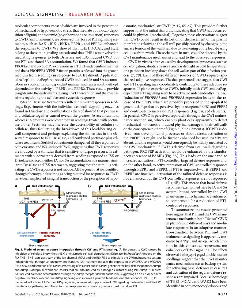
CWD in vivo is often caused by developmental processes, such as cell elongation, abiotic stressors (such as drought or cold temperature), or a pathogen breaking down the cell wall as part of the infection pro-cess (7, 70). Each of these different sources of CWD requires spe-cialized, adaptive responses. The data presented here suggest that CWI and PTI signaling may coordinately contribute to these adaptive re-sponses. If plants experience CWD, initially both CWI and AtPep- dependent PTI signaling seem to be activated independently (Fig. 5A). Induction of PROPEP1 and PROPEP3 expression leads to the re-lease of PROPEPs, which are probably processed in the apoplast to generate AtPeps that are perceived by the receptors PEPR1 and PEPR2 and contribute to increased PTI responses (Fig. 5A, red elements). In parallel, CWD is perceived separately through the CWI mainte-nance mechanism, which enables plant cells apparently to detect mechanical- or osmotic-induced physical damage to their cell walls or the consequences thereof (Fig. 5A, blue elements). If CWD is de-rived from developmental processes or abiotic stress, activation of the PROPEPs might not be further enhanced because PAMPs are absent, and the responses would consequently be mainly mediated by the CWI mechanism. If CWD is derived from a cell wall–degrading pathogen, PROPEP activation would be enhanced by the simulta-neous presence of PAMPs (Fig. 5A). This leads, on the one hand, to increased activation of PTI-controlled, targeted defense responses and, on the other hand, to active repression of CWI-controlled responses through PEPR1 and PEPR2. If PTI is impaired—or if PEPR1 and PEPR2 are inactive—activation of the tailored defense responses is not enhanced, but the CWI-controlled responses are not repressed
(Fig. 5B). This means that basal defense responses (exemplified here by JA and SA accumulation) controlled by the CWI maintenance mechanism are enhanced to compensate for a reduction of PTI- controlled responses.
To summarize, the results presented here suggest that PTI and the CWI main-tenance mechanism both “detect” CWD in plant cells in different ways and modu-late responses in an adaptive manner. Coordination between PTI and CWI maintenance signaling is apparently me-diated by AtPep1 and AtPep3, which func-tion in this context as repressors, not enhancers, of CWI signaling. The effects observed in the pepr1 pepr2 double-mutant seedlings suggest that the CWI mainte-nance mechanism acts as backup system for activating basal defenses in case PTI and activation of the regular defense re-sponses are impaired. Because homologs of THE1, MCA1, and WAK2 have been identified in both monocotyledonous and
Cell wall damage
Cell wall integrity maintenance
Pattern-triggered immunity
PROPEP1/3
Cell wall damage
Cell wall integrity maintenance
PEPR1/2 THE1 MCA1 and FEI2
PROPEP1/3
A B
Other stimuli (pathogen elicitors, hormones, )
Other stimuli (pathogen elicitors,hormones, ) PEPR1/2 THE1
MCA1 and FEI2 ? ?
Stress responses Stress responses Fig. 5. Model of stress response integration through CWI and PTI signaling. (A) Responses to CWD caused by inhibition of cellulose biosynthesis (ISX) or enzymatic cell wall degradation (Driselase) in Arabidopsis depend on the RLK THE1. THE1 acts upstream of the ion channel MCA1 and the RLK FEI2 to stimulate the CWI maintenance system. Independently, through an unknown mechanism, ISX treatment induces the expression of PROPEP1 and PROPEP3 (PROPEP1/3) and secretion of PROPEP3. Processing of PROPEP1 and PROPEP3 generates the host defense peptides AtPep1 and AtPep3 (AtPep1/3), which are DAMPs that are also induced by pathogen elicitors during PTI. AtPep1/3 repress ISX-induced hormone accumulation through the AtPep receptors PEPR1 and PEPR2, suggesting an AtPep-dependent negative feedback mechanism. AtPep signaling also induces a positive feedback loop that enhances PTI. (B) If PTI- mediated induction of AtPeps or AtPep signaling is impaired, suppression of CWI signaling is alleviated, and the CWI maintenance pathway contributes to stress response induction to a greater extent than does PTI.
Engelsdorf et al., Sci. Signal. 11, eaao3070 (2018) 26 June 2018
S C I E N C E S I G N A L I N G | R E S E A R C H A R T I C L E
10 of 14
dicotyledonous plants as well as in more ancient species, the CWI maintenance mechanism—and potentially its interactions with PTI- based defense responses—may be conserved throughout the plant kingdom (28–30).
MATERIALS AND METHODSReagentsAll chemicals and enzymes were purchased from Sigma-Aldrich un-less stated otherwise.
Plant growth and treatmentsWild-type and mutant A. thaliana strains used in this study were ordered from the Nottingham Arabidopsis Stock Centre (http://arabidopsis. info/) or obtained directly from the laboratories previously publishing them. Detailed information is listed in table S2. Seedlings were grown in liquid culture as described (23) with minor modifi-cations. Thirty milligrams of seeds was sterilized by sequential incu-bation with 70% ethanol and 50% bleach on a rotating mixer for 10 min each and washed three times with sterile water. Seeds were then transferred into 250-ml Erlenmeyer flasks containing 125-ml half-strength Murashige and Skoog growth medium [Murashige and Skoog Basal Medium (2.1 g/liter), MES salt (0.5 g/liter), and 1% su-crose at pH 5.7]. Seedlings were grown under long-day conditions (16-hour light, 22°C and 8-hour dark, 18°C) at a photon flux density of 150 mol m−2 s−1 on a IKA KS 501 flask shaker at a constant speed of 130 rpm.
For all experiments, seedlings were grown for 6 days before treat-ment. The following products were used for treatments at the indicated final concentrations throughout the paper, unless stated otherwise: ISX (600 nM; DMSO), mock (DMSO), Driselase (0.03%, w/v; D8037, Sigma-Aldrich), cellulase (0.09%, w/v; C8001, Duchefa), pectinase (0.09%, w/v; 17389, Sigma-Aldrich), xylanase (0.09%, w/v; X2753, Sigma-Aldrich), and sorbitol (300 mM). For heat inactivation, enzymes were boiled for 10 min. Supernatants from treated Col-0 cultures were incubated with ixr1-1 seedlings (DMSO, ISX, and ISX + S) or boiled for 10 min and incubated with Col-0 seedlings (DMSO + S, bDri, bDri + S, Dri, and Dri + S). AtPep1 (ATKVKAKQRGKEKVSSGRP-GQHN), AtPep3 (EIKARGKNKTKPTPSSGKGGKHN), and flg22 (QRLSTGSRINSAKDDAAGLQIA) peptides were obtained from Peptron and dissolved in sterile water.
Confocal laser scanning microscopyWAVE 131Y (52) seedlings used to investigate structural changes in root and cotyledon cells after CWD were placed on microscopy slides, covered with the same medium used for the treatment, and imaged with a Leica SP8 confocal laser scanning microscope. Four Z-stacks were taken for each of the conditions analyzed using HC PL APO 10×/0.40 DRY objective [excitation, 514 nm; BA (barrier filter), 525 to 535], 0.7–airy unit pinhole, and 700-V gain. Z-stacks were trans-formed in two- dimensional images by using the maximum intensity projections (0, threshold) function on LAS X software. To highlight the cell outlines, Z-projection images were transformed in grayscale using GIMP v2.8.22 and presented as insets.
Phytohormone analysisThe JA and SA contents of seedlings were analyzed as described in (71), with minor modifications. Seedlings were sampled at 7 hours after treatment, flash-frozen in liquid nitrogen, and freeze-dried for
24 hours. Aliquots each containing 6 to 7 mg of freeze-dried seed-lings were ground with 5-mm stainless steel beads in a Qiagen Tissue Lyser II for 2 min at 25 Hz. Shaking was repeated after the addition of 400-l extraction buffer (10% methanol and 1% acetic acid) with internal standards (10 ng of jasmonic-d5 acid and 28 ng of salicylic-d4 acid; CDN Isotopes) before samples were incubated on ice for 30 min and centrifuged for 10 min at 16,000g and 4°C. Supernatants were transferred into fresh tubes, and the pellets were reextracted with 400-l extraction buffer without internal standards. Supernatants were combined and centrifuged three times to remove all debris before LC-MS/MS analysis. An extraction control not containing plant material was treated equally to the plant samples. Chromatographic separation was carried out on a Shimadzu UFLC XR, equipped with a Waters Cortecs C18 column (2.7 m; 2.1 × 100 mm). The solvent gradient [acetonitrile (ACN)/water with 0.1 % formic acid each] was adapted to a total run time of 7 min: 0 to 4 min, 20 to 95% ACN; 4 to 5 min, 95% ACN; 5 to 7 min, 95 to 20% ACN; flow rate, 0.4 ml/min. For hormone identification and quantification, an AB SCIEX Triple Quad 5500 system was used. Mass transitions were as follows: JA 209 > 59, D5-JA 214 > 62, SA 137 > 93, D4-SA 141 > 97.
Callose analysisSeedlings were sampled 24 hours after treatment and placed in 70% (v/v) ethanol. For callose staining, samples were incubated in 0.07 M sodium phosphate buffer (pH 9) for 30 min and in 0.005% (w/v) aniline blue [in 0.07 M sodium phosphate buffer (pH 9)] for 60 min. Samples were washed with water, mounted in 50% (v/v) glycerol, and analyzed on a Nikon Eclipse E800 microscope using a UV-2A filter (excitation, 330 to 380 nm; dichroic mirror, 400 nm; BA, 420 nm). Images were taken at ×10 magnification, and callose depositions were quantified using ImageJ software.
Lignin analysisLignification was investigated 12 hours (root tips) and 24 hours (cotyledons) after the start of treatments. Lignin was detected with phloroglucinol-HCl as described (23). Seedlings were photographed using a Zeiss Axio Zoom.V16 stereomicroscope. To assess the ex-tent of lignin production in root tips, phloroglucinol-stained areas and the total root area imaged were quantified using ImageJ (the same root length was maintained in all images taken). The relative lignified area was plotted as fold change compared to wild-type root tips.
Cell death analysisSeedlings were sampled after 24 hours of treatment and incubated in trypan blue staining solution (0.025% trypan blue, 25% phenol, dissolved in equal volumes of lactic acid, glycerol, and water) for 6 hours at room temperature. Samples were destained in chloral hy-drate overnight and transferred into 60% glycerol before microscopy. Images of the cotyledons were obtained with a Zeiss Axio Zoom.V16 stereomicroscope. The percentages of trypan blue–stained areas were quantified from cotyledons using ImageJ color thresholding.
Root growth measurementsAbsolute root lengths were measured immediately before ISX treat-ment (0 hours) to examine root growth phenotypes and 24 hours after start of treatment to determine ISX-dependent RGI. For calcu-lation of %RGI, the following formula was applied: [1 − (ISX 24 hours − ISX 0 hours)/(mock 24 hours − mock 0 hours)]*100.
Engelsdorf et al., Sci. Signal. 11, eaao3070 (2018) 26 June 2018
S C I E N C E S I G N A L I N G | R E S E A R C H A R T I C L E
11 of 14
Hierarchical cluster analysisHierarchical clustering of ISX-dependent phenotypes was per-formed with Cluster 3.0 using the C Clustering Library v1.52 (72). All data from mutant seedlings were normalized to their corre-sponding wild-type control. Log2-transformed data were then used for average linkage clustering with an uncentered correlation simi-larity metric. Results were depicted using Java TreeView v1.1.6r4 and color-coded blue (less than in wild type) or red (more than in wild type) (73).
Genotyping the WAK2 T-DNA insertionSeeds were sown on a six-well plate and grown in 1/2 MS1 for 6 days. Genomic DNA was extracted by grinding the plant material in a 2-ml Eppendorf tube with 5-mm stainless steel beads and 500 l of extraction buffer [0.5 M NaCl, 50 mM EDTA, 0.1 M tris-HCl (pH 8.0)] in a Qiagen TissueLyser II for 1 min at 25 Hz. The lysate was centri-fuged, and 300 l of supernatant was combined with 300 l of iso-propanol to precipitate the DNA. After centrifugation, the pellet was washed with 70% ethanol and dissolved in 100 l of Milli-Q water. One microliter of the isolated DNA was used for PCR using Taq polymerase. The PCR program included an initial denaturation at 95°C for 2 min, followed by 30 cycles with 95°C for 30 s, 57°C for 30 s, and 72°C for 1 min with final elongation at 72°C for 2 min. The PCR products were run on 1% agarose gel containing GelRed dye and imaged with a Syngene G:BOX imaging device.
Quantitative reverse transcription polymerase chain reactionTotal RNA was isolated using a Spectrum Plant Total RNA kit (Sigma- Aldrich). Two micrograms of total RNA was treated with RQ1 RNase- Free DNase (Promega) and processed with the ImProm-II Reverse Transcription System (Promega) for complementary DNA (cDNA) synthesis. qRT-PCR was performed using a LightCycler 480 SYBR Green I Master (Roche) and primers (table S3) diluted according to the manufacturer’s specifications. Four different reference genes (PP2A, ACT2, UBA1, and GRF2) were examined to identify one exhibiting stable expression during ISX treatment. ACT2 was the most stable and used in all experiments as a reference.
RNA-seq analysisTotal RNA was extracted using a Spectrum Plant Total RNA kit (Sigma- Aldrich). RNA concentration was measured using a Qubit RNA HS Assay kit (Thermo Fisher Scientific), and the integrity of the RNA was assessed using an Agilent RNA 6000 Pico kit. RNA-seq libraries were prepared using a TruSeq Stranded mRNA kit (Illumina) ac-cording to the manufacturer’s instructions. Total RNA (500 ng) was used as starting material.
First, index barcodes were ligated for identification of individual samples. mRNA purification, fragmentation, and cDNA synthesis were performed as described in (74). Exonuclease/polymerase was used to produce blunted overhangs. Illumina SR adapter oligonu-cleotides were ligated to the cDNA after 3′ end adenylation. DNA fragments were enriched by 15 cycles of PCR. The libraries were puri-fied using the AMPure XP (Beckman Coulter), quantitated by qPCR using a KAPA Library Quantification kit (Kapa Biosystems), and validated using an Agilent High Sensitivity DNA kit on a Bioanalyzer. The size range of the DNA fragments was measured to be in the range of 200 to 700 base pairs (bp) and peaked around 296 bp. Libraries were normalized and pooled to 2.2 pM and subjected to clustering
on NextSeq 500 high-output flow cells. Finally, single-read sequencing was performed for 75-bp read lengths on a NextSeq 500 instrument (Illumina) according to the manufacturer’s instructions. Base calling has been performed on the NS500 instrument by Illumina RTA v2.4.6. FASTQ files were generated using bcl2fastq2 Conversion Software v1.8.4. Each FASTQ file was subjected to quality control through FastQC v11.1 before technical replicates were combined, and an aver-age of 13.1 million reads was produced for each library. The reads were then aligned to the A. thaliana genome (Ensembl v82) with STAR v2.4.1 in two-pass mode. On average, 96.2% of the reads aligned to the genome. The reads that aligned uniquely to the genome were aggregated into gene counts with featureCounts v1.4.6 using the genome annotations defined in Ensembl v82. Of the 32,000 genes defined in the gene model, a total of 20,750 genes were left for analy-sis after filtering out genes with a CPM (counts per million) value less than 1 in two or more samples.
The filtered gene count table was used as input to the Voom method of the limma R package v3.26.9 for differential expression (75). The sam-ples were normalized using the TMM (trimmed mean of M values nor-malization) method before a linear model was defined (76). Differential expression between groups was tested by empirical Bayesian moderated t tests, and P values were corrected for multiple testing by the Benjamini- Hochberg FDR adjustment. Statistical significance of pairwise compari-sons was determined using a Student’s t test. Genes with significantly altered expression after 1 hour of ISX treatment (data file S1) were an-alyzed for GO enrichment using the PANTHER Overrepresentation Test (release 15 July 2016) and the GO Ontology database (release 28 February 2017) on http://geneontology.org/. Results were filtered by P < 0.05 after Bonferroni correction for multiple testing. Data generated in the transcriptomics experiments are available under the following Gene Expression Omnibus (GEO) submission ID: GSE109613.
Extracellular PROPEP3-Venus assay and peptide mass fingerprintingSeedlings stably expressing pPROPEP3::PROPEP3-Venus were grown for 6 days before mock and ISX treatments (68). After 24 hours, the growth medium was filtered through sterile Miracloth and pH- adjusted to 7.5 with KOH. The number of seedlings per treatment was counted. Protease inhibitor cocktail (P9599, Sigma-Aldrich) and phosphatase inhibitor cocktail 1 (P2850, Sigma-Aldrich) were added to the medium. GFP-Trap agarose beads (ChromoTek) were equilibrated according to the manufacturer’s instructions, and 50 l of bead slurry was added to 2-ml medium. The suspension was tumbled end- over-end for 2 hours at 4°C. Beads were recovered by centrifugation and washed as described. Proteins were dissociated from beads by incubating for 10 min at 95°C in 2× Laemmli buffer, and supernatants were separated via 10% acrylamide gel for SDS-PAGE. Proteins were visualized using a Bio-Rad Silver Stain Plus kit according to the manufacturer’s instructions, and the gel was imaged on a Bio-Rad ChemiDoc XRS+ System.
Gel bands were cut in smaller pieces (3 to 5 mm3) and were destained by incubation for 2 min in 150-l ProteoSilver Destainer solution mix. The gel pieces were washed with ultrapure water and then shrunk with ACN. They were reduced with dithiothreitol at 56°C, alkylated by iodoacetamide at room temperature in the dark, and—after being washed and shrunk—digested by trypsin at 37°C overnight. Peptides were collected, dried in a vacuum concentrator, and reconstituted in 0.1% formic acid.
LC-MS/MS analysis was performed on an EASY-nLC 1200 UPLC system interfaced with a Q Exactive HF mass spectrometer via a
Engelsdorf et al., Sci. Signal. 11, eaao3070 (2018) 26 June 2018
S C I E N C E S I G N A L I N G | R E S E A R C H A R T I C L E
12 of 14
Nanospray Flex ion source. Peptides were injected onto an Acclaim PepMap100 C18 trap column (75-m inside diameter, 2 cm long, 3 m, 100 Å) and further separated on an Acclaim PepMap100 C18 analytical column (75-m inside diameter, 50 cm long, 2 m, 100 Å) using a 60-min gradient (40 min, 5 to 40% B; 7 min, 40 to 100% B; 13 min, 100% B; where B is 0.1% formic acid in CH3CN) at a flow of 250 nl/min. Peptides were analyzed in positive ion mode under data- dependent acquisition using the following parameters: electrospray voltage, 1.9 kV; HCD fragmentation with normalized collision energy, 28%. Each MS1 scan [200 to 2000 mass/charge ratio (m/z), profile] was acquired at a resolution of 120,000 full width at half maximum (FWHM) in the Orbitrap analyzer, followed by 15,000 FWHM MS2 scans (1.2 m/z isolation width, centroid) triggered for the 12 most intense ions, with a 15-s dynamic exclusion. Charge exclusion was set to unassigned, 1, and greater than 5.
Database search was performed in Proteome Discoverer 2.2 using Sequest HT engine against the proteome of A. thaliana at UniProt (UP000006548; 3 September 2017), the amino acid sequence for PROPEP3- Venus, and a list of sequences of usual protein contami-nants. The search allowed up to two missed cleavages, precursor mass tolerance was 10 parts per million, and fragment mass tolerance was 0.02 Da. Carbamidomethyl (M +57.021 Da) was set as static modifi-cation, and up to four dynamic modifications per peptide were allowed. Possible modifications were as follows: oxidation (H,M,W +15.995 Da) and deamidation (N,Q +0.984 Da). Validation was performed at pep-tide spectrum match, peptide, and protein level with high confidence set as 1% FDR, and medium confidence representing 5% FDR. The mass spectrometry–derived proteomics data have been deposited to the ProteomeXchange Consortium via the PRIDE partner repository with the data set identifier PXD009153 (77).
Statistical analysisStatistical significance was assessed using either Student’s t test or one-way ANOVA, followed by post hoc analysis with Tukey’s HSD test. Statistical details of experiments are specified in the figure legends. Statistically significant differences are indicated by *P < 0.05, **P < 0.01, and ***P < 0.001 for Student’s t test and different letters for one-way ANOVA/Tukey’s HSD test at = 0.05. All statistical anal-yses were performed in IBM SPSS Statistics v24.
SUPPLEMENTARY MATERIALSwww.sciencesignaling.org/cgi/content/full/11/536/eaao3070/DC1Fig. S1. Sorbitol modifies root responses to ISX and Driselase.Fig. S2. Sorbitol modifies responses to ISX and Driselase in cotyledons.Fig. S3. CWD-induced cell death and lignin and callose production are sensitive to turgor manipulation.Fig. S4. Sorbitol does not reduce OG- and flg22-induced defense responses.Fig. S5. Individual cell wall–degrading enzymes have specific effects on JA and SA accumulation.Fig. S6. Supernatants from damaged seedlings do not induce pronounced JA and SA accumulation.Fig. S7. ISX-induced CWD responses in osmosensing and mechanoperception mutants.Fig. S8. Isolation of the wak2-12 allele.Fig. S9. JA and SA accumulation, root growth, and ISX resistance phenotypes in mock-treated mutant seedlings.Fig. S10. THE1 is not required for flg22-induced SA accumulation.Fig. S11. PROPEP1-7 expression in ISX-treated seedlings and effects of AtPep1 and AtPep3 treatments on lignin production.Table S1. GO enrichment analysis of genes with ISX-dependent expression changes.Table S2. Arabidopsis genotypes used in this study.Table S3. Primers used in this study.Data file S1. Transcriptomics data.Data file S2. Peptide mass fingerprinting data.References (78–91)
REFERENCES AND NOTES 1. H. Höfte, A. Voxeur, Plant cell walls. Curr. Biol. 27, R865–R870 (2017). 2. M. Somssich, G. A. Khan, S. Persson, Cell wall heterogeneity in root development of
Arabidopsis. Front. Plant Sci. 7, 1242 (2016). 3. F. G. Malinovsky, J. U. Fangel, W. G. T. Willats, The role of the cell wall in plant immunity.
Front. Plant Sci. 5, 178 (2014). 4. K. Hu, J. Cao, J. Zhang, F. Xia, Y. Ke, H. Zhang, W. Xie, H. Liu, Y. Cui, Y. Cao, X. Sun, J. Xiao,
X. Li, Q. Zhang, S. Wang, Improvement of multiple agronomic traits by a disease resistance gene via cell wall reinforcement. Nat. Plants 3, 17009 (2017).
5. G. Shi, Z. Zhang, T. L. Friesen, D. Raats, T. Fahima, R. S. Brueggeman, S. Lu, H. N. Trick, Z. Liu, W. Chao, Z. Frenkel, S. S. Xu, J. B. Rasmussen, J. D. Faris, The hijacking of a receptor kinase–driven pathway by a wheat fungal pathogen leads to disease. Sci. Adv. 2, e1600822 (2016).
6. M. S. Doblin, K. L. Johnson, J. Humphries, E. J. Newbigin, A. T. Bacic, Are designer plant cell walls a realistic aspiration or will the plasticity of the plant’s metabolism win out? Curr. Opin. Biotechnol. 26, 108–114 (2014).
7. S. Wolf, Plant cell wall signalling and receptor-like kinases. Biochem. J. 474, 471–492 (2017).
8. B. D. Kohorn, Cell wall-associated kinases and pectin perception. J. Exp. Bot. 67, 489–494 (2016).
9. K. S. Nissen, W. G. T. Willats, F. G. Malinovsky, Understanding CrRLK1L function: Cell walls and growth control. Trends Plant Sci. 21, 516–527 (2016).
10. C. M. Franck, J. Westermann, A. Boisson-Dernier, Plant malectin-like receptor kinases: From cell wall integrity to immunity and beyond. Annu. Rev. Plant Biol. 69, 301–328 (2018).
11. T. Hamann, M. Bennett, J. Mansfield, C. Somerville, Identification of cell-wall stress as a hexose-dependent and osmosensitive regulator of plant responses. Plant J. 57, 1015–1026 (2009).
12. I. W. Manfield, C. Orfila, L. McCartney, J. Harholt, A. J. Bernal, H. V. Scheller, P. M. Gilmartin, J. D. Mikkelsen, J. Paul Knox, W. G. T. Willats, Novel cell wall architecture of isoxaben-habituated Arabidopsis suspension-cultured cells: Global transcript profiling and cellular analysis. Plant J. 40, 260–275 (2004).
13. E. P. Lorences, S. C. Fry, Sequencing of xyloglucan oligosaccharides by partial Driselase digestion: The preparation and quantitative and qualitative analysis of two new tetrasaccharides. Carbohydr. Res. 263, 285–293 (1994).
14. E. Miedes, R. Vanholme, W. Boerjan, A. Molina, The role of the secondary cell wall in plant resistance to pathogens. Front. Plant Sci. 5, 358 (2014).
15. S. Ferrari, D. V. Savatin, F. Sicilia, G. Gramegna, F. Cervone, G. D. De Lorenzo, Oligogalacturonides: Plant damage-associated molecular patterns and regulators of growth and development. Front. Plant Sci. 4, 49 (2013).
16. C. de Azevedo Souza, S. Li, A. Z. Lin, F. Boutrot, G. Grossmann, C. Zipfel, S. C. Somerville, Cellulose-derived oligomers act as damage-associated molecular patterns and trigger defense-like responses. Plant Physiol. 173, 2383–2398 (2017).
17. O. Hamant, E. S. Haswell, Life behind the wall: Sensing mechanical cues in plants. BMC Biol. 15, 59 (2017).
18. M. Tateno, C. Brabham, S. DeBolt, Cellulose biosynthesis inhibitors—A multifunctional toolbox. J. Exp. Bot. 67, 533–542 (2015).
19. T. Engelsdorf, T. Hamann, An update on receptor-like kinase involvement in the maintenance of plant cell wall integrity. Ann. Bot. 114, 1339–1347 (2014).
20. K. Hematy, P. E. Sado, A. Van Tuinen, S. Rochange, T. Desnos, S. Balzergue, S. Pelletier, J. P. Renou, H. Hofte, A receptor-like kinase mediates the response of Arabidopsis cells to the inhibition of cellulose synthesis. Curr. Biol. 17, 922–931 (2007).
21. W. Feng, D. Kita, A. Peaucelle, H. N. Cartwright, V. Doan, Q. Duan, M.-C. Liu, J. Maman, L. Steinhorst, I. Schmitz-Thom, R. Yvon, J. Kudla, H.-M. Wu, A. Y. Cheung, J. R. Dinneny, The FERONIA receptor kinase maintains cell-wall integrity during salt stress through Ca2+ signaling. Curr. Biol. 28, 666–675.e5 (2018).
22. Y. Nakagawa, T. Katagiri, K. Shinozaki, Z. Qi, H. Tatsumi, T. Furuichi, A. Kishigami, M. Sokabe, I. Kojima, S. Sato, T. Kato, S. Tabata, K. Iida, A. Terashima, M. Nakano, M. Ikeda, T. Yamanaka, H. Iida, Arabidopsis plasma membrane protein crucial for Ca2+ influx and touch sensing in roots. Proc. Natl. Acad. Sci. U.S.A. 104, 3639–3644 (2007).
23. L. Denness, J. F. McKenna, C. Segonzac, A. Wormit, P. Madhou, M. Bennett, J. Mansfield, C. Zipfel, T. Hamann, Cell wall damage-induced lignin biosynthesis is regulated by a reactive oxygen species- and jasmonic acid-dependent process in Arabidopsis. Plant Physiol. 156, 1364–1374 (2011).
24. B. D. Kohorn, S. L. Kohorn, T. Todorova, G. Baptiste, K. Stansky, M. McCullough, A dominant allele of Arabidopsis pectin-binding wall-associated kinase induces a stress response suppressed by MPK6 but not MPK3 mutations. Mol. Plant 5, 841–851 (2012).
25. D. Van der Does, F. Boutrot, T. Engelsdorf, J. Rhodes, J. F. McKenna, S. Vernhettes, I. Koevoets, N. Tintor, M. Veerabagu, E. Miedes, C. Segonzac, M. Roux, A. S. Breda, C. S. Hardtke, A. Molina, M. Rep, C. Testerink, G. Mouille, H. Höfte, T. Hamann, C. Zipfel, The Arabidopsis leucine-rich repeat receptor kinase MIK2/LRR-KISS connects cell wall
Engelsdorf et al., Sci. Signal. 11, eaao3070 (2018) 26 June 2018
S C I E N C E S I G N A L I N G | R E S E A R C H A R T I C L E
13 of 14
integrity sensing, root growth and response to abiotic and biotic stresses. PLOS Genet. 13, e1006832 (2017).
26. A. Brutus, F. Sicilia, A. Macone, F. Cervone, G. De Lorenzo, A domain swap approach reveals a role of the plant wall-associated kinase 1 (WAK1) as a receptor of oligogalacturonides. Proc. Natl. Acad. Sci. U.S.A. 107, 9452–9457 (2010).
27. S. Kamano, S. Kume, K. Iida, K.-J. Lei, M. Nakano, Y. Nakayama, H. Iida, Transmembrane topologies of Ca2+-permeable mechanosensitive channels MCA1 and MCA2 in Arabidopsis thaliana. J. Biol. Chem. 290, 30901–30909 (2015).
28. M. Rosa, M. J. Abraham-Juarez, M. W. Lewis, J. P. Fonseca, W. Tian, V. Ramirez, S. Luan, M. Pauly, S. Hake, The maize MID-COMPLEMENTING ACTIVITY homolog CELL NUMBER REGULATOR13/NARROW ODD DWARF coordinates organ growth and tissue patterning. Plant Cell 29, 474–490 (2017).
29. T. Kurusu, D. Nishikawa, Y. Yamazaki, M. Gotoh, M. Nakano, H. Hamada, T. Yamanaka, K. Iida, Y. Nakagawa, H. Saji, K. Shinozaki, H. Iida, K. Kuchitsu, Plasma membrane protein OsMCA1 is involved in regulation of hypo-osmotic shock-induced Ca2+ influx and modulates generation of reactive oxygen species in cultured rice cells. BMC Plant Biol. 12, 11 (2012).
30. S. Honkanen, V. A. S. Jones, G. Morieri, C. Champion, A. J. Hetherington, S. Kelly, H. Proust, D. Saint-Marcoux, H. Prescott, L. Dolan, The mechanism forming the cell surface of tip-growing rooting cells is conserved among land plants. Curr. Biol. 26, 3238–3244 (2016).
31. S. Du, L.-J. Qu, J. Xiao, Crystal structures of the extracellular domains of the CrRLK1L receptor-like kinases ANXUR1 and ANXUR2. Protein Sci. 27, 886–892 (2018).
32. B. D. Kohorn, S. Johansen, A. Shishido, T. Todorova, R. Martinez, E. Defeo, P. Obregon, Pectin activation of MAP kinase and gene expression is WAK2 dependent. Plant J. 60, 974–982 (2009).
33. S. Masachis, D. Segorbe, D. Turrà, M. Leon-Ruiz, U. Fürst, M. El Ghalid, G. Leonard, M. S. López-Berges, T. A. Richards, G. Felix, A. Di Pietro, A fungal pathogen secretes plant alkalinizing peptides to increase infection. Nat. Microbiol. 1, 16043 (2016).
34. D. L. Tsang, C. Edmond, J. L. Harrington, T. S. Nuhse, Cell wall integrity controls root elongation via a general 1-aminocyclopropane-1-carboxylic acid-dependent, ethylene-independent pathway. Plant Physiol. 156, 596–604 (2011).
35. C. Ellis, I. Karafyllidis, C. Wasternack, J. G. Turner, The Arabidopsis mutant cev1 links cell wall signaling to jasmonate and ethylene responses. Plant Cell 14, 1557–1566 (2002).
36. A. I. Cano-Delgado, K. Metzlaff, M. W. Bevan, The eli1 mutation reveals a link between cell expansion and secondary cell wall formation in Arabidopsis thaliana. Development 127, 3395–3405 (2000).
37. C. Ellis, J. G. Turner, The Arabidopsis mutant cev1 has constitutively active jasmonate and ethylene signal pathways and enhanced resistance to pathogens. Plant Cell 13, 1025–1033 (2001).
38. D. Couto, C. Zipfel, Regulation of pattern recognition receptor signalling in plants. Nat. Rev. Immunol. 16, 537–552 (2016).
39. J. Zhang, W. Li, T. Xiang, Z. Liu, K. Laluk, X. Ding, Y. Zou, M. Gao, X. Zhang, S. Chen, T. Mengiste, Y. Zhang, J.-M. Zhou, Receptor-like cytoplasmic kinases integrate signaling from multiple plant immune receptors and are targeted by a Pseudomonas syringae effector. Cell Host Microbe 7, 290–301 (2010).
40. D. Chinchilla, C. Zipfel, S. Robatzek, B. Kemmerling, T. Nürnberger, J. D. Jones, G. Felix, T. Boller, A flagellin-induced complex of the receptor FLS2 and BAK1 initiates plant defence. Nature 448, 497–500 (2007).
41. A. Heese, D. R. Hann, S. Gimenez-Ibanez, A. M. E. Jones, K. He, J. Li, J. I. Schroeder, S. C. Peck, J. P. Rathjen, The receptor-like kinase SERK3/BAK1 is a central regulator of innate immunity in plants. Proc. Natl. Acad. Sci. U.S.A. 104, 12217–12222 (2007).
42. M. Roux, B. Schwessinger, C. Albrecht, D. Chinchilla, A. Jones, N. Holton, F. G. Malinovsky, M. Tör, S. de Vries, C. Zipfel, The Arabidopsis leucine-rich repeat receptor-like kinases BAK1/SERK3 and BKK1/SERK4 are required for innate immunity to hemibiotrophic and biotrophic pathogens. Plant Cell 23, 2440–2455 (2011).
43. S. Bartels, T. Boller, Quo vadis, Pep? Plant elicitor peptides at the crossroads of immunity, stress and development. J. Exp. Bot. 66, 5183–5193 (2015).
44. B. D. Kohorn, M. Kobayashi, S. Johansen, H. P. Friedman, A. Fischer, N. Byers, Wall-associated kinase 1 (WAK1) is crosslinked in endomembranes, and transport to the cell surface requires correct cell-wall synthesis. J. Cell Sci. 119, 2282–2290 (2006).
45. T. Hamann, Plant cell wall integrity maintenance as an essential component of biotic stress response mechanisms. Front. Plant Sci. 3, 77 (2012).
46. T. S. Nühse, Cell wall integrity signaling and innate immunity in plants. Front. Plant Sci. 3, 280 (2012).
47. C. P. Kubicek, T. L. Starr, N. L. Glass, Plant cell wall–degrading enzymes and their secretion in plant-pathogenic fungi. Annu. Rev. Phytopathol. 52, 427–451 (2014).
48. J. D. Walton, Deconstructing the cell wall. Plant Physiol. 104, 1113–1118 (1994). 49. H. Basham, D. F. Bateman, Killing of plant cells by pectic enzymes: The lack of direct
injurious interaction between pectic enzymes and their soluble reaction products and plant cells. Phytopathology 65, 141–153 (1975).
50. A. Wormit, S. M. Butt, I. Chairam, J. F. McKenna, A. Nunes-Nesi, L. Kjaer, K. O’Donnelly, A. R. Fernie, R. Woscholski, M. C. L. Barter, T. Hamann, Osmosensitive changes of carbohydrate metabolism in response to cellulose biosynthesis inhibition. Plant Physiol. 159, 105–117 (2012).
51. D. E. Levin, Regulation of cell wall biogenesis in Saccharomyces cerevisiae: The cell wall integrity signaling pathway. Genetics 189, 1145–1175 (2011).
52. N. Geldner, V. Denervaud-Tendon, D. L. Hyman, U. Mayer, Y.-D. Stierhof, J. Chory, Rapid, combinatorial analysis of membrane compartments in intact plants with a multicolor marker set. Plant J. 59, 169–178 (2009).
53. W.-R. Scheible, R. Eshed, T. Richmond, D. Delmer, C. Somerville, Modifications of cellulose synthase confer resistance to isoxaben and thiazolidinone herbicides in Arabidopsis Ixr1 mutants. Proc. Natl. Acad. Sci. U.S.A. 98, 10079–10084 (2001).
54. B. Schwessinger, M. Roux, Y. Kadota, V. Ntoukakis, J. Sklenar, A. Jones, C. Zipfel, Phosphorylation-dependent differential regulation of plant growth, cell death, and innate immunity by the regulatory receptor-like kinase BAK1. PLOS Genet. 7, e1002046 (2011).
55. D. C. Prince, C. Drurey, C. Zipfel, S. A. Hogenhout, The leucine-rich repeat receptor-like kinase BRASSINOSTEROID INSENSITIVE1-ASSOCIATED KINASE1 and the cytochrome P450 PHYTOALEXIN DEFICIENT3 contribute to innate immunity to aphids in Arabidopsis. Plant Physiol. 164, 2207–2219 (2014).
56. J. M. Manners, I. A. M. A. Penninckx, K. Vermaere, K. Kazan, R. L. Brown, A. Morgan, D. J. Maclean, M. D. Curtis, B. P. Cammue, W. F. Broekaert, The promoter of the plant defensin gene PDF1.2 from Arabidopsis is systemically activated by fungal pathogens and responds to methyl jasmonate but not to salicylic acid. Plant Mol. Biol. 38, 1071–1080 (1998).
57. J. Braam, M. L. Sistrunk, D. H. Polisensky, W. Xu, M. M. Purugganan, D. M. Antosiewicz, P. Campbell, K. A. Johnson, Life in a changing world: TCH gene regulation of expression and responses to environmental signals. Physiol. Plant. 98, 909–916 (1996).
58. M. Gravino, D. V. Savatin, A. Macone, G. De Lorenzo, Ethylene production in Botrytis cinerea- and oligogalacturonide-induced immunity requires calcium-dependent protein kinases. Plant J. 84, 1073–1086 (2015).
59. O. A. Zabotina, U. Avci, D. Cavalier, S. Pattathil, Y.-H. Chou, S. Eberhard, L. Danhof, K. Keegstra, M. G. Hahn, Mutations in multiple XXT genes of Arabidopsis reveal the complexity of xyloglucan biosynthesis. Plant Physiol. 159, 1367–1384 (2012).
60. D. Merz, J. Richter, M. Gonneau, C. Sanchez-Rodriguez, T. Eder, R. Sormani, M. Martin, K. Hématy, H. Höfte, M.-T. Hauser, T-DNA alleles of the receptor kinase THESEUS1 with opposing effects on cell wall integrity signaling. J. Exp. Bot. 68, 4583–4593 (2017).
61. T. Hamann, The plant cell wall integrity maintenance mechanism—Concepts for organization and mode of action. Plant Cell Physiol. 56, 215–223 (2015).
62. J. A. Lockhart, An analysis of irreversible plant cell elongation. J. Theor. Biol. 8, 264–275 (1965).
63. S. Wolf, D. van der Does, F. Ladwig, C. Sticht, A. Kolbeck, A.-K. Schürholz, S. Augustin, N. Keinath, T. Rausch, S. Greiner, K. Schumacher, K. Harter, C. Zipfel, H. Höfte, A receptor-like protein mediates the response to pectin modification by activating brassinosteroid signaling. Proc. Natl. Acad. Sci. U.S.A. 111, 15261–15266 (2014).
64. M. Stegmann, J. Monaghan, E. Smakowska-Luzan, H. Rovenich, A. Lehner, N. Holton, Y. Belkhadir, C. Zipfel, The receptor kinase FER is a RALF-regulated scaffold controlling plant immune signaling. Science 355, 287–289 (2017).
65. E. S. Haswell, P. E. Verslues, Perspectives on: The response to osmotic challenges: The ongoing search for the molecular basis of plant osmosensing. J. Gen. Physiol. 145, 389–394 (2015).
66. A. Huffaker, G. Pearce, N. Veyrat, M. Erb, T. C. J. Turlings, R. Sartor, Z. Shen, S. P. Briggs, M. M. Vaughan, H. T. Alborn, P. E. A. Teal, E. A. Schmelz, Plant elicitor peptides are conserved signals regulating direct and indirect antiherbivore defense. Proc. Natl. Acad. Sci. U.S.A. 110, 5707–5712 (2013).
67. K. Yamada, M. Yamashita-Yamada, T. Hirase, T. Fujiwara, K. Tsuda, K. Hiruma, Y. Saijo, Danger peptide receptor signaling in plants ensures basal immunity upon pathogen-induced depletion of BAK1. EMBO J. 35, 46–61 (2016).
68. A. Ross, K. Yamada, K. Hiruma, M. Yamashita-Yamada, X. Lu, Y. Takano, K. Tsuda, Y. Saijo, The Arabidopsis PEPR pathway couples local and systemic plant immunity. EMBO J. 33, 62–75 (2014).
69. B. D. Kohorn, M. Kobayashi, S. Johansen, J. Riese, L.-F. Huang, K. Koch, S. Fu, A. Dotson, N. Byers, An Arabidopsis cell wall-associated kinase required for invertase activity and cell growth. Plant J. 46, 307–316.
70. R. Tenhaken, Cell wall remodeling under abiotic stress. Front. Plant Sci. 5, 771 (2014). 71. S. Forcat, M. H. Bennett, J. W. Mansfield, M. R. Grant, A rapid and robust method for
simultaneously measuring changes in the phytohormones ABA, JA and SA in plants following biotic and abiotic stress. Plant Methods 4, 16 (2008).
72. M. J. L. de Hoon, S. Imoto, J. Nolan, S. Miyano, Open source clustering software. Bioinformatics 20, 1453–1454 (2004).
73. A. J. Saldanha, Java Treeview—Extensible visualization of microarray data. Bioinformatics 20, 3246–3248 (2004).
Engelsdorf et al., Sci. Signal. 11, eaao3070 (2018) 26 June 2018
S C I E N C E S I G N A L I N G | R E S E A R C H A R T I C L E
14 of 14
74. N. Ren, G. Gao, Y. Sun, L. Zhang, H. Wang, W. Hua, K. Wan, X. Li, MicroRNA signatures from multidrug-resistant Mycobacterium tuberculosis. Mol. Med. Rep. 12, 6561–6567 (2015).
75. J. A. Law, S. E. Jacobsen, Establishing, maintaining and modifying DNA methylation patterns in plants and animals. Nat. Rev. Genet. 11, 204–220 (2010).
76. M. Robinson, A. Oshlack, A scaling normalization method for differential expression analysis of RNA-seq data. Genome Biol. 11, R25 (2010).
77. J. A. Vizcaíno, A. Csordas, N. del-Toro, J. A. Dianes, J. Griss, I. Lavidas, G. Mayer, Y. Perez-Riverol, F. Reisinger, T. Ternent, Q.-W. Xu, R. Wang, H. Hermjakob, 2016 update of the PRIDE database and its related tools. Nucleic Acids Res. 44, D447–D456 (2016).
78. D. J. Wohlbach, B. F. Quirino, M. R. Sussman, Analysis of the Arabidopsis histidine kinase ATHK1 reveals a connection between vegetative osmotic stress sensing and seed maturation. Plant Cell 20, 1101–1117 (2008).
79. M. Higuchi, M. S. Pischke, A. P. Mahonen, K. Miyawaki, Y. Hashimoto, M. Seki, M. Kobayashi, K. Shinozaki, T. Kato, S. Tabata, Y. Helariutta, M. R. Sussman, T. Kakimoto, In planta functions of the Arabidopsis cytokinin receptor family. Proc. Natl. Acad. Sci. U.S.A. 101, 8821–8826 (2004).
80. P. Veronese, H. Nakagami, B. Bluhm, S. Abuqamar, X. Chen, J. Salmeron, R. A. Dietrich, H. Hirt, T. Mengiste, The membrane-anchored BOTRYTIS-INDUCED KINASE1 plays distinct roles in Arabidopsis resistance to necrotrophic and biotrophic pathogens. Plant Cell 18, 257–273 (2006).
81. E. W. Gachomo, L. Jno Baptiste, T. Kefela, W. M. Saidel, S. O. Kotchoni, The Arabidopsis CURVY1 (CVY1) gene encoding a novel receptor-like protein kinase regulates cell morphogenesis, flowering time and seed production. BMC Plant Biol. 14, 221 (2014).
82. L. Bai, Y. Zhou, X. Ma, L. Gao, C.-P. Song, Arabidopsis CAP1-mediated ammonium sensing required reactive oxygen species in plant cell growth. Plant Signal. Behav. 9, e29582 (2014).
83. M. Haruta, G. Sabat, K. Stecker, B. B. Minkoff, M. R. Sussman, A peptide hormone and its receptor protein kinase regulate plant cell expansion. Science 343, 408–411 (2014).
84. S.-L. Xu, A. Rahman, T. I. Baskin, J. J. Kieber, Two leucine-rich repeat receptor kinases mediate signaling, linking cell wall biosynthesis and ACC synthase in Arabidopsis. Plant Cell 20, 3065–3079 (2008).
85. Q. Duan, D. Kita, C. Li, A. Y. Cheung, H.-M. Wu, FERONIA receptor-like kinase regulates RHO GTPase signaling of root hair development. Proc. Natl. Acad. Sci. U.S.A. 107, 17821–17826 (2010).
86. H. Guo, L. Li, H. Ye, X. Yu, A. Algreen, Y. Yin, Three related receptor-like kinases are required for optimal cell elongation in Arabidopsis thaliana. Proc. Natl. Acad. Sci. U.S.A. 106, 7648–7653 (2009).
87. E. S. Haswell, E. M. Meyerowitz, MscS-like proteins control plastid size and shape in Arabidopsis thaliana. Curr. Biol. 22, 408–413 (2006).
88. E. S. Haswell, R. Peyronnet, H. Barbier-Brygoo, E. M. Meyerowitz, J.-M. Frachisse, Two MscS homologs provide mechanosensitive channel activities in the Arabidopsis root. Curr. Biol. 18, 730–734 (2008).
89. Y. Yamaguchi, A. Huffaker, A. C. Bryan, F. E. Tax, C. A. Ryan, PEPR2 is a second receptor for the Pep1 and Pep2 peptides and contributes to defense responses in Arabidopsis. Plant Cell 22, 508–522 (2010).
90. T. Czechowski, M. Stitt, T. Altmann, M. K. Udvardi, W.-R. Scheible, Genome-wide identification and testing of superior reference genes for transcript normalization in Arabidopsis. Plant Physiol. 139, 5–17 (2005).
91. A. Sessions, E. Burke, G. Presting, G. Aux, J. McElver, D. Patton, B. Dietrich, P. Ho, J. Bacwaden, C. Ko, J. D. Clarke, D. Cotton, D. Bullis, J. Snell, T. Miguel, D. Hutchison, B. Kimmerly, T. Mitzel, F. Katagiri, J. Glazebrook, M. Law, S. A. Goff, A high-throughput Arabidopsis reverse genetics system. Plant Cell 14, 2985–2994 (2002).
Acknowledgments: We would like to thank D. de Miranda Fonseca and the NTNU (Norwegian University of Science and Technology) Proteomics and Metabolomics Core Facility (PROMEC) for support with protein and hormone analysis, the NTNU Genomics Core Facility (GCF) for support with RNA-seq, G. De Lorenzo (University of Rome) for providing OGs, and Y. Saijo and colleagues at the Nara Institute for Science and Technology for making transgenic plant lines available. We are grateful to M. Kuiper and J. Mansfield for constructive comments on the manuscript. Funding: Financial supports from the Deutsche Forschungsgemeinschaft EN 1071/1-1, HORIZON2020 (SugarOsmoSignaling) (to T.E.), Biotechnology and Biological Sciences Research Council (to J.F.M.), NTNU (to M.V.), the Norwegian Financial Mechanism 2009-2014 Project Contract no. MSMT-23681/2015-1 (N.G.-B./T.H.), the Gatsby Charitable Foundation (to C.Z.), and the European Molecular Biology Organization (ALTF 657-2013) (to D.V.d.D.) are acknowledged. Author contributions: T.E., N.G.-B., M.V., L.V., and T.H. contributed to experimental design, generated data, and cowrote the manuscript. J.F.M. and F.A. contributed with experimental data. D.V.d.D. and C.Z. provided unpublished resources and contributed to the writing of the manuscript. Competing interests: The authors declare that they have no competing interests. Data and materials availability: Data generated in the transcriptomics experiments have been deposited in the GEO (www.ncbi.nlm.nih.gov/geo/) with the identifier GSE109613. The mass spectrometry proteomics data have been deposited to the ProteomeXchange Consortium through the PRIDE partner repository (www.ebi.ac.uk/pride/archive/) with the data set identifier PXD009153 (77).
Submitted 6 July 2017Resubmitted 21 December 2017Accepted 4 June 2018Published 26 June 201810.1126/scisignal.aao3070
Citation: T. Engelsdorf, N. Gigli-Bisceglia, M. Veerabagu, J. F. McKenna, L. Vaahtera, F. Augstein, D. Van der Does, C. Zipfel, T. Hamann, The plant cell wall integrity maintenance and immune signaling systems cooperate to control stress responses in Arabidopsis thaliana. Sci. Signal. 11, eaao3070 (2018).
Arabidopsis thalianastress responses in The plant cell wall integrity maintenance and immune signaling systems cooperate to control
Dieuwertje Van der Does, Cyril Zipfel and Thorsten HamannTimo Engelsdorf, Nora Gigli-Bisceglia, Manikandan Veerabagu, Joseph F. McKenna, Lauri Vaahtera, Frauke Augstein,
suggesting that it serves as a failsafe to protect plants even if the immune response is compromised.CWI-dependent processes. The CWI mechanism still elicited damage responses even when PTI was impaired,Cell wall damage also induced the expression of host defense peptides that promoted PTI and dampened kinases THE1 and FEI2 and the ion channel MCA1 were required for stimulating CWI-dependent responses to damage.properties of the cell wall were important for inducing the adaptive responses to cell wall damage. The receptor-like
. found that osmosensitive alterations in the mechanicalet aland systemic responses that limit infection. Engelsdorf of the cell wall and initiates adaptive cellular responses to damage, pattern-triggered immunity (PTI) induces both cellularrepair the walls and prevent infection. Whereas the cell wall integrity (CWI) maintenance mechanism monitors the status
When plant cell walls are damaged by mechanical or biochemical means, the cells initiate adaptive responses toResponding to plant cell wall damage
Terms of ServiceUse of this article is subject to the
is a registered trademark of AAAS.Science SignalingYork Avenue NW, Washington, DC 20005. The title (ISSN 1937-9145) is published by the American Association for the Advancement of Science, 1200 NewScience Signaling