Plant Physiology SITES OF SUGARCANE CALLUS FORMATION lN YOUNG LEAF AND STEM TIP EXPLANTS M.C. Liu, W.H. Chen and S.C. Shih Taiwan Sugar Research Institute Tainan, Taiwan ABSTRACT Sites of callus origin have been histologically traced in the 40day- old sugarcane cv. F168's young leaf and stem lip exphnts culture@on a modified Murashige and Skoog medium containing 3 mg/l 2,4-C/. Panoramically, calli are formed on those regions of the explants yhic have cut wounds and are exposed to the medium. Microscopic ad callus cells arose from the cambium-like cells, phloem parenchyma and the primary xylem in the vascular bundle of the leaf and stem tip ex- plants. The newly formed callus cells are large ad vacuolated in appear- ance but some of them soon grow into a meristemoid. Pressure from the centrifugal growth of the meristemoids results in the appearance of many nodule-like structures along the periphery regions of the ex- plants. INTRODUCTION A number of researchers have investigated the original site of callus formation in various explants of different plant species. Information on this subject would facilitate the culturists to know the potential of different tissues to proliferate in vitro and hence a more efficient working scheme can be undertaken. Gaufheret6 has given a diagram showing callus initiation from a variety of sites including dmbium in a cultured carrot root, but his text presented no convincing histological sectons. White13 has examined the callus origins in a mature spruce tree explant and concluded that callus formed first from the cambium, then from phloem parenchyma and finally from the lining of xylem resin ducts. ~ai' has found that the rice's callus arose from the pericycle and endodermis of roots. Gupta7 traced the hypocotyl callus of fenugreek by paraffin section method and revealed that the callus stemmed from the pericycle region, Dunstan et aI4 examined histo- logically a sorghum explant and found that cell divisions occurred internally and on the periphery of the seed scutellum. Liu and Cheng have made a morphogenetic study on the hormonal effects on the capability of argqno genesis in sugarcane (Saccharurn species hybrid) callus but presented no data to show the exact sites of callus initiation. The present investigation is intended to fill this gap and to provide sufficient histological evidences to verify the origin of the calluscells in theexplants, MATERIALS AND METHODS Sugarcane cv. F168's stem tip tissue including 1st to 9th internodes (ndde 458
Transcript
Plant Physiology
SITES OF SUGARCANE CALLUS FORMATION lN YOUNG LEAF AND STEM TIP EXPLANTS
M.C. Liu, W.H. Chen and S.C. Shih
Taiwan Sugar Research Institute Tainan, Taiwan
ABSTRACT
Sites of callus origin have been histologically traced in the 40day- old sugarcane cv. F168's young leaf and stem lip exphnts culture@ on a modified Murashige and Skoog medium containing 3 mg/l 2,4-C/. Panoramically, calli are formed on those regions of the explants yhic have cut wounds and are exposed to the medium. Microscopic ad callus cells arose from the cambium-like cells, phloem parenchyma and the primary xylem in the vascular bundle of the leaf and stem tip ex- plants. The newly formed callus cells are large ad vacuolated in appear- ance but some of them soon grow into a meristemoid. Pressure from the centrifugal growth of the meristemoids results in the appearance of many nodule-like structures along the periphery regions of the ex- plants.
INTRODUCTION
A number of researchers have investigated the original site of callus formation in various explants of different plant species. Information on this subject would facilitate the culturists to know the potential of different tissues to proliferate in vitro and hence a more efficient working scheme can be undertaken. Gaufheret6 has given a diagram showing callus initiation from a variety of sites including dmbium in a cultured carrot root, but his text presented no convincing histological sectons. White13 has examined the callus origins in a mature spruce tree explant and concluded that callus formed first from the cambium, then from phloem parenchyma and finally from the lining of xylem resin ducts. ~ a i ' has found that the rice's callus arose from the pericycle and endodermis of roots. Gupta7 traced the hypocotyl callus of fenugreek by paraffin section method and revealed that the callus stemmed from the pericycle region, Dunstan e t aI4 examined histo- logically a sorghum explant and found that cell divisions occurred internally and on the periphery of the seed scutellum. Liu and Cheng have made a morphogenetic study on the hormonal effects on the capability of argqno genesis in sugarcane (Saccharurn species hybrid) callus but presented no data to show the exact sites of callus initiation. The present investigation is intended to fill this gap and to provide sufficient histological evidences to verify the origin of the calluscells in theexplants,
MATERIALS AND METHODS
Sugarcane cv. F168's stem tip tissue including 1st to 9th internodes (ndde
458
FIGURE 1. Top part o f sugarcane plant with leaf sheaths cut off and stem tip split open so as to expose the growing point and the leaf portion pro- posed to be excised for explantation.Nodes 14, 13 and 12 have leaf sheaths already mature, d indicates the position of the termination of the leaf sheath of node 11, e of node 10 and f of node 9.
FIGURE 2, A portion of a leaf explant exhibiting the primary interior structures of a leaf blade, a = Protoxylem, b = Chlorophyll-bearing bundle sheath, e = Protophlem, u.e, = Upper epidermis, i.e. = Lower epidermis. ( X 125)
designation is according to ~r tschwa~er ' ) and the 2 to 3 cm portion of the secohd innermost rolled young leaves right above the tip (Fig. 1) were aseptically excised and immediately inoculated on a modifed Murashige and Skoog medium1' con- taining 3 mg/l 2,4-dichlorophenoxyacetic acid (2,4-D) which is designated as non- organ-forming (NOF) medium thereafter (Liu e t a l l 0 ) . About 5 to 7 days, callus cells started to initiate from the cut wounds of the explants. Six weeks later the explants with massive calli were cut into an appropriate size and fixed in formalin- propionic-alcohol (FPA) for 24 hrs., dehydrated with a series of tertiary butyl alcohol before being embedded in paraffin. Serial sections cut at thickness of 1 0 um were stained with safranin-fast green or safranin, tannic acid and orange G (sharmanl 1.
460 PLANT PHYSIOLOGY I
FIGURE 3. Whole view of a leaf explant with massive callus. Note the region where the callus i s proliferating, at the lower epidermis (rectangular areas are enlarged in Figs. 4 and 5). (X 161.
RESULTS AND DISCUSSION I Site of young leaf callus / I
The young leaf when excised was not developed into clear-cut distinction between sheath and blade (~rtschwa~er'). It is composed of cells in meristematic state which are similar in size (cook3). Five to seven days after explanation, the
462 PLANT PHYSIOLOGY
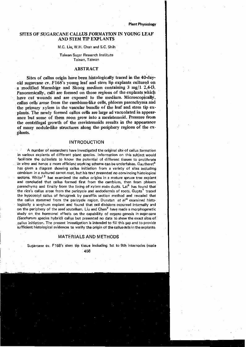
FIGURE 5. Enlarged area of the lower part of Fig. 3 showing the callus cells are mostly derived from the phloem parenchyma-of the smaller vas- cular bundles. The callus cells are vacuolated in appearance. (X 80)
The former is surrounded by the later.
After 40 days in culture, it can be seen that numerous vascular bundles in \ different sizes have emerged along the lower epidermis. The small vascular bundles
are situated near to the lower epidermis than the larger ones (Figs. 2 and 3).
Panoramically, callus is usually formed at the cut wounds of the explant.
FIGURE 6. Whole view of a stem-tip explant with massive callus which is growing on the periphery region (arrowed areas are enlarged in Fig. 7,8,9 and 10). X-15
Histological examination shows that callus is obviously proliferated from the cells near or in the vascular bundle because only those regions of the leaf which house the vascular bundles can produce callus (Fig. 3). This observation is of interest since ~r tschwa~er l has stated that in young developing leaves,cambium-like cells occur between xylem and phloem. They constitute an actively dividing tissue which some-
arises from the vigorous divisions of grotoxylem. Note the secondary wall thickening is conspicuous (X 120).
times takes on the appearance of a typical cambium. Other evidence from the microscopiCa1 slides (Figs. 3, 4 and 5) is that the primary phloem cells possess dense cytoplasm. Thus, the cambium-like cells, the phloem parenchyma and the primary xylem are more easily amenable to proliferation than other parenchymatous cells.
By enlargement of the two portions (labelled by rectangles) on Fig. 3, some callus growth patterns can be observed. Fig. 4 shows that the newly formed cells -
FIGURE 8. Similar to Fig. 7 except that the enlarged area is at b (arrowed) in , Fig. 6 indicating the callus mass is intruding the epidermis. (X 80).
from the primary vascular bundles are larger in size than their mother cells, but some of them soon develop into a nodule-like structure which could be appropriate- ly referred to as "meristemoid" ( ~ u n n i n ~ ~ ) . The meristemoid in i t s early stage is composed of cells in small size but with dense cytoplasm and striking nuclei. The heterogeneous phenomenon exhibited by the sugarcane callus in i t s initial stage was also observed in carrot callus cultures as reported by Gautheret and his co- w o r k e r ~ ~ ' ~ . They found that the callus cultures were actively growing and usually contained a high proportion of vacuolated parenchymatous cells together with
FIGURE 9. Similar to Fig. 7 except that the enlarged area i s at the lower part of the explant (5 arrowed) showing the intruding growth of a meris- temoid toward the periphery region-(>( 80).
more localized groups of smaller and obviously meristematic cells.
Site of stem tip callus
Fig. 6 i s a radial section through a 40-day-old explant with massive callus. The stem tip virtually contains the top 1-9 internodes in he&ht of 6-10 mm. The
FIGURE 10. Enlarged view of a well-developed vascular bundle (cj, arrowed) 6 mm away from the tip, deep embedded in the ground tissue, de- monstrating no callus cell arises at all. (X 125).
468 PLANT PHYSIOLOGY
apical portion i s made of thin-walled meristematic cells which are in a state of active division. Some of them gradually develop into the primordia of vascular bundles and become fullpledged later (~rtschwa~er') . By enlarging the callus-forming site, it is apparent that the callus is incepted form the extensive proliferations of ground vascular bundle in which the protoxylem cells play a very important role. This i s based on the observation that some cells with secondary wall thickening are clearly connected with the cells of a meristemoid as seen in Fig. 7 and 9. Massive callus arising from spruce tree's cambium and young phloem region has also been reported by white1 3 . The development of a meristemoid from the newly born cells i s similar to the case occurring in leaf explant. The meristemoid is virtually a pro- embryoid which will develop into a shoot when transferring it onto an organ- forming medium (Liu and cheng). Phloem parenchyma may also give rise to callus but this i s not clearly shown in these slides. Pressure from the centrifugal growth of the meristemoids has pushed off the epidermis as shown in Fig. 8.
Those vascular bundles which are deeply embedded in the parenchymatous ground tissue of the stem tip explant will not be induced to form callus mass (Fig. 10) whereasthosesituated neare the periphery region can do so (Figs, 3 and 6).
From this studies it can be concluded that callus cells originate from the primary vascular bundles in both the leaf and stem tip explants. Microscpical tracing of callus origin shows that the cambium-like cells, phloem parenchyma and the primary xylem in the vascular bundles are responsible for the callus production. However, any cell which is in meristematic state can, under suitable conditions, be brought to grow and to provide callus.
REFERENCES
1. Artschwager, E. (1925). Anatomy of the vegetative organs of sugarcane. J. Agric. Research 30: 197-221.
2. Bunning, E. (1952). Morphogenesis in plants. Surv. Biol. Progr. 2: 105-138.
3. Cook, M.T. (1926). The unrolling of the leaves of sugarcane. J. Dep. Agric. Puerto Rico 10: 243-245.
8~?5> 4. Dunstan, D.I., K.C. Short and E. Thomas. (1978). The anatomy of secondary
morphogenesis in cultured scutellum tissues of Sorghum bicolor. Protoplasma 97: 251 -260.
5. Gautheret. R.J. (1957). Histogenesis in plant tissue cultures. J. Nat. Cancer Inst. 19: 555.
6. , (1959). "La Culture des Tissus vegetabux, Techniques et Realisations". Masson, Paris.
7. Gupta, K.C. (1972). Histogenesis of fenugreek calli originating from excised cotyl explants. Can. J. BOP. 50: 2687-2688.
M. C. LIU ET AL 469
8. Lai, K. L. (1971). Morphological studies on the callus originated from excised roots of rice plants - The induction and anatomical observation on callus formation. Memoirs coll. Agric. Nat'l. Taiwan Univ. 12: 95-105.
9. biu, M.C. and W.H. Chen. (1974). Histological studies on the origin and process of plantlet differentiation in sugarcane callus.. Proc. Intern. Soc. Sugarcane Technol. (South Africa) 15: 1 18-1 28.
10. Liu, MC., Y.J. Huang and S.C. Shih. (1972). The in virro production of plants from tissues of Saccharum species. J. Agric. Assn. China 77: 52-58.
1 I . Murashige, T. and F. Skoog. (1962). A revised medium for rapid growth and bioassays with tobacco tissue cultures. Physiol, Plantarum 15: 473-497.
12. Sharman, B.C. 11943). Tannic acid and iron alum with safranin and orange G in studies of the shoot apex. Stain Technol. 18: 105-1 1 1.
13. White, P.R. (1967). Sites of callus production in primary explants of spruce tissue. Am. J. Bot. 54: 1055-1059.
SITIOS DE FORMACION CALLOS EN HOJA JOVENES TROZOS DE TALLOS SECCIONADOS DE CARA DE AZUCAR.
M.C. Liu, W.H. Chen and S. C. Shih
RESUMEN
Lugares de origen del callo han sido histologicarnente reconstruidos en una planta de cafia de azucar de 40 dias de edad, clon varietal F 168. Se trabaj6 con hojas jovenes y puntas de 10s tallos de la ex-planta, en el medio de cultivo Murashige y Skook modificado, conteniendo 3 mg/l de 2,4 D.
Panoramicamente, el callo estaba formado en aquellas regiones de la ex-planta que fueron cortadas y ese code expuesto a1 medio citado. Microscopicamente las celulas del callo fueron apareciendo de las celulas parecidas a1 cambium, floema, parenquima y del xilema prirnario en 10s vasos vasculares de 10s organos mencionados a1 principio. La nueva formacion de celulas en 10s callos fueron dargadas y vacuoladas en apariencia pero algunas de elIas crecieron rapidamente a meristemoides.
La presion del crecimiento centrifuge de 10s meristemoides, resultaron en la aparicion de muchas estructuras parecidas a nodulos, a lo largo de la region periferica de la ex-planta.


![Comparative proteomics reveals that central metabolism ...tology [1], morphology [6], physiology, biochemistry [7], as well as genetics of sugarcane [8], to explore smut resistant](https://static.documents.pub/doc/80x56/60b17d1dc3b2c97fd5037b14/comparative-proteomics-reveals-that-central-metabolism-tology-1-morphology.jpg)


