38
Plant Virus Serology Pl. Path. 502/602 PN Sharma Department of Plant Pathology CSK HPAU, Palampur-176062 (HP), INDIA

Plant Virus Serology
Pl. Path. 502/602
PN Sharma
Department of Plant Pathology
CSK HPAU, Palampur-176062 (HP), INDIA

Serology Based on antigen - antibody reaction
Antigen and antibody reaction results in the formation of visible substrate, which is perhaps due to the formation of bridges between the two and thus form an aggregate which precipitate when big enough.

Historical development Coons (1942), developed florescent antibody technique. Precipination in gels (Oudin, 1946; Ouchterolony, 1948). Modified agglutination test (Van Sloglerens, 1955). Double diffusion tests in plates (Octerlony, 1962) in tubes
(Oakelay and Fullthrope, 1963). Bentonite flocculation test (Bozicevich et al., 1963). Kassamis (1972) reported potato mop top virus relatedness with
TMV. Immuno-diffusion tests with Sodium dodecyl sulfate (SDS)
(Purcifall and Batchelor, 1977). Modified latex test (Querfurth and & Paul 1979) Holling and Stone (1973) found groups of 18 viruses serologically
related by immuno-diffusion and other tests. 1975 *Kohler and Milstein, Monoclonal antibodies used in genetic
analysis Clark, M.F. (1975) – developed ELISA technique. Clark & Adam (1978) – used ELISA in Plants Virology. Blanksky and Derrick (1977) reported that seed borne viruses can
be detected serologically. Paul et al. (1980) made serological studies on the relationship of
some isometric virus of graminae.

Discovery of antibodies 1899 *Jules Bordet, Complement and antibody activity in bacteriolysis
1900 *Paul Erlich, Antibody formation theory
1926 Lloyd Felton & GH Bailey, Isolation of pure antibody preparation
1934-8 John Marrack, Antigen-antibody binding hypothesis
1941 Albert Coons, Immunofluorescence technique
1948 Astrid Fagraeus, Demonstration of antibody production in plasma B cells
1959-62 *Rodney Porter et al., Discovery of antibody structure
1963 Jaques Oudin et al., antibody idiotypes
1964-8 Anthony Davis et al., T and B cell cooperation in immune response
1965 Thomas Tomasi et al., Secretory immunoglobulin antibodies
1975 *Kohler and Milstein, Monoclonal antibodies used in genetic analysis
1985 *Tonegawa, Hood et al., Identification of immunoglobulin genes

Principle: It is well known fact that animals that recover from certain infections
diseases rarely catch the same disease again i.e. they become immune to its.
Most of these immunity reactions have common basis. The immunizing agent is called antigen or immunogen (in disease, the
pathogen), It stimulates the animal, so that proteins called the antibodies appear in
its blood serum and they react specifically with the antigen that stimulated their production (the homologous antigens).
Antibodies to a virus may neutralize the infectivity of that virus or precipitates it.
The virus does not have to infect and multiply in animal to elicit antibodies, on many plant viruses are different immunogens.
Many substances induces/causes the production of antibodies when introduced into animal peritoneal, either by infection or injection. The main pre-requisites are:
1. The substances have a molecular structure and 2. That they are not the normal constituents of the animal being
immunized.

Antigen Any substance which evokes the production of antibodies is
called an antigens and includes proteins, polysaccharides, lipids, carbohydrates, nucleic aids enzymes, toxins etc.
induces the formation of antibodies when injected into a warm-blooded animal
Each antigen is made up of distinct sub-regions which have definite spatial and electronic configuration.
These restricted regions of an antigen stimulate the antigenic response and are the regions to which antibodies are/get attached. These regions are called antigenic determinants or Epitopes,
Epitopes, are the actual stimulus for the production of particular antibody and are the combining sites of antibody. In other words antibody molecules are directed against epitopes rather than entire molecule itself.
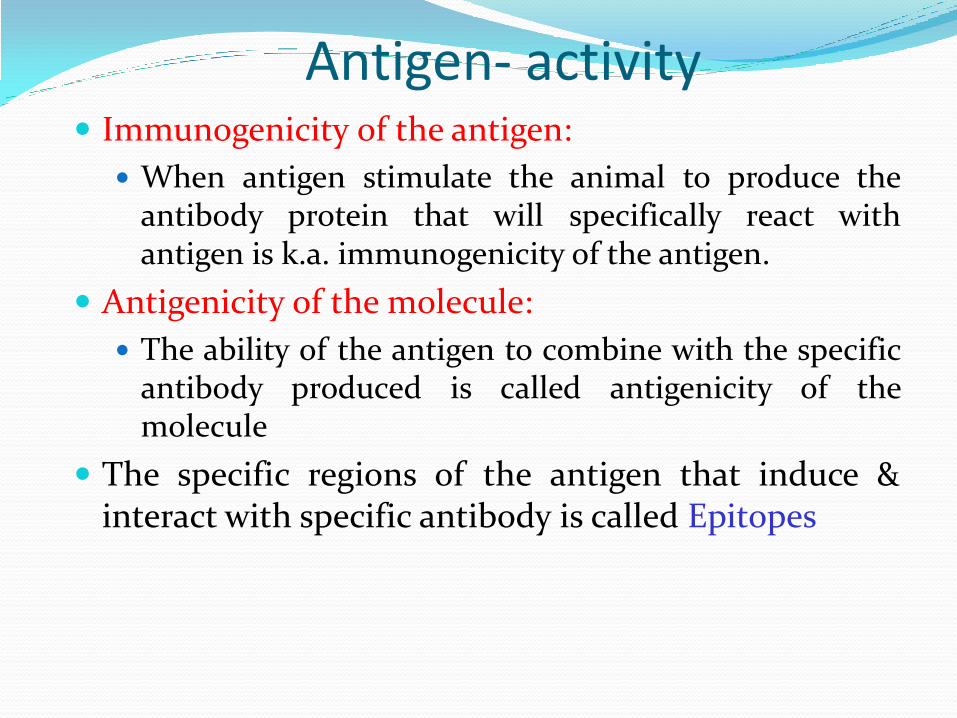
Antigen- activity Immunogenicity of the antigen:
When antigen stimulate the animal to produce the antibody protein that will specifically react with antigen is k.a. immunogenicity of the antigen.
Antigenicity of the molecule:
The ability of the antigen to combine with the specific antibody produced is called antigenicity of the molecule
The specific regions of the antigen that induce & interact with specific antibody is called Epitopes

Antigenic determinants
An antibody will recognize
Epitope: defined segment of an antigen
Immuno-reactivity of epitopes may depend on primary, secondary, tertiary or quaternary structure of an antigen
Define the possible applications
Variability of epitopes depends on the species
Antibodies are antigen themselves

Types of epitopes An epitope, also known as antigenic
determinant, is the part of an antigen that is recognized, specifically by antibodies
The part of an antibody that recognizes the epitope is called a paratope.

Antigenicity of plant viruses Antigenically active part of a plant virus is protein except few viruses
(external part of protein shells which encloses nucleic acid).
Infectivity test depend upon nucleic acid only but serological test depend on its protein though these two occurs together or separate, but differ in their stability.
All the particles do not contribute equally to serological activity as not all parts of peptide chain in each subunit are at the surface of the particle e.g. In TMV, only C-terminal and of the amino acid chain of each sub-unit is serologically active.
Various amino acids are present in a protein of virus and strains differ due to one or more amino acids. In TMV 8 of the 20 amino acids change the serological behaviour of the particles (Van Sengbusch, 1965 & Van Regenmortel, 1967)

Antibody a specific protein formed in the
blood of warm-blooded animals in response to injection of an antigen (protein or polysaccharide)
Responsible for specific recognition and elimination (neutralization) of antigens
Antibodies are proteins termed as γ globulin are built of two types of chains
Heavy chain H. molecular wt. of 50,000 to 70,000 and
light chain-L, with mol. Mt. of 23,000, occurs in pairs linked to one another by disulphide bond. Igh is composed of two light chain and two heavy chains

Antibody
Antibodies found in the globulin fraction of the blood serum proteins of normal animals, after immunization the new kinds appear which differ from those present in normal serum in the sense that these (new kind) can react specifically with the immunizing antigens.
Antibodies have combining sites on their surfaces depending upon shape, charge and hydrophobicity to the antigenic sites and number depends upon size and complexity of antigen e.g. TMV particles have about 2100 similar protein sub-units, each active and have the same antigenic determinants.

The Ig monomer is a "Y"-shaped molecule that consists of four polypeptide chains;
two identical heavy chains and two identical light chains connected by disulfide bonds.
Each chain is composed of structural domains called immunoglobulin domains. These domains contain about 70-110 amino acids and are classified into different categories (for example, variable or IgV, and constant or IgC) according to their size and function.
They have a characteristic immunoglobulin fold in which two beta sheets create a “sandwich” shape, held together by interactions between conserved cysteines and other charged amino acids.
Antibody

Different classes of Immunoglobulin
Name Types Description Antibody
Complexes
IgA 2 Found in mucosal areas, such as the gut, respiratory tract andurogenital tract, and prevents colonization by pathogens. Also found in saliva, tears, and breast milk.
IgD 1
Functions mainly as an antigen receptor on B cells that have not been exposed to antigens.[14] It has been shown to activate basophils and mast cells to produce antimicrobial factors.
IgE 1 Binds to allergens and triggers histamine release from mast cells and basophils, and is involved in allergy. Also protects against parasitic worms.
IgG 4
In its four forms, provides the majority of antibody-based immunity against invading pathogens. The only antibody capable of crossing the placenta to give passive immunity to fetus.
IgM 1
Expressed on the surface of B cells (monomer) and in a secreted form (pentamer) with very high avidity. Eliminates pathogens in the early stages of B cell mediated (humoral) immunity before there is sufficient IgG.

Immunoglobulin domains
Several immunoglobulin domains make up the two
heavy chains (red and blue) and the two light
chains (green and yellow) of an antibody.
The immunoglobulin domains are composed of
between 7 (for constant domains) and 9 (for variable
domains) β-strands

Antigen-antibody interaction
Antigen: foreign molecules that generate antibodies or any substance that can be bound specifically by an antibody molecule
Proteins, sugars, lipids or nucleic acids
Natural or synthetic

Antigen-antibody binding
Hydrogen bonding
Results from the formation of hydrogen bridges between appropriate atoms
Electrostatic forces
Are due to the attraction of oppositely charged groups located on two protein side chains
Van der Waals bonds
Are generated by the interaction between electron clouds (oscillating dipoles)
Hydrophobic bonds
Rely upon the association of non-polar, hydrophobic groups so that contact with water molecules is minimized (may contribute up to half the total strength of the antigen-antibody bond)
Nature of binding forces

Antigen-antibody affinity
•The affinity with which antibody binds antigen results
from a balance between the attractive and repulsive
forces.
•A high affinity antibody implies a good fit and
conversely, a low affinity antibody implies a poor fit and
a lower affinity constant

Antibody production Antibodies are produced in the animal by cells of its
reticula endothelial system.
Especially by cells of lymphocytic and plasma cell series, which are common in lymph nodes, the spleen and bone marrow (Raff, 19=13) in the circulating body fluids of immunized animals so that any moment about half in lymph and other body fluids.
It is not yet known that how the antigenic determinants are recognized by the animals and elicit the production of specific antibodies.
Polyclonal Antibody: react with > one epitopes Monoclonal Antibody : react with a single epitope
(epitopes - antigenic sites)

Polyclonal Antibody Polyclonal antibodies or antiserum (Serum containing
antibodies)
purified or partially purified antigen preparation can be injected
into the animal
The animal body serum contain millions of B-lymptocytes or B-
cell which during immune response recognize one of the many
epitopes' of the antigen. Therefore, many cells clones will be
stimulated and many different antibodies are accumulated in
the serum. Thus serum is heterogeneous.
As a result of polyclonal nature of conventional antiserum:
No antiserum is precisely reproducible
Antisera produced for the same antigenic varies from lab to lab.
Even antiserum obtained from one bleeding differs from that
obtained from the other.
This leads to conflicting results. Therefore, highly specific and well
defined antibodies are required for diagnostic and taxonomic studies.


Monoclonal antibodies
Burnet (1959) put forth the clonal selection hypothesis, which states that each
antibody forming cell (B-lymphocyte) is committed to the production of one type of antibody molecule, which have the potential to react with one or at most a few structurally similar epitopes.
But attempts to culture B-lymphocytes failed because these cells can not be cultured.
The Kohler and Milstein (1975) showed somatic hybridization-between B-lymphocytes (antibody producing cells) and myeloma cells and hybrids rising are called HYBRIDOMAS. The hybrid acquire the ability to produce specific antibody from B-lymphocyte cell and from mycloma cells, the ability to be cultured indefinitely in vitro.
Antibodies produced by single hybridoma are identical and specific for a single epitope.
This technique of producing antibodies is commonly k.a. Hybridoma technology. Which have the potential for producing an unlimited quantity of
monospecific or monoclonal antibodies.

Advantages of monoclonal antibodies over polyclonal
An unlimited quantity of antibodies can be produced
Epoitope specific antibodies are produced.
Specific antibodies can be produced even with impure antigen.
Hybridoma can be stored for long time in liquid nitrogen.
MCA can reveal serological relationship between antigens that are previously unrecognized with polyclonal antibodies.
MCA eliminates quantitative and qualitative variability encountered in different batches of polyclonal serum.

Hybridoma 1975 Kohler and Milstein
http://www.immunecentral.com/images/immune_series/immune29.gif
1.Hyper-immunize mouse
with Ag
2. Fuse B cells with tumor
fusion partner (+ PEG)
3. Limiting dilution (96 wells)
to fractionate fused cells in
HAT media selection (HAT
media (hypoxanthine,
aminopterin, thymidine).
B cells die (mortal, HGPRT-positive)
Tumor cells die (HGPRT deficient and cannot utilise salvage pathway)
Fusions live (HAT Resistant and immortal)

How to purify your MAb
Incubate with
anti-Mouse Ig
polyspecific serum
Monospecific
antibodies
Affinity Purification
•
Or use Ag or epitope to
make affinity column
http://www.tulane.edu/~wiser/methods/handouts/pwpt/17_mab.pdf

Commercial production of antibodies: polyclonal vs monoclonal Host animals can be used to raise antibodies against a given antigen
Selected clones from a polyclonal each recognizing a single epitope can
be fused to a tumor cell (hybridoma) to proliferate indefinitely

Serological Methods Conventional methods
Precipitin test Tube
Slide
Immuno-diffusion
Immuno-precipitation
Ring interface test
Agglutination test Chloroplast (Co-precipitation) or Latex (Flocculation)
Enzyme Linked Immunosorbent Assay ELISA
Dot Immunobinding Assay DIBA
Immunosorbant electron microscopy (ISEM/ IEM)

Immuno-precipitation
Precipitation
Since viruses are of molecular dimensions and their
clumping does not occur, instead a precipitate is formed
which remain suspended in saline solution to make it turbid.
All tests except chloroplast precipitin test are performed with
purified virus preparation or clarified plant extracts.
The precipitation reaction is examined either
macroscopically (e.g. tube and ring interface precipitin test)
or microscopically (e.g. slide and micro precipitin test).
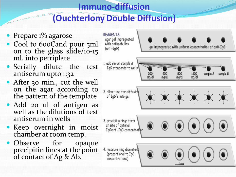
Immuno-diffusion (Ouchterlony Double Diffusion)
Prepare 1% agarose Cool to 60oCand pour 5ml
on to the glass slide/10-15 ml. into petriplate
Serially dilute the test antiserum upto 1:32
After 30 min., cut the well on the agar according to the pattern of the template
Add 20 ul of antigen as well as the dilutions of test antiserum in wells
Keep overnight in moist chamber at room temp.
Observe for opaque precipitin lines at the point of contact of Ag & Ab.

Agar Immunodiffusion Antiserum Antiserum
Spur formation

Enzyme-Linked Immunosorbent Assay - ELISA
sensitivity of the antigen-antibody is increased by attaching an enzyme to one of the two reactions
Ag-Ab-Enzyme conjugates produce color reactions. Used to detect amount of Ag or Ab in a sample. Performed as a solid phase assay.

Double antibody sandwich ELISA (DAS-ELISA)
sap extracted from propagating host, Add 200 µl of IgG diluted in coating buffer (1:200) was to each well of the
microtitre plate. Cover plate with aluminium foil and incubate at 370C in an incubator for 4 hours.
After incubation the wash plate with wash buffer (PBS-T) four times in an automatic ELISA plate washer
Then add 200 µl of the test sample ground in tissue grinding buffer (PBS-T+PVP, Dilution 1:20) to each well and incubate overnight at 40C in refrigerator.
Decant the contents of plate and wash with wash buffer four times in an ELISA plate washer and add 200 µl of enzyme labelled IgG of test virus to each well and incubate at 300C for 4 hours.
Wash plate with wash buffer and add 200 µl of p-nitrophenol phosphate substrate (1 mg/ ml of substrate buffer) to each well of the plate and incubate for 2 hours at room temperature.
After 2 hours reaction is stopped with 50 µl of 3.0 M sodium hydroxide per well Record absorbance at 410 nm wavelength with the help of ELISA reader
calibrated to zero with a blank of substrate buffer. Samples showing 2-3 times more optical density (OD) over healthy control
sample is rated as positive.
Protocol
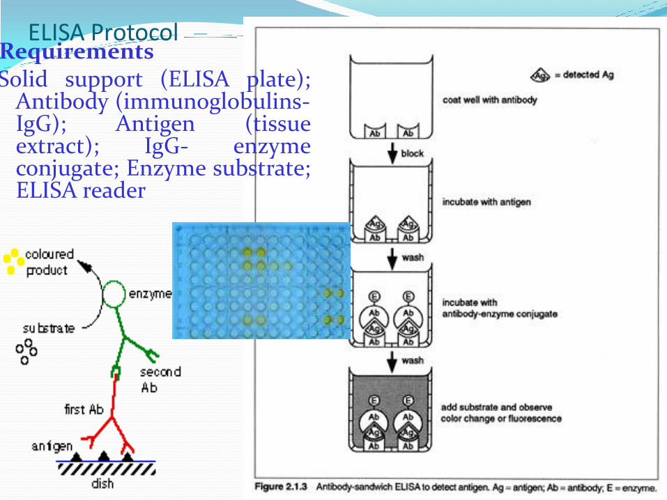
ELISA Protocol Requirements Solid support (ELISA plate);
Antibody (immunoglobulins-IgG); Antigen (tissue extract); IgG- enzyme conjugate; Enzyme substrate; ELISA reader

ELISA Methods 1
Advantages of ELISA
Reasonably sensitive
Handle large number of samples
Can be subjected to automation
Detection kits available commercially

Dot-Immunobinding assay (DIBA)
A variant of ELISA
Nitrocellulose membrane as solid support
Crude antisera can be used
Stains development for revealing the reaction by hydrolysis of the substrate (NBT- nitroblue tetraxolium) or (BCIP- 5-bromo-4-chloro-3-iodyl phosphate p-toludine salt and formamide
Very useful for survey work

DIBA Antigen and antibody immobilized on NCM instead of plate. Antigen is electro- blotted on the membrane or membrane is
coated with antiviral IgG. For final colour, substrate is added, that convert the enzyme
linked to the IgG into an insoluble coloured material. Advantages over ELISA
Easy to transport Detect large no. of samples in the field and Very low amount of antigen and antibody can be used.
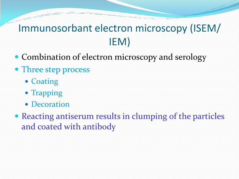
Immunosorbant electron microscopy (ISEM/ IEM)
Combination of electron microscopy and serology
Three step process
Coating
Trapping
Decoration
Reacting antiserum results in clumping of the particles and coated with antibody


