47
PLASMA CELL DISORDERS Dr Vijay Shankar S
| Date post: | 09-Jan-2017 |
| Category: |
Health & Medicine |
| Upload: | vijay-shankar |
| View: | 258 times |
| Download: | 1 times |

PLASMA CELL DISORDERS
Dr Vijay Shankar S

Learning objectives Introduction Multiple myeloma Waldenstorms macroglobulinemia Lymphoplasmacytic lymphoma

PLASMA CELL

B lymphocyte
Reactive B lymphocyte
Plasmablast
Proplasmacyte
Plasmacyte/plasma cell


Proliferation of a B – cell clone that synthesizes and secretes a single homogeneous immunoglobulin or its fragments.
Accounts for 15 % of deaths from white cell neoplasms
Free light chains(Bence Jones Proteins)
Free heavy chains
PLASMA CELL DISORDERS

Monoclonal immunoglogulin – M component
Freely excreted in the urine in the absence of glomerular damage.
Disorders associated with abnormal immunoglobulins – Gammopathy/
Monoclonal gammopathy/
Dysproteinemia/ Paraproteinemia.

Clinicopathologic entities associated with monoclonal
gammopathies1. Multiple myeloma( Plasma cell
myeloma)2. Waldenstrom macroglobulinemia3. Heavy – chain disease.4. Primary or immunocyte associated
Amyloidosis5. Monoclonal gammopathy of
undetermined significance (MGUS)

MULTIPLE MYELOMA Plasma cell neoplasm characterized
by involvement of skeleton at multiple sites.
Multifocal , monoclonal proliferation of plasma cells
1% of all cancer deaths. more frequent in elderly modest male predominance Osseous and extraosseus
manifestations

Proliferation of a plasma cell clone that synthesizes and secretes a single homogeneous immunoglobulin or its fragments.
Free light chains(Bence Jones Proteins)
Free heavy chains

MORPHOLOGY Presents most commonly as
multifocal destructive bone tumors. Bones in axial skeleton affected
most. Most common in vertebral column.

DISTRIBUTION Vertebral column- 66% Ribs - 44% Skull - 41% Pelvis – 28% Femur – 24% Clavicle - 10% Scapula –10%

Round lesions filled with a soft reddish material are indicative of foci of myeloma in this section of vertebral bone.

The skull demonstrates the characteristic rounded "punched out" lesions of multiple myeloma.

The rounded "punched out" lesions of multiple myeloma appear as lucent areas with this skull radiograph.

Lytic lesion in Tibia
Lesions in the lower end of femur
Lesion in the upper end of femur

Bone marrow aspirate

↑↑ no of Plasma cells, usually more than 30% of marrow cellularity

BONE MARROW
At low power, the abnormal plasma cells of multiple myeloma fill the marrow.

BONE MARROW
At medium power, the plasma cells of multiple myeloma here are very similar to normal plasma cells, but they may also be poorly differentiated.

Flame cells

Crystalline inclusionsMott cell

Russel bodies Dutcher bodies

Pathologic rouleaux formation, Multiple myeloma


William Russell Scottish pathologist and physician
(1852 – 1940)

CLINICAL FEATURESManifestations are due to
1. Infiltration of organs by neoplastic plasma cells
2. Production of excessive immunoglobulins with abnormal physiochemical properties.
3. Suppression of normal Humoral immunity
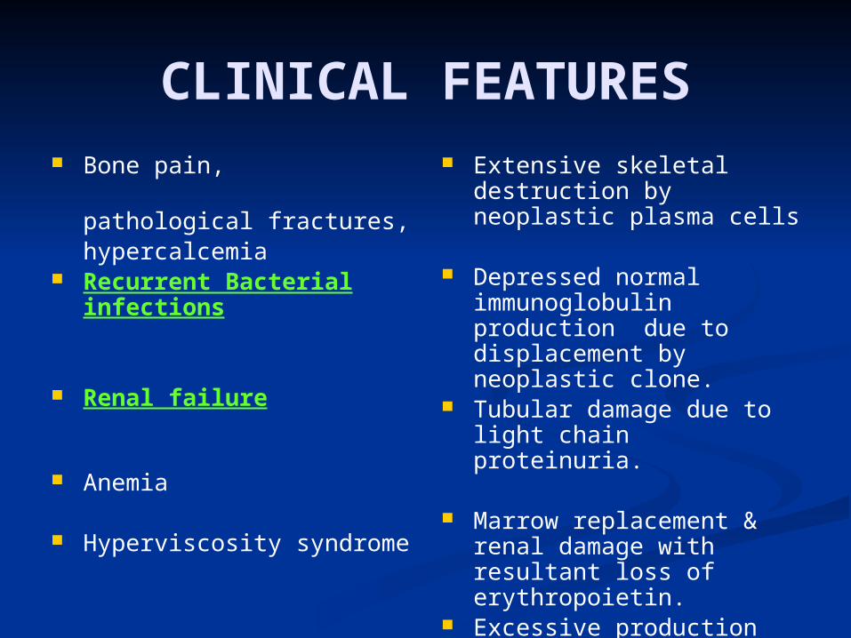
CLINICAL FEATURES Bone pain,
pathological fractures, hypercalcemia
Recurrent Bacterial infections
Renal failure
Anemia
Hyperviscosity syndrome
Extensive skeletal destruction by neoplastic plasma cells
Depressed normal immunoglobulin production due to displacement by neoplastic clone.
Tubular damage due to light chain proteinuria.
Marrow replacement & renal damage with resultant loss of erythropoietin.
Excessive production and aggregation of M proteins

LABORATORY STUDIES
Increased levels of immunoglobulins in the blood
and /or
light chains ( Bence Jones proteins) in urine
in 99% of cases


Most common serum monoclonal immunoglobulin ( M protein) – IgG (55%)
IgA (25%)
IgM, IgD, or IgE - Rare
Both Bence Jones Proteins & serum M
protein : 60 – 70% Only Bence Jones proteins :
20% Nonsecretory :
1%

DIAGNOSIS & PROGNOSIS
Radiographic & laboratory findings Definitive diagnosis – Bone marrow
study
PROGNOSIS - Variable , but generally poor

"The gem cannot be polished without friction, nor man perfected without trials or problems or exams…!."
--Chinese proverb

1944
Two patients with oronasal bleeding, lymphadenopathy, anemia and thrombocytopenia, an elevated ESR, a high serum viscosity level, normal bone radiographs and a bone marrow demonstrating predominately lymphoid cells.

Waldenstrom’s Macroglobulinemia
Dr. Jan Gosta Waldenstrom(Swedish internist) (1906-1996), in 1944

2003 2nd International Workshop on WM,
which was held in Athens, Greece
clinicopathological entity that was represented by the underlying pathological diagnosis of lymphoplasmacytic lymphoma, as defined by the WHO and REAL classification systems which secretes IgM

LYMPHOPLASMACYTIC LYMPHOMA
(SLL/CLL with plasmacytic differentiation., Immunocytoma)
B - cell neoplasm of older adults. 6th or 7th decades of life Resemble CLL/SLL .. But.. Good no
of tumor cells undergo terminal differentiation into plasma cells.
Secrete monoclonal IgM
Hyperviscosity syndrome(W M)
Heavy & light chain synthesis is usually balanced

MORPHOLOGY Bone marrow :heavy infiltrates of
lymphocytes, plasma cells and plasmacytoid lymphocytes.
Russel bodies and Dutcher bodies may be present.
Often involves lymph nodes, liver & spleen.
Infiltration of nerve roots, meninges & brain may be seen.


IMMUNOPHENOTYPE & MOLECULAR GENETICS
B – cell marker – CD20 Plasma cell - expresses monoclonal
immunoglobulin.
MC cytogenetic abnormality – del 6q

CLINICAL FEATURES Non- specific : weakness, fatigue,
weight loss. Hyperviscosity syndrome: Visual impairment Neurologic problems: headache ,
dizziness, deafness etc
Bleeding tendenciesAnemia

Rests on Laboratory data & bone marrow study.
↑↑ Serum proteins
↑↑ Serum monoclonal M component( due to IgM)
↑↑ ESR
Normocytic hypochromic anemia
Characteristic marrow infiltration
DIAGNOSIS

PROGNOSIS Incurable progressive disease.
Plasmapherisis might help
Median survival - 4 yrs.

“Ninety-nine percent of failures come from people
who have a habit of making excuses.”
–George Washington Carver

THE END


