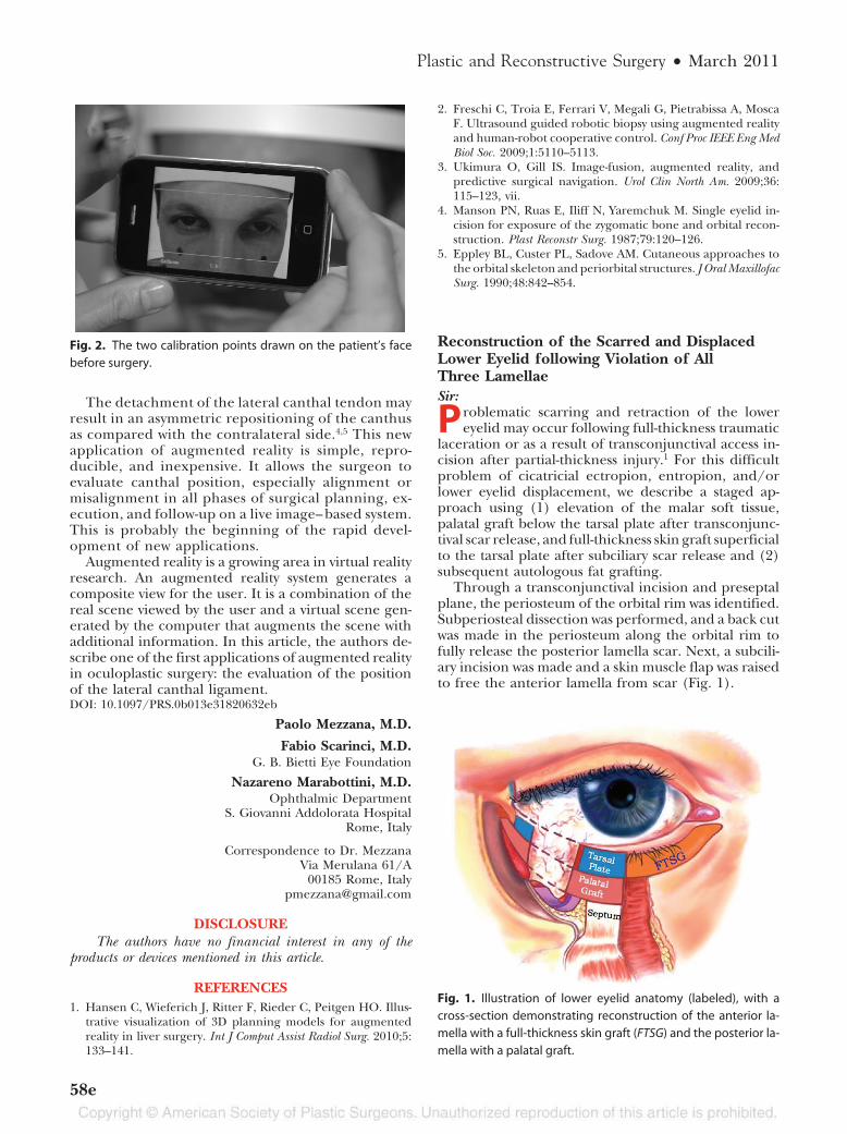
The detachment of the lateral canthal tendon may result in an asymmetric repositioning of the canthus as compared with the contralateral side. 4,5 This new application of augmented reality is simple, repro- ducible, and inexpensive. It allows the surgeon to evaluate canthal position, especially alignment or misalignment in all phases of surgical planning, ex- ecution, and follow-up on a live image– based system. This is probably the beginning of the rapid devel- opment of new applications. Augmented reality is a growing area in virtual reality research. An augmented reality system generates a composite view for the user. It is a combination of the real scene viewed by the user and a virtual scene gen- erated by the computer that augments the scene with additional information. In this article, the authors de- scribe one of the first applications of augmented reality in oculoplastic surgery: the evaluation of the position of the lateral canthal ligament. DOI: 10.1097/PRS.0b013e31820632eb Paolo Mezzana, M.D. Fabio Scarinci, M.D. G. B. Bietti Eye Foundation Nazareno Marabottini, M.D. Ophthalmic Department S. Giovanni Addolorata Hospital Rome, Italy Correspondence to Dr. Mezzana Via Merulana 61/A 00185 Rome, Italy [email protected]DISCLOSURE The authors have no financial interest in any of the products or devices mentioned in this article. REFERENCES 1. Hansen C, Wieferich J, Ritter F, Rieder C, Peitgen HO. Illus- trative visualization of 3D planning models for augmented reality in liver surgery. Int J Comput Assist Radiol Surg. 2010;5: 133–141. 2. Freschi C, Troia E, Ferrari V, Megali G, Pietrabissa A, Mosca F. Ultrasound guided robotic biopsy using augmented reality and human-robot cooperative control. Conf Proc IEEE Eng Med Biol Soc. 2009;1:5110–5113. 3. Ukimura O, Gill IS. Image-fusion, augmented reality, and predictive surgical navigation. Urol Clin North Am. 2009;36: 115–123, vii. 4. Manson PN, Ruas E, Iliff N, Yaremchuk M. Single eyelid in- cision for exposure of the zygomatic bone and orbital recon- struction. Plast Reconstr Surg. 1987;79:120–126. 5. Eppley BL, Custer PL, Sadove AM. Cutaneous approaches to the orbital skeleton and periorbital structures. J Oral Maxillofac Surg. 1990;48:842–854. Reconstruction of the Scarred and Displaced Lower Eyelid following Violation of All Three Lamellae Sir: P roblematic scarring and retraction of the lower eyelid may occur following full-thickness traumatic laceration or as a result of transconjunctival access in- cision after partial-thickness injury. 1 For this difficult problem of cicatricial ectropion, entropion, and/or lower eyelid displacement, we describe a staged ap- proach using (1) elevation of the malar soft tissue, palatal graft below the tarsal plate after transconjunc- tival scar release, and full-thickness skin graft superficial to the tarsal plate after subciliary scar release and (2) subsequent autologous fat grafting. Through a transconjunctival incision and preseptal plane, the periosteum of the orbital rim was identified. Subperiosteal dissection was performed, and a back cut was made in the periosteum along the orbital rim to fully release the posterior lamella scar. Next, a subcili- ary incision was made and a skin muscle flap was raised to free the anterior lamella from scar (Fig. 1). Fig. 2. The two calibration points drawn on the patient’s face before surgery. Fig. 1. Illustration of lower eyelid anatomy (labeled), with a cross-section demonstrating reconstruction of the anterior la- mella with a full-thickness skin graft (FTSG) and the posterior la- mella with a palatal graft. Plastic and Reconstructive Surgery • March 2011 58e
Transcript
The detachment of the lateral canthal tendon mayresult in an asymmetric repositioning of the canthusas compared with the contralateral side.4,5 This newapplication of augmented reality is simple, repro-ducible, and inexpensive. It allows the surgeon toevaluate canthal position, especially alignment ormisalignment in all phases of surgical planning, ex-ecution, and follow-up on a live image– based system.This is probably the beginning of the rapid devel-opment of new applications.
Augmented reality is a growing area in virtual realityresearch. An augmented reality system generates acomposite view for the user. It is a combination of thereal scene viewed by the user and a virtual scene gen-erated by the computer that augments the scene withadditional information. In this article, the authors de-scribe one of the first applications of augmented realityin oculoplastic surgery: the evaluation of the positionof the lateral canthal ligament.DOI: 10.1097/PRS.0b013e31820632eb
DISCLOSUREThe authors have no financial interest in any of the
products or devices mentioned in this article.
REFERENCES
1. Hansen C, Wieferich J, Ritter F, Rieder C, Peitgen HO. Illus-trative visualization of 3D planning models for augmentedreality in liver surgery. Int J Comput Assist Radiol Surg. 2010;5:133–141.
2. Freschi C, Troia E, Ferrari V, Megali G, Pietrabissa A, MoscaF. Ultrasound guided robotic biopsy using augmented realityand human-robot cooperative control. Conf Proc IEEE Eng MedBiol Soc. 2009;1:5110–5113.
3. Ukimura O, Gill IS. Image-fusion, augmented reality, andpredictive surgical navigation. Urol Clin North Am. 2009;36:115–123, vii.
4. Manson PN, Ruas E, Iliff N, Yaremchuk M. Single eyelid in-cision for exposure of the zygomatic bone and orbital recon-struction. Plast Reconstr Surg. 1987;79:120–126.
5. Eppley BL, Custer PL, Sadove AM. Cutaneous approaches tothe orbital skeleton and periorbital structures. J Oral MaxillofacSurg. 1990;48:842–854.
Reconstruction of the Scarred and DisplacedLower Eyelid following Violation of AllThree LamellaeSir:
Problematic scarring and retraction of the lowereyelid may occur following full-thickness traumatic
laceration or as a result of transconjunctival access in-cision after partial-thickness injury.1 For this difficultproblem of cicatricial ectropion, entropion, and/orlower eyelid displacement, we describe a staged ap-proach using (1) elevation of the malar soft tissue,palatal graft below the tarsal plate after transconjunc-tival scar release, and full-thickness skin graft superficialto the tarsal plate after subciliary scar release and (2)subsequent autologous fat grafting.
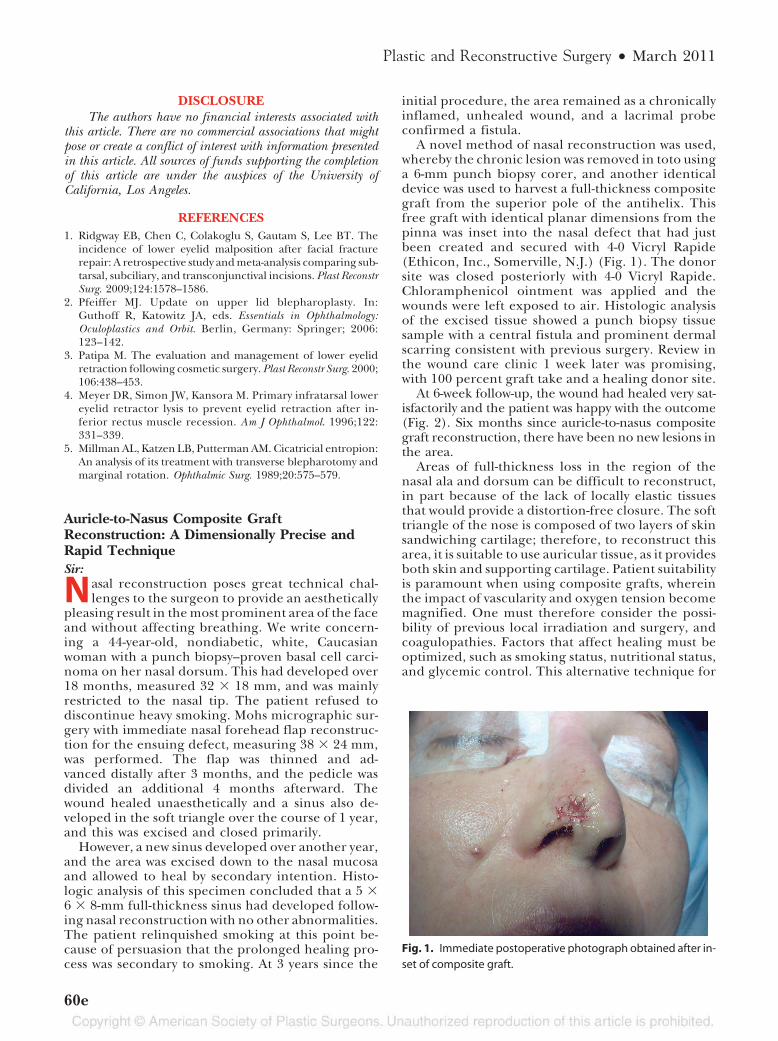
Through a transconjunctival incision and preseptalplane, the periosteum of the orbital rim was identified.Subperiosteal dissection was performed, and a back cutwas made in the periosteum along the orbital rim tofully release the posterior lamella scar. Next, a subcili-ary incision was made and a skin muscle flap was raisedto free the anterior lamella from scar (Fig. 1).
Fig. 2. The two calibration points drawn on the patient’s face
before surgery.
Fig. 1. Illustration of lower eyelid anatomy (labeled), with a
cross-section demonstrating reconstruction of the anterior la-
mella with a full-thickness skin graft (FTSG) and the posterior la-
mella with a palatal graft.
Plastic and Reconstructive Surgery • March 2011
58e
The cheek was elevated by placing a 3-0 Prolenesuture from the soft tissue of the malar prominencethrough a drill hole through the lateral orbital rimthrough an upper supratarsal fold incision. A hardpalate mucoperiosteal graft was harvested to the size
of the posterior lamella defect (6 mm � 25 mm). Thispalatal mucosal graft was then inset into thetransconjunctival defect with interrupted 5-0 chro-mic sutures. A full-thickness skin graft was taken fromeither the contralateral upper eyelid or the postau-ricular area based on a template for the anteriorlamella defect. The full-thickness skin graft was thensutured in with a running 5-0 plain gut suture. Alateral canthoplasty was, at times, performed. Finally,a Frost suture was placed for 5 days for lower eyelidimmobilization.
Three to 6 months later, autologous fat grafting tothe lower eyelid was performed for volume expansion.The Coleman technique of fat transfer was used withatraumatic abdominal harvest, centrifugation for 3minutes at 3000 rpm, and small aliquot injections intodifferent depths of the eyelid. At times, repeated fatinjections were used for further improvements.
Management of the scarred and displaced lower eye-lid using the above operative technique over the pastdecade at our institution led to dramatic improvementsin eyelid position and appearance (Fig. 2). Patients alsoreported improvement or complete resolution of theirsymptoms, including epiphora, tearing, redness, blurryvision, and dryness.
An understanding of lower eyelid anatomy is essen-tial for reconstructing the severely scarred and dis-placed lower eyelid. The lower eyelid has three layers,or lamellae. The anterior lamella contains skin, sub-cutaneous tissue, and the orbicularis oculi.2 The middlelamella includes the orbital septum and subseptal fat.3,4
The posterior lamella includes lower eyelid retractors,the tarsal plate, and conjunctiva.2
Previous publications focused on reconstructionof either the anterior or posterior lamellar plane, butnot both.1,5 We describe an approach that emphasizesrelease of lower lid scar tissue, elevation of the softtissues of the cheek, lengthening of the contractedseptum, support of the posterior lamellae with apalatal graft, and replacement of the anterior lamellawith a full-thickness skin graft. We have documentedsuccess and symptomatic improvement using thisapproach.DOI: 10.1097/PRS.0b013e31820632ae
Jason Roostaeian, M.D.
Emil Kohan, M.D.
Neil Tanna, M.D.
Henry K. Kawamoto, M.D., D.D.S.
James P. Bradley, M.D.Division of Plastic and Reconstructive Surgery
David Geffen School of Medicine at University ofCalifornia, Los Angeles
Los Angeles, Calif.
Correspondence to Dr. Bradley200 Medical Plaza, Suite 465
Los Angeles, Calif. 90095-6960jbradleyjednet.ucla.edu
Fig. 2. (Above) A patient with a severely scarred, retracted left
lower eyelid underwent staged correction. After initial repair of
the displaced zygomaticomaxillary complex, orbital floor frac-
tures, and soft tissue, full-thickness lower eyelid laceration repair
was performed. (Center) The patient is shown 3 months after the
initial injury, with lower eyelid retraction and scar. (Below) The
patient is shown postoperatively, 6 months after lower eyelid re-
construction with cheek elevation, palatal graft, full-thickness
skin graft, and fat grafting.
Volume 127, Number 3 • Viewpoints
59e
DISCLOSUREThe authors have no financial interests associated with
this article. There are no commercial associations that mightpose or create a conflict of interest with information presentedin this article. All sources of funds supporting the completionof this article are under the auspices of the University ofCalifornia, Los Angeles.
REFERENCES
1. Ridgway EB, Chen C, Colakoglu S, Gautam S, Lee BT. Theincidence of lower eyelid malposition after facial fracturerepair: A retrospective study and meta-analysis comparing sub-tarsal, subciliary, and transconjunctival incisions. Plast ReconstrSurg. 2009;124:1578–1586.
2. Pfeiffer MJ. Update on upper lid blepharoplasty. In:Guthoff R, Katowitz JA, eds. Essentials in Ophthalmology:Oculoplastics and Orbit. Berlin, Germany: Springer; 2006:123–142.
3. Patipa M. The evaluation and management of lower eyelidretraction following cosmetic surgery. Plast Reconstr Surg. 2000;106:438–453.
4. Meyer DR, Simon JW, Kansora M. Primary infratarsal lowereyelid retractor lysis to prevent eyelid retraction after in-ferior rectus muscle recession. Am J Ophthalmol. 1996;122:331–339.
5. Millman AL, Katzen LB, Putterman AM. Cicatricial entropion:An analysis of its treatment with transverse blepharotomy andmarginal rotation. Ophthalmic Surg. 1989;20:575–579.
Auricle-to-Nasus Composite GraftReconstruction: A Dimensionally Precise andRapid TechniqueSir:
Nasal reconstruction poses great technical chal-lenges to the surgeon to provide an aesthetically
pleasing result in the most prominent area of the faceand without affecting breathing. We write concern-ing a 44-year-old, nondiabetic, white, Caucasianwoman with a punch biopsy–proven basal cell carci-noma on her nasal dorsum. This had developed over18 months, measured 32 � 18 mm, and was mainlyrestricted to the nasal tip. The patient refused todiscontinue heavy smoking. Mohs micrographic sur-gery with immediate nasal forehead flap reconstruc-tion for the ensuing defect, measuring 38 � 24 mm,was performed. The flap was thinned and ad-vanced distally after 3 months, and the pedicle wasdivided an additional 4 months afterward. Thewound healed unaesthetically and a sinus also de-veloped in the soft triangle over the course of 1 year,and this was excised and closed primarily.
However, a new sinus developed over another year,and the area was excised down to the nasal mucosaand allowed to heal by secondary intention. Histo-logic analysis of this specimen concluded that a 5 �
6 � 8-mm full-thickness sinus had developed follow-ing nasal reconstruction with no other abnormalities.The patient relinquished smoking at this point be-cause of persuasion that the prolonged healing pro-cess was secondary to smoking. At 3 years since the
initial procedure, the area remained as a chronicallyinflamed, unhealed wound, and a lacrimal probeconfirmed a fistula.
A novel method of nasal reconstruction was used,whereby the chronic lesion was removed in toto usinga 6-mm punch biopsy corer, and another identicaldevice was used to harvest a full-thickness compositegraft from the superior pole of the antihelix. Thisfree graft with identical planar dimensions from thepinna was inset into the nasal defect that had justbeen created and secured with 4-0 Vicryl Rapide(Ethicon, Inc., Somerville, N.J.) (Fig. 1). The donorsite was closed posteriorly with 4-0 Vicryl Rapide.Chloramphenicol ointment was applied and thewounds were left exposed to air. Histologic analysisof the excised tissue showed a punch biopsy tissuesample with a central fistula and prominent dermalscarring consistent with previous surgery. Review inthe wound care clinic 1 week later was promising,with 100 percent graft take and a healing donor site.
At 6-week follow-up, the wound had healed very sat-isfactorily and the patient was happy with the outcome(Fig. 2). Six months since auricle-to-nasus compositegraft reconstruction, there have been no new lesions inthe area.
Areas of full-thickness loss in the region of thenasal ala and dorsum can be difficult to reconstruct,in part because of the lack of locally elastic tissuesthat would provide a distortion-free closure. The softtriangle of the nose is composed of two layers of skinsandwiching cartilage; therefore, to reconstruct thisarea, it is suitable to use auricular tissue, as it providesboth skin and supporting cartilage. Patient suitabilityis paramount when using composite grafts, whereinthe impact of vascularity and oxygen tension becomemagnified. One must therefore consider the possi-bility of previous local irradiation and surgery, andcoagulopathies. Factors that affect healing must beoptimized, such as smoking status, nutritional status,and glycemic control. This alternative technique for
Fig. 1. Immediate postoperative photograph obtained after in-