www.elsevier.com/locate/semperi Available online at www.sciencedirect.com Plasticity following early-life brain injury: Insights from quantitative MRI Simona Fiori, MD a , and Andrea Guzzetta, MD, PhD a,b,n a SMILE, Department of Developmental Neuroscience, Stella Maris Scientific Institute, Via dei Giacinti 2, 56018 Calambrone, Pisa, Italy b Department of Clinical and Experimental Medicine, University of Pisa, Pisa, Italy article info Keywords: Neuroplasticity Diffusion imaging fMRI Children Infants Cerebral palsy abstract Over the last decade, the application of novel advanced neuroimaging techniques to study congenital brain damage has provided invaluable insights into the mechanisms underlying early neuroplasticity. The concept that is clearly emerging, both from human and nun- human studies, is that functional reorganization in the immature brain is substantially different from that of the more mature, developed brain. This applies to the reorganization of language, the sensorimotor system, and the visual system. The rapid implementation and development of higher order imaging methods will offer increased, currently unavailable knowledge about the specific mechanisms of cerebral plasticity in infancy, which is essential to support the development of early therapeutic interventions aimed at supporting and enhancing functional reorganization during a time of greatest potential brain plasticity. & 2015 Published by Elsevier Inc. Introduction Neuroplasticity reflects the capability of the brain to modify throughout life by adapting, at different levels, to environ- mental exposition. 1 It underlies the processes of learning and memorizing and the damage-induced processes of brain recov- ery and reorganization. Neuroplastic mechanisms appear to be greatest during infancy, the so-called critical period, at a time when brain maturation has a faster pace. 2,3 Children have, for instance, an enhanced ability to become proficient in speaking a new language or playing a musical instrument, if adequately trained. This enhanced neuroplasticity plays an essential role in functional reorganization following early brain damage. 4–8 Congenital brain lesions provide an interesting model for studying cerebral reorganization, as they naturally encom- pass the different combinations of distribution (focal and diffuse) and timing (early gestation, late gestation, and term) of damage. 9 Several factors influence the efficacy of compen- satory mechanisms of brain functions, in particular, the distribution of the lesion and the timing of the insult. According to the timing of the insult, the brain can be affected (i) during periods of neuronal proliferation, migra- tion, and cortical organization, resulting in brain malforma- tions (first or second trimester of pregnancy) or (ii) during periods of selective vulnerability of white or gray matter and result in periventricular or cortical and deep gray matter gliotic or cystic lesions (third trimester). 10 The stage of cerebral development at the moment of the insult is key for the type of reorganizational response; however, the specific- ities of the young brain's neuroplastic mechanisms expose it to the risk of dysfunctional processes of reorganization (the so-called maladaptive plasticity). 5 A full understanding of the http://dx.doi.org/10.1053/j.semperi.2015.01.007 0146-0005/& 2015 Published by Elsevier Inc. n Corresponding author at: SMILE, Department of Developmental Neuroscience, Stella Maris Scientific Institute, Via dei Giacinti 2, 56128 Calambrone, Pisa, Italy. E-mail address: [email protected](A. Guzzetta). S EMINARS IN P ERINATOLOGY 39 (2015)141 – 146
Transcript
Available online at www.sciencedirect.com
www.elsevier.com/locate/semperi
S E M I N A R S I N P E R I N A T O L O G Y 3 9 ( 2 0 1 5 ) 1 4 1 – 1 4 6
http://dx.doi.org/10.0146-0005/& 2015 Pu
nCorresponding autCalambrone, Pisa, I
E-mail address:
Plasticity following early-life brain injury: Insightsfrom quantitative MRI
Simona Fiori, MDa, and Andrea Guzzetta, MD, PhDa,b,n
aSMILE, Department of Developmental Neuroscience, Stella Maris Scientific Institute, Via dei Giacinti 2, 56018Calambrone, Pisa, ItalybDepartment of Clinical and Experimental Medicine, University of Pisa, Pisa, Italy
Over the last decade, the application of novel advanced neuroimaging techniques to study
congenital brain damage has provided invaluable insights into the mechanisms underlying
early neuroplasticity. The concept that is clearly emerging, both from human and nun-
human studies, is that functional reorganization in the immature brain is substantially
different from that of the more mature, developed brain. This applies to the reorganization
of language, the sensorimotor system, and the visual system. The rapid implementation
and development of higher order imaging methods will offer increased, currently
unavailable knowledge about the specific mechanisms of cerebral plasticity in infancy,
which is essential to support the development of early therapeutic interventions aimed at
supporting and enhancing functional reorganization during a time of greatest potential
brain plasticity.
& 2015 Published by Elsevier Inc.
Introduction
Neuroplasticity reflects the capability of the brain to modifythroughout life by adapting, at different levels, to environ-mental exposition.1 It underlies the processes of learning andmemorizing and the damage-induced processes of brain recov-ery and reorganization. Neuroplastic mechanisms appear to begreatest during infancy, the so-called critical period, at a timewhen brain maturation has a faster pace.2,3 Children have, forinstance, an enhanced ability to become proficient in speakinga new language or playing a musical instrument, if adequatelytrained. This enhanced neuroplasticity plays an essential rolein functional reorganization following early brain damage.4–8
Congenital brain lesions provide an interesting model forstudying cerebral reorganization, as they naturally encom-pass the different combinations of distribution (focal and
7
t of Developmental Neur
. Guzzetta).
diffuse) and timing (early gestation, late gestation, and term)of damage.9 Several factors influence the efficacy of compen-satory mechanisms of brain functions, in particular, thedistribution of the lesion and the timing of the insult.According to the timing of the insult, the brain can beaffected (i) during periods of neuronal proliferation, migra-tion, and cortical organization, resulting in brain malforma-tions (first or second trimester of pregnancy) or (ii) duringperiods of selective vulnerability of white or gray matter andresult in periventricular or cortical and deep gray mattergliotic or cystic lesions (third trimester).10 The stage ofcerebral development at the moment of the insult is key forthe type of reorganizational response; however, the specific-ities of the young brain's neuroplastic mechanisms expose itto the risk of dysfunctional processes of reorganization (theso-called maladaptive plasticity).5 A full understanding of the
oscience, Stella Maris Scientific Institute, Via dei Giacinti 2, 56128
S E M I N A R S I N P E R I N A T O L O G Y 3 9 ( 2 0 1 5 ) 1 4 1 – 1 4 6142
mechanisms underlying the reorganization of brain functionsafter early damage is therefore essential in order to target therehabilitative intervention in terms of intensity, quality, andduration, maximizing the processes of adaptive plasticity andlimiting the maladaptive ones.11
An essential contribution for the understanding of brainabnormalities associated with maldevelopments or lesions isprovided by non-invasive neuroimaging techniques and inparticular by magnetic resonance imaging (MRI). StructuralMRI has been increasingly applied in children, allowing for adetailed study of pathophysiology and timing of cerebrallesions and a strict monitoring of their evolution, thusproviding critical insights into the relation between lesionand function.12 Conventional structural MRI can be comple-mented by advanced MR techniques such as functional MRI(fMRI) or diffusion MRI (dMRI). These techniques allow thestudy of brain functional reorganization beyond structuralimaging (i.e., fMRI) or the in vivo study of microstructure ofwhite matter and brain connectivity (i.e., dMRI) (Fig. 1).The complex and heterogeneous functional corre-
lates observed in children with congenital brain lesionsare the result of different degrees of involvement of thevarious systems and of their supra-modal compensatoryprocesses. Only for the sake of clarity, the principal systemswill be considered independently, focusing on the aspects ofcerebral plasticity explored by the different brain MRItechniques.
The language system
Language processing has been estimated to involve mostlythe left hemisphere of the human brain in healthy adultsubjects. This inborn predisposition of the left hemisphere forlanguage processing is sustained by anatomical studies onstructural asymmetries between the two hemispheres inperisylvian regions13 and in sulcal patterns,14 which becomemore evident during childhood as a proof of a regionalspecialization in the acquisition of language.
Fig. 1 – Whole-brain diffusion tractography in a subject with righthe right hemisphere is shown.
In spite of this innate predisposition, converging evidencefrom fMRI and MEG suggests that a bilateral early devel-opmental involvement for language representation is a fun-damental prerequisite for language neuroplasticity. Indeed,long before the advent of neuroimaging, it has been demon-strated that focal lesions acquired during fetal life or infancycan be compensated for much more effectively comparedto similar lesions acquired later in life. An injury thatoccurs early in the dominant hemisphere can be compen-sated for by a contralateral organization of language net-works.15 During this process, the intact right hemispheredevelops and strengthens a language network that mostlymirrors the typical left-hemispheric topography of healthyindividuals.15,16
This neuroplastic potential was shown for different etio-pathogenic patterns, including periventricular parenchymaldamage,15 arterial stroke,16 cortico-subcortical lesions ofperinatal origin,17 or hemispherectomy.18 The timing ofthe insult or the distribution of the lesion might affectplastic mechanisms of re-mapping. For example, in peri-ventricular damage, the severity of the lesion as assessedby structural MRI metrics, such as the distance between thelateral outline of the ventricle and the midline in specificcoronal projections, was shown to reliably predict the typeof language organization.15 In slightly later damage, thearterial stroke of the term infant, the involvement of Broca'sarea, or the presence of a thalamic lesion are strong pre-dictors of a contralateral organization of language networks16
(Fig. 2).Subtle differences in language outcome in right versus left
congenital lesions are observed and reinforce the idea of aspecialization of the two hemispheres, in spite of their plasticpotential. Convincing data support the hypothesis that aprice for language function plasticity has to be paid by righthemispheric functions, the so-called crowding effect. Forexample, it has been demonstrated by fMRI that childrenwith left-lesion-induced right-hemispheric language organ-ization show deficits in visuospatial abilities, and the degreeof visuospatial dysfunction is proportional to the degree ofright language representation.19
t arterial stroke. A general reduction of tracts stemming from
Fig. 2 – Reorganization of language function following perinatal stroke. fMRI activations following a covert rhyme generationtask in two adolescents with perinatal stroke. In both the subjects, fMRI results supported language reorganization within theright hemisphere. In one case (left column), damage was limited to the basal ganglia and thalamus (lesion is indicated by theblack arrowhead), and in the other case (right column), damage involved the left Broca's area.
Fig. 3 – Reorganization of primary motor function following congenital brain damage. fMRI activations following an active handmotor task in two children with congenital malformation. In one subject (left column), fMRI results supported an ipsilesionalreorganization of primary motor function in a more anterior area of the frontal lobe, confirmed by the results of TMS of theaffected hemisphere. In the other subject (right column), fMRI results supported a contralesional reorganization of primary motorfunction, confirmed by the results of TMS of the unaffected hemisphere. It is of note how the apparent extension of damage tothe primarymotor cortex, more obvious in the patient on the left, would have misled the prediction of the type of reorganization.
S E M I N A R S I N P E R I N A T O L O G Y 3 9 ( 2 0 1 5 ) 1 4 1 – 1 4 6 143
S E M I N A R S I N P E R I N A T O L O G Y 3 9 ( 2 0 1 5 ) 1 4 1 – 1 4 6144
Primary motor and sensory functions
Neuroplastic mechanisms allow the potential recovery ofvoluntary movements after a congenital brain lesion involv-ing the motor system by restoring the cortical impulse to thespinal motor neurons and inter-neurons. Two major mecha-nisms have been described for the corticospinal system torecover its connection to the spinal level after congenitalbrain insult. The first one implicates the motor cortex ipsi-lateral to brain damage to re-organize its functions eitherwithin primary or within adjacent non-primary motor areas.The second mechanism, specific to perinatal lesions, is basedon the persistence of fast-conducting corticospinal connec-tions between each motor cortex and the ipsilateral bodyside5,6,20,21 (Fig. 3).This latter mechanism is made possible by the existence,
during the first weeks of life, of bilateral motor projectionsoriginating from the primary motor areas.6 Physiologically,the great majority of these fibers withdraw during develop-ment, but they can persist in case of unilateral cerebraldamage, giving rise to a contralesional reorganization ofmotor function. This type of reorganization is specific forunilateral perinatal brain lesions, as it requires, at the timewhen damage occurs, the presence of a bilateral innervationof motoneuronal pools and a developmental increase in thenumber of both fast-conducting ipsilateral and contralateralcorticospinal axons from the intact hemisphere. A significanthypertrophy of the corticospinal tract arising from the non-infarcted hemisphere can be found indirectly from MRImeasurements and more directly from post-mortem analysis,
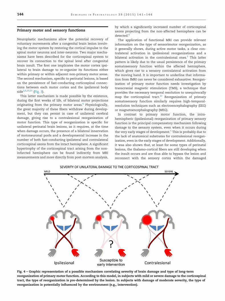
Fig. 4 – Graphic representation of a possible mechanism correlareorganization of primary motor function. According to this modetract, the type of reorganization is pre-determined by the lesionreorganization is potentially influenced by the environment (e.g
by which a significantly increased number of corticospinalaxons projecting from the non-affected hemisphere can bedetected.6
The application of functional MRI can provide relevantinformation on the type of sensorimotor reorganization, asit generally shows, during active motor tasks, a clear con-tralateral activation in ipsilesional reorganizations and abilateral activation in the contralesional ones.5 This latterpattern is likely due to the usual persistence of the primarysomatosensory function within the affected hemisphere,which gives rise to a sensory contralateral activation fromthe moving hand. It is important to underline that informa-tion from fMRI can never be considered exhaustive. Reorgan-ization of primary motor function needs investigation bytranscranial magnetic stimulation (TMS), a technique thatprovides the necessary temporal resolution to unequivocallymap the corticospinal tract.22 Reorganization of primarysomatosensory function similarly requires high-temporal-resolution techniques such as electroencephalography (EEG)or magnetoencephalography (MEG).In contrast to primary motor function, the intra-
hemispheric (ipsilesional) reorganization of primary sensoryfunction is the principal compensatory mechanism followingdamage to the sensory system, even when it occurs duringthe very early stages of development.5 This is probably due tothe lack of anatomical substrates for contralesional reorgan-ization, even in the early stages of development. Additionally,it was also shown that, at least for some types of perinatallesions, the thalamo-cortical fibers are still developing whenthe insult occurs and are thus able to bypass the lesion andreconnect with the sensory cortex within the damaged
ting severity of brain damage and type of long-terml, in subjects with mild or severe damage to the corticospinal. In subjects with damage of moderate severity, the type of., intervention).
S E M I N A R S I N P E R I N A T O L O G Y 3 9 ( 2 0 1 5 ) 1 4 1 – 1 4 6 145
hemisphere.23 This evidence comes from the combined use ofstructural imaging methods, such as diffusion-MRI-basedtractography,24 which identify the likely trajectories ofthalamo-cortical white matter bundles, and functional imag-ing methods, such as MEG, which are capable of reliablymapping the cortical location of primary sensoryprocessing.25
Data obtained from these particular mechanisms of earlyneuroplasticity in primary sensory and motor systems gen-erate distinctive patterns of reorganization that need to beaccurately characterized at the single subject level. Thiscomplexity is further enhanced by the crucial role of theenvironment in driving neuroplasticity. Indeed, the pattern ofsensorimotor reorganization is by no means a mere conse-quence of the size and site of the lesion but is stronglyinfluenced by the experience following damage (action-dependent reorganization). Figure 4 illustrates the complexinteraction between residual motor output from the affectedhemisphere and somatosensory feedback from the affectedlimb. Not surprising, the early structural asymmetries of thecorticospinal tract are not always able to predict the presenceand, most of all, the severity of later hemiplegia.26
This concept is further supported by diffusion studiesperformed during the chronic phases of the disorder, i.e., inadolescents and young adults with unilateral cerebral palsy,which show that the asymmetry of the corticospinal tract isless correlated with sensory and motor function than theasymmetry of other projections such as the thalamo-corticalones. These data suggest an essential role of higher-ordercompensatory mechanisms in shaping the long-term func-tional outcome.24 A growing body of evidence supports thehypothesis that the reorganization of motor function doesnot only take place within the sensorimotor cortex but alsoheavily involves subcortical structures such as the thalamus,brainstem or spinal cord, and cerebellum.27 The complexinterplay between early brain plasticity of the sensory-motor system and its relation to functional outcome remainspoorly understood and the subject of future ongoing studies.
The visual system
Visual impairment is very common after congenital braininjury, given that a substantial part of the brain is directly orindirectly involved in vision. However, visual deficits can varyin terms of the characteristics and severity of the disorder,depending on the site, extension, and timing of braininjury.28,29 The correlation between damage to the opticradiations or the occipital cortex and deficits to the corre-sponding visual field is far less consistent with early lesionsas compared to later ones. This finding may be related to themore powerful cerebral plasticity mechanisms in youngchildren, which have at least in part similar neurophysiolog-ical bases to what we have described for the somatosensorysystem and, in particular, the possibility that thalamo-cortical fibers develop after the lesion, bypassing it.30 Theexact characteristics and limits of this specific type ofplasticity involving thalamo-cortical networks are not fullyunderstood. It is of interest, however, that even when a visualfield deficit is present, the subject with early damage seems
to show fewer difficulties in environmental navigation andexploration. This finding is directly in line with animal modelexperiments, which clearly show how the ablation of thewhole primary visual cortex in newborn animals does notaffect their visual orientation performances, which are mas-sively impaired after a similar lesion in adult animals.31 Thisphenomenon is linked to a reorganization of the pathwaysconnecting subcortical visual structures directly to the extra-striatal visual cortex. Moreover, this occurrence appears totake place in humans as well, as shown, for example, by theincrease in activation of extra-striatal structures on fMRI afterthe stimulation of the affected hemi-field. Taken together,available data in humans support the presence of a moreeffective reorganization of visual function after early braindamage, which may consist of either a “reconnection” withthe targeted structures or secondary to an enhanced use ofcompensating circuitries.28 Even if these circuitries are notable to restore complete vision on the contralesional hemi-sphere, they can allow for good compensation in spatialorienting and localization.
Conclusions
Brain mapping techniques and, in particular, fMRI haveoffered important insights to many critical questions thathad been pending for decades, as it relates to how theimmature brain responds to early-life brain injury. Theconcept that is clearly emerging, both from human andnun-human studies, is that functional reorganization in theimmature brain is complex and substantially different fromthat of a mature brain that has already completed criticalstages of development. Although a complete understandingof the underlying mechanisms of cerebral plasticity in earlyinfancy is still far from understood, a fundamental under-standing of the neurobiological and neurophysiological prin-ciples underlying repair after injury will be essential for moretargeted prescription and tuning of therapeutic interventionprograms. The successful application and refinement ofadvanced MRI techniques to the developing brain (e.g.,ultra-high field MR) will undoubtedly provide important,currently unavailable insights to these intriguing questions.
r e f e r e n c e s
1. Chen R, Cohen LG, Hallett M. Nervous system reorganizationfollowing injury. Neuroscience. 2002;111(4):761–773.
2. Lewis TL, Maurer D. Multiple sensitive periods in humanvisual development: evidence from visually deprived chil-dren. Dev Psychobiol. 2005;46(3):163–183.
3. Berardi N, Pizzorusso T, Maffei L. Critical periods duringsensory development. Curr Opin Neurobiol. 2000;10(1):138–145.
4. Bates E, Reilly J, Wulfeck B, et al. Differential effects ofunilateral lesions on language production in children andadults. Brain Lang. 2001;79(2):223–265.
5. Staudt M, Gerloff C, Grodd W, Holthausen H, Niemann G,Krägeloh-Mann I. Reorganization in congenital hemiparesisacquired at different gestational ages. Ann Neurol. 2004;56(6):854–863.
S E M I N A R S I N P E R I N A T O L O G Y 3 9 ( 2 0 1 5 ) 1 4 1 – 1 4 6146
6. Eyre JA, Smith M, Dabydeen L, et al. Is hemiplegic cerebralpalsy equivalent to amblyopia of the corticospinal system?Ann Neurol. 2007;62(5):493–503.
7. Lidzba K, Staudt M. Development and (re)organization oflanguage after early brain lesions: capacities and limitationof early brain plasticity. Brain Lang. 2008;106(3):165–166.
8. Lidzba K, Wilke M, Staudt M, Krägeloh-Mann I. Early plasticityversus early vulnerability: the problem of heterogeneouslesion types. Brain. 2009;132(Pt 10):e128.
9. Anderson V, Spencer-Smith M, Leventer R, et al. Childhoodbrain insult: can age at insult help us predict outcome? Brain.2010;133(Pt 6):1855 [Brain. 2010 Aug;133(Pt 8):2505].
10. Krägeloh-Mann I, Horber V. The role of magnetic resonanceimaging in elucidating the pathogenesis of cerebral palsy: asystematic review. Dev Med Child Neurol. 2007;49(2):144–151[review].
11. Cioni G, D'Acunto G, Guzzetta A. Perinatal brain damage inchildren: neuroplasticity, early intervention, and molecularmechanisms of recovery. Prog Brain Res. 2011;189:139–154.
12. Arnfield E, Guzzetta A, Boyd R. Relationship between brainstructure on magnetic resonance imaging and motor out-comes in children with cerebral palsy: a systematic review.Res Dev Disabil. 2013;34(7):2234–2250.
13. Geschwind N, Levitsky W. Human brain: left–right asym-metries in temporal speech region. Science. 1968;161(3837):186–187.
14. Sowell ER, Trauner DA, Gamst A, Jernigan TL. Development ofcortical and subcortical brain structures in childhood andadolescence: a structural MRI study. Dev Med Child Neurol.2002;44(1):4–16.
15. Staudt M, Lidzba K, Grodd W, Wildgruber D, Erb M, Krägeloh-Mann I. Right-hemispheric organization of language follow-ing early left-sided brain lesions: functional MRI topography.Neuroimage. 2002;16(4):954–967.
16. Guzzetta A, Pecini C, Biagi L, et al. Language organisation inleft perinatal stroke. Neuropediatrics. 2008;39(3):157–163.
17. Tillema JM, Byars AW, Jacola LM, et al. Cortical reorganizationof language functioning following perinatal left MCA stroke.Brain Lang. 2008;105(2):99–111.
18. Liégeois F, Connelly A, Baldeweg T, Vargha-Khadem F. Speak-ing with a single cerebral hemisphere: fMRI language organ-ization after hemispherectomy in childhood. Brain Lang.2008;106(3):195–203.
19. Lidzba K, Staudt M, Wilke M, Krägeloh-Mann I. Visuospatialdeficits in patients with early left-hemispheric lesions and
functional reorganization of language: consequence of lesionor reorganization? Neuropsychologia. 2006;44(7):1088–1094.
20. Guzzetta A, Bonanni P, Biagi L, et al. Reorganization of thesomatosensory system after early brain damage. Clin Neuro-physiol. 2007;118(5):1110–1121.
21. Thickbroom GW, Byrnes ML, Archer SA, et al. Differences insensory and motor cortical organization following braininjury early in life. Ann Neurol. 2001;49(3):320–327.
22. Zsoter A, Pieper T, Kudernatsch M, Staudt M. Predicting handfunction after hemispherotomy: TMS versus fMRI in hemi-spheric polymicrogyria. Epilepsia. 2012;53(6):e98–e101.
23. Papadelis C, Leonardelli E, Staudt M, Braun C. Can magneto-encephalography track the afferent information flow alongwhite matter thalamo-cortical fibers? Neuroimage. 2012;60(2):1092–1105.
24. Rose S, Guzzetta A, Pannek K, Boyd R. MRI structural con-nectivity, disruption of primary sensorimotor pathways, andhand function in cerebral palsy. Brain Connect. 2011;1(4):309–316.
26. Guzzetta A, Pizzardi A, Belmonti V, et al. Hand movements at3 months predict later hemiplegia in term infants with neo-natal cerebral infarction. Dev Med Child Neurol. 2010;52(8):767–772.
27. Dinomais M, Groeschel S, Staudt M, Krägeloh-Mann I,Wilke M. Relationship between functional connectivity andsensory impairment: red flag or red herring? Hum Brain Mapp.2012;33(3):628–638.
28. Guzzetta A, D'Acunto G, Rose S, Tinelli F, Boyd R, Cioni G.Plasticity of the visual system after early brain damage. DevMed Child Neurol. 2010;52(10):891–900.
29. Ramenghi LA, Ricci D, Mercuri E, et al. Visual performanceand brain structures in the developing brain of pre-terminfants. Early Hum Dev. 2010;86(suppl 1):73–75.
30. Guzzetta A, Fiori S, Scelfo D, Conti E, Bancale A. Reorganiza-tion of visual fields after periventricular haemorrhagic infarc-tion: potentials and limitations. Dev Med Child Neurol. 2013;55(suppl 4):23–26.
31. Fagiolini M, Pizzorusso T, Berardi N, Domenici L, Maffei L.Functional postnatal development of the rat primaryvisual cortex and the role of visual experience: darkrearing and monocular deprivation. Vision Res. 1994;34(6):709–720.