First Department of Internal Medicine, Yokohama CTty Uni~ersity School of Medicine, 3-9 Fukuura, Kanazawa-ku, Yokohama 236, Japan
Received 7 February 1994; revised MS received 10 June 1994; accepted 30 August 1994
Abstract
In the present study, we asked whether platelet-activating factor (PAF) mediates the ozone-induced increase in airway microvascular leakage. To answer this question, we examined the effect of a PAF receptor antagonist on the ozone-induced increase in airway microvascular leakage quantified by the extravasation of Evans blue dye in the guinea pig trachea and main bronchi. Guinea pigs were pretreated with the PAF receptor antagonist, E6123 ((S)-(+)-6-(2-chlorophenyl)-3-cyclopropane- carb•nyl-8•1•-dimethy•-2•3•4•5-tetrahydr•-8H-pyrid•[4'•3':4•5]thien•[3•2-f][••2•4]triaz•••[4•3-a][1•4]diazepine) (0.01, 0.1 and 1.0 mg/kg i.v.) and then exposed to 3 ppm ozone for 30 rain. The PAF receptor antagonist significantly reduced the ozone-induced increase in microvascular leakage in a dose-dependent manner in both the trachea and main bronchi. Our results indicate that PAF mediates the ozone-induced increase in airway microvascular leakage. We therefore suggest that PAF may be involved in ozone-induced airway inflammation.
Ozone, an oxidant pollutant gas, is a major con- stituent of photochemical smog. Ozone exposure has been shown to cause microvascular leakage in the airways (Kaneko et al., 1994), which is an important sign of airway inflammation (Barnes et al., 1990; Pers- son, 1988). However, the mechanism of ozone-induced airway microvascular leakage remains to be elucidated. Recently, we have found that neuropeptides are par- tially involved in the ozone-induced increase in airway microvascular leakage (Kaneko et al., 1994). This find- ing suggested that other factors may be involved in this phenomenon. One possible factor is platelet-activating factor (PAF), which has been reported to be produced by ozone exposure in airway bronchial epithelial cells and in a macrophage-like cell line (Samet et al., 1992). In addition, PAF can cause both airway hyperrespon- siveness and microvascular leakage (McManus and Deavers, 1989), which are also observed after ozone exposure (Kaneko et al., 1994; Nishikawa et al., 1990).
Our objective was to determine whether PAF medi- ates the ozone-induced increase in airway microvascu- lar leakage. We examined the effect of a PAF receptor antagonist on the ozone-induced increase in microvas- cular leakage in both the guinea pig trachea and main bronchi. We used the novel thienodiazepine derivative PAF receptor antagonist, E6123, ((S)-(+)-6-(2-chloro- phenyl)-3-cyclopropanecarbonyl-8,11-dimethyl-2,3,4,5- tetrahydro-8 H-pyrido[4',3' :4,5]thieno[3,2-f ][ 1,2,4]tria- zolo[4,3-a][1,4]diazepine). E6123 has been shown to specifically block PAF-induced responses in a number of biological systems both in vitro and in vivo (Sakuma et al., 1991a,b; Tashiro et al., 1992; Tsunoda et al., 1990).
2. Materials and methods
2.1. Animals and animal preparation
We anesthetized 35 Hartley strain male guinea pigs (Japan SLC, Shizuoka, Japan) weighing 310-450 g with pentobarbital (40 m g / k g injected intraperitoneally) and surgically exposed a femoral vein. A catheter (0.51 mm
252 T. Kaneko et al. / Eur. J. Pharmacol. Ent:iron. Toxicol. Pharmacol. Section 292 (1995) 251-255
i .d.× 0.94 mm o.d., Dow Corning Corporation, Mid- land, MI) filled with anticoagulant solution was in- serted into the femoral vein and advanced to the inferior vena cava. Evans blue dye and medications were injected through this catheter when the guinea pigs were awake. An incision was made at the back of the guinea pig's neck and a tunnel to the femoral side was created with a hemostat to secure the catheter. The free end of the catheter was threaded subcuta- neously through this tunnel and anchored at the back of the neck to prevent the guinea pig from removing the catheter when awake. The end of the catheter was occluded with a piece of copper wire. The femoral and neck incisions were closed with 3.0 silk, which was also used to tie the catheter in place. Surgery was per- formed aseptically.
2.2. Experimental protocol
The effects of 0.01, 0.1 and 1.0 m g / k g concentra- tions of the PAF receptor antagonist E6123 on the ozone-induced increase in airway microvascular leak- age were examined in three groups of five guinea pigs each (Fig. 1A). Another two groups of five guinea pigs each were treated with vehicle and exposed to either 3 ppm ozone or air (control) for 30 min. E6123 or its vehicle was injected 30 min before ozone exposure via the previously inserted catheter when guinea pigs were awake. Airway microvascular leakage was measured in both the trachea and main bronchi, as described above.
To confirm that the highest dose of E6123 (1.0 m g / k g i.v.) we used in the above study was enough to completely inhibit the endogenous PAF-induced in- crease in airway microvascular leakage after ozone exposure, we performed the following study (Fig. 1B). We injected E6123 (1.0 m g / k g i.v.) or its vehicle via a catheter 30 rain before a 30-min exposure to air. We then injected PAF (100 n g / k g i.v.) 30 min after air
A
E6123 or Vehicle
PROTOCOL
Evans blue dye
30 rnln 3 ppm O3or Air for 30mln I
Wash out Anesthesia
4 o,° j lso,\J m
60 min [
E6123 or Wash out Vehicle Evans blue dye Anesthesia
°,o oo,° + I TM °in B
4 ~
60 min - I
Fig. 1. (A) A protocol to study the effect of E6123 on the ozone-in- duced increase in airway microvascular leakage. (B) A protocol to study the effect of E6123 on the PAF-induced increase in airway microvascular leakage.
exposure. The timing of the injection of PAF was chosen because our time course study (Kaneko et al., 1994) showed that airway microvascular leakage in- creased after a time lag of between 30-45 rain after a 30-min exposure to 3 ppm ozone. The dose of PAF (100 n g / k g i.v.) was chosen because it caused larger airway microvascular leakage than a 30-rain exposure to 3 ppm ozone. Airway microvascular leakage was again measured in both the trachea and main bronchi, as described above.
2.3. Ozone exposure
Each guinea pig was exposed to 3 ppm ozone for 30 min while awake, resting quietly, and breathing sponta- neously in a 23.5-1 acrylic chamber. Ozone was pro- duced by an ozone generator (Model MOT-001A; Nip- pon Ozone, Tokyo, Japan) and diluted with clean, filtered air delivered into the exposure chamber at a flow rate of 20 l /rain. The concentration of ozone in the chamber was continuously monitored using an ul- traviolet ozone analyzer (Model DY-1500; Nippon Ozone).
The 30-min 3-ppm dose of ozone was chosen be- cause it has been found to significantly increase airway responsiveness (Lee and Murlas, 1985; Nishikawa et al., 1990). This short-term exposure to a high concen- tration of ozone also allowed us to observe the acute phase of ozone-induced airway inflammation.
2.4. Measurement of airway microc'ascular leakage
Airway microvascular leakage was quantified by the extravasation of Evans blue dye by a modification of the method of Saria and Lundberg (Saria and Lund- berg, 1983), which has been described in our previous study (Kaneko et al., 1994). Evans blue dye (40 m g / m l in 0.9% NaC1, filtered through a 5.0-/.tm Millipore filter, 40 mg/kg) , was injected intravenously via the previously inserted catheter just before the exposure to ozone or air. Guinea pigs were anesthetized intraperi- toneally with 40 m g / k g pentobarbital 45 rain after a 30-rain exposure to ozone or air. Sixty minutes after the exposure, the dye was washed out by perfusing the intravascular space with 200 ml phosphate-buffered saline (pH 7.40) through the abdominal aorta at a perfusion pressure of 100 cm H 2 0 . Sixty minutes was chosen because we observed the largest statistical dif- ference in extravasation of dye between ozone- and air-exposed airways at this time point in our previous study (Kaneko et al., 1994). After all the dye was washed out, a 10-ram section of trachea 10 mm proxi- mal to the tracheal bifurcation and the whole main bronchi were removed, weighed, and placed in I m[ of 100% formamide. Evans blue dye was extracted by incubating tissues at 60°C for 16 h in formamide.
T. Kaneko et aL /Eur. J. Pharrnacol. Environ. Toxicol. Pharmacol. Section 292 (1995) 251-255 253
Tissue content of Evans blue dye was determined by measuring light absorbance at 620 nm with a spectro- photometer (Model V-1100; Hitachi, Tokyo, Japan) and interpolating the data from a standard curve for Evans blue dye. Values are expressed as nanograms of dye per milligram of trachea or main bronchi (wet weight).
2.5. Drugs and chemicals
The following agents were used: Evans blue dye, PAF (Sigma Chemical Co., St. Louis, MO); formamide (Merck, Darmstadt, Germany); and sodium pento- barbital (Abbott Laboratories, North Chicago, IL). E6123 was kindly provided by Eisai Co. (Tokyo, Japan).
2.6. Statistical analysis
to to
E
to t-- t~ > UJ
60
50
40
20
10
0 Control
Main Bronchi
03
*t
0.01 0.1 1
E 6123 (rng/kg)
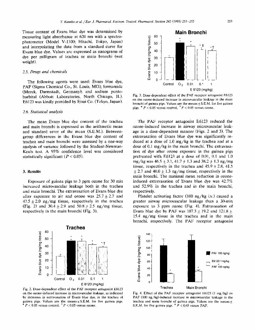
Fig. 3. Dose-dependent effect of the PAF receptor antagonist E6123 on the ozone-induced increase in microvascular leakage in the main bronchi of guinea pigs. Values are the means _ S.E.M. for five guinea pigs. * P < 0.05 versus control, * P < 0.05 versus ozone.
The mean Evans blue dye content of the trachea and main bronchi is expressed as the arithmetic mean and standard error of the mean (S.E.M.). Between- group differences in the Evans blue dye content of trachea and main bronchi were assessed by a one-way analysis of variance followed by the Student-Newman- Keuls test. A 95% confidence level was considered statistically significant (P < 0.05).
3. Results
Exposure of guinea pigs to 3 ppm ozone for 30 min increased microvascular leakage both in the trachea and main bronchi. The extravasation of Evans blue dye after exposure to air and ozone was 25.7 + 2.7 and 47.5 + 2.0 ng/mg tissue, respectively in the trachea (Fig. 2) and 30.4 + 2.9 and 50.8 _+ 2.5 ng/mg tissue, respectively in the main bronchi (Fig. 3).
t~3 E
to
U3
60
50
40
30
10
0 Control
Trachea
03 0.01 0.1 1
E 6123 (mg/kg) Fig. 2. Dose-dependent effect of the PAF receptor antagonist E6123 on the ozone-induced increase in microvascular leakage, as indicated by increases in extravasation of Evans blue dye, in the trachea of guinea pigs, Values are the m e a n s + S . E . M , for five guinea pigs. * P < 0.05 versus control, t p < 0.05 versus ozone.
The PAF receptor antagonist E6123 reduced the ozone-induced increase in airway microvascular leak- age in a dose-dependent manner (Figs. 2 and 3). The extravasation of Evans blue dye was significantly re- duced at a dose of 1.0 mg/kg in the trachea and at a dose of 0.1 mg/kg in the main bronchi. The extravasa- tion of dye after ozone exposure in the guinea pigs pretreated with E6123 at a dose of 0.01, 0.1 and 1.0 mg/kg was 46.5 _+ 3.1, 41.7 _ 1.3 and 38.2 + 1.3 ng/mg tissue, respectively in the trachea and 45.9 _+ 2.8, 41.5 + 2.7 and 40.0 + 1.3 ng/mg tissue, respectively in the main bronchi. The maximal mean reduction in ozone- induced extravasation of Evans blue dye was 42.7% and 52.9% in the trachea and in the main bronchi, respectively.
Platelet activating factor (100 ng/kg i.v.) caused a greater airway microvascular leakage than a 30-min exposure to 3 ppm ozone (Fig. 4). Extravasation of Evans blue dye by PAF was 107.3 + 19.2 and 121.8 + 15.4 ng/mg tissue in the trachea and in the main bronchi, respectively. The PAF receptor antagonist
140-
m 120 - to
t:~ 100-
ao-
>, 60-
4 0 - .£~ to c 2 0 - >
ILl 0 -
Trachea Main Bronchi
• PAF 100 ng/kg
E6123 lmg/kg +
[ ~ PAF 100 ng/kg
Fig. 4. Effect of the PAF receptor antagonist E6123 (1 m g / k g ) on PAF (100 ng/kg)- induced increase in microvascular leakage in the trachea and main bronchi of guinea pigs. Values are the means_+ S.E.M. for five guinea pigs. * P < 0.05 versus PAF.
254 T. Kaneko et al. / Eur. J. Pharmacol. Environ. Toxicol. Pharmacol. Section 292 (1995) 251-255
E6123 (1 mg /kg i.v.) completely inhibited the PAF (100 ng /kg i.v.)-induced increase in airway microvascu- lar leakage. After pretreatment with E6123, extravasa- tion of dye induced by PAF was 24.3_+ 2.2 n g / m g tissue in the trachea and 32.3 + 3.2 rig/rag tissue in the main bronchi, which are not different from the extrava- sation of dye after air exposure (25.7 + 2.7 and 30.4 _+ 2.9 n g / m g tissue in the trachea and in the main bronchi, respectively).
4. Discussion
In this study in guinea pigs, we have shown that PAF mediates the ozone-induced increase in airway microvascular leakage. The evidence is that the PAF receptor antagonist reduced the ozone-induced in- crease in microvascular leakage, as determined by in- creases in the extravasation of Evans blue dye, in the trachea and main bronchi. We suggest that PAF may be involved in ozone-induced airway inflammation.
The most likely sources of PAF in the airways after ozone exposure are airway epithelial cells and macrophages, because human airway epithelial cells and a macrophage-like human cell line have been found to produce PAF after exposure to ozone in vitro (Samet et al., 1992). It is also possible that other resident airway cells, including endothelial cells (Camussi et al., 1983; McIntyre et al., 1986), leukocytes (Clark et al., 1980; Lynch et al., 1979) and platelets (Chignard et al., 1980) could be sources of PAF in the airways after ozone exposure, since these cells have been shown to be able to produce PAF in vitro.
In the present study, the ozone-induced increase in microvascular leakage was reduced but not abolished in both the trachea and main bronchi. The highest dose of the PAF receptor antagonist (1 mg /k g i.v.) used in this study completely inhibited the PAF (100 ng /kg i.v.)-induced increase in airway microvascular leakage, which was a greater leakage than a 30-min exposure to 3 ppm ozone. These findings suggest that the mechanisms involved in the ozone-induced in- crease in airway microvascular leakage in the airway may consist of factors other than PAF. Recently, using the same study design as this study, we have found that neuropeptide depletion by capsaicin treatment par- tially inhibits the ozone-induced increase in airway microvascular leakage in guinea pigs (Kaneko et al., 1994). Therefore, neuropeptides may also have an im- portant role in the ozone-induced increase in airway microvascular leakage. In addition, several other medi- ators, including leukotrienes (Hua et al., 1985; Persson et al., 1986; Woodward et al., 1983), histamine, bradykinin, and serotonin (Saria et al., 1983) have been shown to increase airway microvascular leakage. Some of these mediators could be also involved in the
ozone-induced increase in airway microvascular leak- age.
In contrast to our findings, a recent study found that a PAF receptor antagonist did not alter ozone-induced airway inflammation in guinea pigs (Tan and Bethel, 1992). In that study, airway inflammation was assessed by inflammatory cell infiltration into the airways using bronchoalveolar lavage and was examined over 4-5 h after a 2-h exposure to 3 ppm ozone. We assessed the acute phase of ozone-induced airway inflammation by measuring the increase in microvascular leakage 60 min after a 30-min exposure to 3 ppm ozone. Thus, the marker and time point are completely different from those used in our study. In our acute-phase experi- ments, inflammatory cells may not have yet infiltrated into the airways (Murlas and Roum, 1985). Further- more, it is possible that a number of other mediators are produced at the moment of inflammatory cell mi- gration into the airways, which occurs several hours after ozone exposure, PAF may be only one of the mediators that causes airway inflammation.
In summary, we have shown that PAF mediates the ozone-induced increase in airway microvascular leak- age in guinea pigs. This finding is supported by a reduction in the ozone-induced increase in microvascu- lar leakage in the trachea and main bronchi by pre- treatment with the PAF receptor antagonist. We there- fore suggest that PAF may be involved in ozone-in- duced airway inflammation.
Acknowledgements
We are grateful to Drs. Pierangelo Geppett i and Dale Halliday, Cardiovascular Research Institute, Uni- versity of California San Francisco, for their critical review of this manuscript.
References
Barnes, P.J., P. Boschetto, D.F. Rogers, M. Belvisi, N. Roberts, K.F. Chung and T.W. Evans, 1990, Effects of treatment on airway microvascular leakage., Eur. Respir. J. 3, Suppl. 12, 663S.
Camussi, G., M. Aglietta, F. Malavasi, C. Tetta, W. Piacibello, F. Sanavio and F. Bussolino, 1983, The release of platelet-activating factor from human endothelial cells in culture, J. Immunol. 131, 2397.
Chignard, M., J.P. LeCouedic, B.B. Vargaftig and J. Benveniste, 1980, Platelet-activating factor (PAF-acether) secretion from platelets: effect of aggregating agents, Br. J. Haematol. 46, 455.
Clark, P.O., D.J. Hanahan and R.N. Pinckard, 1980, Physical and chemical properties of platelet-activating factor obtained from human neutrophils and monocytes and rabbit neutrophils and basophils, Biochim. Biophys. Acta 628, 69.
Hua, X.-Y., S.-E. Dahl~n, J.M. Lundberg, S. Hammarstr6m and P. Hedqvist, 1985, Leukotrienes C4, D 4 and E 4 cause widespread and extensive plasma extravasation in the guinea pig, Naunyn- Schmied. Arch. Pharmacol. 330, 136.
T. Kaneko et aL / Eur. J. PharmacoL Era,iron. ToxicoL PharmacoL Section 292 (1995) 251-255 255
Kaneko, T., H. Ikeda, L. Fu, H. Nishiyama, M. Matsuoka, H. Yamakawa and T. Okubo, 1994, Capsaicin reduces ozone-in- duced airway inflammation in guinea pigs, Am. J. Respir. Crit. Care Med. 150, 724.
Lee, H.K. and C. Murlas, 1985, Ozone-induced bronchial hyperreac- tivity in guinea pigs is abolished by BW 755C or FPL 55712 but not by indomethacin, Am. Rev. Respir. Dis. 132, 1005.
Lynch, J.M., G.Z. Lotner, S.J. Betz and P.M. Henson, 1979, The release of a platelet-activating factor by stimulated rabbit neu- trophils, J. Immunol. 123, 1219.
Mclntyre, T.M., G.A. Zimmerman and S.M. Prescott, 1986, Leuko- trienes C 4 and D 4 stimulate human endothelial cells to synthe- size platelet-activating factor and bind neutrophils, Proc. Natl. Acad. Sci. USA 83, 2204.
McManus, L.M. and S.I. Deavers, 1989, Platelet activating factor in pulmonary pathobiology, Clin. Chest. Med. 10, 107.
Murlas, C.G. and J.H. Roum, 1985, Sequence of pathologic changes in the airway mucosa of guinea pigs during ozone-induced bronchial hyperreactivity, Am. Rev. Respir. Dis. 131, 314.
Nishikawa, M., S. Suzuki, H. Ikeda, T. Fukuda, J. Suzuki and T. Okubo, 1990, Dose-response relationship of ozone-induced air- way hyperresponsiveness in unanesthetized guinea pigs, J. Toxi- col. Environ. Health 30, 123.
Persson, C.G.A., 1988, Plasma exudation and asthma, Lung 166, 1. Persson, C.G.A., I. Erjef~ilt and P. Andersson, 1986, Leakage of
macromolecules from guinea-pig tracheobronchial microcircula- tion. Effects of allergen, leukotrienes, tachykinins, and anti- asthma drugs, Acta Physiol. Scand. 127, 95.
Sakuma, Y., M. Shirato, J. Nagaoka, H. Obaishi, H. Tsunoda, S. Katayama, H. Ono and K. Katayama, 1991a, Pharmacological activities of a novel thienodiazepine derivative as a platelet- activating factor antagonist. Effects on microvascular permeabil- ity, hypotension and nephrosis, Arzneim. Forsch. 41, 1255.
Sakuma, Y., H. Tsunoda, M. Shirato, S. Katayama, I. Yamatsu and K. Katayama, 1991b, Pharmacological effects of oral E6123, a novel PAF antagonist, on biological changes induced by PAF inhalation in guinea pigs, Prostaglandins 42, 463.
Samet, J.M., T.L. Noah, R.B. Devlin, J.R. Yankaskas, K. McKinnon, L.A. Dailey and M. Friedman, 1992, Effect of ozone on platelet- activating factor production in phorbol-differentiated HL60 cells, a human bronchial epithelial cell line (BEAS $6), and primary human bronchial epithelial cells, Am. J. Respir. Cell Mol. Biol. 7, 514.
Saria, A. and J.M. Lundberg, 1983, Evans blue fluorescence: quanti- tative and morphological evaluation of vascular permeability in animal tissues, J. Neurosci. Meth. 8, 41.
Saria, A., J.M. Lundberg, G. Skofitsch and F. Lembeck, 1983, Vascular protein leakage in various tissues induced by substance P, capsaicin, bradykinin, serotonin, histamine and by antigen challenge, Naunyn-Schmied. Arch. Pharmacol. 324, 212.
Tan, W.C. and R.A. Bethel, 1992, The effect of platelet activating factor antagonist on ozone-induced airway inflammation and bronchial hyperresponsiveness in guinea pigs, Am. Rev. Respir. Dis. 146, 916.
Tashiro, K., Z. Xie and Y. Ito, 1992, Effects of PAF on excitatory neuro-effector transmission in dog airways, Br. J. Pharmacol. 107, 956.
Tsunoda, H., Y. Sakuma, K. Harada, K. Muramoto, S. Katayama, T. Horie, N. Shimomura, R. Clark, S. Miyazawa, K. Okano, Y. Machida, K. Katayama and I. Yamatsu, 1990, Pharmacological activities of a novel thienodiazepine derivative as a platelet- activating factor antagonist, Arzneim. Forsch. 40, 1201.
Woodward, D.F., B.M. Weichman, C.A. Gill and M.A. Wasserman, 1983, The effect of synthetic leukotrienes on tracheal microvascu- lar permeability, Prostaglandins 25, 131.