Playing games with a thrombus: a dangerousmatch. Paradoxical embolism from a huge centralvenous cathether thrombus: a case reportNuno Cardim1*, Júlia Toste1, Vanessa Carvalho1, Igor Nunes1, Daniel Ferreira1, Vanda Carmelo1, Ana SN Oliveira2,José Ferro2, Sylvie Mariana1, Adelaide Almeida1, Francisco P Machado1, José Roquette1
Abstract
Thromboembolism is a major cause of death in cancer patients. The association between paraneoplastic hypercoa-gulability of oncological patients and long-term central venous catheters (CVC) may result in CVC associatedthrombosis. Patent Foramen Ovale (PFO), especially when associated with atrial septal aneurysm (ASA) is a risk fac-tor for paradoxical embolism. We report a case of paradoxical embolism with stroke in an oncological patient witha huge CVC thrombus playing “ping-pong” with an hypermobile ASA with a PFO. We review the management ofhypercoagulability in oncologic patients and discuss the potential role of routine transthoracic echocardiographybefore the implantation of long term central venous catheters to identify predisposing conditions to paradoxicalembolism and select patients for anticoagulant therapy.
IntroductionIn oncological patients hemostasis abnormalities rangefrom asymptomatic alterations to massive thromboem-bolism. Its clinical importance lies on the fact that itconstitutes the second leading cause of death in cancerpatients [1].This paraneoplastic hypercoagulability syndrome,
although not completely understood, results secondaryfrom a complex imbalance between coagulation andfibrinolysis, for which contributes decreased fibrinogenand platelet catabolism, reduction of antithrombotic fac-tors such as proteins S, C and antithrombin, enhancedplatelet activation and aggregation [2], increased forma-tion of thrombin, and thrombocitosis [3] secondary tooverproduction of thrombopoietin and interleukin 6 bythe tumor [4]. Its severity depends on several factors,including the type and stage of tumor, the effects ofadjuvant chemotherapy, the presence of central venouscatheters (CVC) for its administration, surgical proce-dures and other adjuvant therapies [5].Long-term central venous catheters (CVCs) are com-
monly used in patients with cancer. Their presence may
be complicated by the occurrence of CVC-associatedthrombosis, defined as a mural thrombus extendingfrom the catheter into the lumen of a vessel and leadingto partial or total catheter occlusion. According torecent reviews, the incidence of symptomatic andasymptomatic CVC-associated thrombosis in cancerpatients is much higher than in non selected popula-tions and ranged between 0-28% and between 12-66%,respectively [6].Autopsy reports suggest that a patent Foramen Ovale
(PFO) may occur in 11% to 35% of the normal popula-tion [7], with a 34.3% age-related incidence for agesfrom 0 to 29 years, 25.4% for ages from 30 to 79 years,and 20.2% for ages between 80 and 99 years. Thompsonand Evans consecutively examined 1100 autopsies anddescribed a “probe-PFO” (measuring 0.2 to 0.5 cm) in29% and a “pencil-PFO” (measuring 0.6 to 1.0 cm) in6% of the cases [7].Paradoxical embolism was first described by Cohn-
heim in 1877 [7] and refers to an embolus passing fromthe right to the left heart through a right-to-left shunt,leading to arterial embolism. A PFO accounts for 70%of all right-to-left cardiac shunts. Other reported sourcesinclude a ventricular septal defect, Ebstein’s anomaly,pulmonary arteriovenous malformations, and a patent
* Correspondence: [email protected] Department, Hospital da Luz. Av. Lusíada, 100. 1500-650 Lisbon,Portugal
Cardim et al. Cardiovascular Ultrasound 2010, 8:6http://www.cardiovascularultrasound.com/content/8/1/6
ductus arteriosus. With the introduction of transesopha-geal echocardiography (TEE) the ability to recognizeintracardiac shunting has increased, leading to thedescription of a new clinical, impending paradoxicalembolus [7].A large PFO and the presence of an atrial septal
aneurysm (ASA) have been identified, among others, asmorphological features of PFO with high risk for para-doxical embolism [8].
Case reportFemale patient, 57 years, admitted due to an acute epi-sode of aphasia without apparent motor deficits.She had been diagnosed for a colon adenocarcinoma
(Kras wild-type) three years before, with liver andlung metastasis, being now on chemotherapy
(cetuxime and FOLFOX regimen, which includes foli-nic acid, 5-fluoruracil and oxaliplatin) deliveredthrough a permanent CVC catheter implanted in thesuperior vena cava.Three months before the patient had been hospitalized
due to a Listeria monocytogenes bacteraemia, treatedwith antibiotics (amoxicillin-clavulamic acid + ceftriax-one). The patient recovered and was discharged withoutneurological deficits.Fifteen days before the present admission the catheter
had to be replaced because of fever of unknown origin.The physical examination showed a vigil, lucid and
cooperative patient, with transcortical motor aphasia(word repetition and understanding maintained), with-out focal motor deficits or other changes in the neuro-logical exam.
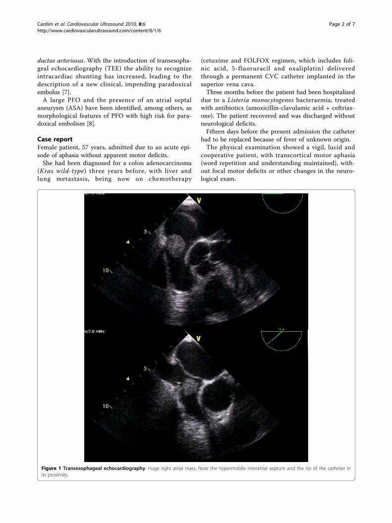
Figure 1 Transesophageal echocardiography. Huge right atrial mass. Note the hypermobile interatrial septum and the tip of the catheter inits proximity.
Cardim et al. Cardiovascular Ultrasound 2010, 8:6http://www.cardiovascularultrasound.com/content/8/1/6
Page 2 of 7
The cranial-encephalic magnetic resonance with diffu-sion study showed lesions in the left frontal area withrestricted diffusion, consistent with acute ischemic cerebralvascular lesion on the left middle cerebral artery territory.The electrocardiogram was normal but the transthoracic
echocardiogram suggested an ASA with a possible PFO.The TEE showed a voluminous round mass inside the
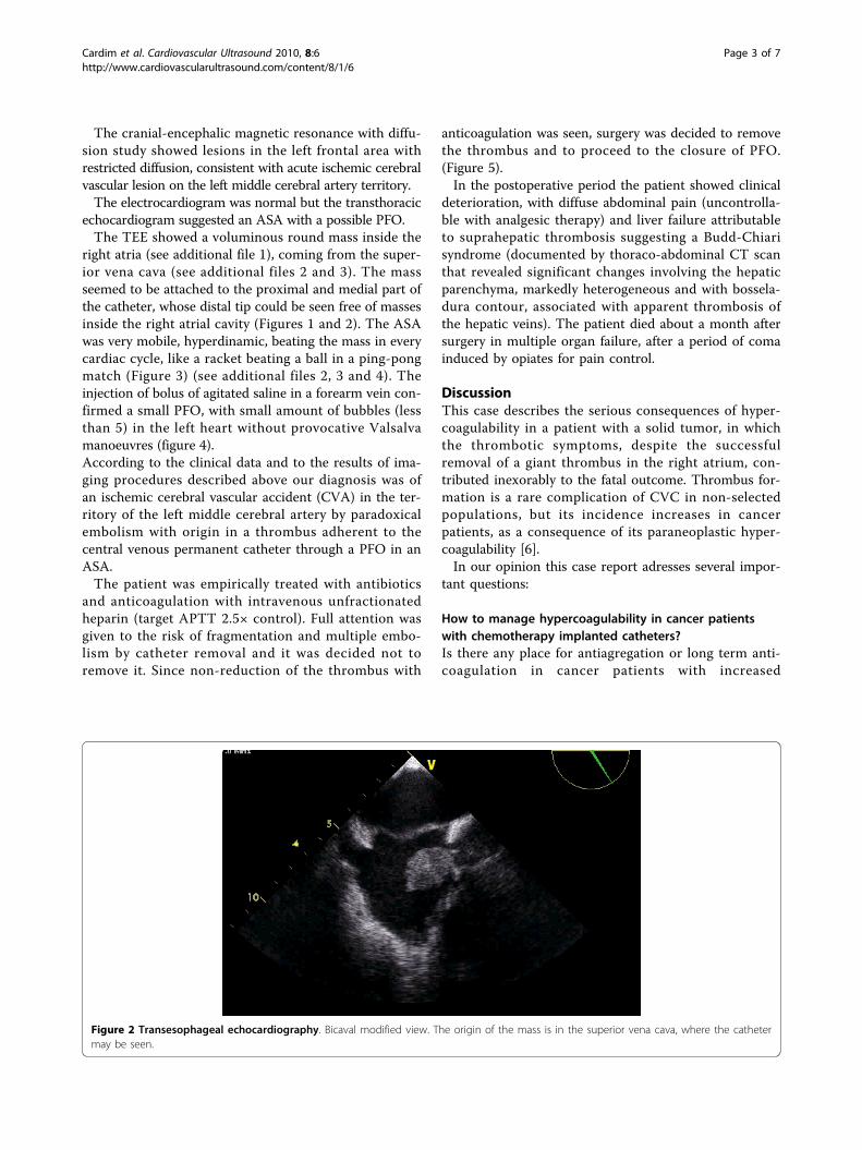
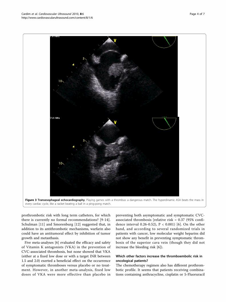
right atria (see additional file 1), coming from the super-ior vena cava (see additional files 2 and 3). The massseemed to be attached to the proximal and medial part ofthe catheter, whose distal tip could be seen free of massesinside the right atrial cavity (Figures 1 and 2). The ASAwas very mobile, hyperdinamic, beating the mass in everycardiac cycle, like a racket beating a ball in a ping-pongmatch (Figure 3) (see additional files 2, 3 and 4). Theinjection of bolus of agitated saline in a forearm vein con-firmed a small PFO, with small amount of bubbles (lessthan 5) in the left heart without provocative Valsalvamanoeuvres (figure 4).According to the clinical data and to the results of ima-ging procedures described above our diagnosis was ofan ischemic cerebral vascular accident (CVA) in the ter-ritory of the left middle cerebral artery by paradoxicalembolism with origin in a thrombus adherent to thecentral venous permanent catheter through a PFO in anASA.The patient was empirically treated with antibiotics
and anticoagulation with intravenous unfractionatedheparin (target APTT 2.5× control). Full attention wasgiven to the risk of fragmentation and multiple embo-lism by catheter removal and it was decided not toremove it. Since non-reduction of the thrombus with
anticoagulation was seen, surgery was decided to removethe thrombus and to proceed to the closure of PFO.(Figure 5).In the postoperative period the patient showed clinical
deterioration, with diffuse abdominal pain (uncontrolla-ble with analgesic therapy) and liver failure attributableto suprahepatic thrombosis suggesting a Budd-Chiarisyndrome (documented by thoraco-abdominal CT scanthat revealed significant changes involving the hepaticparenchyma, markedly heterogeneous and with bossela-dura contour, associated with apparent thrombosis ofthe hepatic veins). The patient died about a month aftersurgery in multiple organ failure, after a period of comainduced by opiates for pain control.
DiscussionThis case describes the serious consequences of hyper-coagulability in a patient with a solid tumor, in whichthe thrombotic symptoms, despite the successfulremoval of a giant thrombus in the right atrium, con-tributed inexorably to the fatal outcome. Thrombus for-mation is a rare complication of CVC in non-selectedpopulations, but its incidence increases in cancerpatients, as a consequence of its paraneoplastic hyper-coagulability [6].In our opinion this case report adresses several impor-
tant questions:
How to manage hypercoagulability in cancer patientswith chemotherapy implanted catheters?Is there any place for antiagregation or long term anti-coagulation in cancer patients with increased
Figure 2 Transesophageal echocardiography. Bicaval modified view. The origin of the mass is in the superior vena cava, where the cathetermay be seen.
Cardim et al. Cardiovascular Ultrasound 2010, 8:6http://www.cardiovascularultrasound.com/content/8/1/6
Page 3 of 7
prothrombotic risk with long term catheters, for whichthere is currently no formal recommendations? [9-14].Schulman [11] and Smorenburg [12] suggested that, inaddition to its antithrombotic mechanisms, warfarin alsocould have an antitumoral effect by inhibition of tumorgrowth and metasthasis.Five meta-analyses [6] evaluated the efficacy and safety
of Vitamin K antagonists (VKA) in the prevention ofCVC-associated thrombosis, but none showed that VKA(either at a fixed low dose or with a target INR between1.5 and 2.0) exerted a beneficial effect on the occurrenceof symptomatic thromboses versus placebo or no treat-ment. However, in another meta-analysis, fixed lowdoses of VKA were more effective than placebo in
preventing both asymptomatic and symptomatic CVC-associated thrombosis [relative risk = 0.37 (95% confi-dence interval 0.26-0.52), P < 0.001) [6]. On the otherhand, and according to several randomized trials inpatients with cancer, low molecular weight heparins didnot show any benefit in preventing symptomatic throm-bosis of the superior cava vein (though they did notincrease the bleeding risk [6]).
Which other factors increase the thromboembolic risk inoncological patients?The chemotherapy regimen also has different prothrom-botic profile. It seems that patients receiving combina-tions containing anthracycline, cisplatin or 5-Fluoruracil
Figure 3 Transesophageal echocardiography. Playing games with a thrombus: a dangerous match. The hyperdinamic ASA beats the mass inevery cardiac cycle, like a racket beating a ball in a ping-pong match.
Cardim et al. Cardiovascular Ultrasound 2010, 8:6http://www.cardiovascularultrasound.com/content/8/1/6
Page 4 of 7
Figure 4 Transesophageal echocardiography. The injection of bolus of agitated saline in a forearm vein confirmed a small PFO, withspontaneous shunt. Note the small amount of bubbles (less than 5) in the left atrium.
Figure 5 The superior vena cava thrombus. Note the similar shape between TEE features (top) and macroscopic data (bottom), with a longand thin border, corresponding to the insertion in the superior vena cava lumen.
Cardim et al. Cardiovascular Ultrasound 2010, 8:6http://www.cardiovascularultrasound.com/content/8/1/6
Page 5 of 7
(as our patient was) have increased thrombotic risk [15](animal experimentation microscopic studies with5-Fluoruracil show severe endothelial damage withthrombotic formation induced by this agent [15]).Another important issue is that our patient had pre-
vious bacteremia. Bacteremia is much more common inpatients with catheter thrombosis and thrombosis isitself a risk factor for catheter infection [15].
Was the surgical approach appropriated in this specificpatient?In the absence of guidelines, all the therapeutic deci-sions, specially the one of carrying on with cardiac sur-gery were intensively debated in multidisciplinaryclinical meetings (internists, neurologists, cardiologistsand cardiac surgeons). To avoid overtreatment, we fol-lowed a rational stepwise approach: our initial optionwas for intravenous non fractionated heparin, which wasinfused during 5 days without any reduction of throm-bus size in a control TEE. We decided not to performtrombolysis because of the risk of thrombus fragmenta-tion and of consequent multiple pulmonary and para-doxical systemic embolization. For the same reason wedecided not to simply pull and remove the CVC,because thrombus diameter was bigger than the lumenof the superior vena cava. So, surgery was decided asthe last and difficult option, taking into account thebenefits (to avoid pulmonary and systemic massiveembolism from a huge CVC thrombus playing ping-pong with an hyperdinamic ASA) versus the risks of theprocedure.
ASA, PFO and paradoxical embolism in oncologicalpatientsParadoxical embolism is a known cause of ischemicCVA in patients with PFO, embolus usually comingfrom venous disease of the lower limbs. However, in thereported case the origin of the thrombus was a bulkyintracardiac mass diagnosed by TEE.The clinical implications of PFO morphology and
associated features are still debated [16,17]. Qualitativeand quantitative and analysis by TEE is helpful in char-acterizing PFO-related risk of paradoxical embolism. Inpatients with cryptogenic CVAs large and tunnelledPFO, with “spontaneous” and severe shunt, with conco-mitant ASA and Chiari network increased the embolicrisk [17]. So, these detailed morphological and func-tional data should be provided in the TEE assessment ofa PFO, and not only its simple presence or absence.
ConclusionThromboembolism is the second major cause of deathin cancer patients and central venous catheter throm-bosis is very frequent in oncological patients.
Routine transthoracic echocardiography may play animportant role in the work-up of oncological patientswith central venous catheters. This technique may iden-tify predisposing conditions to paradoxical embolismand select patients for antiagregant or anticoagulanttherapy. Moreover, in the presence of stroke, if an atrialseptum aneurism and a patent Foramen Ovale isdetected, a transesophageal echocardiogram must beperformed for detailed morphological and functionaldefinition of the PFO related-risk for paradoxicalembolism.In the presence of a high-risk PFO the clinical situa-
tion should be discussed in a multidisciplinary medicalteam (cardiologist, oncologist, neurologist) and the deci-sion to start anticoagulation or not should be taken onan individual basis, taking into account the benefits andrisks of the procedure.
Declaration of Competing interestsThe authors declare that they have no competinginterests.
ConsentA copy of the written consent is available for review bythe Editor-in-Chief of this journal.
Additional file 1: The thrombus. Transesophageal echocardiography.Huge right atrial mass. The origin of the mass is not seen in this view.
Additional file 2: Localizing the mass. Transesophagealechocardiography. Bicaval modified view. The origin of the mass is in thesuperior vena cava, where the catheter may be seen.
Additional file 3: Playing games with a thrombus: a dangerousmatch. Transesophageal echocardiography:The hyperdinamic ASA beatsthe mass in every cardiac cycle, like a racket beating a ball in a ping-pong match.
Additional file 4: Playing games with a thrombus: a dangerousmatch. Transesophageal echocardiography:The hyperdinamic ASA beatsthe mass in every cardiac cycle, like a racket beating a ball in a ping-pong match. Note some thrombus fragmentation.
Author details1Cardiology Department, Hospital da Luz. Av. Lusíada, 100. 1500-650 Lisbon,Portugal. 2Neurology Department, Hospital da Luz. Av. Lusíada, 100. 1500-650 Lisbon, Portugal.
Authors’ contributionsC N carried out TEE and conceived of the paper and participated in itsdesign and coordination and helped to draft the manuscript; T Jparticipated in the sequence alignment and drafted the manuscript; C Vparticipated in the sequence alignment and drafted the manuscript; F Dhelped to draft the manuscript; N I provided clinical medical assistance tothe patient; C V provided clinical assistance to the patient; O A providedneurologic assistance; F J provided neurologic assistance; M S participated inTTE and TEE; A A, participated in TTE and TEE; M F participated in themultidisciplinary clinical meetings; R J carried out cardiac surgery. All authorshave read and approved the final manuscript.
Received: 16 February 2010 Accepted: 16 March 2010Published: 16 March 2010
Cardim et al. Cardiovascular Ultrasound 2010, 8:6http://www.cardiovascularultrasound.com/content/8/1/6
Page 6 of 7
References1. Bayiadzis M, Lieberman F, Geskin L, Foon K: Paraneoplastic syndromes in
Cancer. Principles and practice of oncology. Wolters Klumer, LippincottW&WDe Vita V, Lawrence T, Rosenberg S , 8 2008, II.
2. De Sancho M, Rand J: Bleeding and thrombotic complications in criticallyill patients with cancer. Crit Care Clin 2001, 17:599.
3. Bick R, Strauss J, Frenkel E: Thrombosis and hemorrhage in oncologypatients. Hematol Oncol Clin North Am 1997, 10:875.
4. Estrov Z, Talpaz M, Mavligit G, et al: Elevated plasma thrombopoieticactivity in patients with metastatic cancer-related thrombocytosis. Am JMed 1995, 98:551.
5. Falanga A: The incidence and risk of venous thromboembolismassociated with cancer and nonsurgical cancer treatment. Cancer Investig2009, 27:105-15.
6. Debourdeau P, Chahmi D, Le Gal G, Kriegel I, Desruennes E, Douard M, onbehalf of the working group of the SOR, et al: 2008 SOR guidelines for theprevention and treatment of thrombosis associated with central venouscatheters in patients with cancer: report from the working group. AnnalsOncol 2009, 20:1459-1471.
7. Walker M, Allie D, Lirtzman M, Wyatt C, Dennis A, Vitrella D, Craig : Septicparadoxical embolus through a patent foramen ovale after pacemakerimplantation. Ann Thorac Surg 2000, 69:946-948.
8. Windecker S, Wahl A, Chatterjee T, Garachemani A, Eberli F, Seiler C,Meier B: Percutaneous closure of patent Foramen Ovale in patients withparadoxical embolism: long-term risk of recurrent thromboembolicevents. Circulation 2000, 101:893-898.
9. Geerts W, Bergqvist D, Pineo G: Prevention of venous thromboembolism:American college of chest physicians evidence-based clinical practiceguidelines (8th edition). Chest 2008, 133:381S-453S.
10. National comprehensive cancer network. NCCN clinical practiceguidelines in oncology. Venous thromboembolic disease v2.2008. [http://www.nccn.org/professionals/physician_gls/PDF/vte.pdf].
11. Schulman S, Lindmarker P: Incidence of cancer after prophylaxis withwarfarin against recurrent venous thromboembolism. Duration ofAnticoagulation Trial. N Eng J Med 2000, 342:1953-8.
12. Smorenburg S, Van Noorden C: The complex effects of heparins oncancer progression and metastasis in experimental studies.Pharmacological review 2001, 53:93-105.
13. Akl E, Van Doormaal F, Barba M: Parenteral anticoagulation for prolongingsurvival in patients with cancer who have no other indication foranticoagulation. Cochrane Database Syst Rev 2007, 3:CD006652.
14. Akl E, Karmath G, Yosuico V, Kim S, Barba M, Sperati F, et al:Anticoagulation for thrombosis prophylaxis in cancer patients withcentral venous catheters. Cochrane Database Syst Rev 2007, 3:CD006468.
15. Sanchez J, Battle J, Feijoo J: Catheter related thrombosis: a critical review.Supportive Cancer Therapy 2007, 3:145-151.
16. Goldman R, Fisher D, Fisher E, Budd J, Stacey E: The incidence of patentForamen Ovale in 1000 Consecutive Patients. Chest 1995, 107:1504-1509.
17. Goel S, Tuzcu E, Shishehbor M, Oliveira E, Borek P, Krasuski R, et al:Morphology of the patent Foramen Ovale in asymptomatic versussymptomatic (stroke or transient ischemic attack) patients. Am J Cardiol2009, 1:124-129.
doi:10.1186/1476-7120-8-6Cite this article as: Cardim et al.: Playing games with a thrombus: adangerous match. Paradoxical embolism from a huge central venouscathether thrombus: a case report. Cardiovascular Ultrasound 2010 8:6.
Submit your next manuscript to BioMed Centraland take full advantage of:
• Convenient online submission
• Thorough peer review
• No space constraints or color figure charges
• Immediate publication on acceptance
• Inclusion in PubMed, CAS, Scopus and Google Scholar
• Research which is freely available for redistribution
Submit your manuscript at www.biomedcentral.com/submit
Cardim et al. Cardiovascular Ultrasound 2010, 8:6http://www.cardiovascularultrasound.com/content/8/1/6