626 M ELSHAZLEY et al. Industrial Health 2011, 49, 626–633 Pleural Plaque Profiles on the Chest Radiographs and CT Scans of Asbestos-exposed Japanese Construction Workers Momen ELSHAZLEY 1, 9 * , Eiji SHIBATA 2 , Naomi HISANAGA 3 , Gaku ICHIHARA 4 , Ashraf A. EWIS 5 , Michihiro KAMIJIMA 6 , Sahoko ICHIHARA 7 , Kiyoshi SAKAI 8 , Mitsuo SATO 1 , Masashi KONDO 1 and Yoshinori HASEGAWA 1 1 Department of Respiratory Medicine, Nagoya University Graduate School of Medicine, Nagoya 466-8550, Japan 2 Department of Health and Psychosocial Medicine, Aichi Medical University School of Medicine, Nagakute 480-1195, Japan 3 Center for Campus Health and Environment, Aichi University of Education, Kariya 448-8542, Japan 4 Department of Occupational and Environmental Health, Nagoya University Graduate School of Medicine, Nagoya 466-8550, Japan 5 Department of Occupational Health and Industrial Medicine, Faculty of Medicine, El-Minia University, El-Minia 61111, Egypt 6 Department of Occupational and Environmental Health, Nagoya City University Graduate School of Medical Science, Nagoya 467-8601, Japan 7 Graduate School of Regional Innovation Studies, Mie University, Tsu 514-8507, Japan 8 Environmental Health Department, Nagoya City Public Health Research Institute, Nagoya 467-8615, Japan 9 Department of Industrial Medicine and Occupational Diseases, Faculty of Medicine, Sohag University, Sohag 82524, Egypt Received December 20, 2010 and accepted July 1, 2011 Published online in J-STAGE August 9, 2011 Abstract: Pleural plaques are asymptomatic focal thickenings of the pleura and considered the hallmark of asbestos exposure. However, it is often difficult to detect pleural plaques on chest x-rays (CXR). In a retrospective study, using chest CT scans of 140 Japanese asbestos-exposed construction workers who have probable or definite findings of pleural plaque on CXR; firstly, we proposed plaque morphology-based classification for CXR findings, and then we exam- ined if those classified findings could be confirmed as pleural plaques on CT scans. Our morphology-based classification of pleural plaque findings included nine types. The percent- ages of confirmed pleural plaques on CT scans by type (number of confirmed pleural plaque on CT/number of observed on CXR) were 93% (40/43) for straight, 89% (56/63) for diamond, 88% (7/8) for double, 83% (19/23) for tapered medially, 80% (20/25) for parallel, 77% (23/30) for crescent, 79% (11/14) for tenting, 72% (18/25) for tapered-laterally (long type), and 0% (0/9) for tapered-laterally (short type). When added to the ILO classification, morphology-based classification of CXR pleural plaque findings makes its detection easier and hence chest radio- graph continues to be a suitable tool for screening asbestos-related pleural plaques based on its simplicity, low radiation exposure, wide availability and cost-effectiveness. Key words: Pleural plaques, Construction workers, Chest radiograph, CT scan Industrial Health 2011, 49, 626–633 Original Article *To whom correspondence should be addressed. E-mail: [email protected]
Transcript
626 M ELSHAZLEY et al.
Industrial Health 2011, 49, 626–633
Pleural Plaque Profiles on the Chest Radiographs and CT Scans of Asbestos-exposed Japanese Construction Workers
Mitsuo SATO1, Masashi KONDO1 and Yoshinori HASEGAWA1
1Department of Respiratory Medicine, Nagoya University Graduate School of Medicine, Nagoya 466-8550, Japan
2Department of Health and Psychosocial Medicine, Aichi Medical University School of Medicine, Nagakute 480-1195, Japan
3Center for Campus Health and Environment, Aichi University of Education, Kariya 448-8542, Japan4Department of Occupational and Environmental Health, Nagoya University Graduate School of Medicine,
Nagoya 466-8550, Japan5Department of Occupational Health and Industrial Medicine, Faculty of Medicine, El-Minia University,
El-Minia 61111, Egypt6Department of Occupational and Environmental Health, Nagoya City University Graduate School of
Medical Science, Nagoya 467-8601, Japan7Graduate School of Regional Innovation Studies, Mie University, Tsu 514-8507, Japan8Environmental Health Department, Nagoya City Public Health Research Institute, Nagoya 467-8615, Japan9Department of Industrial Medicine and Occupational Diseases, Faculty of Medicine, Sohag University,
Sohag 82524, Egypt
Received December 20, 2010 and accepted July 1, 2011Published online in J-STAGE August 9, 2011
Abstract: Pleural plaques are asymptomatic focal thickenings of the pleura and considered the hallmark of asbestos exposure. However, it is often difficult to detect pleural plaques on chest x-rays (CXR). In a retrospective study, using chest CT scans of 140 Japanese asbestos-exposed construction workers who have probable or definite findings of pleural plaque on CXR; firstly, we proposed plaque morphology-based classification for CXR findings, and then we exam-ined if those classified findings could be confirmed as pleural plaques on CT scans. Our morphology-based classification of pleural plaque findings included nine types. The percent-ages of confirmed pleural plaques on CT scans by type (number of confirmed pleural plaque on CT/number of observed on CXR) were 93% (40/43) for straight, 89% (56/63) for diamond, 88% (7/8) for double, 83% (19/23) for tapered medially, 80% (20/25) for parallel, 77% (23/30) for crescent, 79% (11/14) for tenting, 72% (18/25) for tapered-laterally (long type), and 0% (0/9) for tapered-laterally (short type). When added to the ILO classification, morphology-based classification of CXR pleural plaque findings makes its detection easier and hence chest radio-graph continues to be a suitable tool for screening asbestos-related pleural plaques based on its simplicity, low radiation exposure, wide availability and cost-effectiveness.
Key words: Pleural plaques, Construction workers, Chest radiograph, CT scan
Industrial Health 2011, 49, 626–633 Original Article
*To whom correspondence should be addressed. E-mail: [email protected]
ASBESTOS PLEURAL PLAQUES PROFILE ON CXR & CT 627
Introduction
In the construction industry, asbestos is found in installed products such as sprayed-on fireproofing, fire-resistant drywall, roofing material, floor tiles, and cement pipes. In Japan, exposure to asbestos likely occurred during construction, renovation or demolition of buildings containing asbestos.
Comparing with asbestosis, a lower inhaled fiber burden results in pleural plaques1). There is a long latency period from 20 to 30 yr between initial expo-sure to asbestos and the development of pleural plaques. Pleural plaques are the commonest radiographic mani-festations of asbestos exposure and the presence of bilateral scattered calcified pleural plaques can be regarded as virtually pathognomonic of asbestos expo-sure2).
Histopathologically, pleural plaques are circumscribed and discrete areas of hyaline or calcified fibrosis local-ized in the parietal pleura of the lateral chest wall, the diaphragm, and/or the mediastinum3).
Pleural plaques tend to be found adjacent to relatively rigid structures such as the ribs, vertebral column and the tendinous portion of the diaphragm. According to radiographic studies, the characteristic sites of pleural plaques include the posterolateral chest wall between the seventh and tenth ribs, the lateral wall between the sixth and ninth ribs, the dome of the diaphragm and the mediastinal pleura particularly over the pericardium4). The sensitivity of the plain chest x-ray (CXR) in detect-ing pleural plaques depends on their size, location, shape, degree of calcification and technical quality of the radiograph5). However, normal anatomic structures, such as extra-pleural muscle and fat, may lead to false positive diagnosis in up to 20% of cases6).
On computed tomography (CT), plaques are readily recognized as circumscribed areas of pleural thickening with well-demarcated edges and are commonly located in the posterolateral and para-spinal regions of the tho-
rax. Many studies showed that CT is more sensitive than CXR for the detection of pleural disease, especially for plaques located in the para-vertebral area7, 8).
The aim of the present study is to improve the screening efficiency of CXR in identifying pleural plaques by proposing a plaque morphology-based clas-sification.
Subjects and Methods
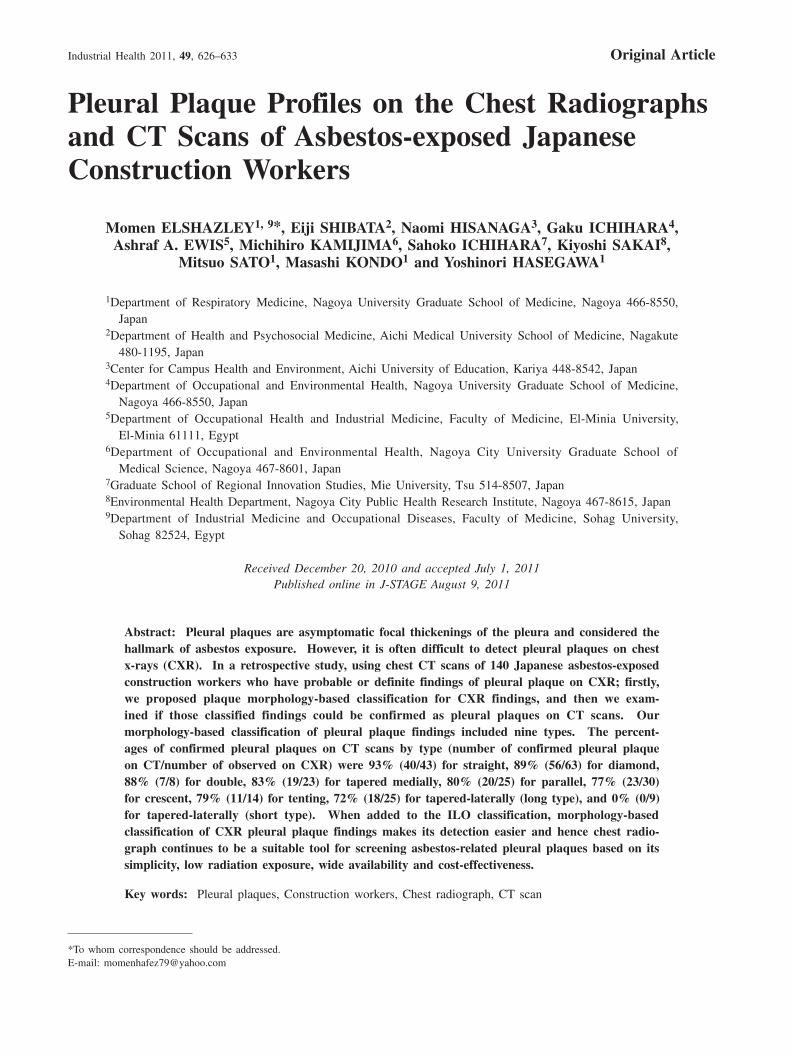
In this study, we hypothesized that the use of a mor-phology-based classification of pleural plaques, in addi-tion to their location, width and extent with the other ILO criteria, may improve the detection rate of pleural plaque findings on CXR. The subjects of this study were 140 Japanese asbestos-exposed construction work-ers with probable and definite findings of pleural plaque on CXR. They were among current Japanese construc-tion workers, and they underwent annual health check up during the period from 2005 to 2007 through the Mie Construction Workers’ Health Insurance Society. The total number of the examined workers was 5,782, 5,771 and 5,346 in 2005, 2006 and 2007 respectively. One of the authors (N.H.) performed screening of their CXR regarding pleural plaque findings. A total of 311 workers with definite or probable findings of pleu-ral plaque on CXR, were asked to perform chest CT scans. Among them, 179 workers agreed to continue in our research. Asbestos exposure was assessed using information obtained from the occupational histories of those workers. We excluded subjects with pulmo-nary pathological changes such as tuberculosis, diffuse pleural thickening and parenchymal fibrosis (irregular opacities corresponding to ILO category of ≥1/0), which may interfere with pleural plaques detection on CXR and CT scans (Table 1). Moreover, cases with non-homogenous shadows were also excluded in order to retain cases with probable or definite pleural plaques only (Fig. 1). Finally, the subject of this study was 140
Table 1. Diagnostic criteria of pleural plaque findings on CXR and CT scans
CT Scans
Lung tissue view Mediastinal view Diagnosis
CXR
In profile pleural plaques
+ + +
+ – ±
– – –
Face-on pleural plaques+ + +
– – –
Diaphragmatic pleural plaques+ + +
– – –
± No clear diagnosis of pleural plaques because only CT lung tissue views showed pleural plaque findings.
628 M ELSHAZLEY et al.
Industrial Health 2011, 49, 626–633
workers. The site (chest wall and diaphragm), width, and extent of pleural plaque findings were recorded separately, according to the International Labour Office (ILO) classification of radiographs of pneumoconiosis9). Moreover, all rib companion shadows were defined by shape for further classification.
Using CT scan as the gold standard for the diagnosis of pleural plaques, we evaluated the reading ability of CXR in detection of pleural plaques. The CXR and CT scans of each construction worker were coded and sepa-rated before being evaluated for pleural plaque shapes, sites, extent and width by three independent occupa-tional physicians. The confirmed pleural plaques repre-sented those that were agreed upon by consensus among the independent readings of them.
Throughout this research, we adhered to the Japanese ethical guidelines for epidemiologic studies; and the study protocol was approved by the Institutional Review Boards of Nagoya University Graduate School of Medicine.
Results
A total of 140 male construction workers were evalu-ated in this study; among them 65 (46.4%) were car-penters, 15 (10.7%) plasterers, 10 (7.1%) steel frame workers, 9 (6.4%) interior finishers, 7 (5.0%) electri-cians, 6 (4.3%) painters, 6 (4.3%) plumbers, 6 (4.3%) roofers, and 16 (11.5%) had performed various other construction-related tasks. The mean age of the workers
was 60.0 ± 7.5 yr; and the majority (about 73.6%) spent long periods (30–49 yr) in the construction-related jobs, during which they were exposed to asbestos (Table 2).
According to the ILO classification of radiographs of pneumoconiosis, among 92 subjects with chest wall pleural plaques (in-profile + face-on), 45 (49%) showed plaques on both sides of the pleura, 16 (17%) on the left side and 31 (34%) on the right side. Among the 61 subjects with diaphragmatic pleural plaques, 18 (30%) had plaques on both sides, 21 (34%) on the left side and 22 (36%) on the right side. Calcification was detected in 34 sites, 21 were located on the diaphragm (9 bilateral, 9 on the right and 3 on the left side), 9 were facing-on, 2 on the cardiac margins, and 2 on the chest wall (Table 3).
Examination of the 140 CT scans showed 427 sites of pleural plaque; 203 in-profile chest wall, 43 face-on, 89 para-vertebral, 6 on the cardiac margins and 86 diaphragmatic pleural plaques. In 22 sites (9 in-profile, 4 face-on, 2 para-cardiac and 7 diaphragmatic sites), the CXR showed no lesion while chest CT scans demon-strated plaques (Table 4). Using CT as the gold stan-dard, the frequency of CXR in-profile, face-on, para-cardiac and diaphragmatic plaques confirmed on CT was 194 out of 203, 39 out of 43, 4 out of 6 and 79 out of 86, respectively.
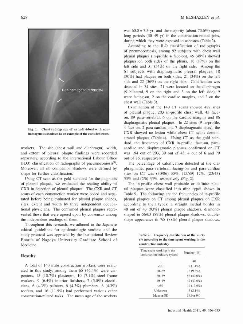
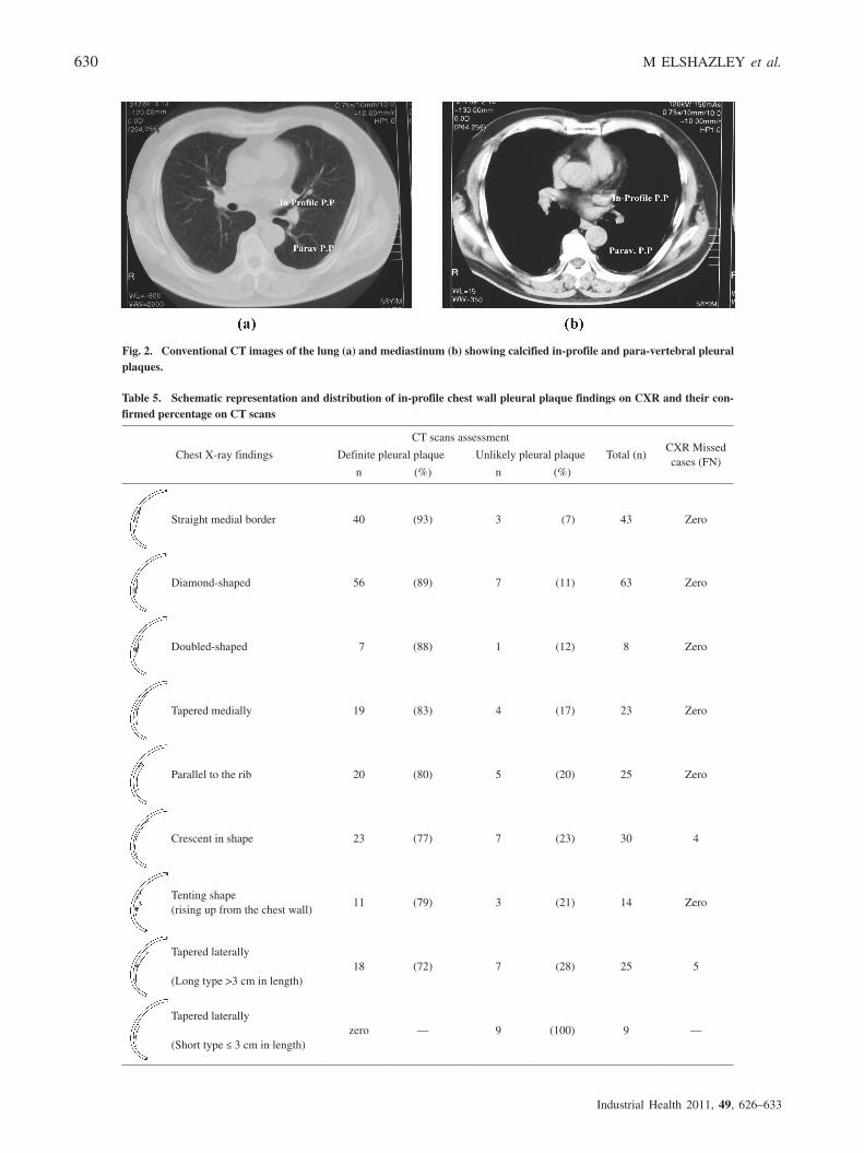
The percentage of calcification detected at the dia-phragmatic, para-vertebral, facing-on and para-cardiac sites on CT was (30/86) 35%, (15/89) 17%, (23/43) 53% and (2/6) 33%, respectively (Fig. 2).
The in-profile chest wall probable or definite pleu-ral plaques were classified into nine types shown in Table 5. The following are the frequencies of in-profile pleural plaques on CT among pleural plaques on CXR according to their types: a straight medial border in 40 out of 43 (93%) pleural plaque shadows, diamond-shaped in 56/63 (89%) pleural plaque shadows, double-shape appearance in 7/8 (88%) pleural plaque shadows,
Fig. 1. Chest radiograph of an individual with non-homogenous shadows as an example of the excluded cases.
Table 2. Frequency distribution of the work-ers according to the time spent working in the construction industry
Time spent working in the construction industry (years)
Number (%)
n 140
<20 2 (1.4%)
20–29 13 (9.3%)
30–39 56 (40.0%)
40–49 47 (33.6%)
≥50 19 (13.6%)
Unknown 3 (2.1%)
Mean ± SD 39.6 ± 9.0
ASBESTOS PLEURAL PLAQUES PROFILE ON CXR & CT 629
medially-tapered in 19/23 (83%) pleural plaque shad-ows, parallel shaped (to the rib margin) in 20/25 (80%) pleural plaque shadows, crescent-shaped in 23/33 (77%) pleural plaque shadows, tenting appearance with base at the chest wall in 11/14 (79%) pleural plaque shadows, tapered laterally (long type >3 cm in length) in 18/25 (72%) pleural plaque shadows and tapered laterally (short type ≤3 cm in length) (Table 5). Pleural plaque shadows with straight medial border and diamond-shape border were the most common on the CXR, comprising 40 and 56 sites, respectively (Fig. 3).
We excluded data obtained from 33 in-profile sites since the diagnosis of pleural plaques was not clear. On the CXR of those 33 sites, we found pleural shad-ows, while there were no corresponding pleural plaque findings on CT mediastinal views; only CT lung tis-sue views showed pleural plaque changes. The percent of those excluded shadows that were confirmed only on CT lung tissue views among the whole diagnosed in-profile pleural plaques was 14% (33/240).
Discussion
In this study, we found that the use of a morphology-based classification of pleural plaques, in addition to their location, width and extent with other ILO criteria, will help pleural plaque identification and improve its reading on CXR. It seems that pleural plaque shadows with straight medial border and diamond-shape were the most common on the CXR and have the highest percentages of confirmed pleural plaques on CT, while shadows which tapered laterally (short type) often appears unlikely in CT. Shadows that were ruled out to be pleural plaques on CT scans had the same CXR shape of the confirmed shadows but they were less in density and width. On the other hand, on CT scans, no corresponding signs could be found for 9 rib com-panion shadows that were detected on CXR with their thick parts directed medially (tapered laterally short type ≤3 cm in length). After applying our morphology-based classification of pleural plaques, CXR couldn’t detect 9 sites of chest wall pleural plaques (9/240=3.7%),
Table 3. Pleural plaque findings according to ILO classification of pneumoconiosis, 2002
Pleural plaques n (%)
SiteIn profile
Bilateral 45 49Right side 16 17Left side 31 34
Face onBilateral 7 44Right side 6 38Left side 3 19
DiaphragmBilateral 18 30Right side 22 36Left side 21 34
Other site(s)Left paracardiac 4 100
CalcificationIn profile
Bilateral 0 0Right side 1 50Left side 1 50
Face onBilateral 5 56Right side 3 33Left side 1 11
DiaphragmBilateral 9 47Right side 9 42Left side 3 11
Other site(s)Left paracardiac 2 100
ExtentRight side
up to ¼ of the lateral chest wall 35 57¼ to ½ of the lateral chest wall 21 34>½ of the lateral chest wall 5 8
Left sideup to ¼ of the lateral chest wall 37 49¼ to ½ of the lateral chest wall 26 34> ½ of the lateral chest wall 13 17
Width (3 mm minimum width)Right side
3 to 5 mm 31 515 to 10 mm 25 41>10 mm 5 8
Left side3 to 5 mm 35 465 to 10 mm 34 45>10 mm 7 9
Table 4. Pleural plaque findings on CXR and CT scans
Fig. 2. Conventional CT images of the lung (a) and mediastinum (b) showing calcified in-profile and para-vertebral pleural plaques.
Table 5. Schematic representation and distribution of in-profile chest wall pleural plaque findings on CXR and their con-firmed percentage on CT scans
Chest X-ray findings
CT scans assessment
Total (n)CXR Missed cases (FN)
Definite pleural plaque Unlikely pleural plaque
n (%) n (%)
Straight medial border 40 (93) 3 (7) 43 Zero
Diamond-shaped 56 (89) 7 (11) 63 Zero
Doubled-shaped 7 (88) 1 (12) 8 Zero
Tapered medially 19 (83) 4 (17) 23 Zero
Parallel to the rib 20 (80) 5 (20) 25 Zero
Crescent in shape 23 (77) 7 (23) 30 4
Tenting shape (rising up from the chest wall)
11 (79) 3 (21) 14 Zero
Tapered laterally
(Long type >3 cm in length) 18 (72) 7 (28) 25 5
Tapered laterally
(Short type ≤ 3 cm in length) zero — 9 (100) 9 —
ASBESTOS PLEURAL PLAQUES PROFILE ON CXR & CT 631
5 shadows have shadows tapered laterally (long type) and 4 shadows have crescent shape but their width is less than 2 mm, taken together, these results suggest better sensitivity of CXR except the above mentioned two types of pleural plaque shadows and this could be a good way to decrease the false negative cases in CXR
by exclusion of these shapes. According to the ILO classification of pneumoco-
niosis radiographs, our results were consistent with the findings of Kikuchi and coworkers9, 10) who reported that 68.6% out of 70 cases with chest wall pleural plaques (in-profile + face-on) were distributed on both
Fig. 3. Chest radiographs of construction workers exposed to asbestos showing different morphologies of the in-profile pleural plaques.
632 M ELSHAZLEY et al.
Industrial Health 2011, 49, 626–633
sides of the chest, while 25.7% were on the left and 5.7% on the right. They also found that among 40 cases with diaphragmatic pleural plaques, 22.5% was bilaterally located, 60% on the left side and 17.5% on the right. In their 22 subjects with calcified plaques, calcification was found in 32 sites; including 26 on the diaphragm (13 on the left and 13 on the right side), 3 on cardiac margins and 3 on the chest wall.
Our results showed 22 false negative sites on the CXR (9 in-profile, 7 diaphragmatic, 4 face-on and 2 para-cardiac sites). The CXR showed no lesions, while the chest CT scans demonstrated hyaline plaques in all 22 sites. These results are similar to those report-ed by Kishimoto et al11).
In this research, data from 33 in-profile sites were excluded because no clear diagnosis of pleural plaques could be made. It is possible that these plaques were early-stage plaques and hence the difficulty in the diag-nosis. Therefore, follow-up of those workers with CXR is recommended. Alternatively, the structures on the CXR could have been false positive findings related to normal anatomical structures (e.g., extra-pleural muscles and fat).
Comparisons of positive findings on both types of imaging (CXR and CT scan) showed that the reading of chest wall, diaphragmatic, face-on and para-cardiac plaques in CXR is quite similar to CT findings. In this regard Suganuma and coworkers13) reported a sensitivity of 0.94 for the CXR and CT scan for pleural plaques detection. This could be a good reason to back up the screening of dust-exposed workers by radiographs, which can be done at a lower cost. Technological inno-vation has allowed accurate diagnosis of dust-related pulmonary diseases, but not all dust-exposed workers can benefit from these new diagnostic modalities. In that sense, simple and inexpensive diagnostic screening tests such as CXR should be further assessed to maxi-mize their sensitivity.
We couldn’t detect pleural plaques located in the para-vetebral site on CXR, while CT examination revealed 89 para-vertebral pleural plaques, in this regards many studies have shown that CT is more sensitive for the detection of pleural disease especially for plaques locat-ed in the paravertebral area12, 13). Our results add fur-ther support to these early studies and showed that CT scan is better than CXR in detection of pleural plaques located in the para-vertebral areas. However, we should take into consideration that the additional information to be gained from CT evaluation must be balanced by the additional costs and time required especially in screen-ing large numbers of dust-exposed individuals over the coming decades. Hence, the CXR will continue to be advantageous as the screening tool of choice for detec-
tion of asbestos-related pleural plaques. In conclusion, while CT scan has its unique advantage in the detection of pleural plaques in the para-vertebral sites, the reading of pleural plaques in CXR can be improved by classifi-cations that include morphology with anatomical loca-tion, width, extent as well as other ILO criteria. CXR will continue to be the primary survey imaging modal-ity of choice because of its simplicity, wide availability, low cost and low radiation exposure.
Acknowledgements
We thank Mrs. Mizutani, from the Department of Occupational and Environmental Health, Nagoya University Graduate School of Medicine, for the gener-ous help in drawing the schematic graphs of rib com-panion shadows.
References
1) Churg A (1982) Fiber counting and analysis in the diagnosis of asbestos-related disease. Hum Pathol 13, 381–92.
2) Hillerdal G, Lindgren A (1980) Pleural plaques: corre-lation of autopsy findings to radiographic findings and occupational history. Eur J Respir Dis 61, 315–9.
3) American Thoracic Society (1986) Medical Section of the American Lung Association: the diagnosis of non-malignant diseases related to asbestos. Am Rev Respir Dis 134, 363–8.
4) Fletcher DE, Edge JR (1970) The early radiological changes in pulmonary and pleural asbestosis. Clin Radiol 21, 355–65.
7) Friedman AC, Fiel SB, Fisher MS, Radecki PD, Lev-Toaff AS, Caroline DF (1988) Asbestos-related pleural disease and asbestosis: a comparison of CT and chest radiography. AJR Am J Roentgenol 150, 269–75.
8) Lozewicz S, Reznek RH, Herdman M, Dacie JE, McLean A, Davies RJ (1989) Role of computed tomography in evaluating asbestos related lung dis-ease. Br J Ind Med 46, 777–81.
9) International Labour Office. Guideline for the use of ILO international classification of radiographs of pneumoconiosis, revised edition. http://www.ilo.org/public/libdoc/ilo/2002/102B09_423_engl.pdf. Accessed March 25, 2003.
10) Kikuchi K, Hiraga Y, Yamamoto A (1986) Pleural thickening caused by asbestos exposure—relation between computed tomography and plain film. Nihon Kyobu Shikkan Gakkai Zasshi 24, 101–8 (in Japanese with English abstract).
ASBESTOS PLEURAL PLAQUES PROFILE ON CXR & CT 633
11) Hosoda Y, Hiraga Y, Sasagawa S (2008) Railways and asbestos in Japan (1928–1987) —epidemiology of pleural plaques, malignancies and pneumoconioses. J Occup Health 50, 297–307.
12) Kishimoto T, Morinaga K, Kira S (2000) The preva-lence of pleural plaques and/or pulmonary changes among construction workers in Okayama, Japan. Am J Ind Med 37, 291–5.
13) Suganuma N, Kusaka Y, Hiraga Y, Hosoda Y, Shida H, Morikubo H, Matsumoto T (2001) Asbestos-related pleural abnormalities detected by chest x-ray: fair
agreement with detection by computed tomography. J Occup Health 43, 365–70.
14) Aberle DR, Gamsu G, Ray CS (1988) High-resolution CT of benign asbestos-related diseases: clinical and radiographic correlation. AJR Am J Roentgenol 151, 883–91.
15) Aberle DR, Gamsu G, Ray CS, Feuerstein IM (1988) Asbestos-related pleural and parenchymal fibrosis: detection with high-resolution CT. Radiology 166, 729–34.