Policy 1.1 Laboratory Diagnosis of Tuberculosis Title Laboratory Diagnosis of Tuberculosis Reference Number WA Tuberculosis Control Program Policy 1.1 Policy Statement This policy aims to outline the laboratory methods used in the diagnosis and management of tuberculosis disease in Western Australia, in particular those performed at the Western Australia Mycobacterium Reference Laboratory. Areas Covered Diagnostic tests used to aid the diagnosis of tuberculosis disease i.e. microscopy, culture, nucleic acid amplification techniques, susceptibility testing and molecular epidemiology. Policy sponsor Medical Director, WA TB Control Program Authors T Keehner, F Haverkort, TJJ Inglis, DJ Speers Review panel Dr I Bastian (SA), Dr C Coulter (Qld), Dr S Pandey (Qld) Issued 02 March 2012 Review Date 02 March 2017 Related WA TB Control Program Policies 1.1 Diagnosis of tuberculosis – Laboratory 1.2 Diagnosis of tuberculosis – Clinical 2.1 Medical treatment of tuberculosis (adults) 2.2 Case management of tuberculosis 3.1 Diagnosis of latent tuberculosis infection 3.2 Treatment of latent tuberculosis infection 4.1 Tuberculosis (active and latent) in children 4.2 Management of tuberculosis in prisoners and immigration detainees 4.3 Tuberculosis (active and latent) in pregnant women 4.4 Tuberculosis and HIV 5.1 BCG Vaccination 6.1 Contact tracing for tuberculosis 6.2 Active surveillance for tuberculosis in recent migrants 6.3 Active surveillance for tuberculosis in health care workers 6.4 Active surveillance for tuberculosis prior to anti-TNF alpha treatment 7.1 Notification of tuberculosis and enhanced surveillance data 8.1 Diagnosis and management of Hansen’s disease 9.1 Management of confidential information for the WA Tuberculosis Control Program 9.2 Client record management policy for the WA Tuberculosis Control Program 9.3 Fees and charges associated with tuberculosis and leprosy treatment Document Control Version Effective Date Author Comment 1.0 01 July 2011 Initial Document 1.1 31 Dec 2011 Consultation and review
Transcript
Policy 1.1 Laboratory Diagnosis of Tuberculosis
Title Laboratory Diagnosis of Tuberculosis Reference Number WA Tuberculosis Control Program Policy 1.1 Policy Statement This policy aims to outline the laboratory methods used in the
diagnosis and management of tuberculosis disease in Western Australia, in particular those performed at the Western Australia Mycobacterium Reference Laboratory.
Areas Covered Diagnostic tests used to aid the diagnosis of tuberculosis disease i.e. microscopy, culture, nucleic acid amplification techniques, susceptibility testing and molecular epidemiology.
Policy sponsor Medical Director, WA TB Control Program Authors T Keehner, F Haverkort, TJJ Inglis, DJ Speers Review panel Dr I Bastian (SA), Dr C Coulter (Qld), Dr S Pandey (Qld) Issued 02 March 2012 Review Date 02 March 2017
Related WA TB Control Program Policies 1.1 Diagnosis of tuberculosis – Laboratory 1.2 Diagnosis of tuberculosis – Clinical 2.1 Medical treatment of tuberculosis (adults) 2.2 Case management of tuberculosis 3.1 Diagnosis of latent tuberculosis infection 3.2 Treatment of latent tuberculosis infection 4.1 Tuberculosis (active and latent) in children 4.2 Management of tuberculosis in prisoners and immigration detainees 4.3 Tuberculosis (active and latent) in pregnant women 4.4 Tuberculosis and HIV 5.1 BCG Vaccination 6.1 Contact tracing for tuberculosis 6.2 Active surveillance for tuberculosis in recent migrants 6.3 Active surveillance for tuberculosis in health care workers 6.4 Active surveillance for tuberculosis prior to anti-TNF alpha treatment 7.1 Notification of tuberculosis and enhanced surveillance data 8.1 Diagnosis and management of Hansen’s disease 9.1 Management of confidential information for the WA Tuberculosis Control Program 9.2 Client record management policy for the WA Tuberculosis Control Program 9.3 Fees and charges associated with tuberculosis and leprosy treatment
Document Control Version Effective Date Author Comment 1.0 01 July 2011 Initial Document 1.1 31 Dec 2011 Consultation and review
Policy 1.1 Laboratory Diagnosis of Tuberculosis Table of Contents TABLE OF CONTENTS ............................................................................................................................................... 2
10.0 QUALITY CONTROL .................................................................................................................................. 10
11.0 NOTIFICATION OF RESULTS .................................................................................................................. 10
13.0 WORKS CITED ............................................................................................................................................ 11
APPENDIX A: MEDIA USED FOR PRIMARY CULTURE AT PATHWEST .................................................... 13 1. B83 MEDIUM ....................................................................................................................................................................... 13 2. GERLOFF MEDIUM ............................................................................................................................................................... 13 3. MYCOBACTERIA GROWTH INDICATOR TUBE (MGIT) ................................................................................................. 13 4. BACTEC MGIT GROWTH SUPPLEMENT/PANTA ...................................................................................................... 14 5. BACTEC MYCO/F LYTIC CULTURE VIALS ..................................................................................................................... 14
APPENDIX D: SUSCEPTIBILITY TESTING OF MTBC. .................................................................................... 18
Public Health and Ambulatory Care, North Metropolitan Health Service Policy 1.1 WA TB Control Program
3
List of Abbreviations AFB acid-fast bacilli BCG bacille Calmette-Guérin CDC Centers for Disease Control (United States) CLSI Clinical and Laboratory Standards Institute DNA Deoxyribonucleic acid EMS early morning specimen FH Fremantle Hospital MAC Mycobacterium avium complex MDR-TB multidrug-resistant tuberculosis MGIT Mycobacteria Growth Indicator Tube MIRU-VNTR Mycobacterial Interspersed Repetitive Unit - Variable Number Tandem
Repeat MRL Mycobacterium Reference Laboratory MTB M. tuberculosis MTBC M. tuberculosis complex NATA National Association of Testing Authorities NAAT nucleic acid amplification techniques NTAC (Australian) National Tuberculosis Advisory Committee NTM non-tuberculous mycobacteria PC3 Physical Containment Level 3 PCR polymerase chain reaction PMH Princess Margaret Hospital QEII Queen Elizabeth II Medical Centre RCPA Royal College of Pathologists Australasia RFLP restriction fragment length polymorphism RIF rifampicin RNA Ribonucleic acid RPH Royal Perth Hospital TB Tuberculosis, disease caused by a member of the MTBC US United States WA Western Australia ZN Ziehl-Nielsen
Public Health and Ambulatory Care, North Metropolitan Health Service Policy 1.1 WA TB Control Program
4
Policy 1.1 Laboratory Diagnosis of Tuberculosis 1.0 Introduction The mycobacteria laboratory situated at PathWest Laboratory Medicine on the Queen Elizabeth II (QEII) hospital site in Nedlands is the state’s Mycobacterium Reference Laboratory (MRL) and supports the WA Tuberculosis Control Program. The laboratory is staffed by a Scientist-in-Charge, who has tertiary and managerial oversight and a Senior Scientist with supervisory responsibility for the daily laboratory activities. There is a variable number of suitable trained technical staff. Pathologist oversight and consultancy is available as required. PathWest laboratories at some other sites (e.g. tertiary hospitals) offer a limited range of tuberculosis (TB) services but otherwise refer all samples to the MRL. The MRL undertakes the following functions:
1. Provision of basic TB diagnostic services (i.e. microscopy & culture) in cooperation with other public & private laboratories;
2. Provision of specialised TB diagnostic services, such as mycobacterial identification, drug susceptibility testing, and rapid molecular detection of drug resistance;
3. Provision of molecular epidemiological typing (genotyping) by nationally-approved methods;
4. Provision of specialised laboratory services for the investigation of clinically-significant non-tuberculous mycobacteria (NTM) infections;
5. Participation in national quality assurance programs; and 6. Training of clinical, public health and laboratory personnel to maintain expertise in
mycobacterial diagnostics in both the public and private sectors. This policy aims to outline the laboratory methods used in the diagnosis of tuberculosis disease in Western Australia (WA), in particular those performed at the Western Australia Mycobacterium Reference Laboratory. 2.0 Overview of the mycobacteria testing process A variety of clinical specimens are processed and cultured to various growth media (see Appendix A and B) and incubated at temperatures appropriate to the requirements of the Mycobacterium species under investigation. Smears are made from concentrated clinical material, examined for acid-fast bacilli (AFB) and reported the same day. Sample-direct molecular testing is carried out according to internationally recommended algorithms using nucleic acid amplification tests (NAAT) for M. tuberculosis (MTB) and the other M. tuberculosis Complex (MTBC) members such as M.bovis. Mycobacteria recovered from culture are fully identified by molecular means.
Public Health and Ambulatory Care, North Metropolitan Health Service Policy 1.1 WA TB Control Program
5
Susceptibility testing of M. tuberculosis and M. bovis to first line anti-tuberculous drugs is undertaken for new cases and for suspected relapse cases, a process that may take 7-14 days from time of positive culture. Second line susceptibility testing is performed on strains of multi-drug resistant TB (MDR-TB), defined as resistance to at least isoniazid (INH) and rifampicin (RIF). Susceptibility testing of rapidly growing NTM and clarithromycin susceptibility testing of Mycobacterium avium complex (MAC) are performed on isolates from significant sites and/or following consultation, in accordance with Clinical and Laboratory Standards Institute (CLSI) guidelines (Clinical and Laboratory Standards Insitute, 2011). Genotyping is performed on all new isolates of MTB using Mycobacterial Interspersed Repetitive Unit - Variable Number Tandem Repeat (MIRU-VNTR) analysis (Supply et al 2000). 3.0 Laboratory Services The MRL is the only laboratory in Western Australia (WA) to provide comprehensive mycobacterial services. Some general bacteriology laboratories may be able to provide direct acid-fast microscopy at short notice, but do not attempt culture for mycobacteria and refer specimens to a higher-level laboratory. One large private laboratory performs both acid-fast microscopy and mycobacterial culture then refers isolates to the MRL for identification. Another private laboratory sends all samples to Adelaide for microscopy and culture, and then refers isolates back to the WA MRL for identification. This present situation could change as laboratory technology evolves. The Centers for Disease Control (CDC), United States, have set demanding criteria for mycobacteriology laboratory performance following the United States tuberculosis epidemic in the early 1990s (Centers for Disease Control, 2000). This includes reporting acid-fast examinations within 24 hours of specimen collection, identification of M. tuberculosis complex (MTBC) within an average of 10-14 days, and reporting of drug susceptibility results within an average of 15-30 days. The Australian Mycobacterium Reference Laboratory Network accepts similar targets to the United States. These are documented in the (Australian) National Tuberculosis Advisory Committee (NTAC) Guidelines for Australian Mycobacteriology Laboratories (National Tuberculosis Advisory Committee Australia, 2006). This extensive document actively promotes high standards of laboratory testing and addresses safety, quality and reporting issues for low- and high-volume laboratories. Physical Containment Level 3 (PC3) is a requirement when dealing with samples from patients with MDR-TB (Standards Australia, 2002).
3.1 After hours services Currently, PathWest sites at Princess Margaret Hospital (PMH), Queen Elizabeth II Medical Centre (QEII), Fremantle Hospital (FH) and Royal Perth Hospital (RPH) can offer an urgent, after-hours AFB (Ziehl-Nielsen) microscopy service. This is performed on sample-direct material to provide a provisional result and is usually only performed
Public Health and Ambulatory Care, North Metropolitan Health Service Policy 1.1 WA TB Control Program
6
following consultation with the on-call Clinical Microbiologist at the respective site. The report will always be followed up by a final microscopy report from the MRL, who perform microscopy on processed samples, i.e. samples subjected to mucolytic decontaminating agents and high-speed centrifugation. Several of the larger PathWest metropolitan sites (RPH, FH and QEII) also offer an after-hours molecular TB detection service utilizing the GeneXpert system (Cepheid, USA). This system detects MTB–specific DNA sequences and can detect some molecular markers for rifampicin resistance. Requests for this test are generally discussed with the Clinical Microbiologist and linked to requests for urgent microscopy. 4.0 Specimen Collection Laboratory guidelines exist that ensure optimal recovery of mycobacteria from samples (see Appendix B). Generally speaking, mycobacteria are robust organisms and tolerate transport and delays in culture quite well. Issues with specimen collection include:
1. Ensuring an adequate sample is provided to capture the low numbers of mycobacteria often present, notably in body fluids. Generally the more organisms present the sooner cultures become positive;
2. Overgrowth by commensal flora, particularly when a highly enriched broth-culture
mycobacteria medium is used or when there is delayed transfer of samples to the laboratory, presents technical problems and delays in laboratory turn-around time. A high quality specimen is therefore important and specimens should reach the laboratory within 24 hours.
5.0 Microscopy Microscopy is performed on all specimens submitted for AFB examination (with the exception of peripheral blood), and an attempt is made to quantify the number of AFB present. Microscopy is a simple and rapid procedure but is much less sensitive than culture. It has been estimated that 5,000-10,000 AFB per millilitre are required before they can be seen in Ziehl-Nielsen (ZN) stained smears. Culture techniques detect 10 to 100 viable mycobacteria per millilitre of sample. Despite this, microscopy remains helpful in several ways. Sputum examination can:
a) Provide a presumptive diagnosis of mycobacterial disease; b) Enable the rapid identification of the most infectious cases, pivotal for infection
control regarding contagiousness; c) Be used to follow the progress of anti-tuberculous chemotherapy; and d) Affect the patient's discharge back into the community.
Public Health and Ambulatory Care, North Metropolitan Health Service Policy 1.1 WA TB Control Program
7
Both fluorochrome (using ultra violet fluorescence) and ZN methods are available. Fluorochrome smears are viewed at lower magnification (250–450x) than ZN (1000x). This lower magnification allows a much larger area of the smear to be scanned. The ZN method remains the reference standard and all new microscopy positive cases are confirmed by this method.
6.0 Culture Culture is performed using a validated commercial broth system, egg-based solid media, or a combination of these (see Appendix A). Species such as M. bovis, M. haemophilum, M. marinum and M. ulcerans have special requirements in media and/or temperature of incubation. Communication between clinicians and the laboratory is required to ensure that appropriate cultures are performed. Direct culture remains the most sensitive and preferred isolation technique. The Mycobacteria Growth Indicator Tube broth system (MGIT; Becton Dickinson) is now widely in use in Western Australia. It contains Middlebrook 7H9 medium with an oxygen sensitive, fluorescent indicator located at the base of the culture tube. Cultures are read by exposing the tubes to long wave ultra violet light (typically 366 nm). Tubes with oxygen depletion emit a bright orange fluorescence. In general, cultures from smear-positive specimens become positive at 37 degrees Celsius within 1-2 weeks, while cultures from smear-negative (mycobacteria-containing) specimens become positive within 2-4 weeks. Respiratory cultures are incubated for 6 weeks before being discarded. Some cultures are retained for 12 weeks. Typically those from non-pulmonary sites are also cultured at 30 to 32 degrees Celsius. 7.0 Identification The MRL identifies all isolates recovered de novo by the MRL or referred from another laboratory by using molecular techniques (as described in Appendix C). M. tuberculosis and closely related species are differentiated from non-tuberculous mycobacteria (NTM) by nucleic acid amplification testing (NAAT). The NTM are then provisionally identified using 16S gene sequencing, with results generally available within a few days. There are limitations associated with 16S gene sequencing and a multi-gene analysis approach is currently under development. The additional resource demands involved in this may limit its use to significant isolates and an adequate clinical history is therefore essential. All MTB isolates (and NTM isolates of clinical significance) are freeze-dried and stored indefinitely. Extracts of processed materials are stored according to National Association of Testing Authorities Australia (NATA) requirements.
Public Health and Ambulatory Care, North Metropolitan Health Service Policy 1.1 WA TB Control Program
8
7.1 Specimen-direct nucleic acid amplification testing (NAAT) Significant improvements in NAAT methods, including automation, have moved these methods from a research environment into routine clinical laboratories. PathWest routinely operates an in-house nested PCR test (see Appendix C) with results available the next day, but can also offer a rapid result (approximately 2 hour turn around time) using Cepheid GeneXpert (see below). NAAT should not take preference over microscopy and culture for tuberculosis, especially if there is a limited amount of sample. NAAT is usually laboratory-initiated following consultation with a consultant Clinical Microbiologist who takes into consideration the test limitations, clinical and public health issues. All new smear-positive clinical samples, regardless of specimen origin and clinical presentation are considered for NAAT. One important factor in NAAT is the smear status of specimens that are culture-positive for MTBC. Although NAAT is very sensitive and can theoretically detect a single bacterial cell, in practice organism load, sample volume and quality are important. The cut-off sensitivity threshold for the in-house test is the same as for microscopy, i.e. ~104 AFB/ml. A further consideration for NAAT is inhibitors sometimes found in a clinical sample. Assays used at PathWest detect their presence and this may cause delays in reporting when re-testing is required. The GeneXpert MTB/RIF is a sensitive cartridge-based, automated real time assay that can detect MTB and resistance to rifampicin, a surrogate marker for MDR strains, within approximately 2 hours from receipt in laboratory. It is said to have similar sensitivity to culture (98-100% in smear-positive cases, 73-91% in smear negative cases). Specificity is ~98%, giving very good positive and negative predictive values, for detection of both MTB presence and rifampicin resistance. The test is available at Royal Perth Hospital, Fremantle Hospital and QEII PathWest sites. It should be noted that the GeneXpert assay is currently only accredited for use on respiratory samples and there is a lack of a true positive control for every assay. Confirmatory methods are therefore needed for both MTB and rifampicin resistance detections and comments to this effect are made at the time of reporting. Issues of when and how often to test have been addressed and the following is the NTAC and CDC recommended algorithm for NAAT that is also applied at PathWest (Centers for Disease Control, 2000 and NTAC 2006). The use of NAAT for screening specimens from patients with suspected TB should be limited to:
• Respiratory smear-positive specimens where the result is likely to influence clinical (treatment) and/or public health (isolation, contact investigation) decisions;
• Respiratory smear-negative specimens from a patient with a high probability of TB, when prompt management and public health decisions are required; and
Public Health and Ambulatory Care, North Metropolitan Health Service Policy 1.1 WA TB Control Program
9
• Selected non-respiratory specimens (e.g. meningeal, some tissue biopsies) where a prompt management decision is necessary (recognising that such tests have not been validated or approved).
The use of NAAT is considered inappropriate in the following instances:
• When a patient is respiratory smear-negative and has a low probability of TB; • When a patient is respiratory smear-positive and has a very high probability of TB;
and • Paucibacillary non-respiratory specimens (e.g. pleural fluid, ascitic fluid).
NAAT should not be used to monitor patients on anti-tuberculosis treatment. Tests may remain positive for an extended period of time regardless of whether DNA or RNA is the target for amplification. 8.0 Susceptibility Testing Any new MTBC isolate has susceptibilities performed to isoniazid, rifampicin ethambutol & pyrazinamide as a matter of urgency using the automated MGIT system. Results are available within 7 to 14 days, dependent on the growth characteristics of the organism. The exception to this is bacille Calmette-Guérin (BCG) associated M. bovis, which is a standardized strain of known lineage. MTB strains that show low-level resistance to INH (at 0.1 µg/ml) are retested at a higher concentration (at 0.4 µg/ml) before classifying the strain as INH resistant. M.bovis is intrinsically resistant to pyrazinamide. If an isolate demonstrates multiple resistance to first-line drugs, additional susceptibility testing is performed against amikacin, kanamycin, capreomycin, ofloxacin, ethionamide and rifabutin (refer to Appendix 4 for additional information on concentrations tested). These tests are also performed using the automated MGIT system. Progress in understanding the genetic mechanisms of drug resistance in MTB has resulted in the development of molecular methods for rapid determination of susceptibility profiles. Resistance to rifampicin is a useful predictor of MDR-TB and molecular methods have demonstrated greater than 90% correlation with established phenotypic methods. Molecular methods have a much poorer correlation with traditional methods for isoniazid, pyrazinamide, and ethambutol. Beyond the GeneXpert test identifying resistance to rifampicin, the laboratory does not currently offer molecular testing for resistance markers. Some non-tuberculous mycobacteria (e.g., M. kansasii) have such uniform susceptibility patterns that there is little to be gained from testing individual isolates. Clarithromycin susceptibility testing of Mycobacterium avium complex can be performed using the automated MGIT system. Rapidly growing species, such as the M. fortuitum and M.chelonae / abscessus groups, are tested by disc diffusion, microbroth dilution or E-test. All susceptibility testing is performed in accordance with Clinical and Laboratory Standards Institute (CLSI) guidelines.
Public Health and Ambulatory Care, North Metropolitan Health Service Policy 1.1 WA TB Control Program
10
9.0 Molecular Epidemiology Repetitive DNA elements present within the genome of mycobacteria undergo genetic rearrangements. DNA typing methods exploit genetic movement or drift in M. tuberculosis and M. bovis and therefore provide an important laboratory tool for understanding the epidemiology of their infections. Automated MIRU-VNTR genotyping is performed on all isolates of MTBC. Fifteen loci are examined in real-time to allow determination of exogenous reinfection versus endogenous reactivation, laboratory cross-contamination, and whether the change in an isolate’s drug resistance profile is due to a reinfection or an acquisition of resistance determinants. Data is added to a comparative (local) database that is forwarded to the Medical Director of the WA Tuberculosis Control Program, whenever new data is added. The genotyping result does not form part of the laboratory report. Confirmation of apparent case-clusters can be achieved by using additional genotyping techniques such as spoligotyping and restriction fragment length polymorphism (RFLP). Isolates requiring this additional analysis are currently referred to another MRL that has this capacity. 10.0 Quality Control All PathWest laboratories are NATA accredited and undergo regular audit. Molecular proficiency is assured through participation in national (Royal College of Pathologists Australasia and the Special Interest Group for Mycobacteria within the Australian Society for Microbiology) and international (QCMD - Quality Control for Molecular Diagnostics, Qnostics Ltd UK) quality assurance programs. These programs cover all aspects of tertiary mycobacteriology. PathWest laboratories offering AFB microscopy also undertake quality assurance to ensure competency. The MRL offers training and quality control materials as required. A tuberculosis subgroup within the PathWest Microbiology and Infectious Diseases Discipline Planning Committee meets regularly to monitor governance and determine and review cross-site policy issues. 11.0 Notification of Results All new AFB smear-positive and new culture-positive MTBC results are communicated by the scientist to the requesting doctor or laboratory. Infection control issues for all inpatients at public hospitals are managed by the Infection Control Officer, who will communicate the result to the requesting doctor and advise as appropriate. A consultancy service is available as required. Hard copy and electronic reports are managed via a laboratory information system.
Public Health and Ambulatory Care, North Metropolitan Health Service Policy 1.1 WA TB Control Program
11
The MRL will notify all new AFB smear-positive and new culture-positive MTBC to the Medical Director, Western Australia TB Control Program. This is done initially by fax, and then followed with a copy of the report. 12.0 Conclusion The key roles of the MRL are rapid detection of MTB, determination of antimicrobial susceptibility and reporting notifiable results. Microscopy and culture remain mandatory procedures in mycobacteriology. A viable culture is necessary for susceptibility testing, specific identification of MTBC to species level and for molecular epidemiological profiling. Nucleic acid amplification testing including GeneXpert represents an important laboratory contribution to patient management and the public health control of tuberculosis but has limitations that preclude its use in direct screening of clinical samples. Rapid advances in laboratory technology, including those in unrelated areas of clinical and public health microbiology, make it vital for the laboratory to remain current with new platforms and technical developments. Regular strategic reviews are undertaken by the MRL to identify capability gaps that may then be integrated into strategic plans, thus ensuring correct alignment with its service delivery obligations to the WA TB Control Program. 13.0 Works Cited
Centers for Disease Control. (2000). Update: Nucleic acid amplification tests for tuberculosis. Morbidity and Mortality Weekly Review, vol. 49 (26), pp. 593-594. Clinical and Laboratory Standards Insitute. (2011). Susceptibility testing of Mycobacteria, Norcardiae and other aerobic Actinomycetes: Approved Standard. 2nd edition. CSLI document M24-A2; vol. 31, no. 5. Retrieved December 20, 2011 from http://www.clsi.org/source/orders/free/m24-a2.pdf National Tuberculosis Advisory Committee Australia (NTAC). (2006). Guidelines for Australian Mycobacteriology Laboratories. Comm Dis Intell, vol. 30 (1), pp. 116-128. Standards Australia. (2002). Safety in laboratories Part 3: Microbiological aspects and containment facilities. AS:NZS 2243.5:2010. Retrieved December 20, 2011 from http://www.saiglobal.com/PDFTemp/Previews/OSH/as/as2000/2200/22433.pdf Supply, P., Mazars, E., Lesjean, S., Vincent, V., Gicquel, B., & Locht, C. (2000). Variable human minisatellite-like regions in the Mycobacterium tuberculosis genome. Mol Microbiol, vol. 36 (3), pp. 762-771.
Public Health and Ambulatory Care, North Metropolitan Health Service Policy 1.1 WA TB Control Program
12
Endorsing Authority Policy or Procedure Sponsor Medical Director, WA TB Control Program Last Reviewed 02 March 2012 Next Review date 02 March 2017 References (Standards) EQuiP EQuiP (5Th edition) criteria 1.1.1, 1.1.8. 1.3, 1.4.1, 1.5.2. 1.5.5.
2.1.1 Legislation WA Health Act (1911) Standards AS:NZS 2243.5:2010 Related Documents WA TB Control Program Policy Documents **Feedback or comments related to this policy should be addressed to the Medical Director, WA TB Control Program [email protected]
Public Health and Ambulatory Care, North Metropolitan Health Service Policy 1.1 WA TB Control Program
13
Appendix A: Media used for primary culture at PathWest 1. B83 Medium
USAGE Mycobacterial media for the growth of fastidious species e g. M.haemophilum, M.bovis. REFERENCE 1. Cousins DV, Francis BR, and Gow BL. Advantages of a New Agar Medium in the Primary Isolation of Mycobacterium bovis. Veterinary Microbiology, 20 (1989) 89-95. 2. Tarshis MS. Blood mediums for the cultivation of Mycobacterium tuberculosis. Am. J. Clin. Path. (1953) 23; 661 – 670 3. Haverkort F. Survey of media used to culture Mycobacterium haemophilum in Australia and New Zealand. Report to the Special Interest Group (Mycobacteria) at the Australian Society for Microbiology’s Annual Scientific Meeting, Auckland NZ September 2003. 4. Haverkort F. Validation of B83 as a culture medium for the isolation of Mycobacterium haemophilum. PathWest Laboratory Medicine WA internal document, March 2004. 2. Gerloff Medium USAGE A general purpose mycobacterial media that will grow both M.bovis and MTB equally well. By the addition of ferric ammonium citrate to the base this medium will also grow M.haemophilum. By the addition of selected antimicrobials (NVAP) to the base it acts as an inhibitory medium for gram positive and negative bacteria and most fungi, for the recovery of mycobacteria from non-sterile sites. REFERENCE 1.Gerloff, W: Entwickling und heutiger Standder Kultivierung des Mycobacteriums tuberculosis unter Berucksichti gun g des Pyruvateffektes.Z. ärtzliche Fortbildun g, 1960, 54, 623 - 627.
3. Mycobacteria Growth Indicator Tube (MGIT)
USAGE The Becton Dickinson (BD) MGIT Mycobacteria Growth Indicator Tube (7 mL) supplemented with BACTEC MGIT Growth Supplement and BD MGIT PANTA antibiotic mixture is intended for the detection and recovery of mycobacteria from clinical specimens using the BACTEC MGIT 960 System. REFERENCE BD product information
Public Health and Ambulatory Care, North Metropolitan Health Service Policy 1.1 WA TB Control Program
14
4. BACTEC MGIT Growth Supplement/PANTA USAGE BACTEC MGIT Growth Supplement/PANTA (Becton Dickinson) is used for the inhibition of gram positive and negative bacteria and most fungi, and provides essential substrates for rapid mycobacterial growth in BBL MGIT 7.0 mL tubes. REFERENCE BD product information 5. BACTEC Myco/F Lytic Culture Vials USAGE The BACTEC Myco/F Lytic culture vial (Becton Dickinson) is designed for the rapid detection of mycobacteria in blood, and yeast and fungi in blood and sterile body fluids for use with BACTEC 9240 system. REFERENCE BD product information **This information is current as at December 2011**
Public Health and Ambulatory Care, North Metropolitan Health Service Policy 1.1 WA TB Control Program
15
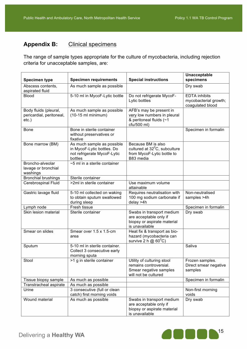
Appendix B: Clinical specimens The range of sample types appropriate for the culture of mycobacteria, including rejection criteria for unacceptable samples, are:
Specimen type Specimen requirements
Special instructions
Unacceptable specimens
Abscess contents, aspirated fluid
As much sample as possible Dry swab
Blood 5-10 ml in MycoF-Lytic bottle Do not refrigerate MycoF-Lytic bottles
Body fluids (pleural, pericardial, peritoneal, etc.)
As much sample as possible (10-15 ml minimum)
AFB’s may be present in very low numbers in pleural & peritoneal fluids (~1 cfu/500 ml)
Bone Bone in sterile container without preservatives or fixative
Specimen in formalin
Bone marrow (BM) As much sample as possible in MycoF-Lytic bottles. Do not refrigerate MycoF-Lytic bottles
Because BM is also cultured at 320C, subculture from MycoF-Lytic bottle to B83 media
Broncho-alveolar lavage or bronchial washings
~5 ml in a sterile container
Bronchial brushings Sterile container Cerebrospinal Fluid >2ml in sterile container Use maximum volume
attainable
Gastric lavage fluid 5-10 ml collected on waking to obtain sputum swallowed during sleep
Requires neutralisation with 100 mg sodium carbonate if delay >4h
Non-neutralised samples >4h
Lymph node Fresh tissue Specimen in formalin Skin lesion material Sterile container Swabs in transport medium
are acceptable only if biopsy or aspirate material is unavailable
Dry swab
Smear on slides Smear over 1.5 x 1.5-cm area
Heat fix & transport as bio-hazard (mycobacteria can survive 2 h @ 600C)
Sputum 5-10 ml in sterile container. Collect 3 consecutive early morning sputa
Saliva
Stool >1 g in sterile container Utility of culturing stool remains controversial. Smear negative samples will not be cultured
Frozen samples. Direct smear negative samples
Tissue biopsy sample As much as possible Specimen in formalin Transtracheal aspirate As much as possible Urine 3 consecutive (full or clean
catch) first morning voids Non-first morning
voids Wound material As much as possible Swabs in transport medium
are acceptable only if biopsy or aspirate material is unavailable
Dry swab
Public Health and Ambulatory Care, North Metropolitan Health Service Policy 1.1 WA TB Control Program
16
Appendix C: Molecular Identification Practices
Current In-House Molecular Identification Practices Used At PathWest for the Identification of Mycobacteria.
1. Identification from culture
Recovered mycobacteria (or referred cultures) are identified using a gel-based multiplex PCR assay (Wilton & Cousins, 1991) that allows rapid screening for MTBC, M.avium and M.intracellulare as well as Mycobacterium “genus”. Whilst it is recognized that some cross-reactions occur in this assay, results are interpreted according to defined algorithms and molecular sequencing of the 16S rRNA gene is undertaken for isolates giving a genus band only and those identifying as M.avium. The MRL is in the process of supplementing 16S-based sequencing with additional targets, in recognition of the limitations inherent in this approach. Qualifying statements are made on the report when an identification of a NTM has been based solely on 16S gene sequence information. A sequence-based assay (oxyR gene, Sreevatsan et al, 1996; Espinosa et al, 1998) is used to differentiate between MTB and M.bovis. Further, isolates identifying as M.bovis are differentiated from M.bovis BCG by examination for the RD1 deletion in a real-time assay (Talbot et al, 1997). Finally, some limited phenotypic identification may be required in special circumstances. The MRL is in the process of supplementing its capacity to more fully identify the other members of the MTBC.
2. Identification of MTBC from fresh tissue
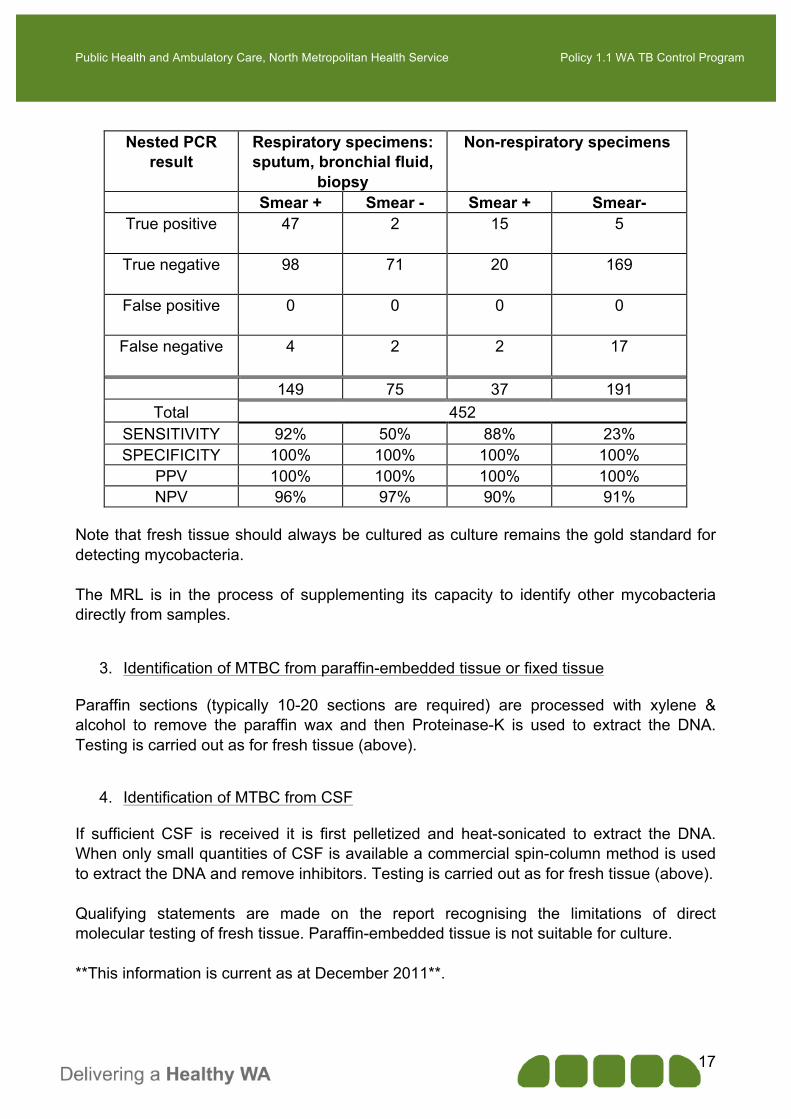
Fresh tissues are first homogenised, processed and centrifuged to maximize the recovery of mycobacteria. DNA is extracted using a heat-sonication process. An in-house nested (double round) gel-based PCR (MPB70 gene; Wilton & Cousins, 1991) allows identification of MTBC. Validation data has given the following specificity and sensitivity for the assay:
Public Health and Ambulatory Care, North Metropolitan Health Service Policy 1.1 WA TB Control Program
Note that fresh tissue should always be cultured as culture remains the gold standard for detecting mycobacteria. The MRL is in the process of supplementing its capacity to identify other mycobacteria directly from samples.
3. Identification of MTBC from paraffin-embedded tissue or fixed tissue Paraffin sections (typically 10-20 sections are required) are processed with xylene & alcohol to remove the paraffin wax and then Proteinase-K is used to extract the DNA. Testing is carried out as for fresh tissue (above).
4. Identification of MTBC from CSF If sufficient CSF is received it is first pelletized and heat-sonicated to extract the DNA. When only small quantities of CSF is available a commercial spin-column method is used to extract the DNA and remove inhibitors. Testing is carried out as for fresh tissue (above). Qualifying statements are made on the report recognising the limitations of direct molecular testing of fresh tissue. Paraffin-embedded tissue is not suitable for culture. **This information is current as at December 2011**.
Public Health and Ambulatory Care, North Metropolitan Health Service Policy 1.1 WA TB Control Program
18
Appendix D: Susceptibility testing of MTBC. First line TB drugs Streptomycin*: 1.0 µg/ml Isoniazid: 0.1 µg/ml then 0.4 µg/ml if 0.1 resistant Rifampicin: 5.0 µg/ml Ethambutol: 1.0 µg/ml Pyrazinamide: 100 µg/ml Second line TB drugs Amikacin: 1.0 µg/ml Kanamycin: 2.5 µg/ml Capreomycin: 2.5 µg/ml Ofloxacin: 2.0 µg/ml Ethionamide: 5.0 µg/ml Rifabutin: 0.5 µg/ml * Streptomycin is tested as it remains a component of the commercial kit used. **This information is current as at December 2011**.